lowoutput cardiac failure
TRANSCRIPT
PATHOPHYSIOLOGICAL BASIS OF HEMODYNAMICS OF LOW OUTPUT HEART
FAILURE
Aniruddha Mandal
Chair personDr. Dipankar Ghosh
Dastidar
Heart failure (HF) is a clinical syndrome that occurs in patients who, because of an inherited or acquired abnormality of cardiac structure and/or function, develop a constellation of clinical symptoms (dyspnea and fatigue) and signs (edema and rales) that lead to frequent hospitalizations, a poor quality of life, and a shortened life expectancy
DEFINITION
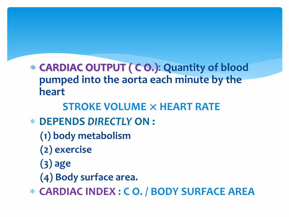
CARDIAC OUTPUT ( C O.): Quantity of blood pumped into the aorta each minute by the heart
STROKE VOLUME × HEART RATE
DEPENDS DIRECTLY ON :(1) body metabolism
(2) exercise
(3) age
(4) Body surface area.
CARDIAC INDEX : C O. / BODY SURFACE AREA
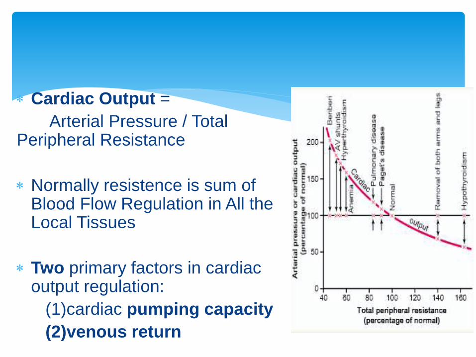
Cardiac Output =
Arterial Pressure / Total Peripheral Resistance
Normally resistence is sum of Blood Flow Regulation in All the Local Tissues
Two primary factors in cardiac output regulation:
(1)cardiac pumping capacity
(2)venous return
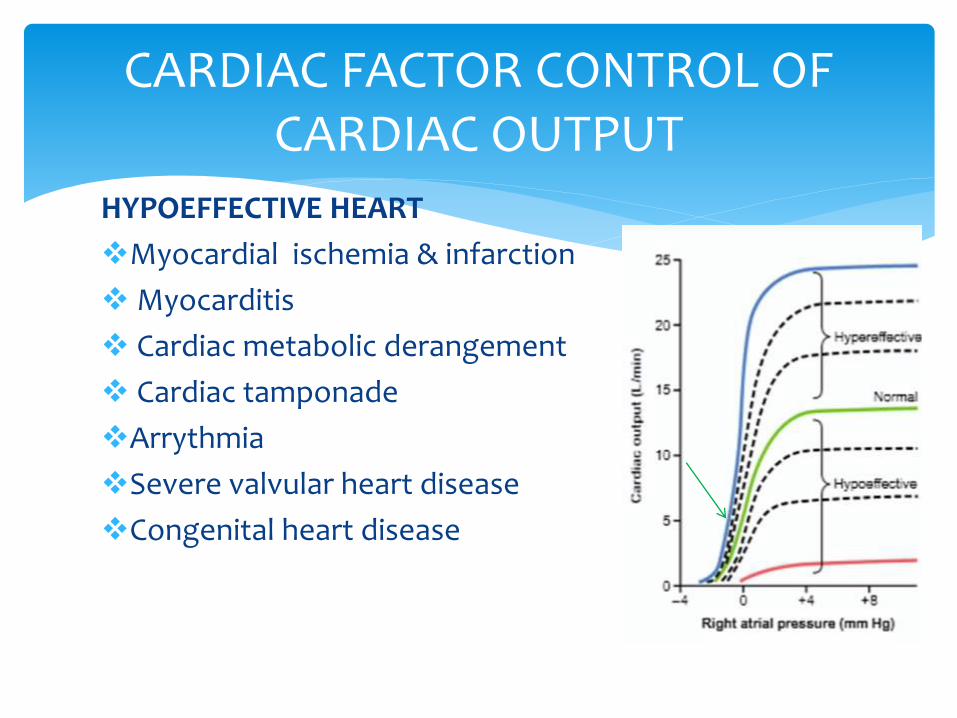
CARDIAC FACTOR CONTROL OF CARDIAC OUTPUT
HYPOEFFECTIVE HEART
Myocardial ischemia & infarction
Myocarditis
Cardiac metabolic derangement
Cardiac tamponade
Arrythmia
Severe valvular heart disease
Congenital heart disease
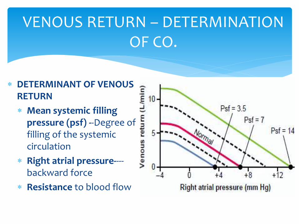
DETERMINANT OF VENOUS RETURN
Mean systemic filling pressure (psf) --Degree of filling of the systemic circulation
Right atrial pressure----backward force
Resistance to blood flow
VENOUS RETURN – DETERMINATION OF CO.
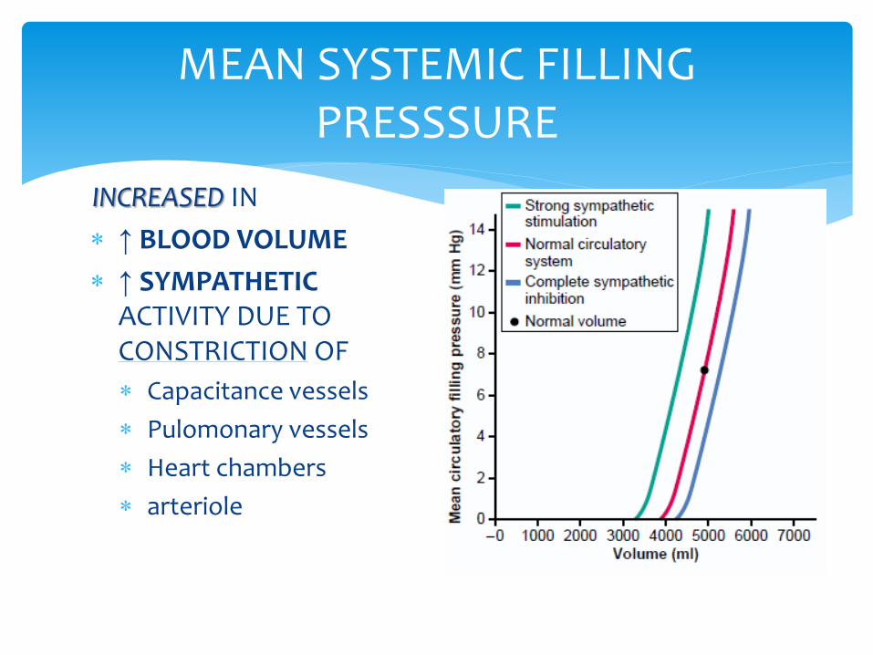
INCREASED IN
↑ BLOOD VOLUME
↑ SYMPATHETIC ACTIVITY DUE TO CONSTRICTION OF
Capacitance vessels
Pulomonary vessels
Heart chambers
arteriole
MEAN SYSTEMIC FILLING PRESSSURE
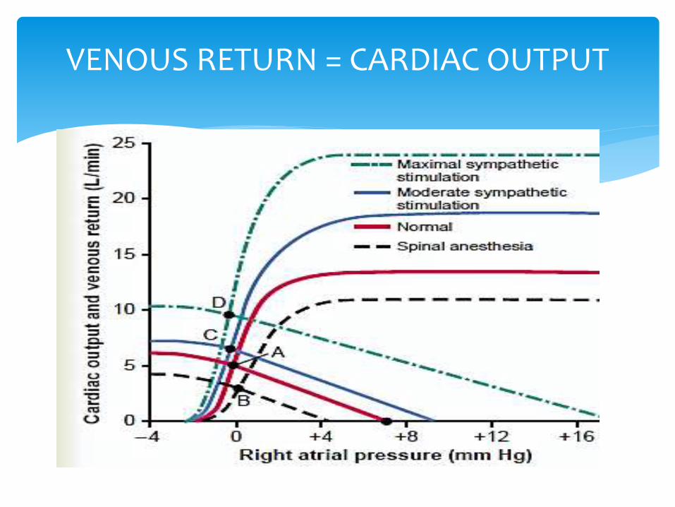
VENOUS RETURN = CARDIAC OUTPUT
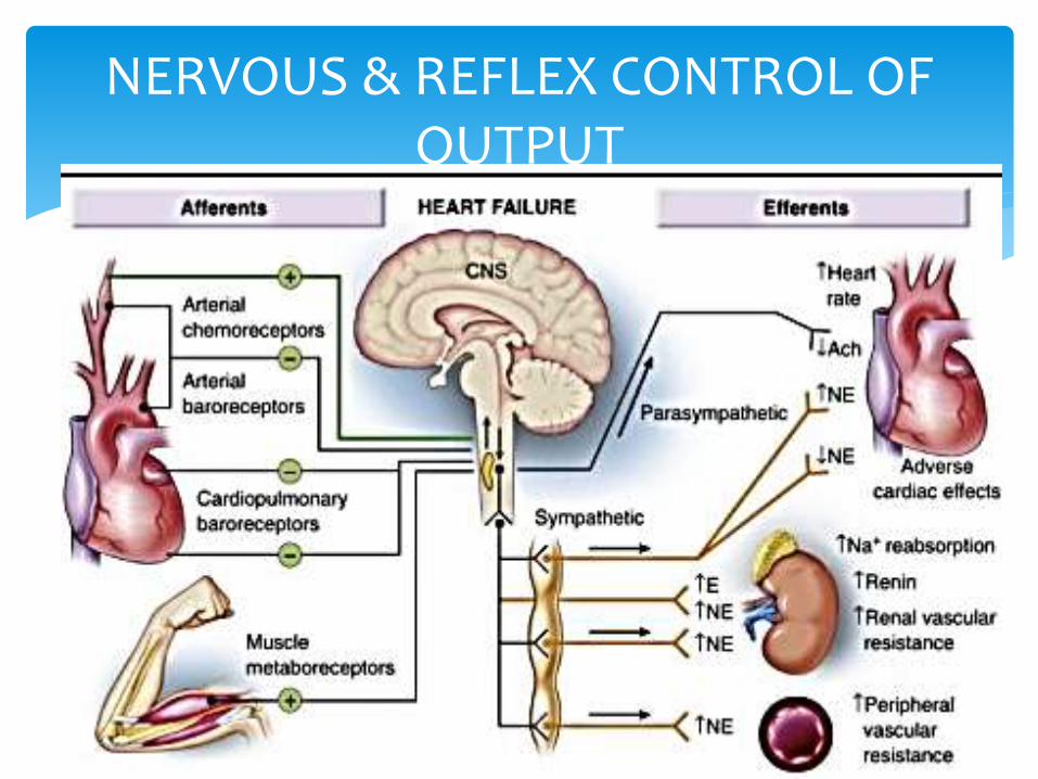
NERVOUS & REFLEX CONTROL OF OUTPUT
Sympathetic stimulation→effect 0n
heart ↑Na+ ,H2O absorption-
kidney
↓ renal blood flow
↑AVP,RAS
↑ 𝑝𝑠𝑓.
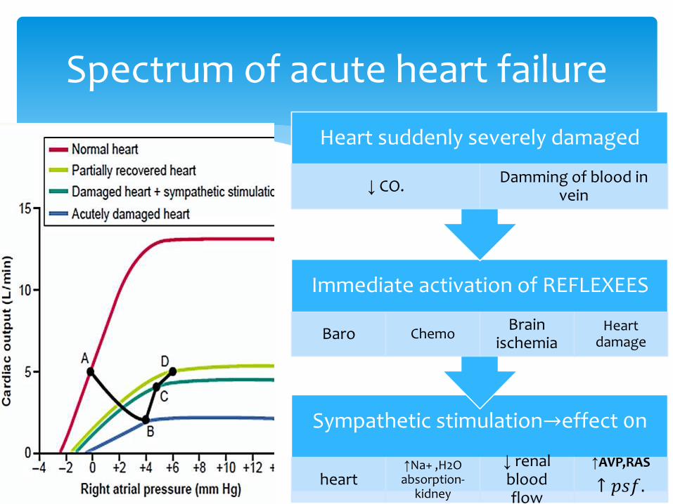
Immediate activation of REFLEXEES
Baro ChemoBrain
ischemiaHeart
damage
Heart suddenly severely damaged
↓ CO.Damming of blood in
vein
Spectrum of acute heart failure
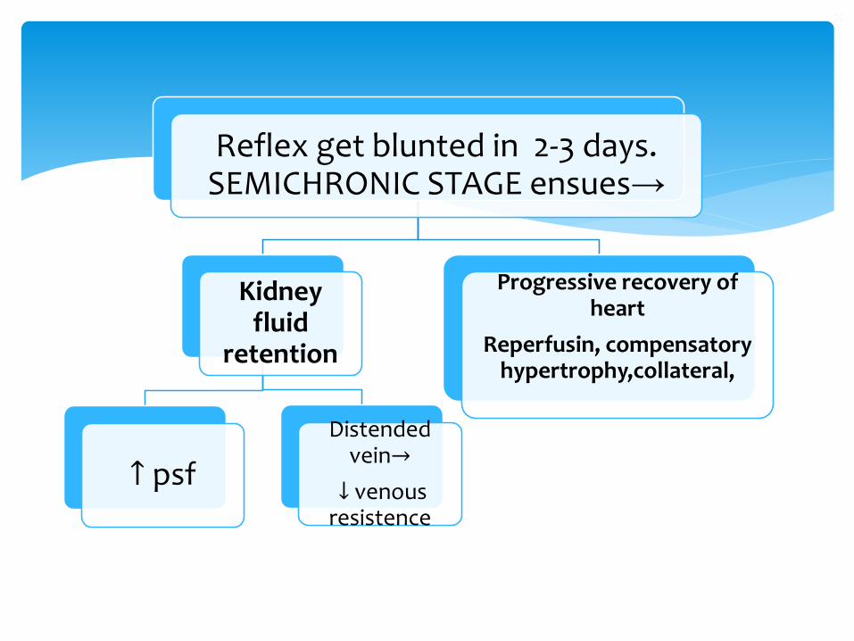
Reflex get blunted in 2-3 days. SEMICHRONIC STAGE ensues→
Kidney fluid
retention
↑ psf
Distended vein→
↓ venous resistence
Progressive recovery of heart
Reperfusin, compensatory hypertrophy,collateral,
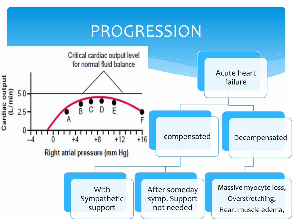
Acute heart failure
compensated
With Sympathetic
support
After someday symp. Support
not needed
Decompensated
Massive myocyte loss,
Overstretching,
Heart muscle edema,
PROGRESSION
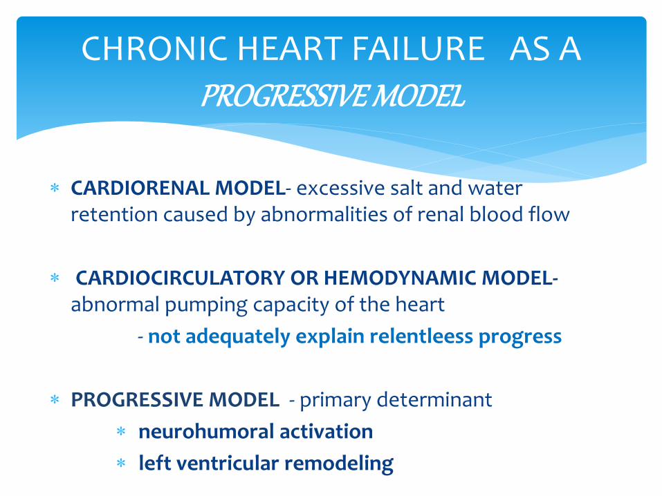
CARDIORENAL MODEL- excessive salt and water retention caused by abnormalities of renal blood flow
CARDIOCIRCULATORY OR HEMODYNAMIC MODEL-abnormal pumping capacity of the heart
- not adequately explain relentleess progress
PROGRESSIVE MODEL - primary determinant
neurohumoral activation
left ventricular remodeling
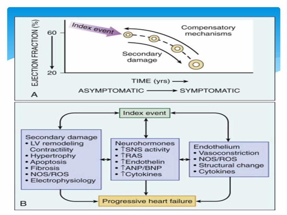
CHRONIC HEART FAILURE AS A PROGRESSIVE MODEL
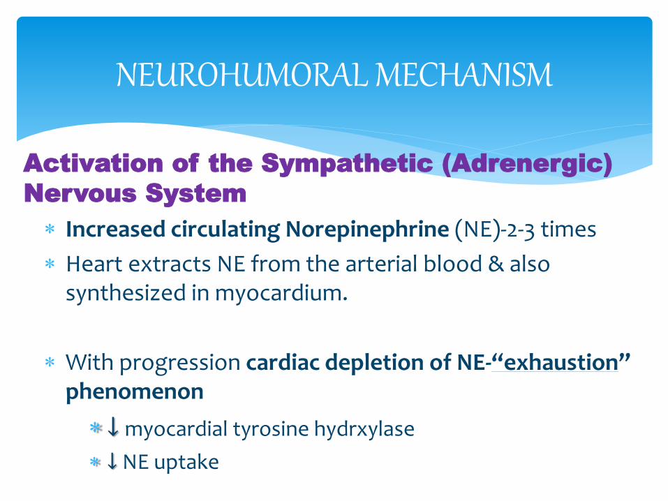
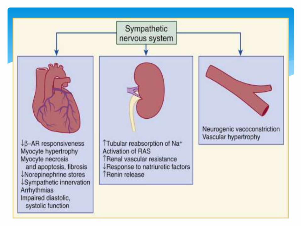
Activation of the Sympathetic (Adrenergic)
Nervous System
Increased circulating Norepinephrine (NE)-2-3 times
Heart extracts NE from the arterial blood & also synthesized in myocardium.
With progression cardiac depletion of NE-“exhaustion” phenomenon
↓ myocardial tyrosine hydrxylase
↓ NE uptake
NEUROHUMORAL MECHANISM
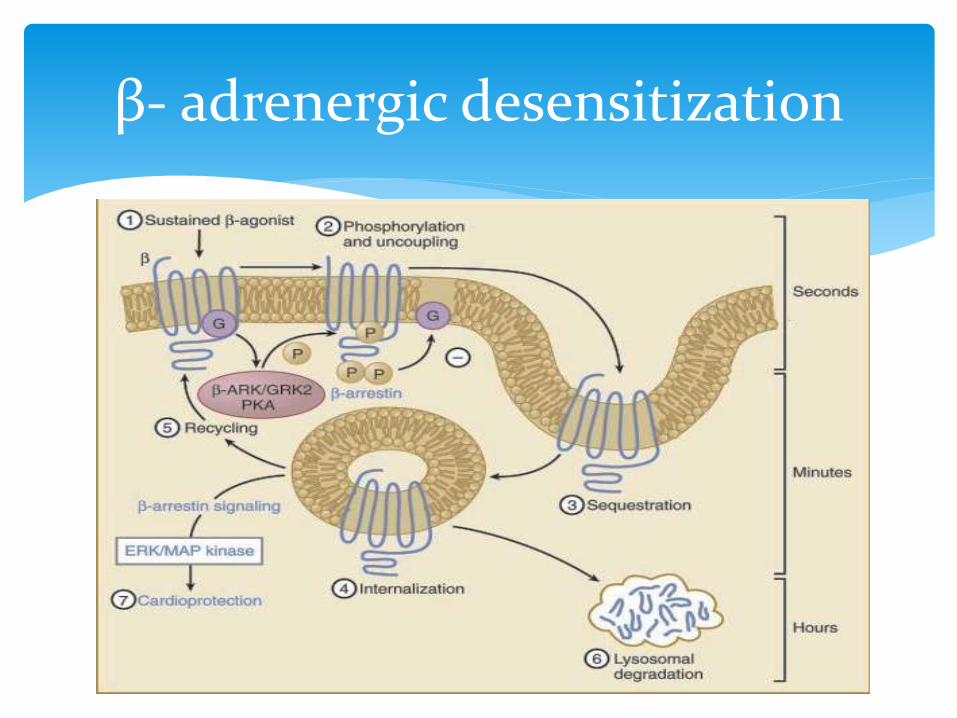
β- adrenergic desensitization
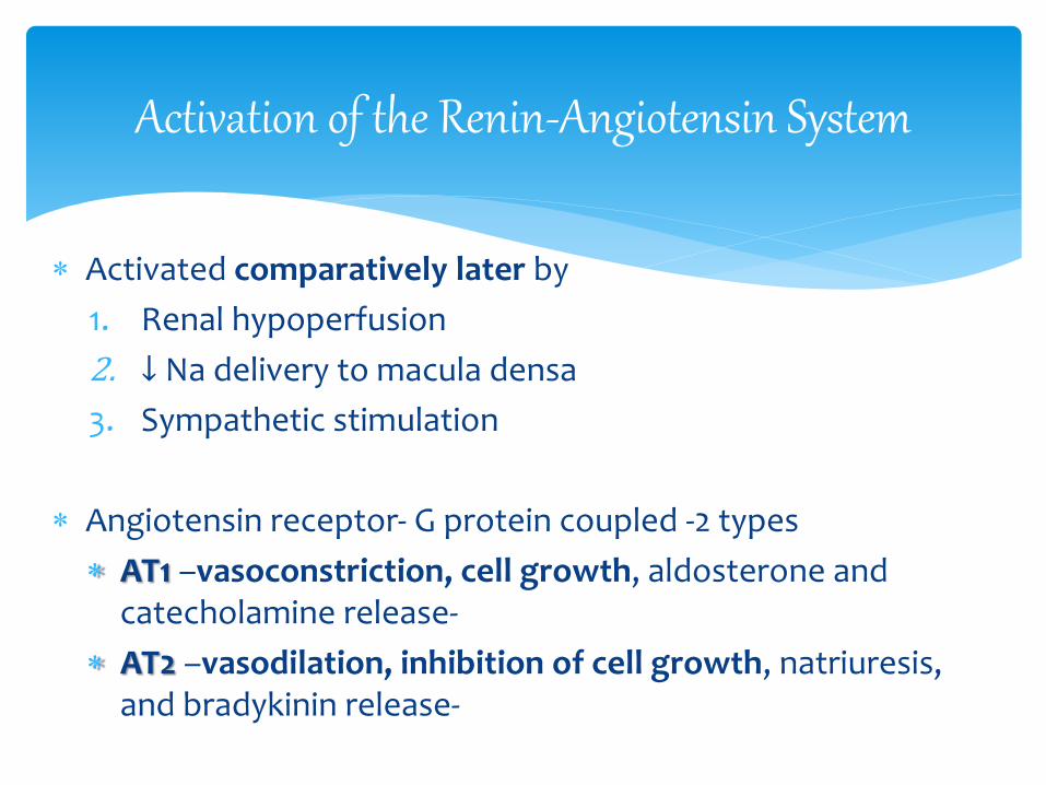
Activated comparatively later by
1. Renal hypoperfusion
2. ↓ Na delivery to macula densa
3. Sympathetic stimulation
Angiotensin receptor- G protein coupled -2 types
AT1 –vasoconstriction, cell growth, aldosterone and catecholamine release-
AT2 –vasodilation, inhibition of cell growth, natriuresis, and bradykinin release-
Activation of the Renin-Angiotensin System
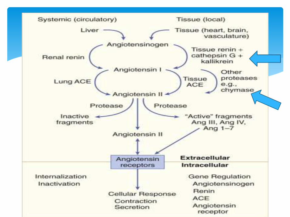
s
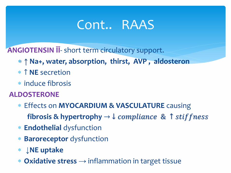
ANGIOTENSIN ĪĪ- short term circulatory support.
↑ Na+, water, absorption, thirst, AVP , aldosteron
↑ NE secretion
induce fibrosis
ALDOSTERONE
Effects on MYOCARDIUM & VASCULATURE causing
fibrosis & hypertrophy → ↓ 𝑐𝑜𝑚𝑝𝑙𝑖𝑎𝑛𝑐𝑒 & ↑ 𝑠𝑡𝑖𝑓𝑓𝑛𝑒𝑠𝑠
Endothelial dysfunction
Baroreceptor dysfunction
↓NE uptake
Oxidative stress → inflammation in target tissue
Cont.. RAAS
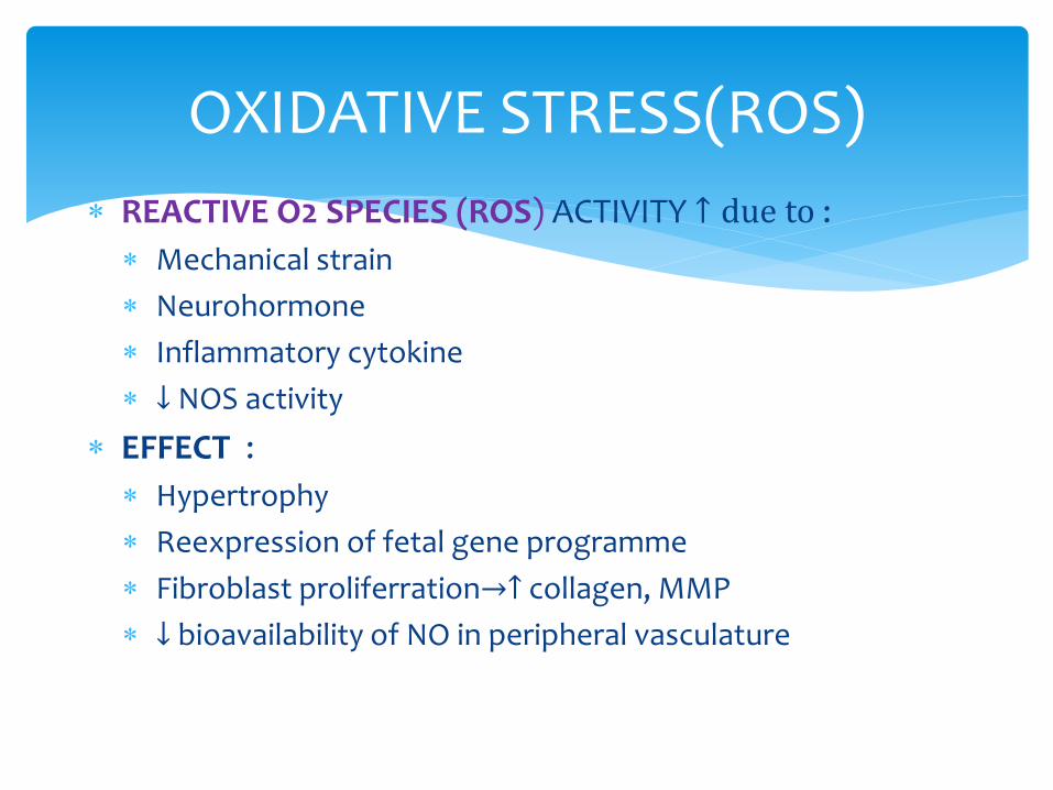
REACTIVE O2 SPECIES (ROS) ACTIVITY ↑ due to :
Mechanical strain
Neurohormone
Inflammatory cytokine
↓ NOS activity
EFFECT :
Hypertrophy
Reexpression of fetal gene programme
Fibroblast proliferration→↑ collagen, MMP
↓ bioavailability of NO in peripheral vasculature
OXIDATIVE STRESS(ROS)
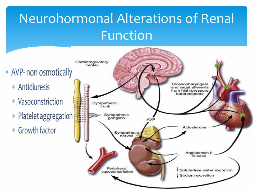
Neurohormonal Alterations of Renal Function
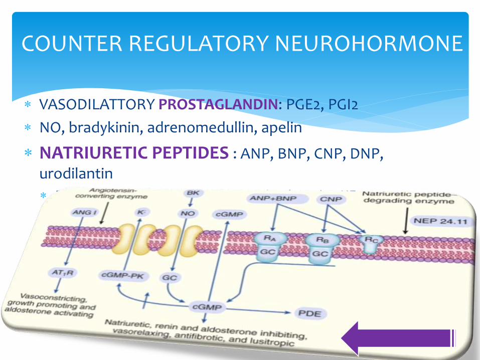
VASODILATTORY PROSTAGLANDIN: PGE2, PGI2
NO, bradykinin, adrenomedullin, apelin
NATRIURETIC PEPTIDES : ANP, BNP, CNP, DNP, urodilantin
Renal effects become blunted with advancing HF
COUNTER REGULATORY NEUROHORMONE
ANP secreted in short burst in ACUTE changes
BNP regulated transcriptionally as CHRONIC response
PROHORMON cleaved to large biologically inactive N-terminal fragments (NT-ANP or NT-
BNP)
smaller biologically active peptides (ANP or BNP)
degraded by neutral endopeptidase
Degraded by NEUTRAL ENDOPEPTIDASE & VASOPEPTIDASE
Candoxatrilat endopeptidase inhibitor
Omapatrilat inhibits both neutral endopeptidase and ACE
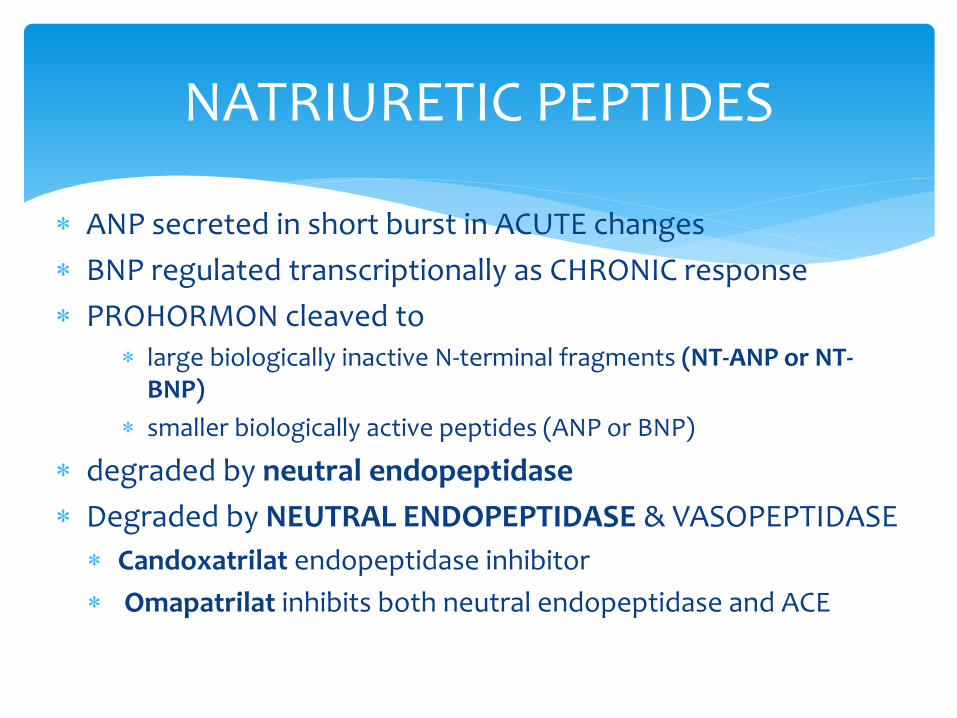
NATRIURETIC PEPTIDES
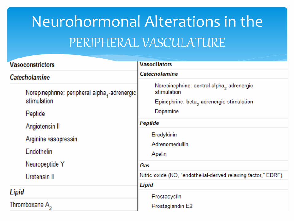
Neurohormonal Alterations in the PERIPHERAL VASCULATURE
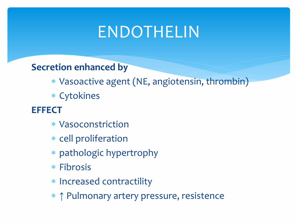
Secretion enhanced by
Vasoactive agent (NE, angiotensin, thrombin)
Cytokines
EFFECT
Vasoconstriction
cell proliferation
pathologic hypertrophy
Fibrosis
Increased contractility
↑ Pulmonary artery pressure, resistence
ENDOTHELIN
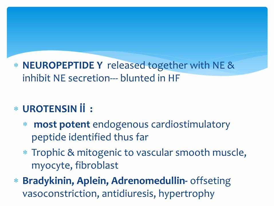
NEUROPEPTIDE Y released together with NE & inhibit NE secretion--- blunted in HF
UROTENSIN İİ :
most potent endogenous cardiostimulatory peptide identified thus far
Trophic & mitogenic to vascular smooth muscle, myocyte, fibroblast
Bradykinin, Aplein, Adrenomedullin- offseting vasoconstriction, antidiuresis, hypertrophy
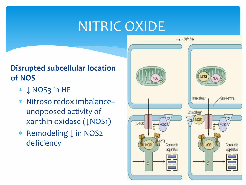
Disrupted subcellular location of NOS
↓ NOS3 in HF
Nitroso redox imbalance–unopposed activity of xanthin oxidase (↓NOS1)
Remodeling ↓ in NOS2 deficiency
NITRIC OXIDE
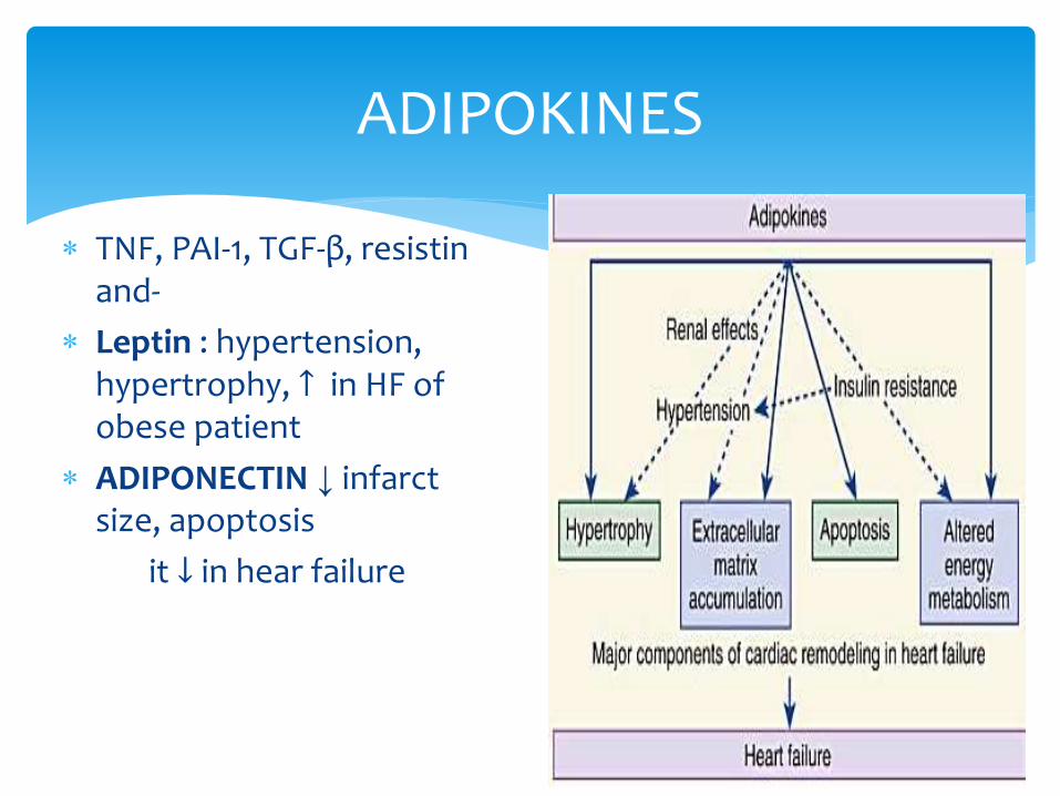
TNF, PAI-1, TGF-β, resistin and-
Leptin : hypertension, hypertrophy, ↑ in HF of obese patient
ADIPONECTIN ↓ infarct size, apoptosis
it ↓ in hear failure
ADIPOKINES

INFLAMMATORY( IL-6, TNF) & ANTIINFLAMMATORY(IL-10) IMBALANCE
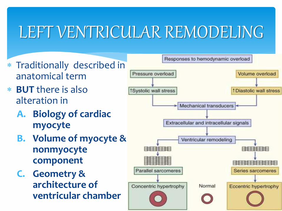
Traditionally described in anatomical term
BUT there is also alteration in
A. Biology of cardiac myocyte
B. Volume of myocyte & nonmyocyte component
C. Geometry & architecture of ventricular chamber
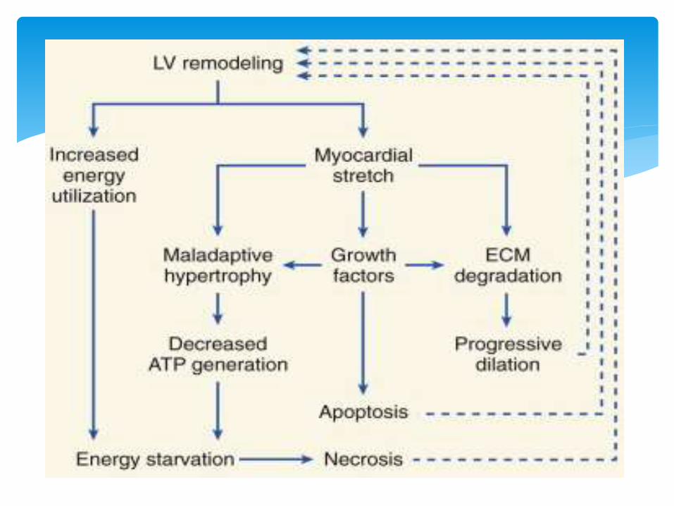
LEFT VENTRICULAR REMODELING
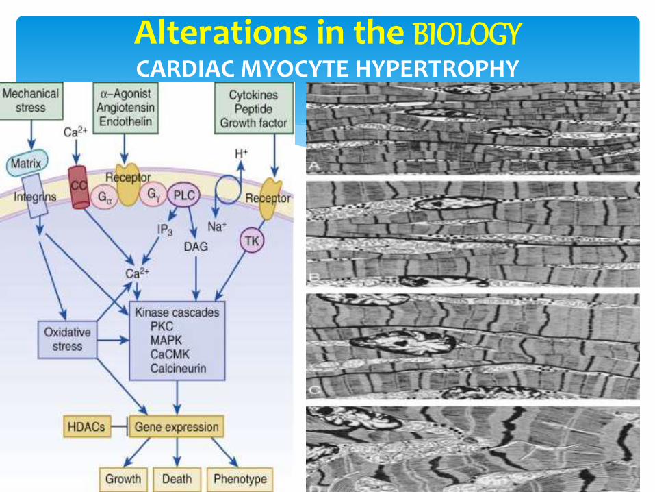
Alterations in the BIOLOGY CARDIAC MYOCYTE HYPERTROPHY
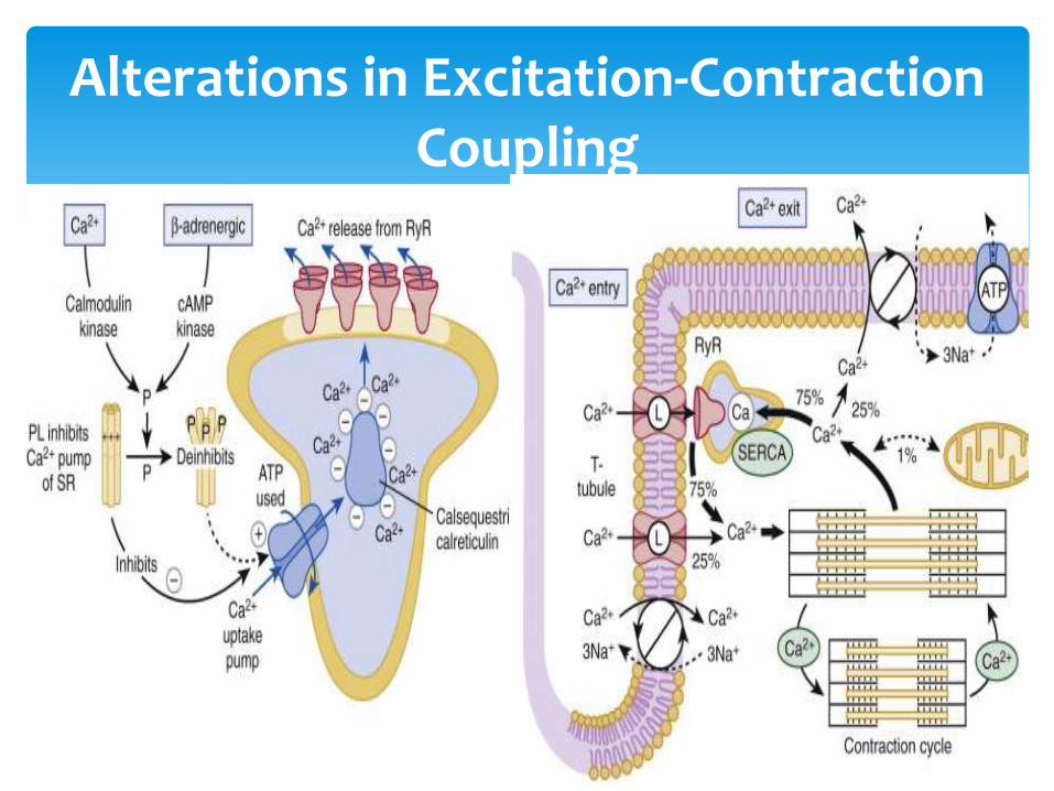
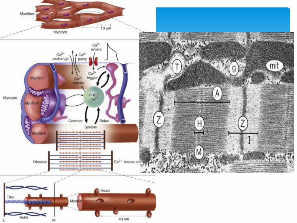
Alterations in Excitation-Contraction Coupling
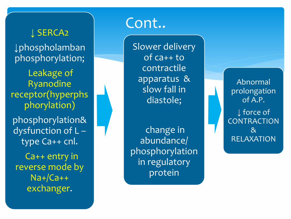
↓ SERCA2
↓phospholamban phosphorylation;
Leakage of Ryanodine
receptor(hyperphsphorylation)
phosphorylation& dysfunction of L –
type Ca++ cnl.
Ca++ entry in reverse mode by
Na+/Ca++ exchanger.
Slower delivery of ca++ to contractile
apparatus & slow fall in diastole;
change in abundance/
phosphorylation in regulatory
protein
Abnormal prolongation
of A.P.
↓ force of CONTRACTION
& RELAXATION
Cont..
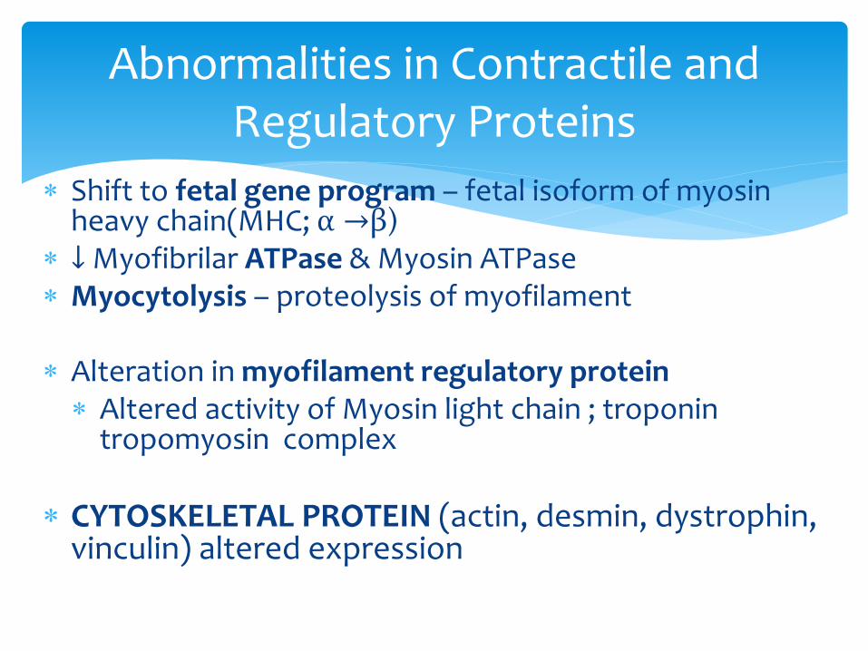
Shift to fetal gene program – fetal isoform of myosin heavy chain(MHC; α →β)
↓ Myofibrilar ATPase & Myosin ATPase Myocytolysis – proteolysis of myofilament
Alteration in myofilament regulatory protein Altered activity of Myosin light chain ; troponin
tropomyosin complex
CYTOSKELETAL PROTEIN (actin, desmin, dystrophin, vinculin) altered expression
Abnormalities in Contractile and Regulatory Proteins
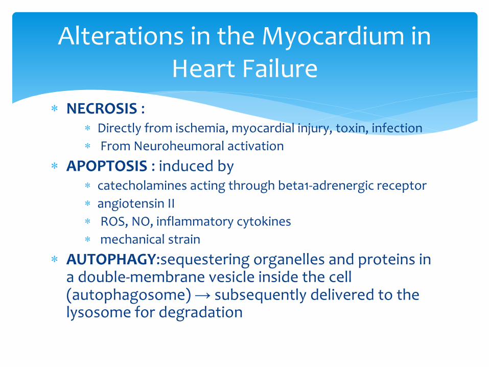
NECROSIS : Directly from ischemia, myocardial injury, toxin, infection
From Neuroheumoral activation
APOPTOSIS : induced by catecholamines acting through beta1-adrenergic receptor
angiotensin II
ROS, NO, inflammatory cytokines
mechanical strain
AUTOPHAGY:sequestering organelles and proteins in a double-membrane vesicle inside the cell (autophagosome) → subsequently delivered to the lysosome for degradation
Alterations in the Myocardium in Heart Failure
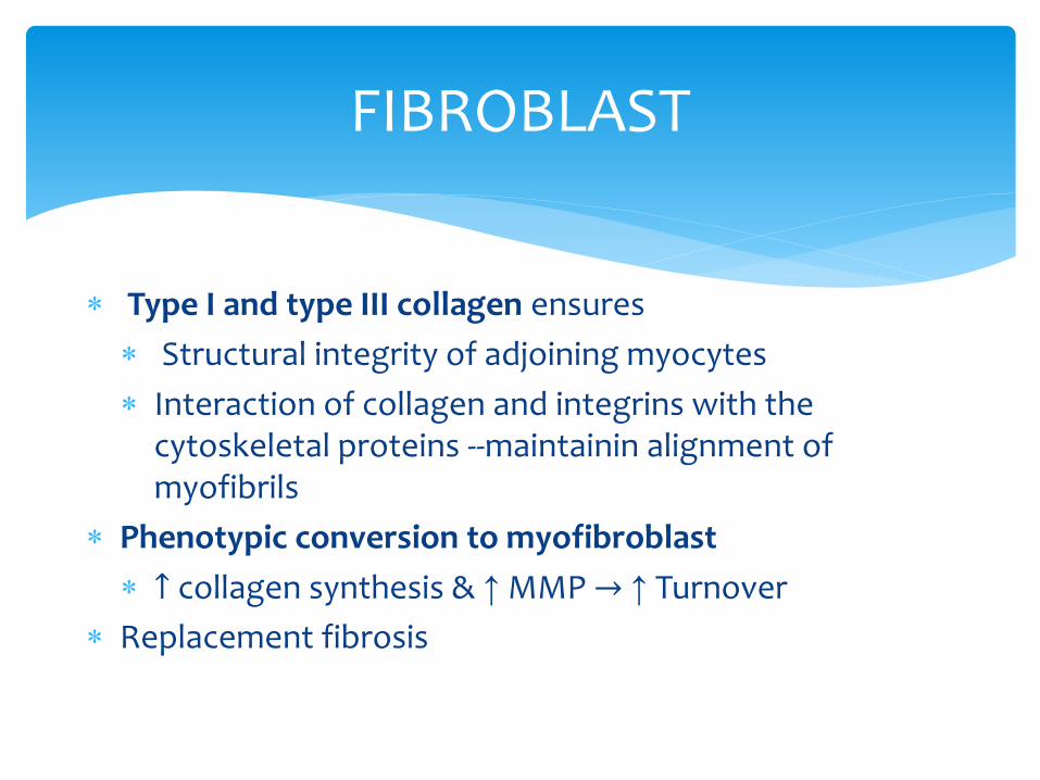
Type I and type III collagen ensures
Structural integrity of adjoining myocytes
Interaction of collagen and integrins with the cytoskeletal proteins --maintainin alignment of myofibrils
Phenotypic conversion to myofibroblast
↑ collagen synthesis & ↑ MMP → ↑ Turnover
Replacement fibrosis
FIBROBLAST
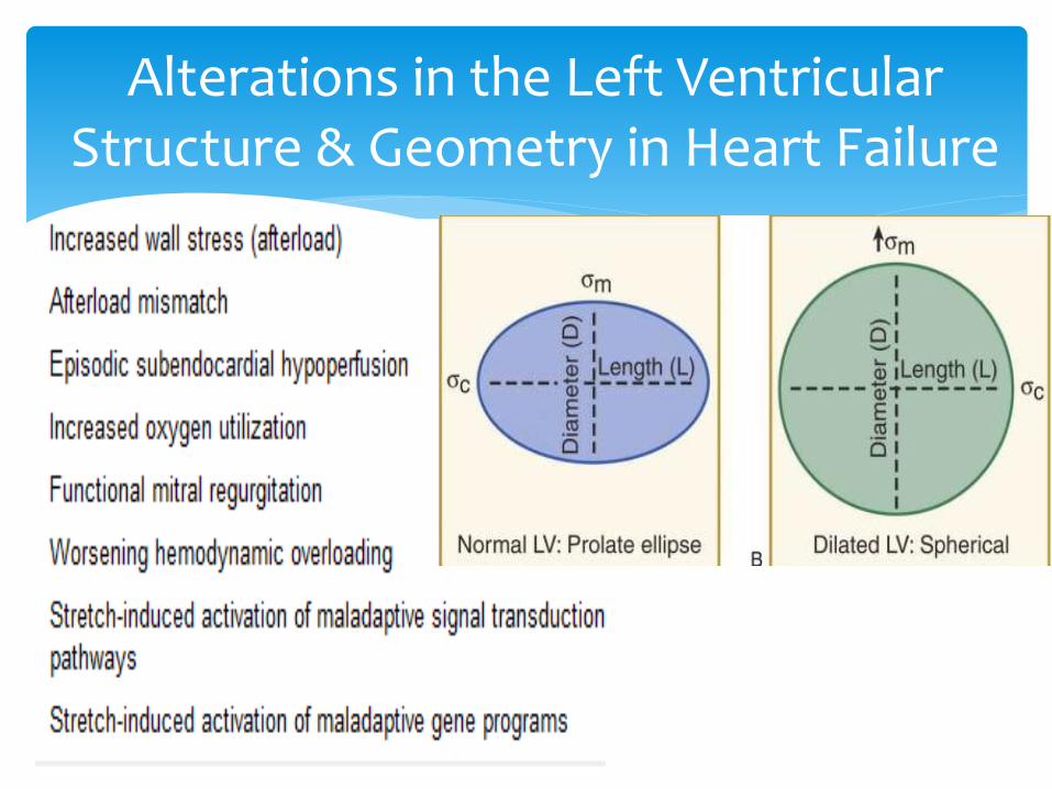
Alterations in the Left Ventricular Structure & Geometry in Heart Failure
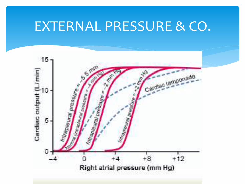
EXTERNAL PRESSURE & CO.
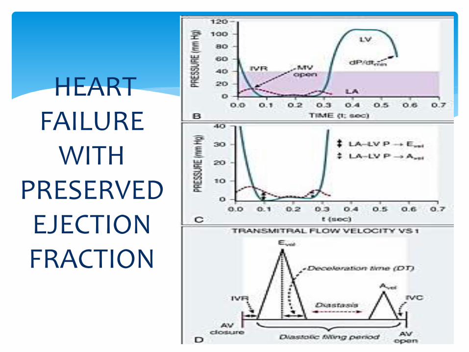
HEART FAILURE
WITH PRESERVED
EJECTIONFRACTION

Thank you