long-term enhancement of skeletal muscle mass and strength by
TRANSCRIPT
Long-term enhancement of skeletal muscle massand strength by single gene administrationof myostatin inhibitorsAmanda M. Haidet*†, Liza Rizo*, Chalonda Handy*†, Priya Umapathi*, Amy Eagle*, Chris Shilling*, Daniel Boue*,Paul T. Martin*†‡, Zarife Sahenk*†‡, Jerry R. Mendell*‡, and Brian K. Kaspar*†‡§
*The Research Institute, Nationwide Children’s Hospital, Columbus, OH 43205; and †Integrated Biomedical Science and ‡Neuroscience Graduate Programs,Ohio State University, Columbus, OH 43210
Edited by Joshua R. Sanes, Harvard University, Cambridge, MA, and approved February 5, 2008 (received for review September 25, 2007)
Increasing the size and strength of muscles represents a promisingtherapeutic strategy for musculoskeletal disorders, and interesthas focused on myostatin, a negative regulator of muscle growth.Various myostatin inhibitor approaches have been identified andtested in models of muscle disease with varying efficacies, depend-ing on the age at which myostatin inhibition occurs. Here, wedescribe a one-time gene administration of myostatin-inhibitor-proteins to enhance muscle mass and strength in normal anddystrophic mouse models for >2 years, even when delivered inaged animals. These results demonstrate a promising therapeuticstrategy that warrants consideration for clinical trials in humanmuscle diseases.
gene therapy � muscular dystrophy
Muscle-enhancing strategies have been proposed for a num-ber of neuromuscular disorders, including muscular dys-
trophies and age-related muscle disorders, and have shownpromising results to build or regenerate stronger, healthiermuscles (1). These strategies have mainly focused on the use oftrophic factors, such as insulin-like growth factor-1 that inducemyocyte precursor proliferation and myofiber hypertrophy (2).Attention has recently highlighted the potential benefit forinhibiting myostatin, resulting in the doubled muscle phenotypeof myostatin deficient cattle (3–5) and myostatin knockout mice(6, 7). Myostatin is a transforming growth factor-� (TGF-�)family member that plays a crucial role in regulating skeletalmuscle mass (8, 9). Myostatin appears to function in two distinctroles: to regulate the number of myofibers formed in develop-ment and to regulate the postnatal growth of muscles. Theregulation of muscle growth postnatally is being explored byvarious pharmacological methods for a number of muscle dis-orders. Delivery of neutralizing antibodies against myostatin hasshown promise in dystrophic mdx mice (10), yet there have beenvarying reports on the efficacy to enhance muscles when deliv-ered in aged animals (11). Furthermore, recent data demon-strated muscle mass enhancement and morphological recoveryin muscular dystrophy mice treated with deacetylase inhibitors.The resulting muscle enhancement was attributed to an increasein the protein follistatin, which has been shown in part to inhibitthe activity of myostatin (12). Trichostatin A (TSA) treatmentrequired daily administration and was not evaluated in agedanimals where off target effects may exist.
The identification of myostatin binding proteins capable ofregulating myostatin activity has led to potential new approachesfor postnatal muscle enhancement and expanded the potentialfor gene therapy to be considered as a method to inhibitmyostatin activity. Follistatin (FS) has been shown to bind tosome TGF-� family members and can function as a potentmyostatin antagonist. Overexpression of follistatin by transgenicapproaches in muscle has been shown to increase muscle growthin vivo (13), and a lack of follistatin results in reduced musclemass at birth (14). Recent data has also shown that follistatin is
capable of controlling muscle mass through pathways indepen-dent of the myostatin signaling cascade. In these studies, myo-statin knockout mice were crossed to mice carrying a follistatintransgene. The resulting mice had a quadrupling of muscle masscompared with the doubling of muscle mass that is observedfrom lack of myostatin alone, confirming a role for follistatin inthe regulation of muscle mass beyond solely myostatin inhibition(15). In addition to follistatin, two other proteins have beenidentified that are involved in the regulation of the myostatin.Follistatin-related gene (FLRG) is highly similar to follistatinand has been shown to inhibit activin and multiple bone mor-phogenic proteins in vitro (16, 17). Growth and differentiationfactor-associated serum protein-1 (GASP-1) is a protein that hasbeen discovered to contain multiple domains associated withprotease-inhibitor proteins and a domain homologous to the10-cysteine repeat found in follistatin. GASP-1 was shown tobind directly to the mature myostatin and myostatin propeptideand inhibits myostatin’s activity (18). Although recombinantprotein injections or myostatin blocking antibodies are feasiblestrategies, gene therapy to express these myostatin inhibitorgenes may prove a more efficacious therapeutic route fornumerous reasons, including the lack of potential immuneresponse to antibody treatment and the requirement for multipleinjections.
Here, we report that a one-time postnatal intramuscularinjection of adeno-associated virus (AAV) encoding myostatin-inhibitor-proteins resulted in long-term improvement of musclesize and strength in wild-type animals. Delivery of a myostatin-inhibitor-protein in dystrophic mdx animals reversed musclepathology and improved strength, even when administered in6.5-month-old animals. Specifically, we show here that follista-tin-344 resulted in the greatest effects on muscle size andfunction and was well tolerated with no untoward effects oncardiac pathology or reproductive capacity in either male orfemale treated animals.
Results and DiscussionAAV-mediated gene delivery to muscle provides a system togenerate high levels of protein in the target tissue or by a secretedproduct carried to remote sites through the circulation (19). Wecloned the known secreted myostatin-inhibiting genes, includinggrowth and differentiation factor-associated serum protein-1(GASP-1) (18), follistatin-related gene (FLRG) (17), and follista-
Author contributions: A.M.H., L.R., P.T.M., Z.S., J.R.M., and B.K.K. designed research;A.M.H., L.R., C.H., P.U., A.E., C.S., D.B., Z.S., J.R.M., and B.K.K. performed research; P.U.contributed new reagents/analytic tools; A.M.H., L.R., C.H., P.U., A.E., C.S., D.B., P.T.M., Z.S.,J.R.M., and B.K.K. analyzed data; and A.M.H., P.T.M., J.R.M., and B.K.K. wrote the paper.
The authors declare no conflict of interest.
This article is a PNAS Direct Submission.
§To whom correspondence should be addressed. E-mail: [email protected].
© 2008 by The National Academy of Sciences of the USA
4318–4322 � PNAS � March 18, 2008 � vol. 105 � no. 11 www.pnas.org�cgi�doi�10.1073�pnas.0709144105
tin-344 (FS) (13) into AAV serotype 1, which have demonstratedhigh muscle transduction capabilities. There are two isoforms offollistatin generated by alternative splicing. The FS-344 variantundergoes peptide cleavage to generate the FS-315 isoform and theother FS-317 variant produces the FS-288 isoform after peptidecleavage. We used the human FS-344 variant, which exclusivelygenerates the serum circulating FS-315 isoform of FS and includesa C-terminal acidic region (20). We chose FS-344 (FS), because theother FS-317 isoform, lacking the C terminus, shows preferentiallocalization to the ovarian follicular fluid and high tissue bindingaffinity through heparin sulfate proteoglycans, which may affectreproductive capacity and bind to other off-target sites (21). FS-288represents the membrane-bound form of follistatin (22), is a potentsuppressor of pituitary follicle stimulating hormone (23), is foundin the follicular fluid of the ovary and in the testes, and demon-strates a high affinity for the granulosa cells of the ovary.
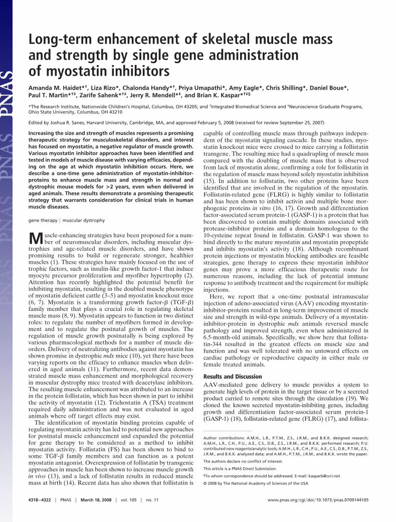
We sought to determine the efficacy of these proteins toincrease muscle mass in normal and dystrophic mice. We ad-ministered 1 � 1011 AAV1 viral particles per animal encodingFS, FLRG, GASP-1, or GFP bilaterally into the quadriceps andtibialis anterior muscles of 4-week-old wild-type C57BL/6 mice.All animals treated with the myostatin inhibitors demonstratedan increase in body mass with an observable gross enhancementof muscles when analyzed at 725-days of age compared withGFP-treated controls (Fig. 1 a and b). Evaluation of individualmuscle weights showed an increase in muscle mass for allmyostatin inhibitor-treated animals, with the greatest increase inFS-treated animals. The increased muscle mass was found in theinjected hindlimb muscles and remote muscles to the injectionsite, such as the triceps. Thus, these inhibitors were secreted intothe circulation from the site of muscle injection, enhancingskeletal muscle mass at remote sites (Fig. 1c). The enlargedmuscle mass was accompanied by functional improvement dem-onstrated by an increase in hindlimb grip strength (Fig. 1d).There was no effect on heart mass or histological appearance ofcardiomyocytes, indicating that myostatin inhibition was selec-tive to skeletal muscle tissue (data not shown). There has been
concern that FS adversely effects gonadal function. We found nochange in reproductive capacity in mice treated with our AAV1carrying the FS344 transgene (AAV1-FS, Table 1) Furthermore,we found no histological/pathological alterations in the gonadaltissue of FS treated-mice compared with controls (data notshown).
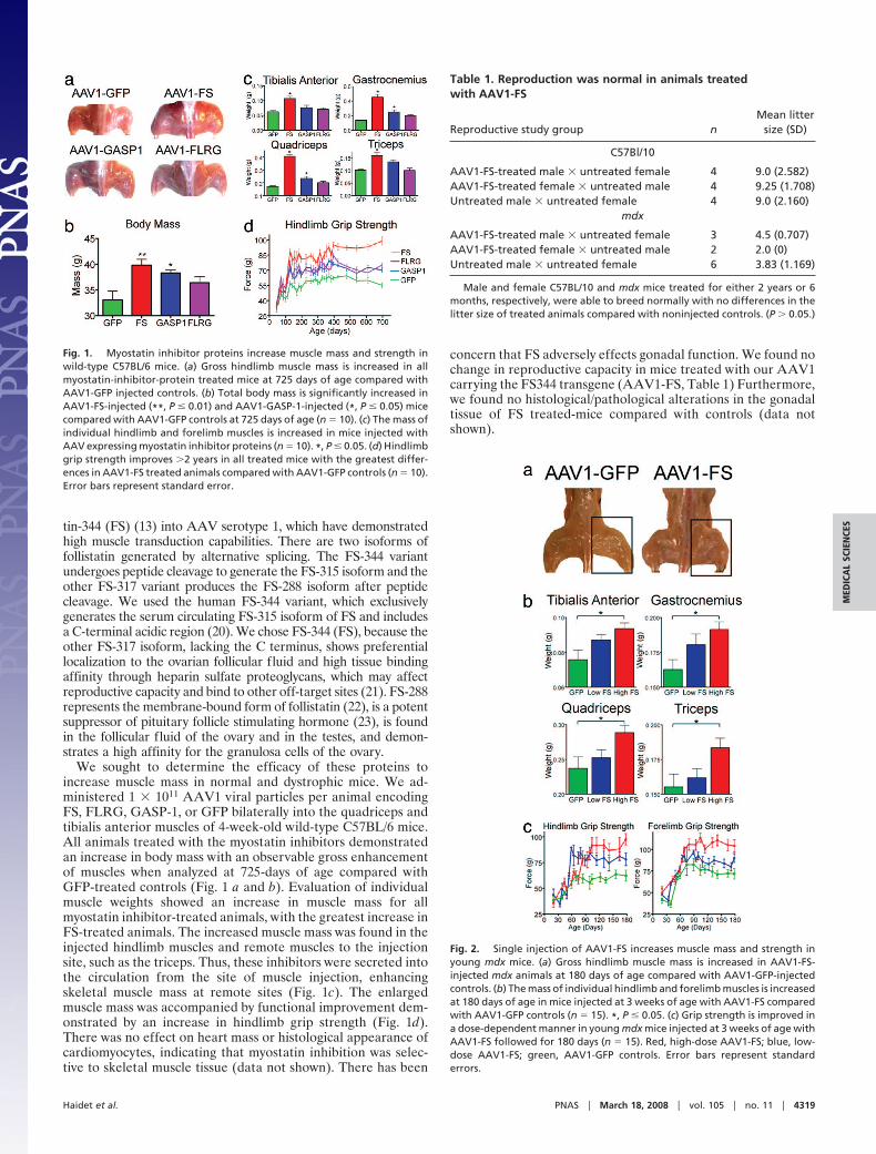
Fig. 2. Single injection of AAV1-FS increases muscle mass and strength inyoung mdx mice. (a) Gross hindlimb muscle mass is increased in AAV1-FS-injected mdx animals at 180 days of age compared with AAV1-GFP-injectedcontrols. (b) The mass of individual hindlimb and forelimb muscles is increasedat 180 days of age in mice injected at 3 weeks of age with AAV1-FS comparedwith AAV1-GFP controls (n � 15). *, P � 0.05. (c) Grip strength is improved ina dose-dependent manner in young mdx mice injected at 3 weeks of age withAAV1-FS followed for 180 days (n � 15). Red, high-dose AAV1-FS; blue, low-dose AAV1-FS; green, AAV1-GFP controls. Error bars represent standarderrors.
Fig. 1. Myostatin inhibitor proteins increase muscle mass and strength inwild-type C57BL/6 mice. (a) Gross hindlimb muscle mass is increased in allmyostatin-inhibitor-protein treated mice at 725 days of age compared withAAV1-GFP injected controls. (b) Total body mass is significantly increased inAAV1-FS-injected (**, P � 0.01) and AAV1-GASP-1-injected (*, P � 0.05) micecompared with AAV1-GFP controls at 725 days of age (n � 10). (c) The mass ofindividual hindlimb and forelimb muscles is increased in mice injected withAAV expressing myostatin inhibitor proteins (n � 10). *, P � 0.05. (d) Hindlimbgrip strength improves �2 years in all treated mice with the greatest differ-ences in AAV1-FS treated animals compared with AAV1-GFP controls (n � 10).Error bars represent standard error.
Table 1. Reproduction was normal in animals treatedwith AAV1-FS
Reproductive study group nMean litter
size (SD)
C57Bl/10
AAV1-FS-treated male � untreated female 4 9.0 (2.582)AAV1-FS-treated female � untreated male 4 9.25 (1.708)Untreated male � untreated female 4 9.0 (2.160)
mdx
AAV1-FS-treated male � untreated female 3 4.5 (0.707)AAV1-FS-treated female � untreated male 2 2.0 (0)Untreated male � untreated female 6 3.83 (1.169)
Male and female C57BL/10 and mdx mice treated for either 2 years or 6months, respectively, were able to breed normally with no differences in thelitter size of treated animals compared with noninjected controls. (P � 0.05.)
Haidet et al. PNAS � March 18, 2008 � vol. 105 � no. 11 � 4319
MED
ICA
LSC
IEN
CES
Given the robust effects of FS delivery, we next tested thepotential for AAV1-FS delivered postnatally in a clinicallymeaningful paradigm to increase muscle mass and strength anddelay muscle deterioration in the mdx mouse model of Duchennemuscular dystrophy (DMD). DMD is an X-linked recessivedisease resulting in the wasting of skeletal muscles and cardiacfunction, ultimately resulting in death. Recently, FS was inves-tigated in mdx animals overexpressing a duplicated domain ofthe follistatin gene. Results demonstrated increased muscle massand attenuated pathology, although the results were only doc-umented to 15 weeks of age (24). In our studies, mdx animalswere injected bilaterally in the quadriceps and tibialis anteriormuscles with a low (1 � 1010 viral particles) or high dose (1 �1011 viral particles) of AAV1-FS at 3 weeks of age and followedfor 5 months before necropsy. Increased levels of circulating FSwere detected in the serum of both low and high dose treatedanimals with the high dose expressing the greatest levels ofserum detected FS (high dose, 15.3 � 2.1 ng/ml; low dose, 6.8 �0.4 ng/ml; GFP controls, 0 � 0.1 ng/ml; n � 8 per group; P �0.01). We demonstrated that AAV1-FS increased body masscompared with GFP treated controls, with the greatest increasein the high dose FS group (data not shown). Gross observationof AAV1-FS treated mice displayed a significant increase inmuscle size compared with AAV1-GFP treated animals (Fig.2a), with the greatest individual muscle weight increase in highdose FS-treated animals (Fig. 2b). Effects were not restricted tothe injected muscles; they were also found at sites remote fromdirectly targeted muscles (Fig. 2b). Increased muscle masstranslated to a dose-dependent improvement in muscle strengthin the hindlimbs and forelimbs of treated animals compared withGFP treated controls (Fig. 2c). Histological and morphometricanalyses of AAV1-FS injected muscles and at remote sitesdemonstrated myofiber hypertrophy, supporting gross observa-tions made at the time of necropsy (Fig. 3 a–c). Furthermore,there was no shift in muscle fiber types in AAV-FS treatedanimals; however, there were fewer total fibers per squaremillimeter of area in the tibialis anterior muscle in animalstreated with the high dose AAV-FS (Fig. 3 d and e). Strikingly,FS-treated mice demonstrated a significant reduction in serum
creatine kinase compared with GFP-treated controls (Fig. 4a).This is of interest, because FS was protective despite its lack ofcorrection of the underlying dystrophin deficiency. The exactmechanism is not clear, but one might speculate that increasingthe strength of individual fibers makes them less susceptible todamage from the stress of normal activities. The involvement ofsatellite cells in postnatal myostatin inhibition remains to be fullyresolved; however, we did not see a statistical change in musclesatellite cell markers for FS-treated animals (data not shown).
We also evaluated the potential for AAV1-FS to increasemuscle strength in mdx animals when treated at an older age. Wefound that AAV1-FS injection at 210 days of age increasedmuscle strength �60 days after administration and that theincreased strength persisted long-term throughout the 560 daysevaluated in this study (Fig. 4b). As early as 180 days of age,before AAV1-FS treatment, there was evident pathology inmuscles of untreated mdx animals, with prominent endomysialconnective tissue proliferation and inflammation (Fig. 4 c and d).Pathological evaluation of gastrocnemius and diaphragm mus-cles at 560 days of age demonstrated that AAV1-FS treatedanimals had substantially fewer focal groups of necrotic musclefibers and mononuclear cell infiltrates. Importantly, AAV1-FStreated animals had significantly reduced focal areas of endomy-sial connective tissue proliferation, which were pronounced inGFP treated animals, demonstrating that fibrosis, a hallmark ofmuscular dystrophy, was decreased in FS-treated animals (Fig.4c). Pathology in the diaphragm also showed that FS-treatmentreduced inflammation and fatty replacement compared withGFP-treated animals (Fig. 4d). Furthermore, AAV1-FS treat-ment demonstrated significant increases in muscle fiber diam-eters at this age compared with control GFP-treated animals(Fig. 4 c and d). These results demonstrated that myostatininhibition by FS treatment was beneficial in aged mdx animalsthat had undergone multiple rounds of muscle degeneration andregeneration. Translation to a clinical parallel suggests thatAAV-mediated FS gene therapy could have potential for theolder DMD patient independent of replacing the missing geneand may have a potential role in combinational therapy similarto that demonstrated for IGF-1 and minidystrophin gene re-placement (25).
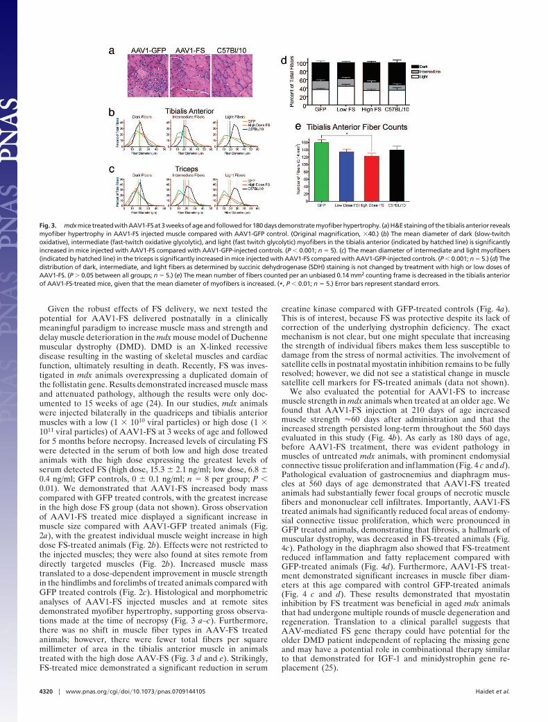
Fig. 3. mdxmicetreatedwithAAV1-FSat3weeksofageandfollowedfor180daysdemonstratemyofiberhypertrophy. (a)H&Estainingof thetibialis anterior revealsmyofiber hypertrophy in AAV1-FS injected muscle compared with AAV1-GFP control. (Original magnification, �40.) (b) The mean diameter of dark (slow-twitchoxidative), intermediate (fast-twitch oxidative glycolytic), and light (fast twitch glycolytic) myofibers in the tibialis anterior (indicated by hatched line) is significantlyincreased in mice injected with AAV1-FS compared with AAV1-GFP-injected controls. (P � 0.001; n � 5). (c) The mean diameter of intermediate and light myofibers(indicated by hatched line) in the triceps is significantly increased in mice injected with AAV1-FS compared with AAV1-GFP-injected controls. (P � 0.001; n � 5.) (d) Thedistribution of dark, intermediate, and light fibers as determined by succinic dehydrogenase (SDH) staining is not changed by treatment with high or low doses ofAAV1-FS. (P � 0.05 between all groups; n � 5.) (e) The mean number of fibers counted per an unbiased 0.14 mm2 counting frame is decreased in the tibialis anteriorof AAV1-FS-treated mice, given that the mean diameter of myofibers is increased. (*, P � 0.01; n � 5.) Error bars represent standard errors.
4320 � www.pnas.org�cgi�doi�10.1073�pnas.0709144105 Haidet et al.
These results suggest that inhibition of myostatin by FS-344,delivered by a single AAV1 injection can enhance muscle sizeand strength and is well tolerated for �2-years. The results ofFS344 may offer a more powerful strategy than others target-ing solely myostatin because of additive effects, such as
follistatin’s involvement in multiple signaling pathways, andthe recent finding demonstrating a reduction in inf lammationin a model of endotoxemia (15, 26). The striking ability of FSto provide gross and functional long-term improvement todystrophic muscles in aged animals warrants its considerationfor clinical development to treat musculoskeletal diseases,including older DMD patients.
Materials and MethodsAnimals. C57BL/6, C57BL/10, and C57BL/10ScSn-DMDmdx/J were purchasedfrom The Jackson Laboratory. All studies were approved by InstitutionalAnimal Care and Use Committee.
Cloning and AAV Production. The cDNA for human follistatin-344 (FS) wasobtained from Origene, follistatin-related gene (FLRG) was obtained fromAmerican Type Culture Collection, and growth and differentiation factor-associated serum protein 1 (GASP-1) was cloned from a human cDNA library(Clontech). Recombinant AAV serotype 1 vectors were produced by a contractmanufacturing company (Virapur).
AAV Injections and Testing. Mice received bilateral intramuscular injections ofa total dose of 1 � 1011 viral particles (high � 1 � 1011, low � 1 � 1010) (n �10–15 per group) at 3–4 weeks of age or at 6.5 months of age. Muscle strengthwas assessed weekly, using a grip strength meter (27). Force measurementswere recorded in three separate trials and averaged. Mouse coordination wastested by using the accelerating rotarod (Columbus Instruments).
Histological Analysis. Muscles were dissected, weighed, snap-frozen in liquidnitrogen-cooled isopentane, cryostat sectioned, and stained by hematox-ylin-eosin (H&E) or succinic dehydrogenase (SDH) for analysis of fiberdiameters. Five animals per group were chosen randomly for muscle fibersize morphometry. For each analysis, five representative pictures (onecentral and four peripheral) were taken of muscle sections, compoundingto 0.7 mm2. Images were captured at �20 magnification, and diameterswere measured with a calibrated micrometer, using the AxioVision 4.2software (Zeiss). Fiber size-distribution histograms were generated andexpressed as percentage of total fibers analyzed.
Creatine Kinase and Follistatin Assay. Serum CK was performed by using a CKtest kit (Pointe Scientific) and expressed as units/liter. Serum was collected at90 days after injection and assayed by using the human follistatin quantikineELISA kit (R&D Systems) with normalization to controls.
Statistical Analysis. All statistical analysis was performed in Graph Pad Prizmsoftware, using one- and two-way ANOVA with Bonferroni post hocanalysis.
ACKNOWLEDGMENTS. We thank Kailin Liu for technical assistance. This workwas supported by Project A.L.S. (to B.K.K.), The Myositis Association (J.R.M.and B.K.K.), Roger Stevens, and a National Research Service Award fellowship(to A.M.H.).
1. Engvall E, Wewer, UM (2003) The new frontier in muscular dystrophy research: Boostergenes. FASEB J 17:1579–1584.
2. Barton ER, Morris L, Musaro A, Rosenthal N, Sweeney HL (2002) Muscle-specificexpression of insulin-like growth factor I counters muscle decline in mdx mice. J CellBiol 157:137–148.
3. Grobet L, et al. (1997) A deletion in the bovine myostatin gene causes the double-muscled phenotype in cattle. Nat Genet 17:71–74.
4. Kambadur R, Sharma M, Smith TP, Bass JJ (1997) Mutations in myostatin (GDF8)in double-muscled Belgian blue and Piedmontese cattle. Genome Res 7:910–916.
5. McPherron AC, Lee SJ (1997) Double muscling in cattle due to mutations in themyostatin gene. Proc Natl Acad Sci USA 94:12457–12461.
6. Wagner KR, Liu X, Chang X, Allen RE (2005) Muscle regeneration in the prolongedabsence of myostatin. Proc Natl Acad Sci USA 102:2519–2524.
7. Wagner KR, McPherron AC, Winik N, Lee SJ (2002) Loss of myostatin attenuates severityof muscular dystrophy in mdx mice. Ann Neurol 52:832–836.
8. McPherron AC, Lawler AM, Lee SJ (1997) Regulation of skeletal muscle mass in mice bya new TGF-beta superfamily member. Nature 387:83–90.
9. Lee SJ (2004) Regulation of muscle mass by myostatin. Annu Rev Cell Dev Biol 20:61–86.10. Bogdanovich S, et al. (2002) Functional improvement of dystrophic muscle by myosta-
tin blockade. Nature 420:418–421.11. Parsons SA, Millay DP, Sargent MA, McNally EM, Molkentin JD (2006) Age-dependent
effect of myostatin blockade on disease severity in a murine model of limb–girdlemuscular dystrophy. Am J Pathol 168:1975–1985.
12. Minetti GC, et al. (2006) Functional and morphological recovery of dystrophic musclesin mice treated with deacetylase inhibitors. Nat Med 12:1147–1150.
13. Lee SJ, McPherron AC (2001) Regulation of myostatin activity and muscle growth. ProcNatl Acad Sci USA 98:9306–9311.
14. Matzuk MM, et al. (1995) Multiple defects and perinatal death in mice deficient infollistatin. Nature 374:360–363.
15. Lee SJ (2007) Quadrupling muscle mass in mice by targeting TGF-beta signalingpathways. PLoS ONE 2:e789.
16. Hayette S, et al. (1998) FLRG (follistatin-related gene), a new target of chromosomalrearrangement in malignant blood disorders. Oncogene 16:2949–2954.
17. Tsuchida K, et al. (2000) Identification and characterization of a novel follistatin-like protein as a binding protein for the TGF-beta family. J Biol Chem 275:40788 –40796.
18. Hill JJ, Qiu Y, Hewick RM, Wolfman NM (2003) Regulation of myostatin in vivo bygrowth and differentiation factor-associated serum protein-1: A novel protein withprotease inhibitor and follistatin domains. Mol Endocrinol 17:1144–1154.
19. Mendell JR, Clark KR (2006) Risks, benefits, and consent in the age of gene therapy.Neurology 66:964–965.
20. Shimasaki S, et al. (1988) Primary structure of the human follistatin precursor and itsgenomic organization. Proc Natl Acad Sci USA 85:4218–4222.
21. Lin SY, Morrison JR, Phillips DJ, de Kretser DM (2003) Regulation of ovarianfunction by the TGF-beta superfamily and follistatin. Reproduction 126:133–148.
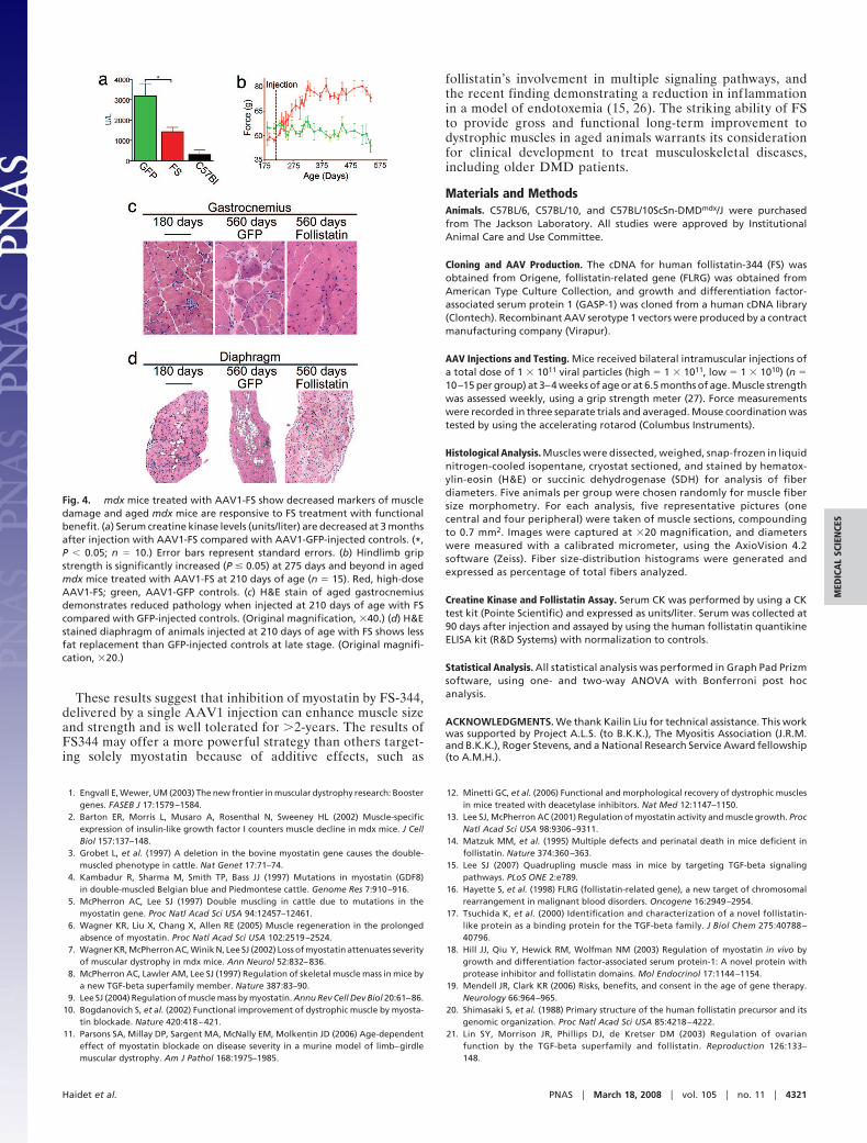
Fig. 4. mdx mice treated with AAV1-FS show decreased markers of muscledamage and aged mdx mice are responsive to FS treatment with functionalbenefit. (a) Serum creatine kinase levels (units/liter) are decreased at 3 monthsafter injection with AAV1-FS compared with AAV1-GFP-injected controls. (*,P � 0.05; n � 10.) Error bars represent standard errors. (b) Hindlimb gripstrength is significantly increased (P � 0.05) at 275 days and beyond in agedmdx mice treated with AAV1-FS at 210 days of age (n � 15). Red, high-doseAAV1-FS; green, AAV1-GFP controls. (c) H&E stain of aged gastrocnemiusdemonstrates reduced pathology when injected at 210 days of age with FScompared with GFP-injected controls. (Original magnification, �40.) (d) H&Estained diaphragm of animals injected at 210 days of age with FS shows lessfat replacement than GFP-injected controls at late stage. (Original magnifi-cation, �20.)
Haidet et al. PNAS � March 18, 2008 � vol. 105 � no. 11 � 4321
MED
ICA
LSC
IEN
CES

22. Sugino K, et al. (1993) Molecular heterogeneity of follistatin, an activin-bindingprotein: Higher affinity of the carboxyl-terminal truncated forms for heparan sulfateproteoglycans on the ovarian granulosa cell. J Biol Chem 268:15579–15587.
23. Inouye S, et al. (1991) Recombinant expression of human follistatin with 315 and 288amino acids: Chemical and biological comparison with native porcine follistatin.Endocrinology 129:815–822.
24. Nakatani M, et al. (2008) Transgenic expression of a myostatin inhibitor derived fromfollistatin increases skeletal muscle mass and ameliorates dystrophic pathology in mdxmice. FASEB J 22:477–487.
25. Abmayr S, Gregorevic P, Allen JM, Chamberlain JS (2005) Phenotypic improvement ofdystrophic muscles by rAAV/microdystrophin vectors is augmented by Igf1 codelivery.Mol Ther 12:441–450.
26. Jones KL, et al. (2007) Activin A is a critical component of the inflammatory response,and its binding protein, follistatin, reduces mortality in endotoxemia. Proc Natl AcadSci USA 104:16239–16244.
27. Miller TM, et al. (2006) Gene transfer demonstrates that muscle is not a primary targetfor non-cell-autonomous toxicity in familial amyotrophic lateral sclerosis. Proc NatlAcad Sci USA 103:19546–19551.
4322 � www.pnas.org�cgi�doi�10.1073�pnas.0709144105 Haidet et al.