long-range structural effects of a charcot–marie– …long-range structural effects of a...
TRANSCRIPT
Long-range structural effects of a Charcot–Marie–Tooth disease-causing mutation in humanglycyl-tRNA synthetaseWei Xie, Leslie A. Nangle, Wei Zhang, Paul Schimmel*, and Xiang-Lei Yang*
The Skaggs Institute for Chemical Biology and Department of Molecular Biology, The Scripps Research Institute, BCC-379, 10550 North Torrey Pines Road,La Jolla, CA 92037
Contributed by Paul Schimmel, April 27, 2007 (sent for review April 23, 2007)
Functional expansion of specific tRNA synthetases in higher or-ganisms is well documented. These additional functions may ex-plain why dominant mutations in glycyl-tRNA synthetase (GlyRS)and tyrosyl-tRNA synthetase cause Charcot–Marie–Tooth (CMT)disease, the most common heritable disease of the peripheralnervous system. At least 10 disease-causing mutant alleles of GlyRShave been annotated. These mutations scatter broadly across theprimary sequence and have no apparent unifying connection. Herewe report the structure of wild type and a CMT-causing mutant(G526R) of homodimeric human GlyRS. The mutation is at the sitefor synthesis of glycyl-adenylate, but the rest of the two structuresare closely similar. Significantly, the mutant form diffracts to ahigher resolution and has a greater dimer interface. The extradimer interactions are located �30 Å away from the G526R muta-tion. Direct experiments confirm the tighter dimer interaction ofthe G526R protein. The results suggest the possible importance ofsubtle, long-range structural effects of CMT-causing mutations atthe dimer interface. From analysis of a third crystal, an appendedmotif, found in higher eukaryote GlyRSs, seems not to have a rolein these long-range effects.
crystal structure � structure-function analysis � dimer interface �inherited peripheral neuropathy � aminoacyl tRNA synthetase
Charcot–Marie–Tooth (CMT) diseases are the most commonheritable disorder of the peripheral nervous system, having a
frequency of occurrence of �1 in 2,500 (1). Although differentgenetic loci associated with the disease have been identified, someof them have provided no obvious explanation for the neuropatho-logical and electrophysiological phenotypes that have been ob-served (2). This lack of understanding may mean that proteinsthought previously to be associated only with non-neuron-specificactivities are part of a broader systems biology where connectionsare made between pathways and activities usually regarded asdiscrete and isolated. For example, CMT-causing dominant muta-tions occur in two of the genes coding for human aminoacyl tRNAsynthetases: YARS [gene for tyrosyl-tRNA synthetase (TyrRS)] (3)and GARS [gene for glycyl-tRNA synthetase (GlyRS)] (4–9). Theseenzymes are representatives of the aminoacyl tRNA synthetasefamily of proteins, which catalyze the first step of protein synthesis,i.e., aminoacylation of tRNAs with their cognate amino acids. Theyare the only two of the 20 tRNA synthetases presently known to becausally linked to CMT disease.
Although GlyRS is an uncommon example of a human tRNAsynthetase, having a single gene encoding both cytoplasmic andmitochondrial forms, the two forms of TyrRS are encoded byseparate genes. CMT-causing mutations are found only in the genefor cytoplasmic TyrRS (3). Thus, even though neuropathies asso-ciated with mitochondrial disorders are known, it seems unlikelythat the CMT disease connection to these two tRNA synthetases isthrough effects on mitochondrial protein synthesis. Alternatively,because several human aminoacyl tRNA synthetases (includinghuman TyrRS) have expanded functions that enable them toparticipate in cell signaling networks (10), these expanded functions
have raised the possibility of tRNA synthetase connections with thenervous system through pathways beyond aminoacylation.
Considering that at least 10 different mutations in GlyRS havebeen linked to CMT disease (4–9), and because the structure of thisenzyme was not known, we were motivated to determine itsstructure and see whether that structure would offer clues into theetiology of the disease. We focused on obtaining crystals ofwild-type and a CMT-causing mutant of cytoplasmic GlyRS, be-cause none of the CMT mutations fall in the mitochondria-specificN-terminal extension. In addition, the cytoplasmic version is easierto express and isolate than the more extended mitochondrial form.GlyRS is one of the 10 members of the class II tRNA synthetasesand, like most members of that class, are �2 dimers. Although thestructure of a bacterial ortholog (Thermus thermophilus) of humanGlyRS is known (11, 12), the latter enzyme has limited sequenceidentity with that ortholog and, in addition, has N- and C-terminalextensions and three insertions not present in the bacterial GlyRSstructure. The distribution of the 10 CMT disease mutations on thesequence of human GlyRS, and rough placement of these muta-tions on the structure of T. thermophilus GlyRS, showed that themutations were widely dispersed across the sequence and structureand, therefore, had no unifying theme to connect them together.Thus, in obtaining the crystal structure of human GlyRS, we wereespecially motivated to look for characteristics in the structure of amutant form that could, in principle, suggest some sort of unifyingconnection between at least some of the CMT disease mutations.
ResultsStructures Determined for Wild-Type and the CMT-Causing MutantG526R GlyRS. Crystals of the wild-type human GlyRS were obtainedearlier (13). These crystals had a modest diffraction quality, and thecollected data did not render a successful structure solution bymolecular replacement, using the structure of the orthologous T.thermophilus GlyRS as the search model. Efforts were then devotedto improving the crystal quality of the wild-type enzyme and tocrystallizing a specific CMT-causing mutant protein. Interestingly,the G526R mutant, under slightly different crystallization condi-tions compared with that used for the wild-type enzyme, yieldedcrystals that diffracted to 2.85 Å and with good data quality (Table1). These crystals, and those formed with the wild-type enzyme, hadthe same space group, P43212, with each asymmetric unit contain-ing one subunit of the dimeric enzyme. Using molecular replace-
Author contributions: P.S. and X.-L.Y. designed research; W.X., L.A.N., and W.Z. performedresearch; W.X., L.A.N., W.Z., P.S., and X.-L.Y. analyzed data; and P.S. and X.-L.Y. wrote thepaper.
The authors declare no conflict of interest.
Abbreviations: CMT, Charcot–Marie–Tooth; GlyRS, glycyl-tRNA synthetase; TyrRS, tyrosyl-tRNA synthetase.
Data deposition: The atomic coordinates have been deposited in the Protein Data Bank,www.pdb.org [PDB ID codes 2PME (for wild-type GlyRS) and 2PMF (for G526R GlyRS)].
*To whom correspondence may be addressed. E-mail: [email protected] [email protected].
© 2007 by The National Academy of Sciences of the USA
9976–9981 � PNAS � June 12, 2007 � vol. 104 � no. 24 www.pnas.org�cgi�doi�10.1073�pnas.0703908104
Dow
nloa
ded
by g
uest
on
Apr
il 15
, 202
0
ment with the structure of T. thermophilus GlyRS, this data setallowed us to solve the structure of the G526R mutant enzyme.Subsequently, the structure of wild-type GlyRS was solved at 2.9 Åusing, in this instance, molecular replacement with the newlyobtained structure of the G526R mutant enzyme.
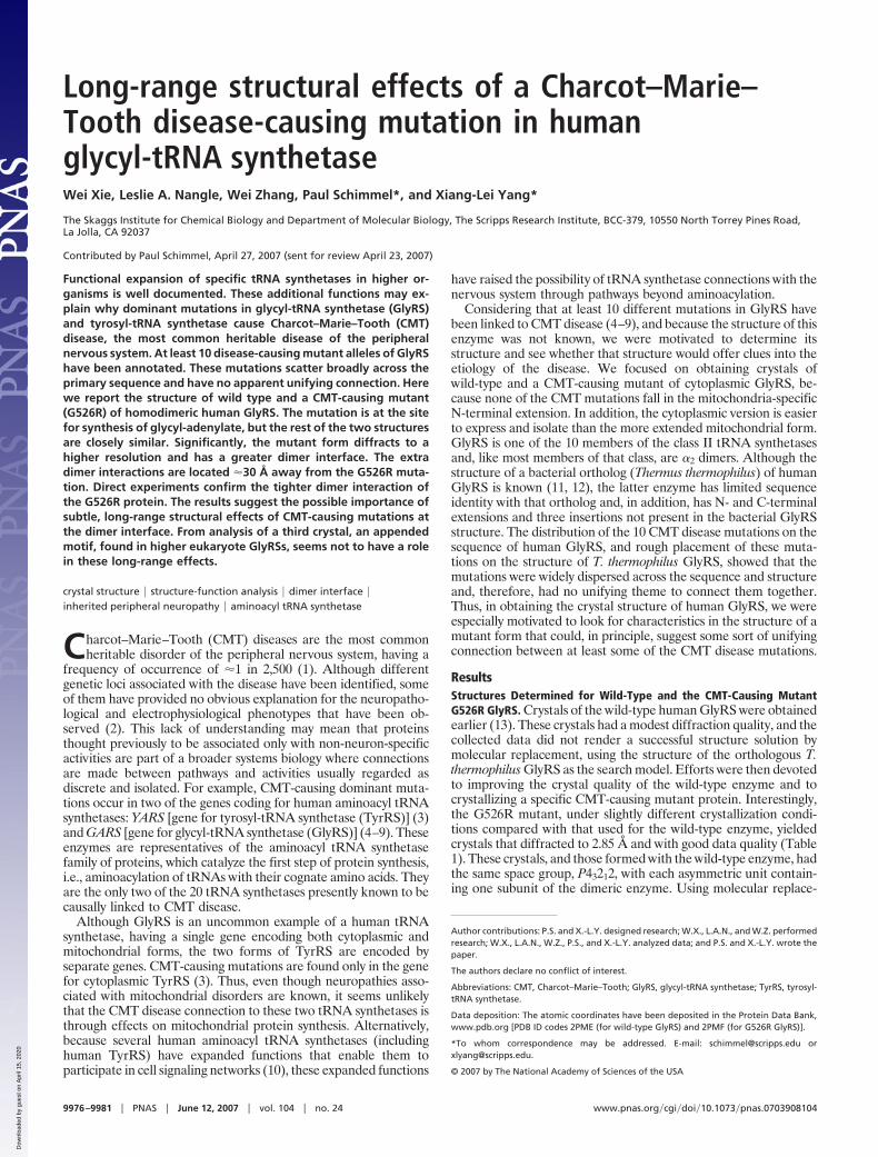
Overall Structural Organization of Human GlyRS. The cytoplasmichuman GlyRS has 685 residues composed of an N-terminal WHEP-TRS domain (M1–D62) that is unique for GlyRS from Metazoans,a catalytic domain (D63–V556), and a C-terminal anticodon bind-ing domain (V557–E685). The catalytic domain has the typical core(for class II enzymes) of an extended �-sheet and the threeconserved motifs, consisting of a helix–strand (motif 1), strand–loop–strand (motif 2), and strand–helix (motif 3), which are char-acteristic of all class II tRNA synthetases. In addition, threeinsertions split the catalytic domain. In comparison to other class IItRNA synthetases, insertion I (F144–N225, between motifs 1 and2) is unique to and shared by GlyRSs through evolution (14).Insertion II (D307–N348, after motif 2) and insertion III (V440–V504, before motif 3) are specific to eukaryotic GlyRSs (15). Exceptfor the disordering of residues E382–Y386 and A421–V504, whichcover the entire insertion III, and of residues D545–E546, ourstructure of wild-type GlyRS resolved most of the catalytic domainincluding insertions I and II (Fig. 1A). The N-terminal WHEP-TRSdomain is completely disordered, whereas the C-terminal antico-don binding domain is well resolved except for 11 residues at the Cterminus (G675–E685).
Interestingly, whereas insertion I is well resolved in humanGlyRS, it was not resolved in the structure of T. thermophilus GlyRS(11, 12). Apart from the absence of insertions II and III in T.thermophilus GlyRS, the overall structure of human GlyRS issimilar to that of its bacterial ortholog. The rmsd between the twostructures is 1.9 Å for 400 C�s with a similarity Z-score of 36.9 (16).
Fig. 2 gives the structure-based sequence alignment of the humanand T. thermophilus GlyRSs. Insertion I of the catalytic domaincontains a compact three-helix bundle (�4–�5–�6) that is flankedat one end by a three-strand antiparallel �-sheet (�5–�6–�7). Thisinsertion forms a domain-like entity that is virtually separated fromthe rest of the catalytic domain (Fig. 1A) and protrudes itself away
from the dimer interface (Fig. 1B). Insertion I is likely to be involvedin acceptor stem recognition (W.X., unpublished data).
The eukaryote-specific insertion II associates closely with thecatalytic core by extending the central seven-strand �-sheet, byadding �12 and �13, to give a nine-strand �-sheet. The spatiallocation of this insertion suggests that it contributes to neithersubstrate binding nor dimerization (Fig. 1B). However, the almostcompletely disordered insertion III could be involved with tRNAbinding across the dimer interface and make contact with theanticodon stem or variable or D-loops (W.X., unpublished data). Inthat connection, mammalian GlyRS is more discriminating than theT. thermophilus enzyme in that T. thermophilus GlyRS chargestRNAGly from both bacteria and eukaryotes, whereas mammalianGlyRS can charge only eukaryotic tRNAGly (17). Thus, insertion IIIcould provide additional tRNA recognition that is eukaryote-specific.
Compared with T. thermophilus GlyRS, human GlyRS has 27additional residues at the C terminus (Fig. 2). Most of these areresolved, with the first part folding into helix �18 and a short strand�27. The final 11 residues are disordered, with more than half ofthem being either positively or negatively charged. In our tRNAcomplex model (W.X., unpublished data), the extra C-terminalsequences bring the charged C terminus into a space that it couldinteract with tRNA at the anticodon and thus provide additionaltRNA binding and recognition capacity to the anticodon-bindingdomain.
The disordered N-terminal WHEP domain is predicted to havea helix–turn–helix structure, as shown for human TrpRS (18) andfor human Glu-ProRS (19). The WHEP domain is present inseveral higher eukaryotic tRNA synthetases in different configu-rations, for example, as an N-terminal appended domain (as inGlyRS or TrpRS), as part of the linker that fuses together the twosynthetases of the novel Glu-ProRS, and as a C-terminal appendeddomain (as in MetRS) (20). The domain usually is flexibly linkedto the core enzyme and, probably for that reason, tends to bedisordered in crystal structures, if not provided with crystal latticeinteractions. We found that the WHEP domain of human GlyRScould be clipped off by limited protease digestion (data not shown).The resulting truncated protein was crystallized. Interestingly, thesecrystals have the same lattice and space group parameters as thoseof the crystals for full-length GlyRS. This observation suggested
Table 1. Data collection and refinement statistics
Wild type G526R
Space group P43212 P43212Molecules in asymmetric unit 1 1Data collection
Resolution, Å 50.0–2.90 (3.0–2.9) 50.0–2.85 (2.95–2.85)a, Å 91.74 91.41c, Å 247.18 246.81Wavelength, Å 0.97945Rsym,* % 9.9 (52.4) 6.6 (37.0)I/�(I) 17.6 (3.1) 36.6 (7.7)Redundancy 8.9 (7.8) 13.8 (14.0)No. of unique reflections 18,765 25,080Completeness, % 77.2 (80.2) 98.7 (98.5)
RefinementResolution, Å 36.8–2.90 29.2–2.85No. of reflections used for
refinement17,766 23,727
No. of reflections used fortest set
961 1,238
Final R, % 23.5 23.1Final Rfree,† % 27.0 27.1rmsd bonds, Å 0.003 0.003rmsd angles, Å 0.756 0.847Mean B value, Å2 55.0 58.2
Values in parentheses refer to the highest-resolution shell.*Rmerge � [�h�iI i(h) � �I(h)� / �h�iIi(h)] � 100.†Calculated from 5% of the reflections that were excluded before refinement.
90
Anticodon Binding Domain
Insertion II
Catalytic Domain
N
C
Insertion I
Insertion III (disordered)
G526
A B
β8 β9
β8'β9'
G526
Μ1
Μ3Μ2
Fig. 1. Overall structure of human GlyRS. (A) ‘‘Front’’ view of the asymmetricunit that contains one subunit of the dimeric enzyme. Different domains andinsertions of the subunit are colored as indicated. The three conserved se-quence motifs (M1, M2, and M3) in the catalytic domain are colored magenta,pink, and orange, respectively. (B) A 90° rotation along the x axis to view theinterface between two subunits of the GlyRS dimer. The second subunit isgenerated by symmetry operation and is not colored. The �8–�9 hairpins arelabeled on both subunits to show their interaction on top of the dimerinterface.
Xie et al. PNAS � June 12, 2007 � vol. 104 � no. 24 � 9977
BIO
CHEM
ISTR
Y
Dow
nloa
ded
by g
uest
on
Apr
il 15
, 202
0
that the absence of the WHEP domain does not likely cause aconformational change in the rest of the enzyme. [A similarobservation was made with human TrpRS. And like what wasobserved for TrpRS (21), deletion of the WHEP domain in GlyRSdoes not affect aminoacylation activity (data not shown).] Inaddition, because exactly the same crystals were obtained with bothfull-length and WHEP-truncated GlyRS, the WHEP domain itselfmust make little or no contribution to crystal lattice formation.These observations are consistent with this extra domain beingcompletely disordered in the structure of wild-type GlyRS shownhere.
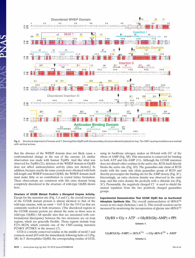
Structure of G526R Mutant Predicts a Disrupted Enzyme Activity.Except for the mutation site (Fig. 3 A and C), the overall structureof the G526R mutant protein is almost identical to that of thewild-type enzyme, with an rmsd � 0.65 Å for the 519 C�s that arecommonly resolved in both structures. (The disordered regions inthe G526R mutant protein are almost the same as those seen inwild-type GlyRS.) All specific sites that are associated with con-formational discrepancy between the two structures are on loopregions, which are generally flexible. Those regions include loopF231–M238, which contains one of the CMT-causing mutationP234KY (P278KY in the mouse) (7).
G526 is a strictly conserved residue in the middle of motif 3 andconnects strand �19 with the immediately following helix �13 (Fig.3B). In T. thermophilus GlyRS, the corresponding residue of G526,
using its backbone nitrogen, makes an H-bond with O2� of theribose of AMP (Fig. 3B). This interaction is conserved for bindingto both ATP and Gly-AMP (11). Although the G526R mutationdoes not disturb other residues in the active site, R526 itself directlyblocks the active site (Fig. 3D). The guanadino side chain of R526stacks with the same side chain guanadino group of R529 andthereby preoccupies the binding site for the AMP moiety (Fig. 3C).Interestingly, an extra electron density was observed in the omitmap, and this extra density fits perfectly with a chloride ion (Fig.3C). Presumably, the negatively charged Cl� is used to shield themutual repulsion from the two positively charged guanadinogroups.
Experimental Demonstration That G526R GlyRS Has an InactivatedAdenylate Synthesis Site. The overall aminoacylation of tRNAGly
occurs in two steps (Schemes 1 and 2). This overall reaction can bemeasured by monitoring the incorporation of glycine into tRNAGly
Scheme 1.
Scheme 2.
Insertion I
Catalytic Domain
Anticodon Binding Domain
Insertion II
Motif 1
Motif 2
Motif 3Disordered Insertion III
Disordered WHEP DomainG
KY
N R
L A
P
R
F
R
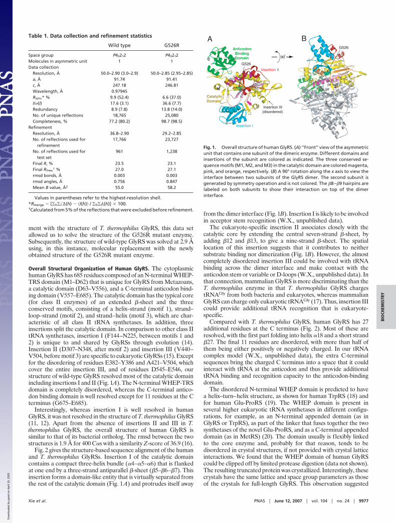
Fig. 2. Structural alignment of human and T. thermophilus GlyRS with the secondary structure elements placed on top. Ten CMT-causing mutations are markedwith vertical arrows.
9978 � www.pnas.org�cgi�doi�10.1073�pnas.0703908104 Xie et al.
Dow
nloa
ded
by g
uest
on
Apr
il 15
, 202
0
to form Gly-tRNAGly. Use of this assay showed clearly that, whereasthe wild-type enzyme was active as expected, G526R GlyRS wasinactive (Fig. 3E).
Either the first or second reaction (Scheme 1 or Scheme 2) canbe responsible for the defect in aminoacylation of G526R GlyRS.For example, a failure to bind tRNAGly, or a failure to transfer theglycyl moiety from Gly-AMP to tRNAGly, would cause inactivationof aminoacylation. Alternatively, disruption of the first reaction,formation of the adenylate, would be another way to disruptaminoacylation.
We focused on monitoring the first step in two different ways.The most common assay is measurement of glycine-dependentATP–PPi exchange in the presence of GlyRS. We found that,whereas the wild-type enzyme was fully active in this assay, G526RGlyRS was inactive (Fig. 3F). This inactivity could be due to afailure to synthesize Gly-AMP or, additionally or alternatively, anextremely slow back-reaction of PPi with the bound Gly-AMP.These possibilities can be distinguished by a second assay thatmonitors the ‘‘adenylate burst’’ (22). In this experiment, GlyRS ismixed with ATP and Gly to form bound Gly-AMP in an initialburst. As the adenylate is slowly released from the enzyme, addi-tional rounds of synthesis can occur. These additional rounds are ata slower rate than the initial burst. We found that, whereaswild-type GlyRS gave the expected burst of synthesis of Gly-AMP(as measured by consumption of ATP), no such burst was observedwith the mutant enzyme (Fig. 3F Inset). Thus, the defect inATP–PPi exchange is caused by an inability to synthesize Gly-AMP,
presumably because of the blockage of the AMP binding site byR526.
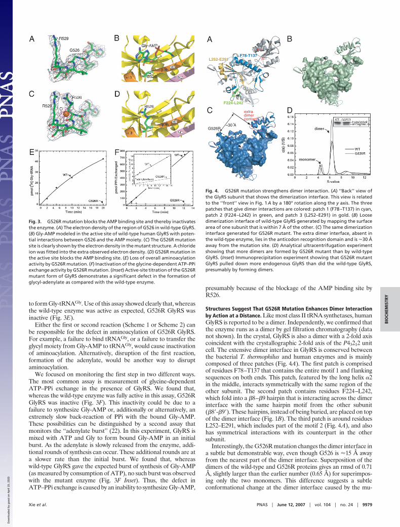
Structures Suggest That G526R Mutation Enhances Dimer Interactionby Action at a Distance. Like most class II tRNA synthetases, humanGlyRS is reported to be a dimer. Independently, we confirmed thatthe enzyme runs as a dimer by gel filtration chromatography (datanot shown). In the crystal, GlyRS is also a dimer with a 2-fold axiscoincident with the crystallographic 2-fold axis of the P43212 unitcell. The extensive dimer interface in GlyRS is conserved betweenthe bacterial T. thermophilus and human enzymes and is mainlycomposed of three patches (Fig. 4A). The first patch is comprisedof residues F78–T137 that contains the entire motif 1 and flankingsequences on both ends. This patch, featured by the long helix �2in the middle, interacts symmetrically with the same region of theother subunit. The second patch contains residues F224–L242,which fold into a �8–�9 hairpin that is interacting across the dimerinterface with the same hairpin motif from the other subunit(�8�-�9�). These hairpins, instead of being buried, are placed on topof the dimer interface (Fig. 1B). The third patch is around residuesL252–E291, which includes part of the motif 2 (Fig. 4A), and alsohas symmetrical interactions with its counterpart in the othersubunit.
Interestingly, the G526R mutation changes the dimer interface ina subtle but demonstrable way, even though G526 is �15 Å awayfrom the nearest part of the dimer interface. Superposition of thedimers of the wild-type and G526R proteins gives an rmsd of 0.71Å, slightly larger than the earlier number (0.65 Å) for superimpos-ing only the two monomers. This difference suggests a subtleconformational change at the dimer interface caused by the mu-
Fig. 3. G526R mutation blocks the AMP binding site and thereby inactivatesthe enzyme. (A) The electron density of the region of G526 in wild-type GlyRS.(B) Gly-AMP modeled in the active site of wild-type human GlyRS with poten-tial interactions between G526 and the AMP moiety. (C) The G526R mutationsite is clearly shown by the electron density in the mutant structure. A chlorideion was fitted into the extra observed electron density. (D) G526R mutation inthe active site blocks the AMP binding site. (E) Loss of overall aminoacylationactivity by G526R mutation. (F) Inactivation of the glycine-dependent ATP–PPiexchange activity by G526R mutation. (Inset) Active-site titration of the G526Rmutant form of GlyRS demonstrates a significant defect in the formation ofglycyl-adenylate as compared with the wild-type enzyme.
F78-T137
F224-L242
L252-E291
A B
DC extra dimer contact
G526R~30 A
β8β9
α2
α8
β10 β1β2
β4α3
β23
α14
Fig. 4. G526R mutation strengthens dimer interaction. (A) ‘‘Back’’ view ofthe GlyRS subunit that shows the dimerization interface. This view is relatedto the ‘‘front’’ view in Fig. 1A by a 180° rotation along the y axis. The threepatches that give dimer interactions are colored: patch 1 (F78–T137) in cyan,patch 2 (F224–L242) in green, and patch 3 (L252–E291) in gold. (B) Loosedimerization interface of wild-type GlyRS generated by mapping the surfacearea of one subunit that is within 7 Å of the other. (C) The same dimerizationinterface generated for G526R mutant. The extra dimer interface, absent inthe wild-type enzyme, lies in the anticodon recognition domain and is �30 Åaway from the mutation site. (D) Analytical ultracentrifugation experimentshowing that more dimers are formed by G526R mutant than by wild-typeGlyRS. (Inset) Immunoprecipitation experiment showing that G526R mutantGlyRS pulled down more endogenous GlyRS than did the wild-type GlyRS,presumably by forming dimers.
Xie et al. PNAS � June 12, 2007 � vol. 104 � no. 24 � 9979
BIO
CHEM
ISTR
Y
Dow
nloa
ded
by g
uest
on
Apr
il 15
, 202
0
tation. When the surface area of one subunit that is within 7 Å ofthe other is then considered, apparent differences between G526Rand wild-type GlyRS are more specifically demonstrated (Fig. 4 Band C). This ‘‘loose’’ (7-Å band either side of interface) dimercontact area for the G526R mutant protein is 7% larger than thesame area calculated for the wild-type enzyme. This observationsuggested that G526R GlyRS formed a more stable dimer than doesthe wild-type enzyme and, for that reason, the G526R mutantenzyme formed a slightly more compact crystal lattice that dif-fracted to a higher resolution than did crystals of wild-type GlyRS(Table 1). Some of the extra dimer contact area for G526R GlyRSis from the anticodon binding domain, �30 Å away from the G526Rmutation site (Fig. 4C). The extra contact from the anticodonbinding domain is contributed from residues on helix �14 and thefollowing strand, �23 (Fig. 4A), including S581, whose mutation(S581L) is associated with CMT disease. Thus, this long-rangestructural effect, caused by the active site mutation, has manifesteditself through an apparent strengthened interaction with the othersubunit.
Two Independent Measurements Demonstrate Tighter Dimer Forma-tion for G526R Enzyme. To see whether G526R GlyRS formed atighter dimer than did the wild-type enzyme, analytical ultracen-trifugation was first carried out to evaluate the quaternary struc-tures of the two GlyRSs (Fig. 4D). By sedimentation velocityanalysis, two peaks were observed for both wild-type and G526Rmutant GlyRS, with the smaller peak (S values of �4.30 for the wildtype and �4.66 for G526R GlyRS) corresponding to the monomersof GlyRS and the larger peak (S values of �6.23 for wild type and�6.35 for the G526R GlyRS) being at the expected position for thedimer. Remarkably, the G526R mutant protein had a larger peakfor the dimer, showing that, under the experimental conditions, itformed more dimers than did the wild-type protein (Fig. 4D).
A second assay was developed to give a picture more represen-tative of those that occur in vivo. Genes encoding wild-type andG526R mutant human GlyRS were transfected into mouse neuro-blastoma N2a cells that also expressed endogenous mouse GlyRS.The transfected GlyRSs were labeled with a V5-epitope tag that wasplaced at the N terminus. (This tag and the linker add a total of 41aa to the N terminus and, therefore, give a small increase to the sizeof GlyRS.) Twenty-four hours after the cells were transfected, theywere lysed and immunoprecipitated with anti-V5 antibodies to pulldown the GlyRSs that were expressed from the recombinant,transfected genes. The precipitates were then redissolved and runon SDS/PAGE. The resolved proteins on the gel were then visu-alized by Western blot analysis using anti-GlyRS (not anti-V5-tagged) polyclonal antibodies.
For both wild-type and G526R GlyRS gene transfections, thestarting lysate (before immunoprecipitation) had a small amount oftransfected GlyRS relative to the endogenous GlyRS (�5%).Therefore, the chance for the transfected GlyRS to form a ho-modimer with itself was low. After immunoprecipitation, two bandsthat differ slightly in molecular weight were seen using the anti-GlyRS antibodies (Fig. 4D Inset). In addition to the V5-taggedGlyRS expressed from the transfected DNA, a smaller band wasobserved. This band corresponded to endogenous GlyRS that was
coimmunoprecipitated with transfected GlyRS through formationof a heterodimer. For transfections with the wild-type GlyRS gene,the amount of endogenous GlyRS pulled down was �14% of thetransfected GlyRS. This number is �35% for the transfections withthe G526R GlyRS gene (Fig. 4D Inset). By comparing heterodimerformation in these experiments, the results support strongly the ideathat the G526R mutation strengthens the dimer interface.
DiscussionThe structures presented here raise the possibility that the disper-sion of CMT-causing mutations across the sequence of GlyRS couldbe understood in terms of effects on the dimer interface. Remark-ably, the G526R mutation at the active site causes a long-rangestructural effect that perpetrates into distal areas to strengthen thedimer interface. That the wild-type subunit interacts more stronglywith the G526R mutant protein provides a rationale for a dominantphenotype, that is, ‘‘subunit poisoning’’ by heterodimer formation.In studies of a CMT-causing mutant TyrRS (normally an �2 dimer),heterodimers were seen as well and were suggested to be involvedin some way in the disease phenotype (3). Not clear, however, iswhether it is a change in the interface per se, or the formation ofheterodimers, or both, that are relevant. Thus, our results point toa need for further investigations of subunits’ interactions and theirrelationship, if any, to CMT disease.
At the start of our investigations we were interested in the WHEPdomain at the N terminus of GlyRS. Our thought was that thisdomain could bring in another neuron-specific function. However,no CMT-causing mutations have been mapped to the WHEPdomain. Thus, if the WHEP domain has a role in CMT disease, thenit should in some way communicate with the rest of the enzymewhere the mutations are located. However, we obtained crystals ofGlyRS with the WHEP domain clipped off. Although we did notsolve the structure, the crystals had almost the same crystal latticeparameters as those for wild-type GlyRS, slightly bigger than thosefor G526R GlyRS (data not shown). This observation suggests thatthe structure of the non-WHEP-containing main body of GlyRS isindependent of the WHEP domain and, therefore, that this domainmost likely has little role in CMT disease.
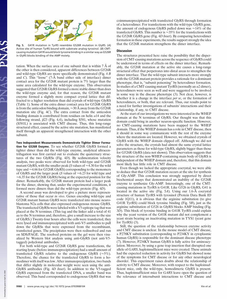
Even though we lacked the appropriate cocrystal, we were ableto deduce that that G526R mutation occurs at the site for synthesisof Gly-AMP. This conclusion was strongly supported by directbiochemical assays that demonstrated an inability of the G526Renzyme to synthesize Gly-AMP. Interestingly, one of the CMT-causing mutations in TyrRS is G41R. Like G526 in GlyRS, G41 islocated in the active site (Fig. 5A). Using our 1.6-Å cocrystalstructure of human TyrRS with tyrosinol (Protein Data Bank IDcode 1Q11), it is obvious that the arginine substitution (to giveG41R TyrRS) could block tyrosine binding (Fig. 5B), just as thearginine substitution of G526 in GlyRS blocks AMP binding (Fig.3D). This block of tyrosine binding in G41R TyrRS could explainwhy the yeast version of the G41R mutant did not complement ayeast strain bearing an inactivating mutation in TYS1 (yeast genefor TyrRS) (3).
Still, the question of the relationship between enzyme activityand CMT disease is unclear. In the mouse model of CMT disease,a P278KY substitution (corresponding to P234KY in cytoplasmichuman GlyRS) is responsible for the dominant disease phenotype(7). However, P234KY human GlyRS is fully active for aminoacy-lation. Moreover, by using a gene trap insertion that disrupted oneallele of GARS, haploinsufficient mice were created. These animalshad the expected reduction in activity for GlyRS but showed noneof the symptoms for CMT disease or for any other neurologicaldisorder. This experiment raises doubts about the relationship ofactivity to CMT disease. Moreover, with respect to the haploinsuf-ficient mice, only the wild-type, homodimeric GlyRS is present.Thus, haploinsufficient mice for GARS leave open the question ofthe relevance of intersubunit interactions to CMT disease and
A BTyrosine
G41
G41R
Fig. 5. G41R mutation in TyrRS resembles G526R mutation in GlyRS. (A)Active site of human TyrRS bound with substrate analog tyrosinol. (B) CMT-causing mutation G41R would block tyrosine binding in a similar way as G526Rin GlyRS blocks binding of the AMP moiety.
9980 � www.pnas.org�cgi�doi�10.1073�pnas.0703908104 Xie et al.
Dow
nloa
ded
by g
uest
on
Apr
il 15
, 202
0
further raise the motivation to investigate the significance of thedimer interface.
Materials and MethodsCrystallization and Data Collection. Wild-type GlyRS was cloned,expressed, and crystallized as described (13). The G526R mutationwas introduced by standard site-directed mutagenesis. PurifiedG526R GlyRS protein was crystallized in 13–15% PEG 6000, 0.1 Msodium citrate buffer (pH 5.5), and 0.1 M NaCl. Crystals were flashfrozen at 100 K, with 20% ethylene glycol added to the reservoirsolution as a cryoprotectant. Both wild-type and G526R GlyRSdata were collected on beamline 11-1 at the Stanford SynchrotronRadiation Laboratory (Menlo Park, CA).
Structure Determination. The diffraction data were processed andscaled with HKL2000. The crystals belong to space group P43212,with one molecule of GlyRS per asymmetric unit and a solventcontent of 69.2%. The structure of G526R GlyRS was determinedby molecular replacement with Phaser (23) using the T. thermophi-lus GlyRS structure (Protein Data Bank ID code 1ATI) as a searchmodel. Density modification with solvent flattening was done byusing DM (24). Iterative model building and refinement wereperformed by using Coot (25) and Refmac5 (26), respectively. Therefined G526R GlyRS structure was used as the starting model tosolve and refine the wild-type GlyRS structure. Data collection andrefinement statistics are given in Table 1.
Aminoacylation Assays. A mixture containing 100 mM Hepes (pH7.5), 20 mM KCl, 10 mM MgCl2, 2 mM DTT, 2 mM ATP, 20 �ML-glycine, and 0.7 �M L-[3H]glycine was added to �200 �M totalbovine tRNA (�10 �M tRNAGly) plus 100 nM purified GlyRS (toinitiate the reaction), and the sample was incubated at 37°C.Aliquots were removed at specified time points, spotted ontotrichloroacetic acid-soaked filter pads, washed with cold 5% tri-chloroacetic acid, and measured by scintillation counting (27).
Pyrophosphate Exchange Assays. Glycyl adenylate synthesis wasmeasured by using the glycine-dependent ATP-pyrophosphate(PPi) exchange assay (28). A mixture containing 100 mM Hepes(pH 7.5), 20 mM KCl, 2 mM ATP, 1 mM NaPPi, 2 mM DTT, 1 mML-glycine, 10 mM MgCl2, and �0.01 mCi/ml Na[32P]PPi was addedto 0.1–1 mM purified GlyRS. The reaction was incubated at roomtemperature, and aliquots were removed at specified times andquenched in a mixture containing 40 mM NaPPi, 1.4% HClO4,0.4% HCl, and 8% (wt/vol) activated charcoal. The charcoalmixture was filtered and washed with a solution of 7% HClO4 and200 mM NaPPi using Spin-X Centrifuge Filters (Corning, Corning,NY) containing 0.45-�m pore-size cellulose acetate filters. Radio-
activity from the centrifuge filters containing the remaining char-coal mixture was measured by scintillation counting.
Active-Site Titration Assays. A mixture containing 100 mM Hepes(pH 7.5), 20 mM KCl, 10 mM MgCl2, 2 mM DTT, 5 �M ATP, 5mM L-glycine, and [�-32P]ATP was added to 5 �M purified GlyRS(to initiate the reaction). The reaction was incubated at 37°C, andaliquots were removed at specified times and quenched in PDVFMultiScreen filter plates (Millipore, Billerica, MA) containing amixture of 10 mM NaPPi, 7% HClO4, 0.5% HCl, and 10% (wt/vol)activated charcoal. Quenched reactions were mixed well and cen-trifuged through the filter plate into a collection plate, and 200 �lwas transferred to scintillation vials for counting.
Analytical Ultracentrifugation. Sedimentation velocity and equilib-rium analytical ultracentrifugation of wild-type and G526R humanGlyRS were performed by using a temperature-controlled Beck-man XL-I analytical ultracentrifuge equipped with an An60Ti rotorand photoelectric scanner (Beckman Coulter, Fullerton, CA) Sam-ples (�400 �l) were loaded by using a Hamilton syringe into adouble-sector cell equipped with a 12-mm Epon centerpiece andsapphire windows.
Immunoprecipitation for Dimer Detection. Mammalian cell expres-sion vector pcDNA3.1/nV5-DEST (Invitrogen, Carlsbad, CA) wasused to clone and express wild-type GlyRS and G526R GlyRS withan N-terminal V5 tag. Wild-type or mutant GlyRS was transfectedinto mouse neuroblastoma cell line N2a, which was cultured inminimum essential medium (Eagle) containing 2 mM L-glutamine,Earle’s BSS, 1.5 g/liter sodium bicarbonate, 0.1 mM nonessentialamino acids, 1.0 mM sodium pyruvate, and 10% FCS. Twenty-fourhours after transfection, cell lysates were prepared in mammalianprotein extraction reagent (M-PER; Pierce, Rockford, IL) contain-ing mixture protease inhibitors (Roche, Indianapolis, IN). The celllysates were concentrated, precleared, and then added into theprotein G agarose beads, which were preincubated overnight withanti-V5 antibody (Invitrogen). After 1 h of incubation, beads werewashed four times by PBS containing 1% Nonidet P-40, boiled in2� SDS loading buffer, and then analyzed in Western blots usingpolyclonal anti-GlyRS as the primary antibody to detect bothtransfected and endogenous GlyRS.
We thank Dr. Robert Burgess (The Jackson Laboratory, Bar Harbor,ME) and Prof. Alexander Rich (Massachusetts Institute of Technology,Cambridge, MA) for helpful comments on the manuscript. This work wassupported by National Cancer Institute Grant CA92577, National Insti-tutes of Health Grant GM15539, and a grant from the NationalFoundation for Cancer Research.
1. Skre H (1974) Clin Genet 6:98–118.2. Shy ME (2004) Curr Opin Neurol 17:579–585.3. Jordanova A, Irobi J, Thomas FP, Van Dijck P, Meerschaert K, Dewil M, Dierick
I, Jacobs A, De Vriendt E, Guergueltcheva V, et al. (2006) Nat Genet 38:197–202.4. Antonellis A, Ellsworth RE, Sambuughin N, Puls I, Abel A, Lee-Lin SQ,
Jordanova A, Kremensky I, Christodoulou K, Middleton LT, et al. (2003) Am JHum Genet 72:1293–1299.
5. Sivakumar K, Kyriakides T, Puls I, Nicholson GA, Funalot B, Antonellis A,Sambuughin N, Christodoulou K, Beggs JL, Zamba-Papanicolaou E, et al.(2005) Brain 128:2304–2314.
6. Del Bo R, Locatelli F, Corti S, Scarlato M, Ghezzi S, Prelle A, Fagiolari G,Moggio M, Carpo M, Bresolin N, Comi GP (2006) Neurology 66:752–754.
7. Seburn KL, Nangle LA, Cox GA, Schimmel P, Burgess RW (2006) Neuron51:715–726.
8. Antonellis A, Lee-Lin SQ, Wasterlain A, Leo P, Quezado M, Goldfarb LG, MyungK, Burgess S, Fischbeck KH, Green ED (2006) J Neurosci 26:10397–10406.
9. James PA, Cader MZ, Muntoni F, Childs AM, Crow YJ, Talbot K (2006)Neurology 67:1710–1712.
10. Park SG, Ewalt KL, Kim S (2005) Trends Biochem Sci 30:569–574.11. Arnez JG, Dock-Bregeon AC, Moras D (1999) J Mol Biol 286:1449–1459.12. Logan DT, Mazauric MH, Kern D, Moras D (1995) EMBO J 14:4156–4167.
13. Xie W, Schimmel P, Yang X-L (2006) Acta Crystallogr F 62:1243–1246.14. Cusack S (1995) Nat Struct Biol 2:824–831.15. Freist W, Logan DT, Gauss DH (1996) Biol Chem Hoppe Seyler 377:343–356.16. Holm L, Sander C (1993) J Mol Biol 233:123–138.17. Mazauric MH, Keith G, Logan D, Kreutzer R, Giege R, Kern D (1998) Eur
J Biochem 251:744–757.18. Yang X-L, Otero FJ, Skene RJ, McRee DE, Schimmel P, Ribas de Pouplana
L (2003) Proc Natl Acad Sci USA 100:15376–15380.19. Jeong EJ, Hwang GS, Kim KH, Kim MJ, Kim S, Kim KS (2000) Biochemistry
39:15775–15782.20. Lee SW, Cho BH, Park SG, Kim S (2004) J Cell Sci 117:3725–3734.21. Yang X-L, Otero FJ, Ewalt KL, Liu J, Swairjo MA, Kohrer C, RajBhandary
UL, Skene RJ, McRee DE, Schimmel P (2006) EMBO J 25:2919–2929.22. Fersht AR, Ashford JS, Bruton CJ, Jakes R, Koch GL, Hartley BS (1975)
Biochemistry 14:1–4.23. McCoy AJ (2007) Acta Crystallogr D 63:32–41.24. Cowtan K, Main P (1998) Acta Crystallogr D 54:487–493.25. Emsley P, Cowtan K (2004) Acta Crystallogr D 60:2126–2132.26. Murshudov GN, Vagin AA, Dodson EJ (1997) Acta Crystallogr D 53:240–255.27. Schreier AA, Schimmel PR (1972) Biochemistry 11:1582–1589.28. Calendar R, Berg P (1966) Biochemistry 5:1690–1695.
Xie et al. PNAS � June 12, 2007 � vol. 104 � no. 24 � 9981
BIO
CHEM
ISTR
Y
Dow
nloa
ded
by g
uest
on
Apr
il 15
, 202
0