long posterior ciliary arterial blood flow and systemic blood pressure
TRANSCRIPT
Investigative Ophthalmology & Visual Science, Vol. 31, No. 5, May 1990Copyright © Association for Research in Vision and Ophthalmology
Long Posterior Ciliary Arterial Blood Flowand Systemic Blood Pressure
Hirofumi Okubo, Tseggoi Gherezghiher, and Michael C. Koss
The purpose of the current study was to investigate the relationship between systemic blood pressure(BP) and long posterior ciliary arterial (LPCA) blood flow (BF) in response to autonomic drugs, and todetermine the types of receptors involved. LPCA BF was measured in anesthetized rabbits at theretrobulbar region using laser Doppler flowmetry. The observation that most of the short posteriorciliary arteries diverge from two LPCAs in rabbits was confirmed using a vascular casting technique.Therefore, the LPCA BF at the retrobulbar region should be representative of total uveal BF. Norepi-nephrine (IV) increased systemic BP and decreased LPCA BF. These responses were suppressed onlyslightly by yohimbine, but were inhibited markedly by prazosin and by the combination of yohimbineand prazosin. B-HT 920 (IV), a selective alpha-2 adrenoceptor agonist, increased systemic BP anddecreased LPCA BF. B-HT 920 (IA) also decreased LPCA BF, with all effects of B-HT 920 antago-nized by yohimbine. Methacholine (IV) decreased systemic BP and increased LPCA BF whetheradministered IV or IA. These effects were blocked uniformly by atropine. The current results suggestthat LPCA BF is controlled both by systemic BP and local ocular vascular tone. There are vasocon-strictive alpha-1 and alpha-2 adrenoceptors and vasodilative muscarinic receptors in the rabbit ocularvascular tree. Invest Ophthalmol Vis Sci 31:819-826,1990
Earlier studies have shown that topical epinephrinedecreases blood flow in the anterior segment1 andthat intravenous administration of epinephrine pro-duces vasoconstriction of choroidal blood vessels andof long posterior ciliary arteries.2 In contrast, sys-temic administration of epinephrine3 and norepi-nephrine4"6 have been reported to increase choroidalblood flow (BF) accompanied by a drug-induced in-crease in systemic arterial blood pressure (BP). Thus,ocular BF may be related both to systemic BP levelsas well as to local ocular vascular tone. In a similarfashion, systemic administration of acetylcholine hasbeen shown to increase choroidal BF of cats5'7 andrabbits,7 but when administered subcutaneouslyto cats, methacholine decreases choroidal bloodvolume.4
The purpose of the current study was to investigatefurther the relationship between systemic BP and oc-ular BF changes in response to autonomic drugs. In
From the Dean A. McGee Eye Institute and Departments ofOphthalmology and of Pharmacology, University of Oklahoma,Health Sciences Center, Oklahoma City, Oklahoma.
Supported in part by grants from the National Science Founda-tion (OK 8710676) and Research to Prevent Blindness, Inc.
Presented in part at the 1989 meeting of the Association forResearch in Vision and Ophthalmology, Sarasota, Florida.
Submitted for publication: April 27, 1989; accepted September29, 1989.
Reprint requests: Hirofumi Okubo, MD, Dean A. McGee EyeInstitute, 608 Stanton L. Young Boulevard, Oklahoma City, OK73104.
these experiments, ocular BF was measured along theretrobulbar lateral long posterior ciliary artery(LPCA) with laser Doppler flowmetry.8'9 Effects ofnorepinephrine (NE), B-HT 920, and methacholine(MC) were investigated.
Materials and Methods
Adult New Zealand White rabbits (Mel WishartRabbit Ranch, Sand Springs, OK) of either sex weresedated with ketamine (35 mg/kg, IM) and Xylazine(5 mg/kg, IM), and anesthetized with pentobarbital(15-30 mg/kg, IV) through a marginal ear vein. Afemoral vein and artery were cannulated for drug ad-ministration and monitoring of systemic BP, respec-tively. After tracheotomy, the animals were paralyzedwith gallamine triethiodide (2-5 mg/kg, IV) and arti-ficially ventilated with room air. In some experi-ments, an external maxillary or an ascending pharyn-geal artery was cannulated for close intraarterial drugadministration. Additional doses of pentobarbitalwere given intravenously as required.
A 360° peritomy was performed, and four rectusand a portion of the retractor muscles were dissectedcarefully so as not to damage the vortex veins. Thesuperior and laterial orbital margins sometimes wereremoved in order to more easily expose the LPCA inthe retrobulbar region. Connective tissue around theLPCA was carefully removed.
For BF measurement, we used a laser Dopplerflowmeter (Model Pf2; Perimed, Sweden), which de-
819
Downloaded From: http://iovs.arvojournals.org/pdfaccess.ashx?url=/data/journals/iovs/933153/ on 03/25/2018
820 INVESTIGATIVE OPHTHALMOLOGY & VISUAL SCIENCE / May 1990 Vol. 31
100
50
10 20 40 SO 60
IOP (mmHg)
70 B0 90
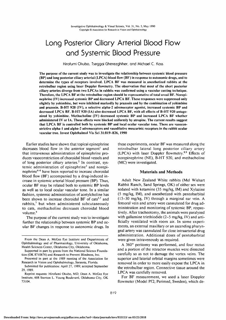
Fig. 1. Relationship between percent of the BF in the LPCA andIOP. IOP was artificially increased by intracameral infusion ofnormal saline at a rate of 50 jtl/min during continuous measure-ment of LPCA blood flow. Note that there was an inverse relation-ship between LPCA blood flow and an increase in IOP. n = 5-6.
tects the velocity and the number of moving redblood cells by using the Doppler phenomenon.8-9
That is, the frequency and the magnitude of theshifted Doppler signal are related to the velocity andthe number of red blood cells, respectively. The bloodflow is calculated internally by multiplying the veloc-
ity by number. The instrument measures the BF in atissue hemisphere with a radius of approximately 1mm.8-9 A black vinyl membrane was inserted beneaththe LPCA at the posterior aspect of the eyeball closeto the optic nerve in order to prevent the detection offlow from surrounding tissues. The reliability of theinstrument to detect LPCA BF was tested by measur-ing LPCA BF against a graded increase of intraocularpressure (IOP) (Fig. 1).
Drugs used were as follows: NE hydrochloride(Sigma, St. Louis, MO), MC bromide (Sigma), B-HT920 (C. W. Boehringer Sohn, Ingelheim, Rhein, WestGermany),10 prazosin hydrochloride (Pfizer, Groton,CT), yohimbine hydrochloride (Aldrich, Milwaukee,WI), atropine sulfate (Sigma). Drugs were adminis-tered either intravenously or by close intraarterial in-jection.
The data was presented as means ± SEM. Statisti-cal differences between the means were determinedby the student t-test for paired observations.
In some experiments, ocular vascular structureswere examined with a casting technique.'' After anes-thesia with pentobarbital, a common carotid arterywas cannulated and all blood was washed out withnormal saline. Subsequent to injection of 5 ml of 2%glutaraldehyde, 20 ml of base resin (Mercox CL-2R;Dainippon Ink and Chemicals, Tokyo, Japan) and 1ml of polymerizer were mixed and injected. The eyewas enucleated after the animal was left at room tem-perature for 20 min. The eye was placed in the warmwater for 24 hr and subsequently immersed in a 20%
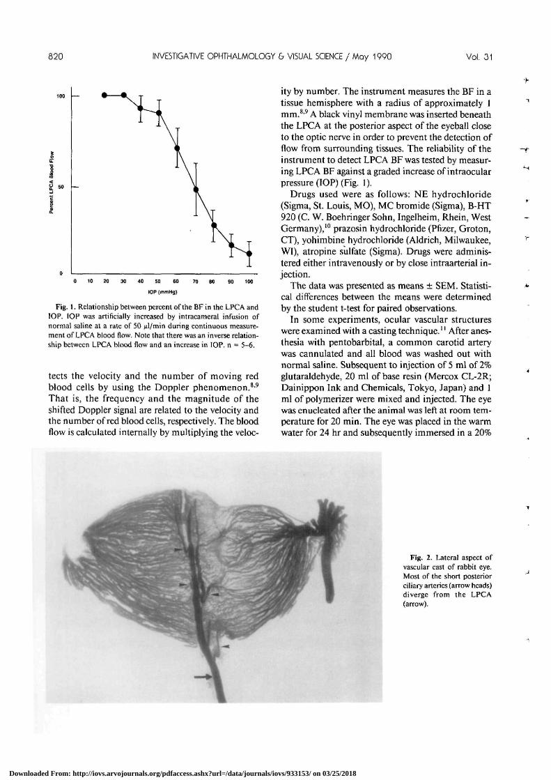
Fig. 2. Lateral aspect ofvascular cast of rabbit eye.Most of the short posteriorciliary arteries (arrow heads)diverge from the LPCA(arrow).
Downloaded From: http://iovs.arvojournals.org/pdfaccess.ashx?url=/data/journals/iovs/933153/ on 03/25/2018
No. 5 OCULAR BLOOD FLOW / OKubo er ol 821
KOH for one week in order to dissolve the oculartissues. The vascular resin casts were washed and ob-served in distilled water using an operation micro-scope.
All animals used in these studies were treated inaccordance with the ARVO Resolution on the Use ofAnimals in Research.
Results
The vascular casts of rabbit eyes showed that mostshort posterior ciliary arteries (SPCAs) diverge from
the LPCA (Fig. 2). As SPCAs run close to the LPCAat the retrobulbar region, a black vinyl membraneusually was inserted beneath the lateral LPCA and aportion of one or more SPCAs.
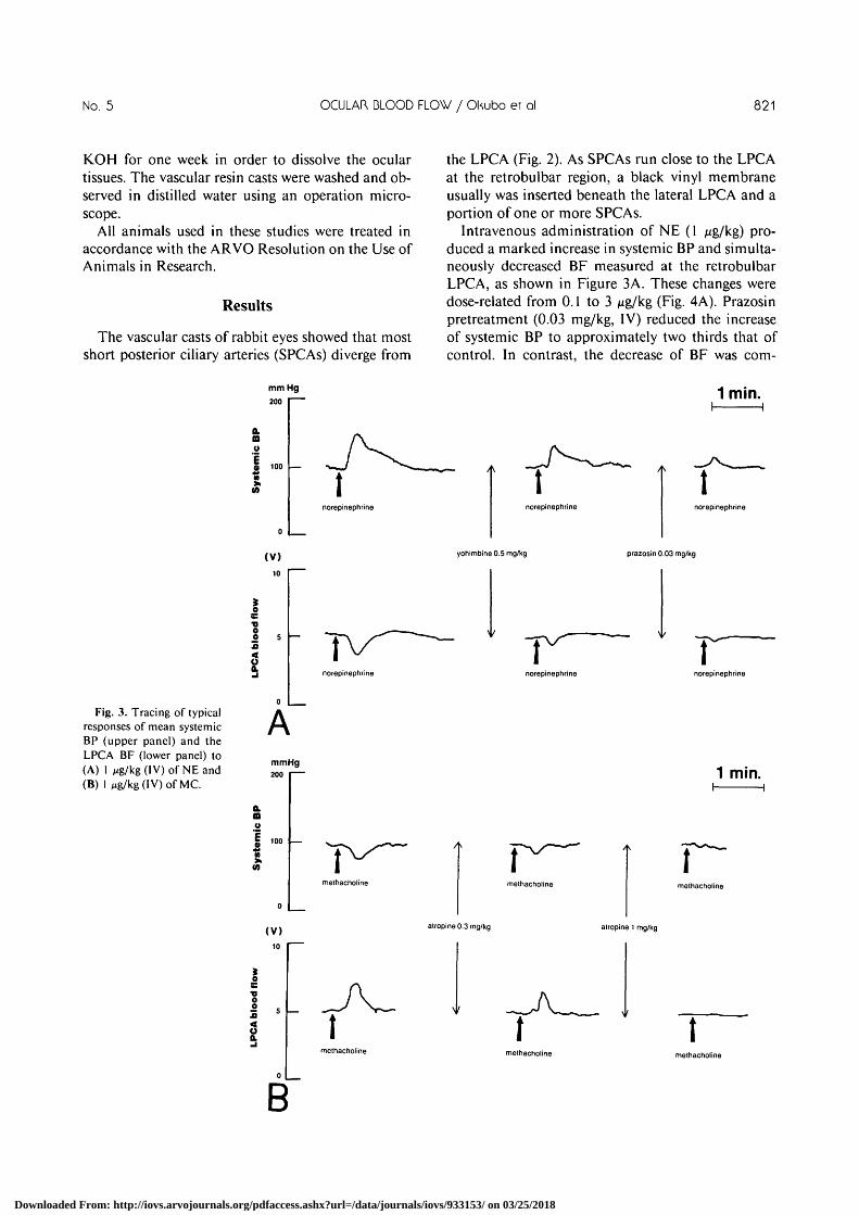
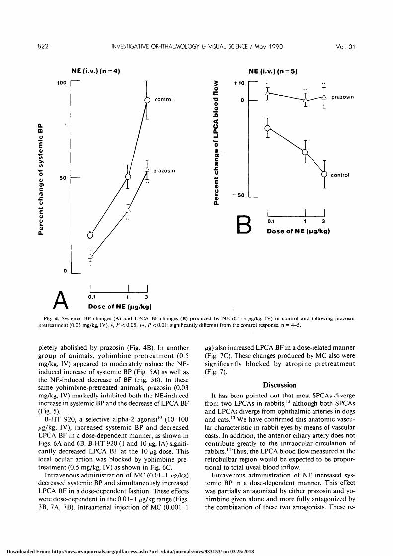
Intravenous administration of NE (1 Mg/kg) pro-duced a marked increase in systemic BP and simulta-neously decreased BF measured at the retrobulbarLPCA, as shown in Figure 3A. These changes weredose-related from 0.1 to 3 Mg/kg (Fig. 4A). Prazosinpretreatment (0.03 mg/kg, IV) reduced the increaseof systemic BP to approximately two thirds that ofcontrol. In contrast, the decrease of BF was corn-
Fig. 3. Tracing of typicalresponses of mean systemicBP (upper panel) and theLPCA BF (lower panel) to(A) 1 Mg/kg (IV) of NE and(B) 1 Mg/kg (IV) of MC.
mm Hg200
(V)10
AmmHg200
(V)10
norepinephrine
norepinephrine
methacholine
norepinephrine
yohimbine 0.5 mg/kg
norepinephrine
1 min.
norepinephrine
prazosin 0.03 mg/kg
norepinephrine
1 min.
atropine 0.3 mg/kg alropine 1 mg/kg
methacholine
Downloaded From: http://iovs.arvojournals.org/pdfaccess.ashx?url=/data/journals/iovs/933153/ on 03/25/2018
822 INVESTIGATIVE OPHTHALMOLOGY & VISUAL SCIENCE / Moy 1990 Vol. 31
NE(i.v.)(n NE(i.v.)(n = 5)
100
Q.aoE
co0)
Q.
50
control
prazosin
O
•oo2
<oQ.- I<̂O0)
c
+ 10
0
- 50
prazosin
control
B 0.1 1 3
Dose of NE (/ug/kg)
A 0.1 1 3
Dose of NE (jjg/kg)
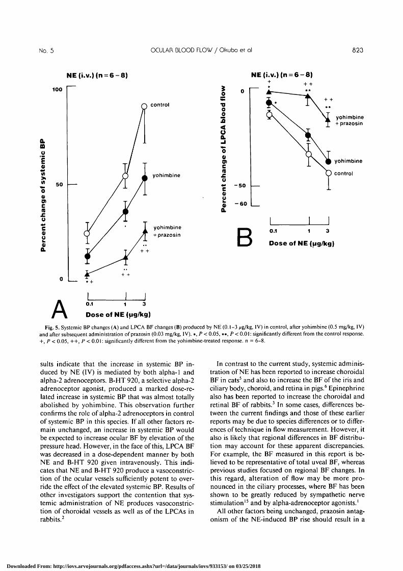
Fig. 4. Systemic BP changes (A) and LPCA BF changes (B) produced by NE (0.1-3 Mg/kg, IV) in control and following prazosinpretreatment (0.03 mg/kg, IV). *, P < 0.05, •*, P < 0.01: significantly different from the control response, n = 4-5.
pletely abolished by prazosin (Fig. 4B). In anothergroup of animals, yohimbine pretreatment (0.5mg/kg, IV) appeared to moderately reduce the NE-induced increase of systemic BP (Fig. 5A) as well asthe NE-induced decrease of BF (Fig. 5B). In thesesame yohimbine-pretreated animals, prazosin (0.03mg/kg, IV) markedly inhibited both the NE-inducedincrease in systemic BP and the decrease of LPCA BF(Fig. 5).
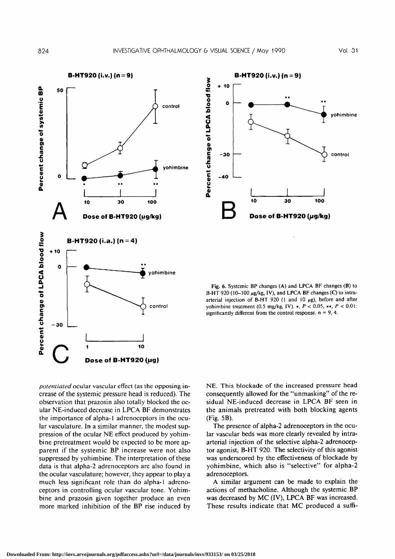
B-HT 920, a selective alpha-2 agonist10 (10-100Mg/kg, IV), increased systemic BP and decreasedLPCA BF in a dose-dependent manner, as shown inFigs. 6A and 6B. B-HT 920 (1 and 10 fig, IA) signifi-cantly decreased LPCA BF at the 10-/*g dose. Thislocal ocular action was blocked by yohimbine pre-treatment (0.5 mg/kg, IV) as shown in Fig. 6C.
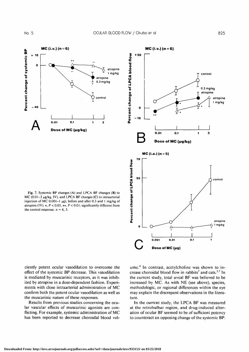
Intravenous administration of MC (0.01-1 /ug/kg)decreased systemic BP and simultaneously increasedLPCA BF in a dose-dependent fashion. These effectswere dose-dependent in the 0.01-1 /xg/kg range (Figs.3B, 7A, 7B). Intraarterial injection of MC (0.001-1
Hg) also increased LPCA BF in a dose-related manner(Fig. 7C). These changes produced by MC also weresignificantly blocked by atropine pretreatment(Fig. 7).
DiscussionIt has been pointed out that most SPCAs diverge
from two LPCAs in rabbits,12 although both SPCAsand LPCAs diverge from ophthalmic arteries in dogsand cats.13 We have confirmed this anatomic vascu-lar characteristic in rabbit eyes by means of vascularcasts. In addition, the anterior ciliary artery does notcontribute greatly to the intraocular circulation ofrabbits.14 Thus, the LPCA blood flow measured at theretrobulbar region would be expected to be propor-tional to total uveal blood inflow.
Intravenous administration of NE increased sys-temic BP in a dose-dependent manner. This effectwas partially antagonized by either prazosin and yo-himbine given alone and more fully antagonized bythe combination of these two antagonists. These re-
Downloaded From: http://iovs.arvojournals.org/pdfaccess.ashx?url=/data/journals/iovs/933153/ on 03/25/2018
No. 5 OCULAR DLOOD FLOW / Okubo er ol 823
NE(i.v.)(n = 6-8) NE(i.v.)(n = 6-8)
100
Q.CO
uE0)(A>; soO0)O>cn
c0)ooQ.
control
yohimbine
O
TJO£
<o
4>O)
c(0
c0)o4)Q.
- 5 0
- 6 0
yohimbine+ prazosin B
yohimbine+ prazosin
yohimbine
control
0.1 1 3
Dose of NE (pg/kg)
* +
A 0.1 1
Dose of NE
Fig. 5. Systemic BP changes (A) and LPCA BF changes (B) produced by NE (0.1-3 Mg/kg, IV) in control, after yohimbine (0.5 mg/kg, IV)and after subsequent administration of prazosin (0.03 mg/kg, IV). *, P < 0.05, •*, P < 0.01: significantly different from the control response.+, P < 0.05, ++, P < 0.01: significantly different from the yohimbine-treated response, n = 6-8.
suits indicate that the increase in systemic BP in-duced by NE (IV) is mediated by both alpha-1 andalpha-2 adrenoceptors. B-HT 920, a selective alpha-2adrenoceptor agonist, produced a marked dose-re-lated increase in systemic BP that was almost totallyabolished by yohimbine. This observation furtherconfirms the role of alpha-2 adrenoceptors in controlof systemic BP in this species. If all other factors re-main unchanged, an increase in systemic BP wouldbe expected to increase ocular BF by elevation of thepressure head. However, in the face of this, LPCA BFwas decreased in a dose-dependent manner by bothNE and B-HT 920 given intravenously. This indi-cates that NE and B-HT 920 produce a vasoconstric-tion of the ocular vessels sufficiently potent to over-ride the effect of the elevated systemic BP. Results ofother investigators support the contention that sys-temic administration of NE produces vasoconstric-tion of choroidal vessels as well as of the LPCAs inrabbits.2
In contrast to the current study, systemic adminis-tration of NE has been reported to increase choroidalBF in cats5 and also to increase the BF of the iris andciliary body, choroid, and retina in pigs.6 Epinephrinealso has been reported to increase the choroidal andretinal BF of rabbits.3 In some cases, differences be-tween the current findings and those of these earlierreports may be due to species differences or to differ-ences of technique in flow measurement. However, italso is likely that regional differences in BF distribu-tion may account for these apparent discrepancies.For example, the BF measured in this report is be-lieved to be representative of total uveal BF, whereasprevious studies focused on regional BF changes. Inthis regard, alteration of flow may be more pro-nounced in the ciliary processes, where BF has beenshown to be greatly reduced by sympathetic nervestimulation15 and by alpha-adrenoceptor agonists.1
All other factors being unchanged, prazosin antag-onism of the NE-induced BP rise should result in a
Downloaded From: http://iovs.arvojournals.org/pdfaccess.ashx?url=/data/journals/iovs/933153/ on 03/25/2018
824 INVESTIGATIVE OPHTHALMOLOGY b VISUAL SCIENCE / May 1990 Vol. 31
B-HT920(i.v.)(n =
Q.ao
4)
Wcnroc4)O
4)Q.
50
control
yohimbine
A10 30 100
Dose of B-HT920 (pg/kg)
,0 +10
<o0.
o0)O)£ -30re
- 4 0
B-HT920(i.v.)(n =
yohimbine
control
B10 30 100
Dose of B-HT920
B-HT920(i.a.)(n =
"2oo
<aQ.
O0)
c
+1 0
- 3 0
yohimbine
control
C 10
Dose of B-HT920(^jg)
Fig. 6. Systemic BP changes (A) and LPCA BF changes (B) toB-HT 920 (10-100 jtg/kg, IV), and LPCA BF changes (C) to intra-arterial injection of B-HT 920 (I and 10 /ig)> before and afteryohimbine treatment (0.5 mg/kg, IV). *, P < 0.05, **, P < 0.01:significantly different from the control response, n = 9, 4.
potentiated ocular vascular effect (as the opposing in-crease of the systemic pressure head is reduced). Theobservation that prazosin also totally blocked the oc-ular NE-induced decrease in LPCA BF demonstratesthe importance of alpha-1 adrenoceptors in the ocu-lar vasculature. In a similar manner, the modest sup-pression of the ocular NE effect produced by yohim-bine pretreatment would be expected to be more ap-parent if the systemic BP increase were not alsosuppressed by yohimbine. The interpretation of thesedata is that alpha-2 adrenoceptors are also found inthe ocular vasculature; however, they appear to play amuch less significant role than do alpha-1 adreno-ceptors in controlling ocular vascular tone. Yohim-bine and prazosin given together produce an evenmore marked inhibition of the BP rise induced by
NE. This blockade of the increased pressure headconsequently allowed for the "unmasking" of the re-sidual NE-induced decrease in LPCA BF seen inthe animals pretreated with both blocking agents(Fig. 5B).
The presence of alpha-2 adrenoceptors in the ocu-lar vascular beds was more clearly revealed by intra-arterial injection of the selective alpha-2 adrenocep-tor agonist, B-HT 920. The selectivity of this agonistwas underscored by the effectiveness of blockade byyohimbine, which also is "selective" for alpha-2adrenoceptors.
A similar argument can be made to explain theactions of methacholine. Although the systemic BPwas decreased by MC (IV), LPCA BF was increased.These results indicate that MC produced a sufii-
Downloaded From: http://iovs.arvojournals.org/pdfaccess.ashx?url=/data/journals/iovs/933153/ on 03/25/2018
No. 5 OCULAR BLOOD FLOW / OKubo er ol 825
a.u
i0)
>(A
"o0Olcraoc0)ow0)Q.
+ 10
0
- 4 0
MC(i.v.)(n = 6)
* *v^^r ^̂ i_
\ ^ ^ " \ . """"-^ atropine\ . " ^ ^ T 1 mg/kg
^ ^ ^fc atropine\ j -L 0.3 mg/kg
i \^ \
\ T
KJ control
I I I
AI I I0.01 0.1 1 3
Dose of MC (ug/kg)
3 +5C_0
"O00
J5s0.•J
"oa>c
£g•* 0
05 - ioQ.
MC(i
control
0.3 mg/kgatropine
atropinemg/kg
0.01 0.1 1
Dose of MC (fig/kg)
MC(i.a.)(n =
Fig. 7. Systemic BP changes (A) and LPCA BF changes (B) toMC (0.01-3 Mg/kg, IV), and LPCA BF changes (C) to intraarterialinjection of MC 0.00l-l ng), before and after 0.3 and I mg/kg ofatropine (IV). *, P < 0.05, **, P < 0.01: significantly different fromthe control response, n = 6, 5.
o
•ooo
<o.J
"o0)O)
c10
oc0)o0)Q.
70
50 control
atropine1 mg/kg
0.001 0.01
Dose of MC
0.1
ciently potent ocular vasodilation to overcome theeffect of the systemic BP decrease. This vasodilationis mediated by muscarinic receptors, as it was inhib-ited by atropine in a dose-dependent fashion. Experi-ments with close intraarterial administration of MCconfirm both the potent ocular vasodilation as well asthe muscarinic nature of these responses.
Results from previous studies concerning the ocu-lar vascular effects of muscarinic agonists are con-flicting. For example, systemic administration of MChas been reported to decrease choroidal blood vol-
ume.4 In contrast, acetylcholine was shown to in-crease choroidal blood flow in rabbits7 and cats.57 Inthe current study, total uveal BF was believed to beincreased by MC. As with NE (see above), species,methodologic, or regional differences within the eyemay explain the discrepant observations in the litera-ture.
In the current study, the LPCA BF was measuredat the retrobulbar region, and drug-induced alter-ation of ocular BF seemed to be of sufficient potencyto counteract an opposing change of the systemic BP.
Downloaded From: http://iovs.arvojournals.org/pdfaccess.ashx?url=/data/journals/iovs/933153/ on 03/25/2018
826 INVESTIGATIVE OPHTHALMOLOGY b VISUAL SCIENCE / May 1990 Vol. 01
Key words: ocular circulation, laser Doppler flowmetry,alpha-adrenoceptors, muscarinic receptors, vascular struc-tures
Acknowledgments
The authors would like to thank Ms. Marlene Richard-son for typing the manuscript; Ms. Linda Turner, Ms.Linda Kuhlman, Hanh Nguyen, and Mr. Robert Adamsfor technical assistance; and Ms. Terri Hamby for preparingthe illustrations.
References
1. Aim A: The effect of topical /-epinephrine on regional ocularblood flow in monkeys. Invest Ophthalmol Vis Sci 19:487,1980.
2. Wudka E and Leopold IH: Experimental studies of the choroi-dal vessels: IV. Pharmacologic observations. AMA ArchOphthalmol 55:857, 1956.
3. Chiou GCY, Girgis Z, and Chiou PY: Effects of epinephrineon retinal and choroidal blood flow through different routes ofdrug administration. Ophthalmic Res 20:293, 1988.
4. Bettman JW and Fellows V: Effects of peripheral vasodilatorand vasoconstrictor drugs on the intraocular (choroidal) bloodvolume. Trans Am Ophthalmol Otolaryngol 66:480, 1962.
5. Chandra SR and Friedman E: Choroidal blood flow: II. Theeffects of autonomic agents. Arch Ophthalmol 87:67, 1972.
6. Malik AB, VanHeuven WAJ, and Satler LF: Effects of isopro-
terenol and norepinephrine on regional ocular blood flows.Invest Ophthalmol 15:492, 1976.
7. Bill A: Autonomic nervous control of uveal blood flow. ActaPhysiol Scand 56:70, 1962.
8. Nilsson GE, Tenland T, and Oberg PA: Evaluation of a laserDoppler flowmeter for measurement of tissue blood flow.IEEE Trans Biomed Eng 27:597, 1980.
9. Nilsson GE: Signal processor for laser Doppler tissue flow-meters. Med Biol Eng Comput 22:343, 1984.
10. Koblinger W and Pichler L: Investigation into different typesof post- and presynaptic a-adrenoceptors at cardiovascularsites in rats. Eur J Pharmacol 65:393, 1980.
11. Matsusaka T: Angio-architecture of the choroid. Jpn J Oph-thalmol 20:330, 1976.
12. Wudka E and Leopold IH: Experimental studies of the choroi-dal vessels: III. Anatomical observations. AMA Arch Ophthal-mol 55:617, 1956.
13. Fujii I: Research on comparative anatomy of mammals inrespect to their vascular systems of the eye: Comparative studyof long posterior ciliary arteries and its connecting arteries,using acrylic resin casts. Acta Soc Ophthalmol Jpn 72:1905,1968.
14. Morrison JC, DeFrank MP, and VanBuskirk EM: Regionalmicrovascular anatomy of the rabbit ciliary body. InvestOphthalmol Vis Sci 28:1314, 1987.
15. Aim A and Bill A: The effects of stimulation of the cervicalsympathetic chain on retinal oxygen tension and on uveal,retinal and cerebral blood flow in cats. Acta Physiol Scand88:84, 1973.
Downloaded From: http://iovs.arvojournals.org/pdfaccess.ashx?url=/data/journals/iovs/933153/ on 03/25/2018