long noncoding rna derived from cd244 signaling epigenetically controls ... · long noncoding rna...
TRANSCRIPT
Long noncoding RNA derived from CD244 signalingepigenetically controls CD8+ T-cell immuneresponses in tuberculosis infectionYangWanga,1, Huiling Zhonga,1, Xiaodan Xiea,1, Crystal Y. Chenb, Dan Huangb, Ling Shenb, Hui Zhangc, ZhengW. Chenb,and Gucheng Zenga,2
aDepartment of Microbiology, Zhongshan School of Medicine, Key Laboratory for Tropical Diseases Control of the Ministry of Education, Sun Yat-senUniversity, Guangzhou 510080, China; bDepartment of Microbiology and Immunology, Center for Primate Biomedical Research, University of Illinois Collegeof Medicine, Chicago, IL 60612; and cInstitute of Human Virology, Zhongshan School of Medicine, Key Laboratory for Tropical Diseases Control of theMinistry of Education, Sun Yat-sen University, Guangzhou 510080, China
Edited by Barry R. Bloom, Harvard School of Public Health, Boston, MA, and approved June 8, 2015 (received for review January 26, 2015)
Molecular mechanisms for T-cell immune responses modulated by Tcell-inhibitory molecules during tuberculosis (TB) infection remainunclear. Here, we show that active human TB infection up-regulatesCD244 and CD244 signaling-associated molecules in CD8+ T cells andthat blockade of CD244 signaling enhances production of IFN-γ andTNF-α. CD244 expression/signaling in TB correlates with high levels ofa long noncoding RNA (lncRNA)-BC050410 [named as lncRNA-AS-GSTT1(1-72) or lncRNA-CD244] in the CD244+CD8+ T-cell subpopula-tion. CD244 signaling drives lncRNA-CD244 expression via sustaininga permissive chromatin state in the lncRNA-CD244 locus. By recruitingpolycomb protein enhancer of zeste homolog 2 (EZH2) to infg/tnfapromoters, lncRNA-CD244mediates H3K27 trimethylation at infg/tnfaloci toward repressive chromatin states and inhibits IFN-γ/TNF-α ex-pression in CD8+ T cells. Such inhibition can be reversed by knockdown of lncRNA-CD244. Interestingly, adoptive transfer of lncRNA-CD244–depressed CD8+ T cells to Mycobacterium tuberculosis (MTB)-infected mice reduced MTB infection and TB pathology comparedwith lncRNA-CD244–expressed controls. Thus, this work uncoverspreviously unidentified mechanisms in which T cell-inhibitory signal-ing and lncRNAs regulate T-cell responses and host defense againstTB infection.
tuberculosis | lncRNA | CD8+ T cells
Tuberculosis (TB) caused by Mycobacterium tuberculosis (MTB)infection remains a leading public health threat with high
morbidity and mortality around the world (1, 2). CD4+ T cells,CD8+ T cells, and γδ T cells played critical roles in mountingadaptive immune response against MTB infection (3–8). Deci-phering the molecular mechanisms for host responses linked to TBpathogenesis and prognosis is of great importance for developingnew vaccines and therapeutics and for diagnosis.Activation and effector functions of T cells are regulated by
CD3/T-cell receptor (TCR) signal upon antigenic engagementand by a group of signals from costimulatory molecules, in-cluding CD28, cytotoxic T-lymphocyte–associated protein4 (CTLA4), inducible T-cell costimulator (ICOS), programmeddeath-1 (PD-1), T cell immunoglobulin mucin-3 (Tim-3), andCD244 (2B4) (9–14). Accumulating evidence suggests that avariety of pathogens, including HIV, simian immunodeficiencyvirus, hepatitis C virus (HCV), lymphocytic choriomeningitisvirus, and Plasmodium, induce immune evasion by up-regulatingcostimulatory molecules such as PD-1, CTLA4, and Tim-3 asa result of repeated antigenic stimulation of T cells. However,T-cell immune responses regulated by these “inhibitory re-ceptors” during TB infection appear to be more complex thanwhat have been observed in chronic viral infections. In fact, wehave recently reported that up-regulation of the T cell-inhibitory molecule Tim-3 would lead to an enhanced but notinhibitory anti-TB effector function during active human TB(15), and others have also found that Tim-3 signaling may
benefit innate immunity against intracellular MTB (16). Thus,it is necessary to elucidate the mechanisms by which T cell-inhibitory molecules regulate T-cell effector functions pro-ducing cytokines during active microbial infection.Although heritable changes in gene regulation that occur via
modification of the DNA without changes to the DNA sequenceare often referred to as epigenetic programming, noncoding RNA(ncRNA)-mediated transcriptional or posttranscriptional regula-tion is one of the major regulation mechanisms for epigeneticprogramming (17–30). Recent studies have identified thousands oflong ncRNAs (lncRNAs) (17, 23) in mammalian genomes thatregulate gene expression in a variety of immunological processes(31–35), such as differentiation of T cells (33, 36) and dendriticcells (DCs) (35). Thus, diverse functions among T-cell sub-populations may manifest through highly dynamic changes inlncRNA-regulated epigenetic programming, and lncRNA-regulatedepigenetic reprogramming is emerging as a novel mechanism toexplain functional plasticity and diversity of T cells. Despite theseadvances, it remains unclear whether and how lncRNAs act asregulators of T-cell immune response during TB infection.CD244 (2B4) is a costimulatory receptor regulating im-
mune functions of natural killer (NK) cells (37), and it may also
Significance
Tuberculosis (TB) infection induces up-regulation of T cell-inhibitorymolecules on CD8+ T cells, which may induce impairment of CD8+
T-cell immunity. However, how T cell-inhibitory molecules regulateCD8+ T-cell immune responses during TB infection remains unclear.Here, we demonstrate that CD244, a T cell-inhibitory molecule,mediates inhibition of IFN-γ and TNF-α expression through in-ducing expression of a long noncoding RNA (lncRNA)-CD244.lncRNA-CD244 physically interacts with a chromatin-modificationenzyme, enhancer of zeste homolog 2 (EZH2), and mediatesmodification of a more repressive chromatin state in infg and tnfaloci. Knock down of lncRNA-CD244 significantly enhances IFN-γ andTNF-α expression and improves protective immunity of CD8+ Tcells. This study therefore uncovers a previously unknown mech-anism for T-cell immune responses regulated by lncRNA duringTB infection.
Author contributions: Z.W.C. and G.Z. designed research; Y.W., H. Zhong, and X.X. per-formed research; H. Zhang contributed new reagents/analytic tools; Y.W., H. Zhong, X.X.,C.Y.C., D.H., L.S., and G.Z. analyzed data; and Z.W.C. and G.Z. wrote the paper.
The authors declare no conflict of interest.
This article is a PNAS Direct Submission.
Freely available online through the PNAS open access option.1Y.W., H. Zhong, and X.X. contributed equally to this work.2To whom correspondence should be addressed. Email: [email protected].
This article contains supporting information online at www.pnas.org/lookup/suppl/doi:10.1073/pnas.1501662112/-/DCSupplemental.
www.pnas.org/cgi/doi/10.1073/pnas.1501662112 PNAS | Published online July 6, 2015 | E3883–E3892
IMMUNOLO
GYAND
INFLAMMATION
PNASPL
US
provide a negative signal that counters the activation signal providedby TCR engagement in CD8+ T cells (38). It has recently beenshown that CD244 is expressed on virus-specific CD8+ T cells (9,39–41) and that CD244 signaling correlates with viral persistenceof hepatitis B virus (HBV) and HCV in humans (9, 39, 40).However, little is known about the molecular mechanism andconsequence for CD244 regulation of T-cell effector functionsduring active TB infection. In this study, we examine whetherand how CD244 signaling regulates T-cell effector function andimpacts homeostasis and host defense against MTB infection.We hypothesize that sustained CD244 signaling directly orindirectly induces epigenetic changes to regulate the expres-sion of proinflammatory cytokines by TB-specific T cells. Ourdata provide previously unidentified insights into the mecha-nism by which T cell-inhibitory signaling-derived lncRNA actsas an epigenetic regulator of IFN-γ and TNF-α production inCD8+ T cells and impacts CD8+ T-cell immunity against activeMTB infection.
ResultsActive TB Infection Induces CD244 Signaling Cascades in CD8+ T Cells.To determine whether CD244 signaling is involved anti-TB
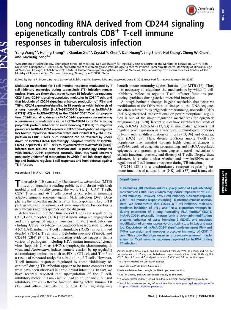
immune responses, we first examined CD244 expression levelsin CD4+ T cells, CD8+ T cells, and NK cells. Flow cytometricanalysis showed that, compared with healthy controls, activeTB infection induced significant increases in CD244+CD8+ Tcells but not CD244+CD3−CD56Bright NK cells (Fig. 1 A andB). In addition, percentages of CD244+CD8+ T cells weremuch higher than those of CD244+CD4+ T cells in peripheralblood mononuclear cells (PBMCs) from either healthy controlsor patients with active TB, regardless of ex vivo stimulationwith MTB lysates (Fig. 1 A and B). Furthermore, ex vivostimulation with MTB lysate induced further increases in per-centages of CD244+CD8+ T cells during active TB infection,suggesting that up-regulation of CD244 in CD8+ T cells is atleast partially TB-driven (Fig. 1 A and B). These data collec-tively suggested the importance of CD244 signaling in regu-lating CD8+ T-cell immune responses during active TBinfection. The role for CD8+ T cells in anti-TB immunity (5)led us to examine the expression of molecules associated withthe CD244 signaling pathway in CD8+ T cells during active TBinfection. Because signaling lymphocytic activation molecule(SLAM)-associated protein (SAP) and EWS-Fli1-activated tran-script 2 (EAT-2) are potential downstream molecules associated
0
200
400
600
pg/m
l
anti-CD244Isotype Ctrl
Medium + + +
++-
- --
** *NS
0
200
400
600
800
1000
TFN-
pg/m
l
anti-CD244Isotype Ctrl
Medium + + +
++-
- --
* *NS
0
500
1000
1500
2000
2500
IL-6
pg/m
l
anti-CD244Isotype Ctrl
Medium + ++
++-
- --
* *NS
TNF-IFN-
APC
PE
+Med
ium
+ant
iCD
244
+iso
type
Ctr
l
C D
F
ECD4
CD
244
Medium +Mtb lysates
CD56CD8 CD4 CD8 CD56
0.8
96.2
3.8
98.399.3
0.72.6
99.2
2
98 97.4
1.7
CD4CD
244
Medium +Mtb lysates
CD56CD8 CD4 CD8 CD56CD4
0.2
97.698.2
1.8
99.8
0.2
99.8 99.1
0.9
95.8
4.2
Healthy Control
2.4
Active TB
A
B
0.00.51.01.52.0
2468
101214
Healthy Control Active TBMedium Medium+Mtb lysates +Mtb lysates
CD3+CD4+
CD3+CD8+
CD3-CD56Bright***
*** ***
***
*** ******
CD
244+ c
ells
(% o
f CD
4+ or
CD
8+ T c
ells
or N
K c
ells)
Fig. 1. CD244 is preferentially up-regulated on CD8+ T cells during active MTB infection, and blockade of CD244 signaling enhances production of IFN-γ and TNF-αby CD8+ T cells. (A) Representative flow cytometric dot plots show the ex vivo expression of CD244 on CD4+ and CD8+ T cells and CD3-CD56Bright NK cells from onehealthy control and one patient with active TB. Data were gated on CD3+CD4+, CD3+CD8+, and CD3-CD56Bright. Percentages of CD244+ T (or NK) cells are shown inthe upright quadruple in each dot plot. PBMCs were treated either with or without ex vivo restimulation with MTB lysates. (B) Pooled data show the percentages ofCD244+CD4+ T cells, CD244+CD4+ T cells, or CD244+NK cells among total CD3+CD4+ T cells, CD3+CD8+ T cells, and NK cells (n = 15). Error bars represent SEM.(C) Representative CBA assays of a patient with active TB showing that treatment of anti-CD244 mAb induced significant increase of concentration of IFN-γ and TNF-α in culture supernatants of CD8+ T cells purified from PBMCs of patients with active TB. The red and green squares mark the TNF-α and IFN-γ, respectively. The dashedlines mark relative fluorescent intensity of TNF-α and IFN-γ. Treatment of anti-CD244 mAb increased the concentrations of TNF-α and IFN-γ (i.e., the fluorescentintensity of phycoerythrin (PE) increased, and squares shift toward right). (D–F) Pooled data show the concentrations of IFN-γ, TNF-α, and IL-6 in the presence ofindicated antibody treatment (n = 7). *P < 0.05; **P < 0.01; NS, no statistical significance. Error bars represent SEM from three independent experiments.
E3884 | www.pnas.org/cgi/doi/10.1073/pnas.1501662112 Wang et al.
with CD244 signaling (42), we sought to determine whether SAPand EAT-2 could be affected by CD244 signaling in CD8+ T cellsduring active TB infection. To address this, PBMCs from patientswith active TB were transfected with siRNA targeting CD244(siRNA-CD244), siRNA control (siRNA-Ctrl), or transfection me-dium. Although we detected significant percentages of SAP+CD8+
T-cell subsets and EAT-2+CD8+ T-cell subsets in CD8+ T cells frompatients with active TB (Fig. S1), siRNA-CD244 transfection sig-nificantly decreased percentages of CD244+CD8+ T, CD244+SAP+
CD8+ T, and CD244+EAT-2+CD8+ T-cell subsets in total CD8+ Tcells (Fig. S1). The data support the idea that SAP and EAT-2 (42–44) may be associated with CD244 signaling in CD8+ T cells duringactive TB infection.
Anti-CD244 mAb Modulation of CD244 Signaling in CD8+ T Cells fromTB Patients Leads to Increased Production of IFN-γ and TNF-α. Wethen examined the role of CD244 signaling in mediating theeffector function of CD8+ T cells. We found that anti-CD244mAb but not control IgG significantly increased concentration ofIFN-γ, TNF-α, and IL-6 in supernatants of cultured CD8+ T cellsfrom patients with active TB (Fig. 1 C–F). These data suggestedthat antibody modulation of CD244 on CD8+ T cells of patientswith active TB could signal an increase in effector function forcytokine production.
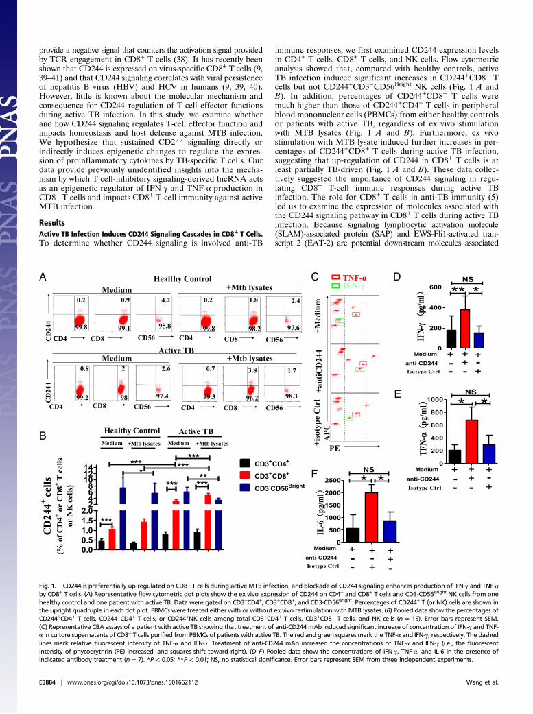
CD244 Signaling Epigenetically Inhibits EZH2 Expression, but theDifferential EZH2 Expression Itself Is Not Efficient Enough to InhibitIFN-γ and TNF-α Production in CD8+ T Cells. We then sought to ex-plore whether CD244 expression or signaling in CD8+ T cellscorrelated with altered expression of regulatory molecules inactive TB infection. We purified CD244+CD8+ T cells andCD244−CD8+ T cells from PBMCs of patients with active TB fordifferential expression of genes, and the hierarchical clusteringanalysis revealed differential gene expression profiles betweenCD244+CD8+ T and CD244−CD8+ T cells (Fig. 2A). Particu-larly, two genes encoding histone-modification enzymes, in-cluding polycomb protein enhancer of zeste homolog 2 (EZH2)and histone deacetylases 11 (HDAC11), were differentiallyexpressed between CD244+CD8+ T and CD244−CD8+ T cells(Fig. 2B). Confocal microscopic images of immunofluorescencestaining of EZH2 verified that CD8+ T cells expressed significantamounts of EZH2 in the nucleus (Fig. 2C).It has been suggested that EZH2 forms two closely related
PRC2 complexes that can trimethylate H3K27 (45, 46); thisprocess marks a repressive chromatin state coinciding with genesilencing (47). We thus examined whether differential EZH2expression between CD244+ and CD244− subpopulations mightimpact effector functions of CD8+ T cells during active TB. In-deed, quantitative (q)PCR validation indicated that CD244−CD8+
anti-C
D244
Isotyp
e
Medium
0.0
0.5
1.0
1.5
2.0
2.5
Rel
ativ
e E
xpre
ssio
n ofezh2
Nor
mal
ized
to G
APD
H)
024
306090
CD244- CD244+ CD244+CD244-
Medium +Mtb lysates
EZH2+EZH2-*
*
*
*
**
**
CD
8+ T c
ells
(% o
f gat
ed C
D8+ T
cells
)
P#1,CD244-
P#2,CD244-
P#3,CD244-
P#4,CD244-
P#1,CD244+
P#2,CD244+
P#6,CD244-
P#5,CD244-
P#3,CD244+
P#4,CD244+
P#5,CD244+
P#6,CD244+
A B
F
D
GE
92
1.5
0.8
3.1
0.488
CD244EZH
2
+Mtb lysatesMedium
CCD8 CD244 EZH2 Nucleus Merge
mRNA Folds of Change ##( CD244-/CD244+)
p value(Student t-test)
EZH2 2.3 0.01HDAC11 5.3 0.01
+
CD244
-
CD244
02468
10
CD8+T cells
***
Rel
ativ
eezh2
Qua
ntifi
catio
n(N
orm
aliz
ed )
Fig. 2. EZH2 correlates negatively with CD244 signaling. (A) Unsupervised clustering analysis of differentially expressed genes between CD244+CD8+ T cellsand CD244−CD8+ T cells that were purified from PBMCs of patients with active TB. Individual squares represent the relative gene expression intensity of thegiven genes (rows) in each of six patients (columns), with red indicating an increase in expression and blue a decrease. (B) A table shows the fold of changes ofexpression of EZH2 and HDAC11 in CD244+CD8+ T cells overexpression in CD244−CD8+ T cells. (C) Typical confocal microscopic images show the expression ofEZH2 in CD244high CD8+ T cells in PBMCs derived from patients with active TB. (Scale Bar: 5 μm.) (D) qPCR validation of differential expression of EZH2 genebetween CD244+CD8+ T cells and CD244−CD8+ T cells. (E) qPCR analysis of the EZH2 gene in PBMCs from patients with active TB treated with anti-CD244 mAbor control antibody for 5 d. Data are presented as relative expression levels of ezh2 in PBMCs treated with anti-CD244 mAb or control antibody over ex-pression levels of ezh2 in PBMCs treated with medium (n = 7). Data were normalized to GAPDH. (F) Representative flow cytometric dot plot data showingexpression of EZH2 and CD244 in CD8+ T cells of PBMCs with or without ex vivo restimulation with MTB lysates. Data were gated on CD8+ T cells. (G) Pooleddata show the frequency of EZH2+CD244−, EZH2-CD244+, and EZH2+CD244+ subpopulations of CD8+ T cells over total CD8+ T cells (n = 6). **P < 0.01; NS, nostatistical significance. Except for A, error bars represent SEM from two independent experiments.
Wang et al. PNAS | Published online July 6, 2015 | E3885
IMMUNOLO
GYAND
INFLAMMATION
PNASPL
US
T cells expressed much higher levels of EZH2 than their CD244+
counterparts (Fig. 2D). Consistently, ex vivo anti-CD244 mAbmodulation of CD244 signaling in PBMCs of patients with activeTB significantly enhanced ezh2 gene expression (Fig. 2E). Inaddition, immune costaining of CD244 and EZH2 in CD8+
T cells and flow cytometric analysis showed that percentages ofEZH2+CD244−CD8+ T-cell subsets or EZH2−CD244+CD8+
T-cell subsets are much higher than those of EZH2+CD244+CD8+
T-cell subsets regardless of MTB lysate ex vivo restimulation,suggesting that EZH2 and CD244 tend to be expressed in distinctCD8+ T-cell subpopulations during active TB infection (Fig. 2 Fand G). Thus, these results suggest that CD244 signaling nega-tively regulates EZH2 expression in CD8+ T cells during activeTB infection.
A B C
D
G F
H
E
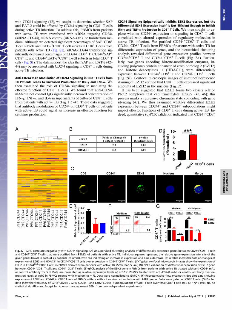
Fig. 3. lncRNA-CD244 is highly expressed in CD244+CD8+ T cells during active TB. (A) Unsupervised clustering analysis of differentially expressed lncRNAs betweenCD244+CD8+ T cells and CD244−CD8+ T cells that were purified from PBMCs of patients with active TB. Individual squares represent the relative lncRNA expressionintensity of the given lncRNAs (rows) in each of the patients (columns), with red indicating an increase in expression and blue a decrease. (B) Supervised clusteringanalysis using differentially expressed lncRNAs that can distinguish CD244+CD8+ T cells from CD244−CD8+ T cells. (C) A table shows the folds of change and P values(Student t test) of eight lncRNAs that could distinguish CD244+CD8+ T-cell subpopulation from CD244−CD8+ T-cell subpopulation of six patients with active TB. (D)qPCR validation of differential expression of lncRNA-CD244 between CD244+CD8+ T cells and CD244−CD8+ T cells. (E) qPCR analysis of lncRNA-CD244 expression inCD244+CD8+ T cells purified from PBMCs of patients with active TB upon stimulation withMTB ESAT-6 (10 μg/mL) or CFP-10 (10 μg/mL) peptide pools for 5 d in vitro. (F)Schematic diagram of the lncRNA-CD244 genomic locus in human chromosome 22. The bars represent exons, dashed lines represent intron, red line represents 5′ (or3′) UTR, and arrows indicate the direction of transcription. The length of lncRNA-CD244 is 796 bases, which overlapped 5′ UTR of GST θ1 (GSTT1) by 72 bases. Nohomologs of lncRNA-CD244 were found in mouse (Fig. S3 A and B). (G) Northern blot analysis show the expression of full-length lncRNA-CD244 in CD244+CD8+ T cellsfrom patients with active TB using specific probe but not antisense control probe. No lncRNA-CD244 was detected in HEK293T cells. U6 RNA served as a control. (H)Plasmids as schematically shown at Left were transfected to HEK293T cells (Right). Immunoblotting using antibody specific to EGFP and fluorescent imaging (Fig. S5)showed that lncRNA-CD244-EGFP plasmid and lncRNA-CD244 plasmid did not express GFP. Error bars represent SEM. Data shown in D, E, G, and H are representativeof at least two independent experiments.
E3886 | www.pnas.org/cgi/doi/10.1073/pnas.1501662112 Wang et al.

We then determined whether differential expression of EZH2contributes to inhibition of IFN-γ and TNF-α production byCD8+ T cells during active TB infection. PBMCs from patientswith active TB were transfected with siRNA targeting EZH2(siRNA-EZH2), control siRNA (siRNA-Ctrl), or transfectionmedium only. Compared with PBMCs transfected with siRNA-Ctrl or medium, transfection of siRNA-EZH2 did not signifi-cantly increase production of IFN-γ and TNF-α in PBMC culturesupernatants or percentages of IFN-γ+CD8+ T cells and TNF-α+CD8+ T cells among total CD8+ T cells (Fig. S2 A–G). Consis-tently, ChIP-qPCR also showed that, compared with siRNA-Ctrl,siRNA-EZH2 was not able to induce a significant decrease in theamounts of H3K27Me3 at promoter regions of infg and tnfa loci(Fig. S2H). Thus, the differential expression of EZH2 itself inCD244+ T cells was not efficient enough to depress productionof IFN-γ and TNF-α by CD8+ T cells in active TB.
CD244 Signaling Positively Correlates with High-Level Expression ofTB-Specific lncRNA-BC050410 [lncRNA-AS-GSTT1(1-72) or lncRNA-CD244]in CD8+ T Cells. Because differential EZH2 expression was not effi-cient enough to inhibit production of IFN-γ and TNF-α by CD8+ Tcells, we then investigated the possibility that EZH2 might berecruited to the promoters of IFN-γ and TNF-α in CD8+ T cells toinduce repressive chromatin states at infg and tnfa loci in CD244+
CD8+ T cells. This consideration was supported by the finding thatlncRNA might mediate targeted recruitment of repressive histone-modifying activities to epigenetically silence transcription (48–52).We used human lncRNA microarray and hierarchical clusteringanalyses to compare lncRNA expression in CD244+CD8+ T cells andCD244−CD8+ T cells. The comparative analysis between these twosubsets allowed us to display a distinct lncRNA expression profile inCD244+CD8+ T cells (Fig. 3A). The supervised hierarchal clusteringsegregation analysis then identified dominant groups of lncRNAsselectively expressed in CD244+CD8+ T cells (Fig. 3B). Interestingly,
lncRNA-BC050410 [named as lncRNA-AS-GSTT1(1-72) based onits genomic context (53) and termed as lncRNA-CD244 here forsimplicity] with genomic overlapping to 5′UTR of GST θ1 (GSTT1)was one of the eight lncRNAs that could distinguish the CD244+
CD8+ T-cell subpopulation from its CD244− counterpart, with alargest-fold difference and a most-significant P value (Fig. 3 C and Eand Fig. S3 A and B). Note that lncRNA-CR592555, a lncRNAmostly down-regulated in CD244+CD8+ T cells, was located between79,946,861 bp and ∼79,947,776 bp in chromosome 5 (Fig. S4). Suchdifferential expression of lncRNA-CD244 in CD244+CD8+ T cells inactive TB was also validated by qPCR (Fig. 3D). Furthermore,stimulation with peptide pools of 6-kDa early secretory antigenictarget (ESAT-6) or 10-kDa culture filtrate protein (CFP-10) of MTBinduced higher expression levels of lncRNA-CD244 in purifiedCD244+CD8+ T cells, suggesting that expression of lncRNA-CD244is at least partially TB-specific (Fig. 3E). Also, expression of full-length lncRNA-CD244 was confirmed by Northern blot analysis (Fig.3F). We then assessed whether lncRNA-CD244 has protein-codingcapability. As with lncRNA-DC (35), analysis based on coding po-tential calculator (CPC) (Fig. S3 C–E) suggests that the overallprotein-coding potential of lncRNA-CD244 is weak [e.g., the hitscore of lncRNA-CD244 (∼23.56) is as small as that of lncRNA-DC(∼24.35), which suggests that lncRNA-CD244, like lncRNA-DC(35), is unlikely protein-coding; lncRNA-CD244 has five potentialORFs (Fig. S3F), but ORF coverage of lncRNA-CD244 (∼33.54%)is much smaller than that of lncRNA-DC (∼50.72%), which suggeststhat the ORF quality of lncRNA-CD244 is worse than that oflncRNA-DC]. In addition, analysis of the ratio of the number ofnonsynonymous substitutions per nonsynonymous site to the numberof synonymous substitutions per synonymous site (Ka/Ks or dN/dS)between two different species of Hominoidea (e.g., dN/dS ratio be-tween human and chimpanzee is 0.48; P = 0.068 > 0.05) (Fig. S3G)suggests that the ORFs of lncRNA-CD244 lack negative selection inspecies of Hominoidea. Thus, no bioinformatics evidence implicates
A
B C F
D E G
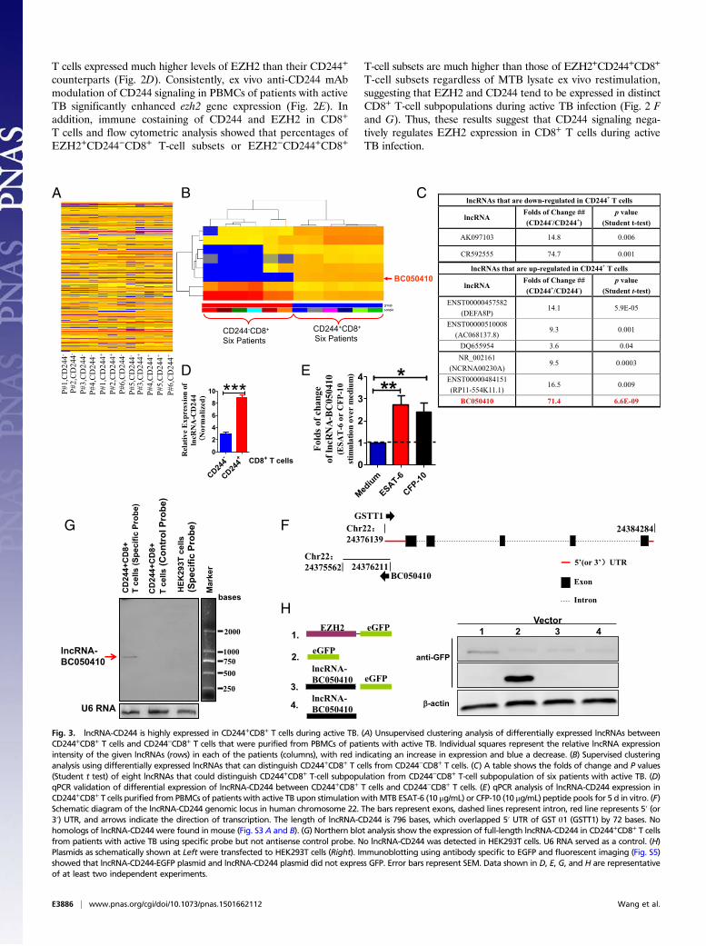
Fig. 4. Knock down of CD244 or blockade of CD244 signaling induces a more repressive chromatin state in lncRNA-CD244 locus and inhibits expression oflncRNA-CD244. Seven regions (capital letters A to G) across lncRNA-CD244 locus, as shown in A, were analyzed in ChIP-qPCR analyses for H3K27Me3 (B and D)histone modification and EZH2 (C and E) in PBMCs from patients with active TB. PBMCs were transfected with siRNA-CD244 or siRNA-Ctrl or treated with anti-CD244 mAb or IgG control as indicated in each of subfigure. Values derived from three independent experiments were normalized by background signals andinput chromatin. (F and G) qPCR analysis of lncRNA-CD244 and/or the cd244 gene in PBMCs from patients with active TB transfected (or treated) with in-dicated siRNAs (F) or antibodies (G). Data are presented as relative expression levels of lncRNA-CD244 (or cd244) (normalized to GAPDH) in siRNA-CD244–transfected (or anti-CD244–treated) PBMCs over expression levels of lncRNA-CD244 (or cd244) in siRNA-Ctrl–transfected (or IgG-treated) PBMCs (n = 7). *P <0.05; **P < 0.01; ***P < 0.001; NS, no statistical significance. Error bars represent SEM from three independent experiments.
Wang et al. PNAS | Published online July 6, 2015 | E3887
IMMUNOLO
GYAND
INFLAMMATION
PNASPL
US
lncRNA-CD244 as having protein-coding capability. Furthermore, avector construct comprising full-length lncRNA-CD244 and EGFPtag was developed and assessed for expression using immunoblotting(IB) and fluorescent imaging. The lncRNA-CD244 did not have anydetectable protein-coding ability because neither lncRNA-CD244-EGFP vector nor lncRNA-CD244 vector expressed EGFP (Fig. 3Hand Fig. S5). Thus, lncRNA-CD244 preferentially expressed inCD244+CD8+ T cells during active human TB infection.
CD244 Signaling Drives lncRNA-CD244 Expression via Sustaining aMore Permissive Chromatin State in lncRNA-CD244 Locus. To de-termine the mechanisms underlying the preferential expressionof lncRNA-CD244 mediated by CD244 signaling, PBMCs ofpatients with active TB were transfected with siRNA targetingCD244 (siRNA-CD244) and control siRNA (siRNA-Ctrl) ortreated with anti-CD244 and control IgG. ChIP-qPCR analysisshowed that EZH2 and trimethylation at H3K27, a histone modi-fication that negatively regulates transcription, markedly increasedin lncRNA-CD244 loci after treatment with siRNA-CD244 butnot siRNA-Ctrl (Fig. 4 A–C). Consistently, siRNA-CD244, notsiRNA-Ctrl, decreased expression of the cd244 gene and lncRNA-CD244 (Fig. 4F). Furthermore, anti-CD244 but not IgG controlinduced significant increases of H3K27Me3 and EZH2 inlncRNA-CD244 loci (Fig. 4 A, D, and E), and anti-CD244 but notIgG significantly decreased the expression of lncRNA-CD244(Fig. 4G). Taken together, knock down of CD244 or blockade ofCD244 signaling induces a more repressive chromatin state inlncRNA-CD244 locus and reduces expression of lncRNA-CD244.Thus, these results implicate CD244 signaling as driving expres-sion of lncRNA-CD244 in CD8+ T cells most likely through sus-taining a more permissive chromatin state in the lncRNA-CD244locus during active TB infection.
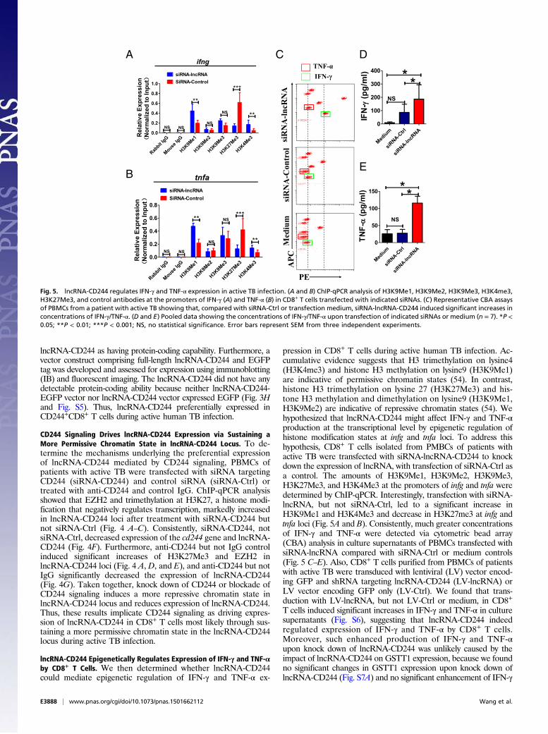
lncRNA-CD244 Epigenetically Regulates Expression of IFN-γ and TNF-αby CD8+ T Cells. We then determined whether lncRNA-CD244could mediate epigenetic regulation of IFN-γ and TNF-α ex-
pression in CD8+ T cells during active human TB infection. Ac-cumulative evidence suggests that H3 trimethylation on lysine4(H3K4me3) and histone H3 methylation on lysine9 (H3K9Me1)are indicative of permissive chromatin states (54). In contrast,histone H3 trimethylation on lysine 27 (H3K27Me3) and his-tone H3 methylation and dimethylation on lysine9 (H3K9Me1,H3K9Me2) are indicative of repressive chromatin states (54). Wehypothesized that lncRNA-CD244 might affect IFN-γ and TNF-αproduction at the transcriptional level by epigenetic regulation ofhistone modification states at infg and tnfa loci. To address thishypothesis, CD8+ T cells isolated from PMBCs of patients withactive TB were transfected with siRNA-lncRNA-CD244 to knockdown the expression of lncRNA, with transfection of siRNA-Ctrl asa control. The amounts of H3K9Me1, H3K9Me2, H3K9Me3,H3K27Me3, and H3K4Me3 at the promoters of infg and tnfa weredetermined by ChIP-qPCR. Interestingly, transfection with siRNA-lncRNA, but not siRNA-Ctrl, led to a significant increase inH3K9Me1 and H3K4Me3 and decrease in H3K27me3 at infg andtnfa loci (Fig. 5A and B). Consistently, much greater concentrationsof IFN-γ and TNF-α were detected via cytometric bead array(CBA) analysis in culture supernatants of PBMCs transfected withsiRNA-lncRNA compared with siRNA-Ctrl or medium controls(Fig. 5 C–E). Also, CD8+ T cells purified from PBMCs of patientswith active TB were transduced with lentiviral (LV) vector encod-ing GFP and shRNA targeting lncRNA-CD244 (LV-lncRNA) orLV vector encoding GFP only (LV-Ctrl). We found that trans-duction with LV-lncRNA, but not LV-Ctrl or medium, in CD8+
T cells induced significant increases in IFN-γ and TNF-α in culturesupernatants (Fig. S6), suggesting that lncRNA-CD244 indeedregulated expression of IFN-γ and TNF-α by CD8+ T cells.Moreover, such enhanced production of IFN-γ and TNF-αupon knock down of lncRNA-CD244 was unlikely caused by theimpact of lncRNA-CD244 on GSTT1 expression, because we foundno significant changes in GSTT1 expression upon knock down oflncRNA-CD244 (Fig. S7A) and no significant enhancement of IFN-γ
A C D
EB
Fig. 5. lncRNA-CD244 regulates IFN-γ and TNF-α expression in active TB infection. (A and B) ChIP-qPCR analysis of H3K9Me1, H3K9Me2, H3K9Me3, H3K4me3,H3K27Me3, and control antibodies at the promoters of IFN-γ (A) and TNF-α (B) in CD8+ T cells transfected with indicated siRNAs. (C) Representative CBA assaysof PBMCs from a patient with active TB showing that, compared with siRNA-Ctrl or transfection medium, siRNA-lncRNA-CD244 induced significant increases inconcentrations of IFN-γ/TNF-α. (D and E) Pooled data showing the concentrations of IFN-γ/TNF-α upon transfection of indicated siRNAs or medium (n = 7). *P <0.05; **P < 0.01; ***P < 0.001; NS, no statistical significance. Error bars represent SEM from three independent experiments.
E3888 | www.pnas.org/cgi/doi/10.1073/pnas.1501662112 Wang et al.
and TNF-α production upon GSTT1 knock down by shRNA (Fig.S7 B and C). Furthermore, such enhanced production of IFN-γand TNF-α was not attributable to changes in viability of CD8+
T cells because CD8+ T cells did not exhibit enhanced apoptosisafter lncRNA-CD244 knockdown (Fig. S7 D and E). Thus, theseresults collectively suggest that whereas TB-driven up-regulation
A
F
G H
B
E
C D
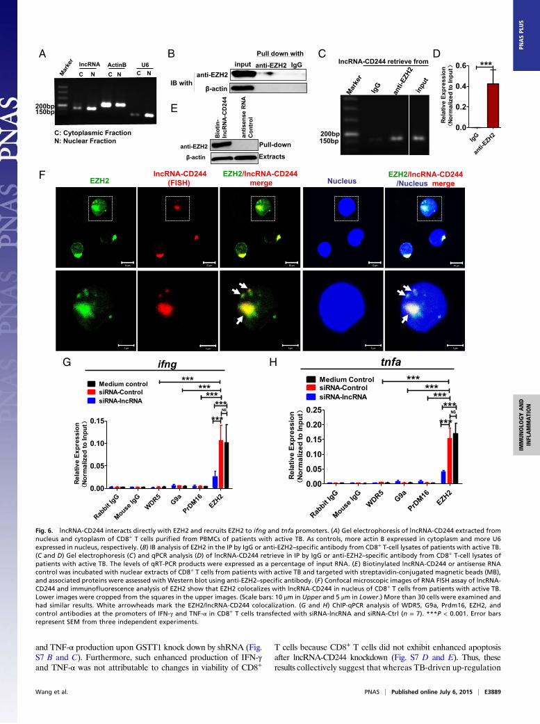
Fig. 6. lncRNA-CD244 interacts directly with EZH2 and recruits EZH2 to ifng and tnfa promoters. (A) Gel electrophoresis of lncRNA-CD244 extracted fromnucleus and cytoplasm of CD8+ T cells purified from PBMCs of patients with active TB. As controls, more actin B expressed in cytoplasm and more U6expressed in nucleus, respectively. (B) IB analysis of EZH2 in the IP by IgG or anti-EZH2–specific antibody from CD8+ T-cell lysates of patients with active TB.(C and D) Gel electrophoresis (C ) and qPCR analysis (D) of lncRNA-CD244 retrieve in IP by IgG or anti-EZH2–specific antibody from CD8+ T-cell lysates ofpatients with active TB. The levels of qRT-PCR products were expressed as a percentage of input RNA. (E ) Biotinylated lncRNA-CD244 or antisense RNAcontrol was incubated with nuclear extracts of CD8+ T cells from patients with active TB and targeted with streptavidin-conjugated magnetic beads (MB),and associated proteins were assessed with Western blot using anti-EZH2–specific antibody. (F ) Confocal microscopic images of RNA FISH assay of lncRNA-CD244 and immunofluorescence analysis of EZH2 show that EZH2 colocalizes with lncRNA-CD244 in nucleus of CD8+ T cells from patients with active TB.Lower images were cropped from the squares in the upper images. (Scale bars: 10 μm in Upper and 5 μm in Lower.) More than 30 cells were examined andhad similar results. White arrowheads mark the EZH2/lncRNA-CD244 colocalization. (G and H) ChIP-qPCR analysis of WDR5, G9a, Prdm16, EZH2, andcontrol antibodies at the promoters of IFN-γ and TNF-α in CD8+ T cells transfected with siRNA-lncRNA and siRNA-Ctrl (n = 7). ***P < 0.001. Error barsrepresent SEM from three independent experiments.
Wang et al. PNAS | Published online July 6, 2015 | E3889
IMMUNOLO
GYAND
INFLAMMATION
PNASPL
US
of lncRNA-CD244 might influence repressive chromatin states atinfg and tnfa loci, silence or down-regulation of lncRNA-CD244could confer permissive chromatin states at infg and tnfa loci andenhance expression of IFN-γ and TNF-α.
lncRNA-CD244 Associates Physically with EZH2 and Mediates Recruitmentof EZH2 to ifng and tnfa Loci for Repressive Chromatin States. Next, wesought to examine the interrelation of lncRNA-CD244, repressivechromatin states at infg and tnfa loci, and altered expression ofEZH2 (Fig. 2). We presumed that lncRNA-CD244 could mediatethe recruitment of the histone-modifying enzyme EZH2, whichcatalyzed the trimethylation modification of H3K27 at promoters ofinfg and tnfa. Of note, while performing gel electrophoresis of RNAextracts of nuclear and cytoplasmic fractions of CD8+ T cells frompatients with active TB, we found that most of the lncRNA-CD244localized in the nuclear fraction of CD8+ T cells (Fig. 6A). In ad-dition, when we performed immunoprecipitation (IP) of EZH2 inextracts of CD8+ T cells from TB patients or in HEK293T cells withexogenous expression of EZH2 and full-length lncRNA-CD244, wefound that EZH2-specific mAb, but not control IgG, could actuallycoprecipitate lncRNA-CD244 molecules, as detected by qRT-PCR(Fig. 6 B–D and Fig. S8). Furthermore, biotinylated lncRNA-CD244 and an antisense control RNA were incubated with nuclearextracts of CD8+ T cells from patients with active TB, and Westernblotting showed that lncRNA-CD244 but not antisense controlRNA specifically bound to EZH2 (Fig. 6E). Consistently, when weperformed confocal microscope-based FISH analysis of lncRNA-CD244 and immunofluorescent analysis of EZH2, we found thatsignificant amounts of EZH2 colocalized with lncRNA-CD244 inthe nucleus of CD8+ T cells from patients with active TB (Fig. 6F).These results suggest that lncRNA-CD244 is physically associatedwith EZH2 during active TB. Furthermore, transfection of CD8+ Tcells with siRNA-lncRNA-CD244, but not siRNA-Ctrl, led to sig-nificant decreases in EZH2, but not WDR5, Prdm16, and G9a, at
either the infg or tnfa promoter (Fig. 6 G and H). ChIP-qPCRanalysis also demonstrated that EZH2 accumulated at promoters ofinfg or tnfa at much greater levels than WDR5, PRDM16, and G9ain CD8+ T cells from patients with active TB (Fig. 6 G and H).These results suggest that EZH2 and lncRNA-CD244 complexformation may lead to trimethylation of H3K27, which contributesto inducing repressive chromatin states at infg or tnfa loci in CD8+ Tcells during active TB infection. The data also suggest a hypotheticalmodel in which expression of lncRNA-CD244 may physically recruitEZH2 to control H3K27Me3 at the ifng and tnfa loci and thereforeallow chromatin to program repressive states and inhibit tran-scription of infg and tnfa genes in CD244+CD8+ T cells.
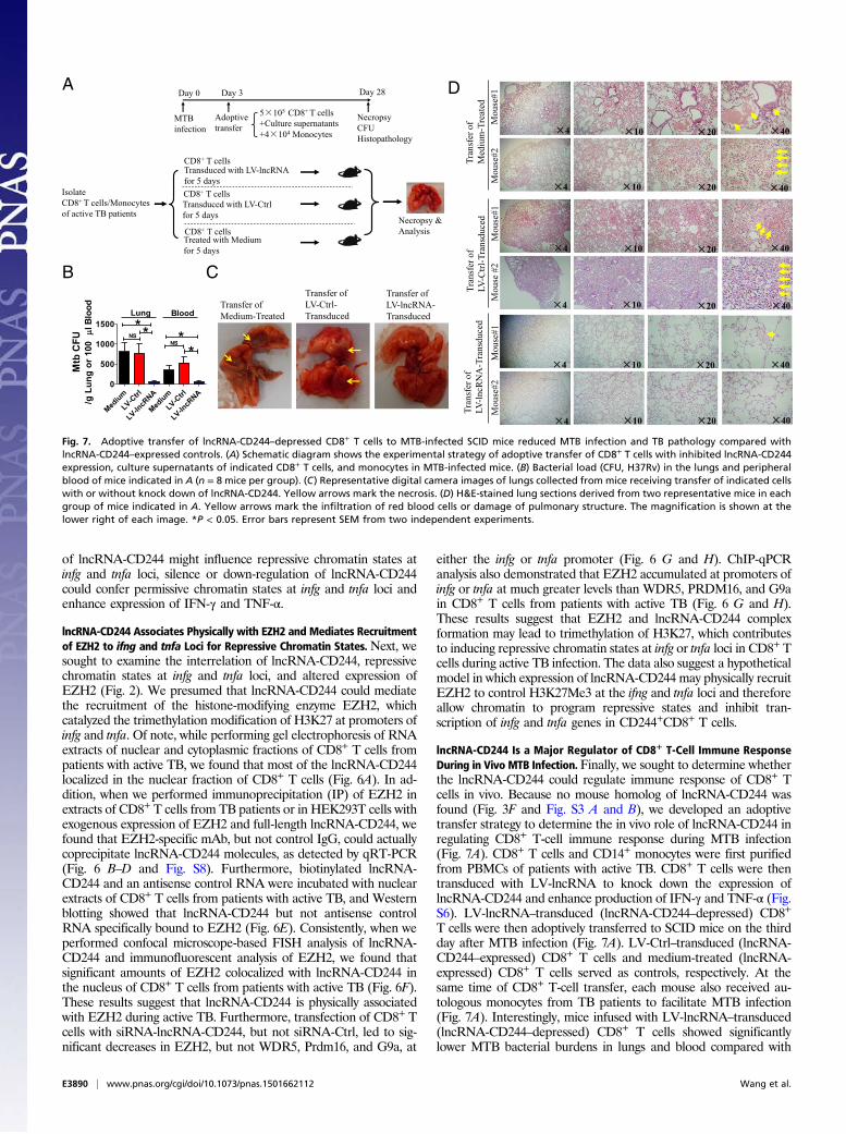
lncRNA-CD244 Is a Major Regulator of CD8+ T-Cell Immune ResponseDuring in Vivo MTB Infection. Finally, we sought to determine whetherthe lncRNA-CD244 could regulate immune response of CD8+ Tcells in vivo. Because no mouse homolog of lncRNA-CD244 wasfound (Fig. 3F and Fig. S3 A and B), we developed an adoptivetransfer strategy to determine the in vivo role of lncRNA-CD244 inregulating CD8+ T-cell immune response during MTB infection(Fig. 7A). CD8+ T cells and CD14+ monocytes were first purifiedfrom PBMCs of patients with active TB. CD8+ T cells were thentransduced with LV-lncRNA to knock down the expression oflncRNA-CD244 and enhance production of IFN-γ and TNF-α (Fig.S6). LV-lncRNA–transduced (lncRNA-CD244–depressed) CD8+
T cells were then adoptively transferred to SCID mice on the thirdday after MTB infection (Fig. 7A). LV-Ctrl–transduced (lncRNA-CD244–expressed) CD8+ T cells and medium-treated (lncRNA-expressed) CD8+ T cells served as controls, respectively. At thesame time of CD8+ T-cell transfer, each mouse also received au-tologous monocytes from TB patients to facilitate MTB infection(Fig. 7A). Interestingly, mice infused with LV-lncRNA–transduced(lncRNA-CD244–depressed) CD8+ T cells showed significantlylower MTB bacterial burdens in lungs and blood compared with
A
B C
D
Fig. 7. Adoptive transfer of lncRNA-CD244–depressed CD8+ T cells to MTB-infected SCID mice reduced MTB infection and TB pathology compared withlncRNA-CD244–expressed controls. (A) Schematic diagram shows the experimental strategy of adoptive transfer of CD8+ T cells with inhibited lncRNA-CD244expression, culture supernatants of indicated CD8+ T cells, and monocytes in MTB-infected mice. (B) Bacterial load (CFU, H37Rv) in the lungs and peripheralblood of mice indicated in A (n = 8 mice per group). (C) Representative digital camera images of lungs collected from mice receiving transfer of indicated cellswith or without knock down of lncRNA-CD244. Yellow arrows mark the necrosis. (D) H&E-stained lung sections derived from two representative mice in eachgroup of mice indicated in A. Yellow arrows mark the infiltration of red blood cells or damage of pulmonary structure. The magnification is shown at thelower right of each image. *P < 0.05. Error bars represent SEM from two independent experiments.
E3890 | www.pnas.org/cgi/doi/10.1073/pnas.1501662112 Wang et al.
control mice infused with LV-Ctrl–transduced (lncRNA-CD244–expressed) CD8+ T cells or medium-treated (lncRNA-CD244–expressed) CD8+ T cells (Fig. 7B). The control mice infused withLV-Ctrl–transduced CD8+ T cells or medium-treated CD8+ T cellsexhibited severer lung necrosis (Fig. 7C) and histopathology char-acterized by significant hemorrhages or infiltration of RBCs inalveoli and apparent damage of pulmonary structures (Fig. 7D). Incontrast, only mild changes in alveoli were observed in lung sectionsof mice infused with LV-lncRNA-CD244–transduced CD8+ T cells(Fig. 7D). Thus, lncRNA-CD244–depressed CD8+ T cells can morepotently control in vivo MTB infection than lncRNA-CD244–expressed CD8+ T cells in SCID mice, suggesting that lncRNA-CD244 may play a role in regulating in vivo immune response ofCD8+ T cells during MTB infection.
DiscussionMolecular mechanisms for CD244 modulation of T-cell responsesin infections remain unknown, although CD244 appears to regu-late NK cell function that correlates with HBV or HCV persistencein humans (40, 41). In this study, we find that human TB can up-regulate CD244 and CD244 signaling-related molecules in CD8+ Tcells. Whereas the CD244+CD8+ T-cell subset from TB patientsexpresses much higher levels of lncRNA-CD244 than its CD244−
CD8+ T-cell subset counterpart, siRNA or shRNA knock downof lncRNA-CD244 leads to increased production of IFN-γ andTNF-α. The blockade of CD244 signaling by anti-CD244 mAb cansimilarly lead to in vitro enhancement of IFN-γ and TNF-α pro-duction. Thus, we demonstrate the interrelation between CD244signaling, lncRNA-CD244 expression, and regulation of effectorfunction in CD8+ T cells at epigenetic levels in TB patients.The current study provides a previously unidentified mechanism
by which CD244 signaling regulates IFN-γ and TNF-α productionin human CD8+ T cells in MTB infection. CD244 signaling duringactive MTB infection can exploit lncRNA and histone-modifyingenzymes to regulate the effector functions of CD244+CD8+ T cells.Often, CD244 expression/signaling in CD244+CD8+ T cellsfrom TB patients leads to remarkable increases in expression oflncRNA-CD244. Interestingly, lncRNA-CD244 appears to phys-ically interact with a polycomb protein, EZH2. This interactionmediates recruitment of EZH2 to infg and tnfa loci and trime-thylates H3K27 at promoters of infg and tnfa toward repressivechromatin states and suppression of infg and tnfa expression. Ourfindings support the hypothesis that ncRNA can interact withchromatin and mediate targeted recruitment of repressive his-tone-modifying activities to epigenetically silence transcription(49, 51). In fact, it has been recently shown that a number ofintronic RNA sequences are capable of binding to the corecomponent EZH2 and regulating the transcriptional output of itsgenomic counterpart (55). In addition, the ncRNAs HOTAIR(56), Xist (57), and RepA (58) might recruit the polycombcomplex to the HoxD locus or the X chromosome, respectively,where they mediate trimethylation of H3K27 and induce het-erochromatin formation and repression of gene expression.Furthermore, EZH2 is also shown to bind strongly to genesencoding the transcription factors T-bet, Eomes, and Gata3;controls differentiation into Th1 and Th2 effector cells; andregulates plasticity of these subsets after differentiation (59).However, we cannot rule out the possibility that lncRNA-CD244 in our model may regulate the repression of infg andtnfa via other undefined epigenetic mechanisms (48, 50).The data from the current study also suggest that down-regulation
of lncRNA-CD244 in CD8+ T cells from TB patients can yielda favorable in vivo consequence during MTB infection at SCIDmodel. Adoptive transfer of lncRNA-CD244–depressed CD8+ Tcells with the enhanced IFN-γ/TNF-α production could attenuateMTB infection and TB pathology in SCID mice compared withlncRNA-CD244–expressed controls. Enhanced CTL activity andIFN-γ/TNF-α production of lncRNA-CD244–depressed CD8+ T
cells may contribute to inhibition of intracellular MTB replicationin infused autologous monocytes/macrophages. This notion is sup-ported by the previous studies demonstrating CD8+ T-cell-mediatedanti-TB immunity (5). Thus, the identification of the CD244 andlncRNA-CD244 axis in the modulation of IFN-γ and TNF-α ex-pression provides an lncRNA-driven epigenetic program of T-cellimmunity against MTB infection.lncRNAs have recently been in the spotlight for their critical
roles in human biology and diseases (21, 22, 34, 50, 60, 61). Theyhave been explored as biomarkers for cancers and potentialtargets for disease or dysregulated expression of genes or phe-notypes (49). Here, we demonstrate that lncRNA-CD244 servesas one of the mechanisms by which CD244 signaling regulatesthe ability of human CD8+ T cells to produce IFN-γ and TNF-αat epigenetic levels in MTB infection. To our knowledge, this isthe first evidence that lncRNA is one of the major epigeneticfactors modulating Th1 immune response in human TB.Thus, this study allows us to demonstrate that CD244 signaling
in active human TB regulates repression of IFN-γ and TNF-αthrough a mechanism in which lncRNA-CD244 modulates re-cruitment of EHZ2 to promoters of IFN-γ and TNF-α for po-tential trimethylation of H3K27 and repression of infg and tnfaexpression (Fig. S9). The CD244 signaling and lncRNA-CD244modulation of IFN-γ and TNF-α expression presents an lncRNA-driven epigenetic program of T-cell immunity against microbialinfection. Our findings also suggest that lncRNA-CD244 may be apotential target for therapeutic intervention of TB.
Materials and MethodsStudy Subjects. Active TB infections were confirmed based on clinical symp-toms, chest radiography, sputum staining for acid-fast bacilli (AFB), and labo-ratory culture and PCR for MTB that were carried out in the Institute for ChronicDiseases Prevention of Huadu District in Guangzhou, China. All patients were notreceiving anti-TB therapy at the time of analysis. Patients with active TB wererecruited in our study and gave written informed consent according to theprotocols approved by the institutional review and the ethics boards of theZhongshan School of Medicine of Sun Yat-sen University (SYSU).
Animal study protocols were also reviewed and approved by the SYSUInstitutional Animal Care and Use Committee.
Statistics. Statistical analysis was performed using GraphPad Prism. Statisticalsignificance was determined with Student t test. A value of P < 0.05 wasconsidered significant. Asterisks in the figures represent the following:*P < 0.05, **P < 0.01, and ***P < 0.001. “NS” in the figures indicates nostatistical significance.
Full materials and methods and any associated references for followingexperiments are described in detail in SI Materials and Methods: isolation ofPBMCs; antibodies for flow cytometry; CBA analysis of the cell culture su-pernatants; blocking experiments using anti-CD244 mAb; intracellular cytokinestaining (ICS) and flow cytometric assay; siRNA transfection; purification ofmonocytes, CD8+CD244− T cells, and CD8+CD244+ T cells; mRNA and lncRNAmicroarray analysis, data analysis, and statistics; lentivirus-mediated knock downof lncRNA-CD244 or GSTT1; qPCR; ChIP-qPCR; RNA IP (RIP)-qPCR; nuclear andcytoplasmic extraction of lncRNA; RNA FISH and immunofluorescence micros-copy; confocal microscopic analysis; bioinformatics analyses of evolutionaryconservation and coding potential of lncRNA-CD244 and plasmid constructions;Northern blot assay of lncRNA; lncRNA pull-down assay; MTB infection of mice;adoptive transfer; and histopathological, bacterial, and immune analyses ofMTB-infected mice.
ACKNOWLEDGMENTS. We thank members of the Sun Yat-sen University Bio-safety Level-3 Laboratory for biosafety management; Xiaobo Li for flow cyto-metric analysis; Dr. Yijun Zhang, Shaoyuan Li, Sufen Zhang, and Dongting Maofor technical assistance; and Jun Liu, JianguoWang, and Dr. Xionglei He for helpwith bioinformatics analysis. This work was supported by National Natural Sci-ence Foundation of China–National Institutes of Health (NSFC-NIH) BiomedicalCollaborative Research Program Grant 81361120379 (to G.Z.), NSFC Grant31170847 (to G.Z.), the Guangzhou Municipality Commission for Science andTechnology Innovation (Pearl River S&T Nova Program) Grant 201506010034(to G.Z.), Guangdong Innovative R&D Team Program Grant 2009010058 (toH. Zhang), United States–China Collaborative Research Program Grants NIHAI106590 and NSFC 31129002 (to Z.W.C.), and NIH R01 Grants HL64560 andOD015092 (RR13601) and AI106590 (to Z.W.C.).
Wang et al. PNAS | Published online July 6, 2015 | E3891
IMMUNOLO
GYAND
INFLAMMATION
PNASPL
US

1. Rubin EJ (2014) Troubles with tuberculosis prevention. N Engl J Med 370(4):375–376.2. Zumla A, George A, Sharma V, Herbert N; Baroness Masham of Ilton (2013) WHO’s
2013 global report on tuberculosis: Successes, threats, and opportunities. Lancet382(9907):1765–1767.
3. Bold TD, Ernst JD (2012) CD4+ T cell-dependent IFN-γ production by CD8+ effector Tcells in Mycobacterium tuberculosis infection. J Immunol 189(5):2530–2536.
4. Cooper AM (2009) Cell-mediated immune responses in tuberculosis. Annu Rev Im-munol 27:393–422.
5. Chen CY, et al. (2009) A critical role for CD8 T cells in a nonhuman primate model oftuberculosis. PLoS Pathog 5(4):e1000392.
6. Kaufmann SH (2001) How can immunology contribute to the control of tuberculosis?Nat Rev Immunol 1(1):20–30.
7. Chen CY, et al. (2013) Phosphoantigen/IL2 expansion and differentiation of Vγ2Vδ2 Tcells increase resistance to tuberculosis in nonhuman primates. PLoS Pathog 9(8):e1003501.
8. Nunes-Alves C, et al. (2014) In search of a new paradigm for protective immunity toTB. Nat Rev Microbiol 12(4):289–299.
9. Bengsch B, et al. (2010) Coexpression of PD-1, 2B4, CD160 and KLRG1 on exhaustedHCV-specific CD8+ T cells is linked to antigen recognition and T cell differentiation.PLoS Pathog 6(6):e1000947.
10. Wherry EJ, et al. (2007) Molecular signature of CD8+ T cell exhaustion during chronicviral infection. Immunity 27(4):670–684.
11. Ha SJ, et al. (2008) Enhancing therapeutic vaccination by blocking PD-1-mediatedinhibitory signals during chronic infection. J Exp Med 205(3):543–555.
12. Blackburn SD, et al. (2009) Coregulation of CD8+ T cell exhaustion by multiple in-hibitory receptors during chronic viral infection. Nat Immunol 10(1):29–37.
13. Zhu Y, Yao S, Chen L (2011) Cell surface signaling molecules in the control of immuneresponses: A tide model. Immunity 34(4):466–478.
14. Velu V, et al. (2009) Enhancing SIV-specific immunity in vivo by PD-1 blockade. Nature458(7235):206–210.
15. Qiu Y, et al. (2012) Tim-3-expressing CD4+ and CD8+ T cells in human tuberculosis (TB)exhibit polarized effector memory phenotypes and stronger anti-TB effector func-tions. PLoS Pathog 8(11):e1002984.
16. Jayaraman P, et al. (2010) Tim3 binding to galectin-9 stimulates antimicrobial im-munity. J Exp Med 207(11):2343–2354.
17. Mattick JS, Gagen MJ (2001) The evolution of controlled multitasked gene networks:The role of introns and other noncoding RNAs in the development of complex or-ganisms. Mol Biol Evol 18(9):1611–1630.
18. Guttman M, et al. (2009) Chromatin signature reveals over a thousand highly con-served large non-coding RNAs in mammals. Nature 458(7235):223–227.
19. Gupta RA, et al. (2010) Long non-coding RNA HOTAIR reprograms chromatin state topromote cancer metastasis. Nature 464(7291):1071–1076.
20. Huarte M, et al. (2010) A large intergenic noncoding RNA induced by p53 mediatesglobal gene repression in the p53 response. Cell 142(3):409–419.
21. Cesana M, et al. (2011) A long noncoding RNA controls muscle differentiation byfunctioning as a competing endogenous RNA. Cell 147(4):358–369.
22. Rinn JL, Chang HY (2012) Genome regulation by long noncoding RNAs. Annu RevBiochem 81(81):145–166.
23. Mercer TR, Mattick JS (2013) Structure and function of long noncoding RNAs in epi-genetic regulation. Nat Struct Mol Biol 20(3):300–307.
24. Engreitz JM, et al. (2013) The Xist lncRNA exploits three-dimensional genome archi-tecture to spread across the X chromosome. Science 341(6147):1237973.
25. Monnier P, et al. (2013) H19 lncRNA controls gene expression of the imprinted genenetwork by recruiting MBD1. Proc Natl Acad Sci USA 110(51):20693–20698.
26. Li W, et al. (2013) Functional roles of enhancer RNAs for oestrogen-dependenttranscriptional activation. Nature 498(7455):516–520.
27. Klattenhoff CA, et al. (2013) Braveheart, a long noncoding RNA required for car-diovascular lineage commitment. Cell 152(3):570–583.
28. Garzon R, et al. (2014) Expression and prognostic impact of lncRNAs in acute myeloidleukemia. Proc Natl Acad Sci USA 111(52):18679–18684.
29. Giovarelli M, et al. (2014) H19 long noncoding RNA controls the mRNA decay pro-moting function of KSRP. Proc Natl Acad Sci USA 111(47):E5023–E5028.
30. Trimarchi T, et al. (2014) Genome-wide mapping and characterization of Notch-reg-ulated long noncoding RNAs in acute leukemia. Cell 158(3):593–606.
31. Turner M, Galloway A, Vigorito E (2014) Noncoding RNA and its associated proteins asregulatory elements of the immune system. Nat Immunol 15(6):484–491.
32. Gomez JA, et al. (2013) The NeST long ncRNA controls microbial susceptibility andepigenetic activation of the interferon-γ locus. Cell 152(4):743–754.
33. Hu G, et al. (2013) Expression and regulation of intergenic long noncoding RNAsduring T cell development and differentiation. Nat Immunol 14(11):1190–1198.
34. Carpenter S, et al. (2013) A long noncoding RNA mediates both activation and re-
pression of immune response genes. Science 341(6147):789–792.35. Wang P, et al. (2014) The STAT3-binding long noncoding RNA lnc-DC controls human
dendritic cell differentiation. Science 344(6181):310–313.36. Pang KC, et al. (2009) Genome-wide identification of long noncoding RNAs in CD8+ T
cells. J Immunol 182(12):7738–7748.37. Waggoner SN, Taniguchi RT, Mathew PA, Kumar V, Welsh RM (2010) Absence of
mouse 2B4 promotes NK cell-mediated killing of activated CD8+ T cells, leading to
prolonged viral persistence and altered pathogenesis. J Clin Invest 120(6):1925–1938.38. Kambayashi T, Assarsson E, Chambers BJ, Ljunggren HG (2001) Cutting edge: Regu-
lation of CD8(+) T cell proliferation by 2B4/CD48 interactions. J Immunol 167(12):
6706–6710.39. Raziorrouh B, et al. (2010) The immunoregulatory role of CD244 in chronic hepatitis B
infection and its inhibitory potential on virus-specific CD8+ T-cell function. Hepatol-
ogy 52(6):1934–1947.40. Sun C, et al. (2012) TGF-β1 down-regulation of NKG2D/DAP10 and 2B4/SAP expression
on human NK cells contributes to HBV persistence. PLoS Pathog 8(3):e1002594.41. Schlaphoff V, et al. (2011) Dual function of the NK cell receptor 2B4 (CD244) in the
regulation of HCV-specific CD8+ T cells. PLoS Pathog 7(5):e1002045.42. Ma CS, Nichols KE, Tangye SG (2007) Regulation of cellular and humoral immune
responses by the SLAM and SAP families of molecules. Annu Rev Immunol 25:
337–379.43. Veillette A (2006) Immune regulation by SLAM family receptors and SAP-related
adaptors. Nat Rev Immunol 6(1):56–66.44. Veillette A, Dong Z, Latour S (2007) Consequence of the SLAM-SAP signaling pathway
in innate-like and conventional lymphocytes. Immunity 27(5):698–710.45. Di Meglio T, et al. (2013) Ezh2 orchestrates topographic migration and connectivity of
mouse precerebellar neurons. Science 339(6116):204–207.46. Ezhkova E, et al. (2009) Ezh2 orchestrates gene expression for the stepwise differ-
entiation of tissue-specific stem cells. Cell 136(6):1122–1135.47. Allan RS, et al. (2012) An epigenetic silencing pathway controlling T helper 2 cell
lineage commitment. Nature 487(7406):249–253.48. Mercer TR, Dinger ME, Mattick JS (2009) Long non-coding RNAs: Insights into func-
tions. Nat Rev Genet 10(3):155–159.49. Nagano T, Fraser P (2011) No-nonsense functions for long noncoding RNAs. Cell
145(2):178–181.50. Martin L, Chang HY (2012) Uncovering the role of genomic “dark matter” in human
disease. J Clin Invest 122(5):1589–1595.51. Guil S, Esteller M (2012) Cis-acting noncoding RNAs: Friends and foes. Nat Struct Mol
Biol 19(11):1068–1075.52. Dinger ME, et al. (2008) Long noncoding RNAs in mouse embryonic stem cell pluri-
potency and differentiation. Genome Res 18(9):1433–1445.53. Mattick JS, Rinn JL (2015) Discovery and annotation of long noncoding RNAs. Nat
Struct Mol Biol 22(1):5–7.54. Wang J, et al. (2012) Sequence features and chromatin structure around the genomic
regions bound by 119 human transcription factors. Genome Res 22(9):1798–1812.55. Guil S, et al. (2012) Intronic RNAs mediate EZH2 regulation of epigenetic targets. Nat
Struct Mol Biol 19(7):664–670.56. Rinn JL, et al. (2007) Functional demarcation of active and silent chromatin domains
in human HOX loci by noncoding RNAs. Cell 129(7):1311–1323.57. Fatica A, Bozzoni I (2014) Long non-coding RNAs: New players in cell differentiation
and development. Nat Rev Genet 15(1):7–21.58. Zhao J, Sun BK, Erwin JA, Song JJ, Lee JT (2008) Polycomb proteins targeted by a short
repeat RNA to the mouse X chromosome. Science 322(5902):750–756.59. Tumes DJ, et al. (2013) The polycomb protein Ezh2 regulates differentiation and
plasticity of CD4(+) T helper type 1 and type 2 cells. Immunity 39(5):819–832.60. Yang L, et al. (2013) lncRNA-dependent mechanisms of androgen-receptor-regulated
gene activation programs. Nature 500(7464):598–602.61. Guttman M, Rinn JL (2012) Modular regulatory principles of large non-coding RNAs.
Nature 482(7385):339–346.62. Yao S, et al. (2010) Differentiation, distribution and gammadelta T cell-driven regu-
lation of IL-22-producing T cells in tuberculosis. PLoS Pathog 6(2):e1000789.63. Zeng G, et al. (2011) Membrane-bound IL-22 after de novo production in tuberculosis
and anti-Mycobacterium tuberculosis effector function of IL-22+ CD4+ T cells.
J Immunol 187(1):190–199.64. Zhang Z, et al. (2006) KaKs_Calculator: calculating Ka and Ks through model selection
and model averaging. Genomics Proteomics Bioinformatics 4(4):259–263.
E3892 | www.pnas.org/cgi/doi/10.1073/pnas.1501662112 Wang et al.