lithographing of biomolecules on a substrate surface using an enzyme-immobilized afm tip
TRANSCRIPT
Lithographing of Biomolecules on aSubstrate Surface Using anEnzyme-Immobilized AFM TipSeiji Takeda,† Chikashi Nakamura,*,†,‡ Chie Miyamoto,‡ Noriyuki Nakamura,†,‡
Masami Kageshima,§ Hiroshi Tokumoto,§ and Jun Miyake†,‡
Tissue Engineering Research Center (TERC), National Institute of AdVanced IndustrialScience and Technology (AIST), 3-11-46 Nakoji, Amagasaki, Hyogo 661-0974, Japan,Department of Biotechnology, Tokyo UniVersity of Agriculture and Technology(TUAT), 2-24-16 Naka-cho, Koganei, Tokyo 184-8588, Japan, and NanotechnologyResearch Institute (NRI), National Institute of AdVanced Industrial Science andTechnology (AIST), 1-1-4 Higashi, Tsukuba, Ibaraki 305-8562, Japan
Received June 26, 2003; Revised Manuscript Received September 8, 2003
ABSTRACT
A method of enzymatic lithography was successfully performed in a buffered solution using Staphylococcal serine V8 protease and atomicforce microscopy (AFM). The retained activity of the protease immobilized on the AFM tip was confirmed by force measurement to rupture theenzyme−substrate complex. After contact scanning using the enzyme-immobilized tip to the substrate peptide layer, a square shape of thelithographed area was clearly observed by subsequent AFM imaging.
The controlled alignment of biomolecules on solid surfacesat the nanometer to micrometer scale is useful not only forfundamental studies examining the interactions betweenenzymes and substrates, ligands and receptors, and antigensand antibodies,1-4 but also for its potential role in the designof peptide-based sensors and biomaterials.
Atomic force microscopy (AFM) is a technique that notonly measures the topography of a surface but which canalso be used for manipulating biomolecules.5-10 For these
reasons, together with the fact that the average diameter ofan AFM tip is generally between 5 and 50 nm, AFM hasbecome a useful tool for aligning or modifying biomolecules.A typical method for aligning biomaterials on substratesurfaces using AFM at nanometer levels is by dip pennanolithography.11-12 Such methods are available for lithog-raphy of organic and inorganic materials. However, modi-fication of biomolecules requires buffered conditions. Mi-crocontact printing has also been used for lithography at themicrometer level, but it is not suitable for use with biomol-ecules. The nanostructure of the DNA surface was con-structed by scratching a self-assembled monolayer of DNAwith an AFM tip.13 This method could be performed inbuffered solutions, but it is used only for simple detachment
* Corresponding author. Phone+81-6-6494-7858; Fax+81-6-6494-7862; E-mail [email protected]
† TERC/AIST.‡ TUAT.§ NRI/AIST.
VOLUME 3, NUMBER 11, NOVEMBER 2003
© Copyright 2003 by the American Chemical Society
10.1021/nl034448k CCC: $25.00 © 2003 American Chemical SocietyPublished on Web 10/01/2003
of molecules. A novel method for alignment and modificationof biomolecules on a substrate would be a powerful tool forbiomolecular fabrication.
Recently, it has been reported that the enzyme shikimatekinase, when covalently immobilized to an AFM tip, retainedits enzymatic activity.14 If enzymes such as proteases,polymerases, and the like could be covalently immobilizedto an AFM tip without denaturation, such enzyme-im-mobilized tips would be powerful tools for modification ofbiomolecules on surfaces. In this study we chose the enzymeStaphylococcalserine V8 protease to be immobilized ontoan AFM tip. This enzyme is a monomer and remains activeeven in the presence of 4 M urea or 0.2% sodium dodecylsulfate. From this, we demonstrated thatStaphylococcalserine V8 protease, when immobilized onto an AFM tip, isable to digest a peptide on a mica surface and thatlithographing is possible by scanning the surface with anenzyme-immobilized AFM tip.
Staphylococcalserine V8 protease recognizes eitherglutamic or aspartic acid residues in the peptide and digeststhe peptides carboxyl acid terminus. The enzyme wascovalently immobilized to the tip via amide bonds usingN-(6-maleimidocaproyloxy)succinimide (EMCS), a hetero-bifunctional reagent, after the tip was silanized using3-mercaptoproplytrimethoxysilane. A Nanoscope IIIa (DigitalInstruments, Santa Barbara, CA) with a fluid cell wasemployed to measure force curves and surface images. Forthe AFM measurements, an optical head is used to sensethe tip deflection by sensing the change in position of a laserbeam that is reflected off the back of the cantilever. Thecantilevers used in this study had a nominal spring constantof 0.06 or 0.12 N/m. The retained activity of the proteaseimmobilized on AFM tip was investigated by force measure-ment to rupture enzyme-substrate complex.
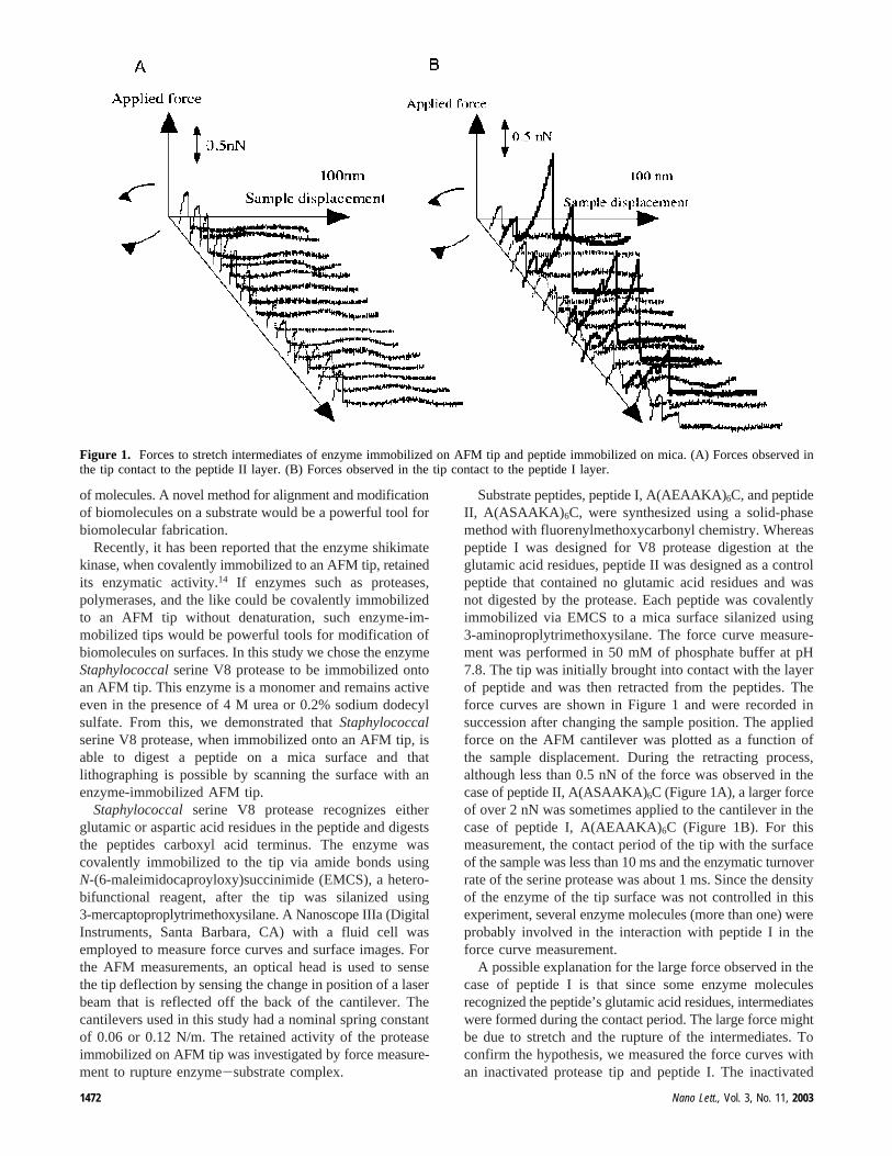
Substrate peptides, peptide I, A(AEAAKA)6C, and peptideII, A(ASAAKA) 6C, were synthesized using a solid-phasemethod with fluorenylmethoxycarbonyl chemistry. Whereaspeptide I was designed for V8 protease digestion at theglutamic acid residues, peptide II was designed as a controlpeptide that contained no glutamic acid residues and wasnot digested by the protease. Each peptide was covalentlyimmobilized via EMCS to a mica surface silanized using3-aminoproplytrimethoxysilane. The force curve measure-ment was performed in 50 mM of phosphate buffer at pH7.8. The tip was initially brought into contact with the layerof peptide and was then retracted from the peptides. Theforce curves are shown in Figure 1 and were recorded insuccession after changing the sample position. The appliedforce on the AFM cantilever was plotted as a function ofthe sample displacement. During the retracting process,although less than 0.5 nN of the force was observed in thecase of peptide II, A(ASAAKA)6C (Figure 1A), a larger forceof over 2 nN was sometimes applied to the cantilever in thecase of peptide I, A(AEAAKA)6C (Figure 1B). For thismeasurement, the contact period of the tip with the surfaceof the sample was less than 10 ms and the enzymatic turnoverrate of the serine protease was about 1 ms. Since the densityof the enzyme of the tip surface was not controlled in thisexperiment, several enzyme molecules (more than one) wereprobably involved in the interaction with peptide I in theforce curve measurement.
A possible explanation for the large force observed in thecase of peptide I is that since some enzyme moleculesrecognized the peptide’s glutamic acid residues, intermediateswere formed during the contact period. The large force mightbe due to stretch and the rupture of the intermediates. Toconfirm the hypothesis, we measured the force curves withan inactivated protease tip and peptide I. The inactivated
Figure 1. Forces to stretch intermediates of enzyme immobilized on AFM tip and peptide immobilized on mica. (A) Forces observed inthe tip contact to the peptide II layer. (B) Forces observed in the tip contact to the peptide I layer.
1472 Nano Lett., Vol. 3, No. 11, 2003
enzyme-immobilized tip was prepared by immersing the tipin a phenylmethylsulfonyl fluoride (PMSF) solution, whichirreversibly inactivated the serine proteases. A force of lessthan 0.4 nN was observed during the retracting period aftercontact with the surface of peptide I (data not shown). Itwould appear that the large force was therefore due tostretching of the intermediate.
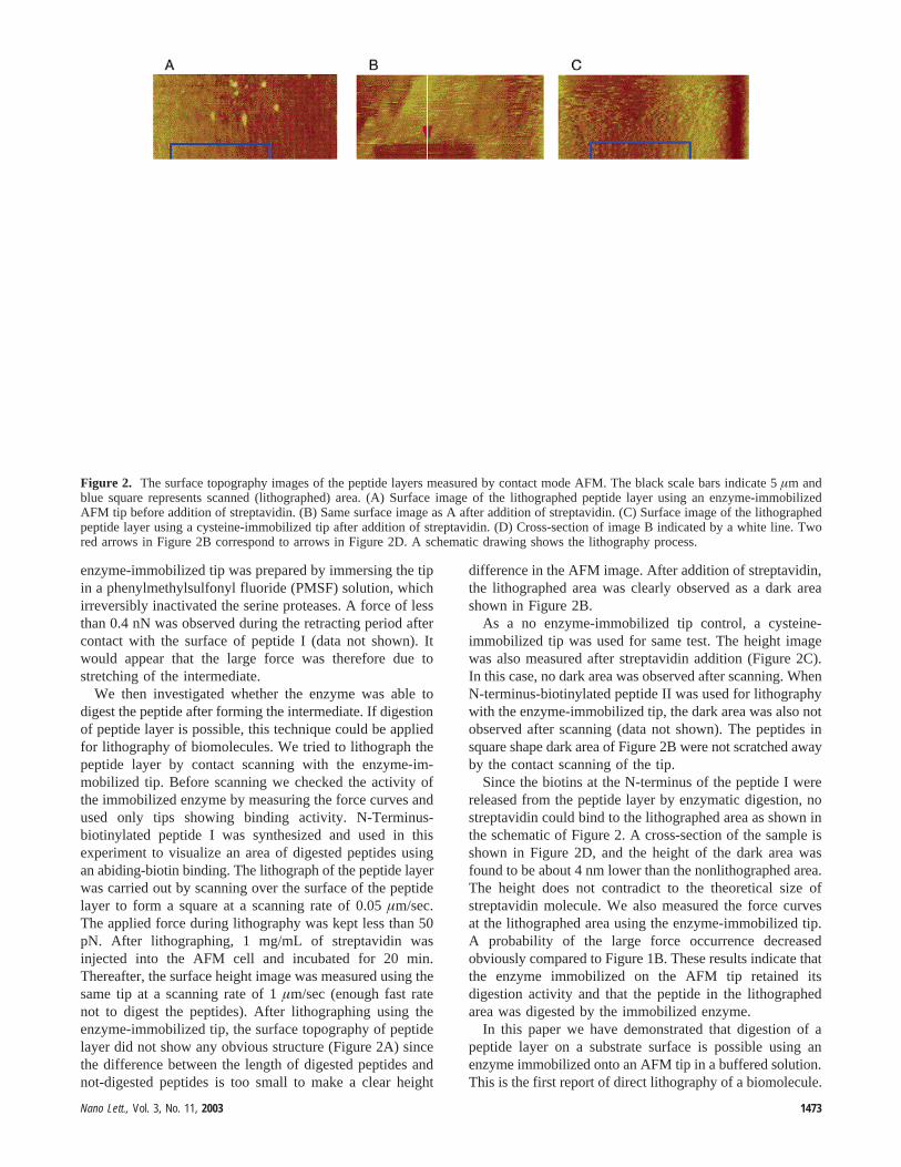
We then investigated whether the enzyme was able todigest the peptide after forming the intermediate. If digestionof peptide layer is possible, this technique could be appliedfor lithography of biomolecules. We tried to lithograph thepeptide layer by contact scanning with the enzyme-im-mobilized tip. Before scanning we checked the activity ofthe immobilized enzyme by measuring the force curves andused only tips showing binding activity. N-Terminus-biotinylated peptide I was synthesized and used in thisexperiment to visualize an area of digested peptides usingan abiding-biotin binding. The lithograph of the peptide layerwas carried out by scanning over the surface of the peptidelayer to form a square at a scanning rate of 0.05µm/sec.The applied force during lithography was kept less than 50pN. After lithographing, 1 mg/mL of streptavidin wasinjected into the AFM cell and incubated for 20 min.Thereafter, the surface height image was measured using thesame tip at a scanning rate of 1µm/sec (enough fast ratenot to digest the peptides). After lithographing using theenzyme-immobilized tip, the surface topography of peptidelayer did not show any obvious structure (Figure 2A) sincethe difference between the length of digested peptides andnot-digested peptides is too small to make a clear height
difference in the AFM image. After addition of streptavidin,the lithographed area was clearly observed as a dark areashown in Figure 2B.
As a no enzyme-immobilized tip control, a cysteine-immobilized tip was used for same test. The height imagewas also measured after streptavidin addition (Figure 2C).In this case, no dark area was observed after scanning. WhenN-terminus-biotinylated peptide II was used for lithographywith the enzyme-immobilized tip, the dark area was also notobserved after scanning (data not shown). The peptides insquare shape dark area of Figure 2B were not scratched awayby the contact scanning of the tip.
Since the biotins at the N-terminus of the peptide I werereleased from the peptide layer by enzymatic digestion, nostreptavidin could bind to the lithographed area as shown inthe schematic of Figure 2. A cross-section of the sample isshown in Figure 2D, and the height of the dark area wasfound to be about 4 nm lower than the nonlithographed area.The height does not contradict to the theoretical size ofstreptavidin molecule. We also measured the force curvesat the lithographed area using the enzyme-immobilized tip.A probability of the large force occurrence decreasedobviously compared to Figure 1B. These results indicate thatthe enzyme immobilized on the AFM tip retained itsdigestion activity and that the peptide in the lithographedarea was digested by the immobilized enzyme.
In this paper we have demonstrated that digestion of apeptide layer on a substrate surface is possible using anenzyme immobilized onto an AFM tip in a buffered solution.This is the first report of direct lithography of a biomolecule.
Figure 2. The surface topography images of the peptide layers measured by contact mode AFM. The black scale bars indicate 5µm andblue square represents scanned (lithographed) area. (A) Surface image of the lithographed peptide layer using an enzyme-immobilizedAFM tip before addition of streptavidin. (B) Same surface image as A after addition of streptavidin. (C) Surface image of the lithographedpeptide layer using a cysteine-immobilized tip after addition of streptavidin. (D) Cross-section of image B indicated by a white line. Twored arrows in Figure 2B correspond to arrows in Figure 2D. A schematic drawing shows the lithography process.
Nano Lett., Vol. 3, No. 11, 2003 1473
To apply this technique for other enzymes, more studieswould be needed; however, the technique would be devel-oped to the precision machining of biomolecules at thesubmicrometer to nanometer scale. It means that not onlydigestion molecules but also build-up of or modification ofmolecules would be possible using this technique. Westrongly believe that our lithography method has potentialto be used in a wide variety of applications, such asdevelopment of a DNA chip, bioMEMS, microTAS, micro-biosensor, etc., especially in the field of biotechnology.
Acknowledgment. This work was partially supported byMillennium Project of Ministry of Economy, Trade andIndustry of Japan.
References
(1) Mckendry, R.; Theoclitou, M. E.; Rayment, T.; Abell, C.Nature1998,391, 566.
(2) Kaasgaard, T.; Mouritsen, O. G.; Jorgensn, K.FEBS Lett. 2002, 515,29.
(3) Kienberger, F.; Kada, G.; Gruber, H. J.; Pastushenko, V. P.; Riener,C.; Trieb, M.; Knaus, H. G.; Schindler, H.; Hiterdorfer, P.SingleMol. 2000, 1, 59.
(4) Hinterdorfer, P.; Schilcher, K.; Baumgartner, W.; Gruber, H. J.;Schidler, H.Nanobiology1998, 4, 177.
(5) Grandbois, M.; Beyer, M.; Rief, M.; Clausen-Schaumann, H.; Gaub,H. E. Science1999, 283, 1727.
(6) Mitsui, K.; Hara, M.; Ikai, A.FEBS Lett. 1996, 385, 29.(7) Oesterhelt, F.; Oesterhelt, D.; Pfeiffer, M.; Engel, A.; Gaub, H. E.;
Muller, D. J.Science2000, 288, 143.(8) Takeda, S.; Ptak, A.; Nakamura, C.; Miyake, J.; Kageshima, M.;
Jarvis, S. P.; Tokumoto, H.Chem. Pharm. Bull. 2001, 49, 1512.(9) Ptak, A.; Takeda, S.; Nakamura, C.; Miyake, J.; Kageshima, M.;
Jarvis, S. P.; Tokumoto, H.J. Appl. Phys.2001, 90, 3095.(10) Kageshima, M.; Lantz, M. A.; Jarvis, S. P.; Tokumoto, H.; Takeda,
S.; Ptak, A.; Nakamura, C.; Miyake, J.Chem. Phys. Lett.2001, 343(1/2), 77.
(11) Piner, R.; Zhu, J.; Xu, F.; Hong, S.; Mirkin, C. A.Science1999,283, 661.
(12) Hyun, J.; Ahn, S. J.; Lee, W. K.; Chilkoti, A.; Zauscher, S.NanoLett. 2002, 2, 1203.
(13) Liu, M.; Amro, N. A.; Chow, C. S.; Liu, G.Nano Lett.2002, 2, 863.(14) Fiorini, M.; Mckendry, R.; Cooper, M. A.; Rayment, T.; Abell, C.
Biophy. J. 2001, 80, 2471.
NL034448K
1474 Nano Lett., Vol. 3, No. 11, 2003