light reaction path of photosynthesis - uni … · light reaction path of photosynthesis edited by...
TRANSCRIPT
Light Reaction Path of Photosynthesis Edited by Francis K. Fong
With Contributions by G.S. Beddard • R.H. Clarke • R.J. Cogdell F.K Fong • A. Hoff • H. Levanon J.R. Norris • H. Scheer • M.R. Wasielewski
With 118 Figures
Springer-Verlag Berlin Heidelberg New York 1982
Contents
Chapter 1: Free Energy Change for Quantum Storage in Photosynthesis F.K. Fong 1 Introduction 1 2 Definition of the Primary Photochemical Reaction 2 3 Photopotential, Photooverpotential and Free Energy Change for
Quantum Storage in Photosynthesis 3 4 Outline of this Book 5 References 6
Chapter 2: Phycobiliproteins: Molecular Aspects of a Photosynthetic Antenna System. H. Scheer (With 8 Figures) 1 Introduction 7 2 Morphology 9 3 Energy Transfer 14 4 Chromophore Structure 17
4.1 Chromophores Cleaved from Biliproteins 17 4.2 Chromophores Bound to the Protein 19
5 Noncovalent Protein Chromophore Interactions 22 5.1 Topology of the Chromophore 24 5.2 Conformational Mobility 27
6 The Proteins 31 7 Biosynthesis 33 8 Concluding Remarks 35 Notes Added in Proof 38 References 39
Chapter 3: Structure and Excitation Dynamics of Light-harvesting Protein Complexes. G.S. Beddard and R J . Cogdell (With 19 Figures) 1 Introduction 46
1.1 General Discussion of Excitation Migration 47 1.2 Coherence 52
2 Experimental Methods 52 2.1 Streak Camera and Neodymium Laser 54 2.2 Single Photon Counting 54
VIII Contents
3 Excited State Annihilation 55 4 Anaerobic Photosynthetic Bacteria 56
4.1 The B800-850 Light-harvesting Pigment-Protein Complex Isolated Isolated from Rps. sphaeroides 57
4.2 Kinetic Studies 60 4.3 The Water-soluble Bchl-tf Antenna Complex fromP. aestuarii,
Strain2K 63 5 LowerAlgae 65
5.1 The Perdinin-Chl a Protein from Glenodinium 65 5.2 The Phycobiliproteins of the Red Algae 67 5.3 Kinetic Studies 69
6 Antenna Pigment-Protein Complexes from Higher Plants 71 References 77
Chapter 4: Photooxidation of the Reaction Center Chlorophylls and Structural Properties of Photosynthetic Reaction Centers. A J . Hoff (With 27 Figures)
Abbreviations and Symbols 80 1 Introduction 81
1.1 Energetics of Photosynthesis 82 1.2 Chlorophylls, Quinones and Related Molecules 84
2 The Photosystem of Purple Bacteria 84 2.1 Optical Investigations 84
2.1.1 Absorption Difference Spectroscopy 84 2.1.2 Spectroscopic Nomenclature of Behl 90 2.1.3 Circular Dichroism 92 2.1.4 Linear Dichroism 94 2.1.5 Nano- and Picosecond Spectroscopy 105
2.2 ESR and ENDOR 108 2.2.1 Characteristics of the ESR Signal of P860+ 108 2.2.2 ENDOR of the Primary Donor 110 2.2.3 ESR and ENDOR of the Reduced Intermediary
Acceptor, I" 113 2.3 The Triplet State of the Primary Donor 115
3 The Plant Photosystems 117 3.1 Optical Investigations of the Primary Donor of Photosystems
land2 117 3.1.1 Absorption Difference Spectroscopy of P700 118 3.1.2 Absorption Difference Spectroscopy of P680 122 3.1.3 Circular and Linear Dichroism of Photosystems 1 and 2 . 124
3.2 ESR and ENDOR 128 3.2.1 P700+ 128 3.2.2 P680+ 129
3.3 The Intermediary Acceptors of Photosystems 1 and 2 130
Contents IX
3.3.1 Photosystem 1 130 3.3.2 Photosystem 2 131 3.3.3 Triplet States 132
4 Structure of the Bacterial Primary Donor-Acceptor Complex 132 4.1 Electron Transfer Rates 133 4.2 Configuration of Primary Reactants 137
Bibliography 142 References 143 Notes Added in Proof (In Connection with Chapter 8) 322
Chapter 5: Triplet State and Chlorophylls. H. Levanon and J.R. Norris (With 10 Figures)
Abbreviations 152 1 Introduction 153 2 Optical-Magnetic Resonance Spectroscopy 155
2.1 Triplet Detection. Zero Field Experiments 155 2.2 The Triplet State and the EPR Experiment 158 2.3 The Triplet Yield vs Magnetic Field 160 2.4 Triplet Photochemistry. The CIDEP Method 160
2.4.1 Whatis CIDEP? 160 2.4.2 Triplet Precursor vs Triplet Mechanism 160 2.4.3 The Triplet Mechanism 164 2.4.4 The Radical Pair Mechanism: ST ± 1 Mixing 165 2.4.5 The Radical Pair Mechanism: ST 0 Mixing 166
3 Triplet State Studies of Model Chlorophyll Compounds 169 4 In-Vivo Chlorophyll Triplets 173
4.1 Introduction 173 4.2 Bacterial Photosynthesis 174
4.2.1 The Triplet State in Bacterial Photosynthesis 174 4.2.2 Zero Field Splitting Parameters 175 4.2.3 Electron Spin Polarization in Triplets 179
4.3 Green Plant Photosynthesis 184 4.3.1 Photoexcited Triplet State Detection in Green Plants... 184 4.3.2 CIDEP Studies of Photosynthesis 184
5 Summary 186 References and Notes 187
Chapter 6: The Chlorophyll Triplet State and the Structure of Chlorophyll Aggregates. R.H. Clarke (With 9 Figures)
1 Introduction 196 2 Optically Detected Magnetic Resonance in the Triplet State 197 3 Application of ODMR to the Chlorophyll Triplet State 204
3.1 Chlorophyll Triplet State Zero-Field Splittings 206 3.2 Chlorophyll T1 -> S 0 Intersystem Crossing Rates 208
X Contents
4 Application of Triplet State ODMR to Chlorophyll Aggregate Structure 214 4.1 The Triplet Exciton Model 216 4.2 Application to the Chlorophyll Dimer In Vitro 220 4.3 Chlorophyll Aggregate Structure In Vivo 226
References 231
Chapter 7: Synthetic Approaches to Photoreaction Center Structure and Function. M.R. Wasielewski (With 31 Figures)
1 Introduction 234 2 Porphyrin Models of Photoreaction Center Chlorophylls 236 3 Noncovalent Chlorophyll Special Pair Models 242 4 Preparation of Singly Linked Covalent Chlorophyll Dimers 246 5 Solvent-dependent Structure of Singly Linked Covalent
Chlorophyll Dimers 249 6 Photophysical Properties of Singly Linked Covalent Chlorophyll
Dimers 254 7 Photochemical Properties of Singly Linked Covalent Chlorophyll
Dimers 262 8 Biomimetic Charge Separation Photochemistry 264 9 Doubly Linked Chlorophyll Cyclophane Models of Special Pair
Structure 269 10 Concluding Remarks 274 References 274
Chapter 8: Light Path of Carbon Reduction in Photosynthesis F.K. Fong (With 14 Figures)
1 Introduction 277 2 Scope 279
2.1 Originof0 2 Evolution 280 2.2 Light and DarkPathsof Carbon 280 2.3 Submolecular Interactions of Chl-a Light Reactions 281
3 Model for Chlorophyll Light Reactions in Photosynthesis 282 3.1 Long-Wavelength Shifts of Chlorophyll Aggregates 282 3.2 Postulates 285 3.3 Path of Electrons from Water 288
4 Dimer Model ofP700 289 4.1 Chlorophyll Purification 289 4.2 Mg . . . 0(H)H Interactions 290 4.3 Model P700 Structure and Properties 295
5 P680 Model and Water Splitting 297 6 Carbon Reduction by Water 301 7 Two-Photon Activation of Water Splitting 303 8 Primary and Secondary Processes of Photosynthesis 307
Contents XI
8.1 Comparison of Models for P680 and P700 308 8.2 Light Reaction Sequence 309 8.3 Photochemical Reduction of C0 2 310 8.4 Time Sequence and Branching of Electron Flow from Water . . 313
9 Further Conclusions 314 9.1 SpatialRelationshipsofP680andP700 314 9.2 Quantum Requirement of Oxygen Evolution 315 9.3 PGA Reduction as Mechanism for Photoregulation 316
References 317
Notes Added in Proof for Chapter 4 322
Author Index 327
Subject Index 337
Chapter 2 Phycobiliproteins: Molecular Aspects of Photosynthetic Antenna System
Hugo Scheer1
1 Introduction
Harvesting the sun requires both the ab Sorption of the dilute energy, light, and its trans-formation into chemical energy. With one exception (e. g., the halobacteria), the orga-nisms capable of photosynthesis have these two functions also physically divided. Antenna Systems collect the light and guide the excitation energy to the reaction centers, where it is transformed into electrochemical energy. The reaction centers are the con-servative part of the photosynthetic apparatus, whereas the size, Organization and com-position of the antenna varies widely as a developmental and often also individual response to the environmental light quality (Table 1). The reaction centers are always integral parts of the photosynthetic membranes. The antenna may be part of the mem-brane, too, but it may also be attached on either its inner or outer surface, or even in separate particles or vesicles.
Irrespective of their location, the function of all antenna Systems is to störe excitation as a temporary buffer, and at the same time guide it to the reaction centers. De-pending on the sign and magnitude of the energy gap, as well as on the distance be-tween the antenna chromophores and the reaction center, either of these function is more strongly expressed, or at least more obvious.
The subject of this article may be properly described as a funnel for collecting and feeding excitation energy into the reaction center. It is the biliprotein antenna Systems of cyanobacteria and red algae, and of the cryptophytan algae. These pigments are only loosely attached to the photosynthetic membrane and water-soluble, which greatly facilitated their investigation and made especially the former two the hitherto probably best understood antennas on a molecular basis.
This review is focused on the molecular aspects of the process. For recent reviews on biliproteins written from various points of view and citing earlier literature, the reader is referred to the articles of: Bennett and Siegelman (1979), Berns (1971), Bog-orad (1975), Chapman (1973), Gantt (1975, 1979), Glazer (1977), MacColl and Berns (1979), O'Carta and O'hEocha (1976), Rüdiger (1971, 1975, 1978, 1979), Scheer (1978, 1981), and Troxler (1975). Biliproteins containing structurally very similar chromophores, the phytochromes and phycochromes, are also involved as reaction center pigments in sensory transduction of green plants and many algae. For recent surveys on these subjects, see: Björn (1979), Hartmann and Haupt (1978), Lazaroff (1973),
1 Botanisches Institut der Universität München, Menzinger Straße 67, 8000 München 19, FRG
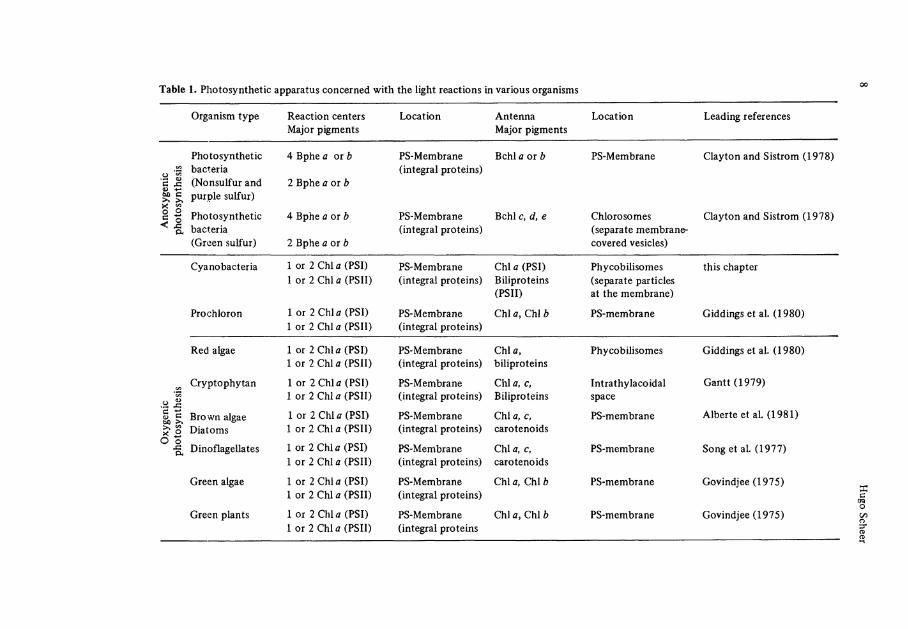
Table 1. Photosynthetic apparatus concerned with the light reactions in various organisms
Organism type Reaction centers Major pigments
Location Antenna Major pigments
Location Leading references
tyge
nic
1 sy
nthe
sis Photosynthetic
bacteria (Nonsulfur and purple sulfur)
4 Bphe a oib
2Bphe a or b
PS-Membrane (integral proteins)
Behl a or b PS-Membrane Clayton and Sistrom (1978)
§ <
phot
o Photosynthetic bacteria (Green sulfur)
4 Bphe a or b
2 Bphe a or b
PS-Membrane (integral proteins)
Behl c, d, e Chlorosomes (separate membrane-covered vesicles)
Clayton and Sistrom (1978)
Cyano bacteria 1 or 2 Chi <z (PSI) 1 or 2 Chi* (PSII)
PS-Membrane (integral proteins)
Chi a (PSI) Biliproteins (PSII)
Phycobilisomes (separate particles at the membrane)
this chapter
Prochloron 1 or2Chla (PSI) 1 or2Chlß(PSII)
PS-Membrane (integral proteins)
Chlfl, Chlfc PS-membrane Giddings et al. (1980)
Red algae 1 or 2Chifl (PSI) 1 or2Chlß(PSII)
PS-Membrane (integral proteins)
Chlö, biliproteins
Phycobilisomes Giddings et al. (1980)
o '5
Cryptophytan lor2Chlfl(PSI) 1 or 2 Chi a (PSII)
PS-Membrane (integral proteins)
C h U c, Biliproteins
Intrathylacoidal space
Gantt (1979)
)xyg
eni
tosy
ntl
Brown algae Dialoms
1 or 2 Chi A (PSI) 1 or 2 Chi a (PSII)
PS-Membrane (integral proteins)
C h U c, carotenoids
PS-membrane Alberte et al. (1981)
o Dinoflagellates 1 or 2Chlfl (PSI) lor2Chlö(PSII)
PS-Membrane (integral proteins)
Chi a, c, carotenoids
PS-membrane Song et al. (1977)
Green algae 1 or 2 Chi <z (PSI) 1 or 2 Chi a (PSII)
PS-Membrane (integral proteins)
Chi a, ChU> PS-membrane Govindjee (1975)
Green plants 1 or 2 Chi* (PSI) lor2Chlfl(PSII)
PS-Membrane (integral proteins
Chi Ä , Chi b PS-membrane Govindjee(1975)
Phycobiliproteins: Molecular Aspects of a Photosynthetic Antenna System 9
Marme (1977), Mitrakos and Shropshire (19720' Mohr (1972), Pratt (1978), Rüdiger (1971, 1980), Scheer (1981), Smith (1975), and Smith and Kendrick (1976).
2 Morphology
In electron micrographs of the unicellular red algae, Porphyridium c r u e n t u m , Gantt and Conti described in 1965 a new particle of oblong shape (^ 4 nm diameter), which was arranged in a rather regulär fashion on the outside of the thylakoid membrane. Subsequent investigations by several research groups (Gantt 1979; Glazer et al. 1979; Koller et al. 1978; Wildman and Bowen 1974) revealed similar particles, although of varying size and arrangement, as a main characteristic of the photosynthetic apparatus of cyanobacteria and red algae.
Both classes of photosynthetic organisms owe their coloration to büiproteins, which had been shown by bichromatic action spectroscopy to be major light-harvesting pigments, feeding excitation energy mainly to photosystem II (Emerson 1958; Haxo 1960). The early suspicion that the phycobiliproteins are contained in these particles was confirmed after their isolation as integral entities and the analysis of their composi-tion (Bryant et al. 1976; Gantt and Lipschultz 1974; Gantt et al. 1979; Glazer et al. 1979; Gray and Gantt 1975; Koller et al. 1978; Rigbi et al. 1980; Wanner and Köst 1980). They are almost entirely (Koller et al. 1978; Tandeau de Marsac and Cohen-Buzire 1977; Yamanaka et al. 1978) composed of phycobiliproteins, and are thus prop-erly termed phycobilisomes (Gantt and Conti 1966).
The phycobilisomes contain three different types of biliproteins2, the phycoery-thrins (PE) absorbing in the ränge between 480 and 580 nm, the phycocyanins (PC) ab-sorbing in the ränge between 570 and 630 nm, and the allophycocyanins (APC) absorbing in the ränge between 610 and 670 nm (Table 2). In addition, minor amounts of un-colored proteins have been reported by several workers (Koller et al. 1978; Tandeau de Marsac and Cohen-Bazire 1977; Yamanaka et al. 1978). The majority of the pigment content is PE and PC. The ratio between the two is variable within different species, and in spite of many exceptions, the blue PC's are predominant in the cyanobacteria ("blue algae"), and the red PE's are predominant in the red algae. The ratio between the two is often also variable within a given species in response to the environmental light quality, the relative proportion of the PC's absorbing red light being higher in red and lower in green light (see "chromatic adaptation"). The sizes and fine structures of the phycobilisomes vary accordingly, although a common construction principle is cur-rently evolving.
The detailed investigation of the phycobilisomes revealed a striking morphology. From dissociation experiments, Gantt and co-workers arrived at a model for P. cruen-t u m phycobilisomes in which an APC core in contact with the photosynthetic membrane is covered by a roughly hemispherical layer of PC, which in turn is covered by
2 Abbreviations: PC = Phycocyanin, PE = Phycoerythrin, APC = Ailophycocyanin. The prefixes C - , R - and B - stand for Cyanobacteria, red algae and iangiales, an order of the red algae. Chi = Chlorophyll

10 Hugo Scheer
Table 2. Properties of phycobiliproteins and Classification according to occurrence, spectra, and subunit composition
Typea Occurence Xmax (nm) in the visible spectral ränge
Chromophores
a - ß- 7-Chain
APC-I Cyanobacteria and red algae
656 I x l a I x l a 7
- II,
-III
Cyanobacteria and red algae
650 I x l a I x l a
- B Cyanobacteria and red algae
670 I x l a I x l a
C-PC Cyanobacteria 635d,620,(590)e I x l a I x l a -
PEC b Cyanobacteria 590,568 lxPXßg 2 x l a -
R-PC Red algae 620,555 i x l a l x l a , l x 2 -
Cr-PC Cryptophytan algae
645,610 580 (and others)
l x l a h
(and others) I x l a , 1x2 (? ) -
C-PE Cyanobacteria 575d,560,540 2x2 3-4x2 -
R - P E C Red algae 568,540,4988 2x2 ?x2 ?xPUB
?xPU&i 1x2
b-PE Red algae 575d,565,540 2x2 4x2 -
B-PE Red algaeJ 565,545,498f 2x2 4x2 2xPU&) 2x2
Cr-PE Cryptophytan algae
545-565 ?x2 ?x2 -
a Prefixes according to their occurence: B = Brangiales, an order of the red algae; Cr = Cryptophytan algae; C = Cyanobacteria, R = Red algae
b Phycoerythrocyanin c R-PE has been reported to be a glycochromoprotein (Chapman 1973; Raftery and O'hEocha
1965) d Possibly an aggregate form (Brown et al. 1975; Zilinskas et al. 1978) e Shoulder, resolved at low temperatures (Frackowiak et al. 1975; Friedrich et al. 1981, Giay
and Gantt 1975; Scheer and Kufer 1977)
Phycobiliproteins: Molecular Aspects of a Photosynthetic Antenna System 11
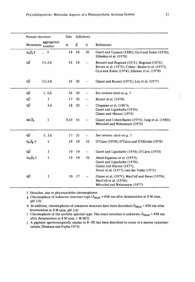
Protein structure Size (kDalton)
Monomers aggregation number a ß 7 References
. 1 18 18 30 Gantt and Canaani (1980); Gysi and Zuber (1976); Zilinskas et al. (1978)
aß ü),3,6 16 18 Bennett and Bogorad (1971); Bogorad (1975); Brown et al. (1975); Cohen-Bazire et al. (1977); Gysi and Zuber (1974); Zilinkas et al. (1978)
aß (D,3,6 16 20 - Glazer and Bryant (1975); Ley et al. (1977)
aß 1, 3,6 16 20 - See reviews cited on p. 7
aß 3 17 20 - Bryant et al. (1978)
aß 3,6 18 20 Chapman et al. (1967); Gantt and Lipschultz (1974); Glazer and Hixson (1975)
1 9,10 16 Glazer and Cohen-Bazire (1975); Jung et al. (1980); Mörschel and Wehrmeyer (1975)
aß 1, 3,6 17 21 - See reviews cited on p. 7
<kße7 1 19 19 35 O'Carra (1970); O'Carra and O'hEocha (1976)
aß 3 19 19 - Gantt and Lipschultz (1974); O'Carra (1970)
a-eßel 1 19 19 35 Abad-Zapatero et al. (1977); Gantt and Lipschultz (1974); Glazer and Hixson (1977); Sweet et al. (1977); van der Velde (1973)
aß 2 10 17 Glazer et al. (1971); MacColl and Berns (1979); MacColl et al. (1976); Mörschel and Wehrmeyer (1977)
f Shoulder, due to phycourobilin chromophores g Chromophore of unknown structure type ( X m a x = 600 nm after denaturation in 8 M urea,
pH 3.0) h In addition, chromophores of unknown structure have been described (A^x = 690 nm after
denaturation in 8 M urea, pH 2.0) i Chromophore of the urobilin spectral type. The exact structure is unknown (\nax = 498 nm
after denaturation in 8 M urea, 1 M HCl) j A pigment spectroscopically similar to B-PE has been described to occur in a marine cyanobac-
terium (Shimura and Fujita 1975)
12 Hugo Scheer
PE. Phycobilisomes can be isolated intact in high ionic strength buffer both with and without parts of the thylacoid membrane still attached, and the dissociation has been studied both by fluorescence spectroscopy (Gantt and Zilinskas 1976; see below) and immunoelectron microscopy (Gantt and Zilinskas 1978; Gantt and Lipschultz 1977). The phycobilisomes of P. c r u e n t u m are rather large. Indications of a fine structure have been obtained only recently, including a small stalk which may function as an an-chor to the membrane (Wanner and Köst 1980).
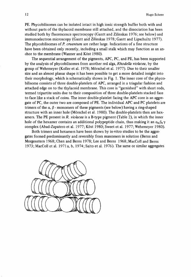
The sequential arrangement of the pigments, APC, PC, and PE, has been supported by the analysis of phycobilisomes from another red alga, Rhodeila violacea, by the group of Wehrmeyer (Koller et al. 1978; Mörschel et al. 1977). Due to their smaller size and an almost planar shape it has been possible to get a more detailed insight into their morphology, which is schematically shown in Fig. 1. The inner core of the phyco-bilisome consists of three double-platelets of APC, arranged in a triagular fashion and attached edge on to the thylacoid membrane. This core is "garnished" with short rods, termed tripartite units due to their composition of three double-platelets stacked face to face like a Stack of coins. The inner double-platelet facing the APC core is an aggre-gate of PC, the outer two are composed of PE. The individual APC and PC platelets are trimers of the a, ß - monomers of these pigments (see below) having a ring-shaped structure with an inner hole (Mörschel et al. 1980). The double-platelets then are hex-amers. The PE present in R. violacea is a B-type pigment (Table 2), in which the inner hole of the hexamer contains an additional Polypeptide chain, thus making it an a6ß6y complex (Abad-Zapatero et al. 1977; Köst 1980; Sweet et al. 1977; Wehrmeyer 1980).
Both trimers and hexamers have been shown by in-vitro studies to be the aggre-gates formed predominantly and reversibly from manomers in Solution (Berns and Morgenstern 1968; Chen and Berns 1978; Lee and Berns 1968;MacColl and Berns 1973; MacColl et al. 1971 a, b, 1974; Saito et al. 1976). The same or similar aggregates
Phycobiliproteins: Molecular Aspects of a Photosynthetic Antenna System 13
have also been suggested as the building blocs of phycobiliprotein crystals (Bryant et al. 1976; Dobler et al. 1972; Sweet et al. 1977). Some apparently confliction results between morphological, biochemical and biophysical investigations on biliprotein aggregates have been discussed recently Mörschel et al. (1980).
The phycobilisomes of cyanobacteria have been characterized in comparable detail only recently, due to isolation problems (Gantt et al. 1979; Gray and Gantt 1975; Gray et al. 1973; Rigbi et al. 1980; Yamanaka et al. 1978). Electron microscopy of three species has yielded basically the same fine structure, with the number of the central APC platelets or the lenght of the rods being somewhat variable (Glazer et al. 1979).
The phycobilisomes are probably products of a complex self-assembly process which does not only require the aggregation of identical biliproteins (homo-aggrega-tion), but also of different types of biliproteins with each other (hetero-aggregation) and with the membrane, in a highly ordered and regulated fashion. The organizing prin-ciples have only very recently begun to emerge. Homo-aggregates larger than the hexa-mer and heteroaggregates have been isolated by several groups from partly dissociated phycobilisomes (Kessel et al. 1973; Koller et al. 1978; Mörschel et al. 1977, 1980a, b; Yamanaka et al. 1978; Gantt et al. 1979; Rigbi et al. 1980; Grabowski et al. 1981), but their reassociation was rarely observed and difficult to achieve in a reproducible way.
After the identification of colorless proteins as integral components of phycobilisomes, at least some of them have tentatively been related to an ordering function (Tandeau de Marsac and Cohen-Bazire 1977 b). An indirect evidence to this is the find-ing, that crude dissociates of phycobilisomes containing larger fragments still bearing colorless proteins can be reassociated into functional phycobilisomes (Canaani et al. 1980; Katoh, private communication). The largest one of the colorless proteins (75-90 kDalton) is probably involved in the attachment of phycobilisomes to the photosynthetic membrane (see Gantt 1981). A protein of this size has recently been identified both in isolated phycobilisomes and in membranes from which the biliproteins had been dissociated (Redlinger and Gantt 1980). This protein is blue, however, with the fluorescence characteristics of an APC (-B? ), and its relation to the colorless proteins is still unresolved (Gantt 1981). Irrespective of its coloration, an "anchor" protein has also been suggested from electron-micrographs of phycobilisomes fromP. c r u e n t u m showing a footlike Protrusion (Wanner and Köst 1980).
Three different smaller colorless proteins (nominally 33, 30 and 27 kDalton) have recently been isolated from Synechococcus 6301 phycobilisomes (Lundell et al. 1981). From reassociation experiments with isolated phycocyanin and one or more of the rather hydrophobic colorless proteins, specific functions for the latter have been suggested. The 33 and 30 kDalton species are involved in stacking hexamers into rods, which may grow to exceptional and unnatural lengths. The 27 kDalton protein is rather related to rod termination. Only small Stacks are formed in its presence, even if the 30 and/or 33 kDalton proteins are present as well. A small colorless protein has also been found in PC-PE complexes isolated from P. sordidum phycobilisomes, where it appears to function as a linker between the different pigments (Lipschultz and Gantt 1981).
These findings do assign specific functions to at least some of the colorless proteins (group II). As an intriguing aspect of these results, one can imagine, that the self-assembly of phycobilisomes is controlled by the relative concentrations of the different biliproteins and colorless proteins, which in turn is controlled by their biosynthesis and degradation.
14 Hugo Scheer
In many cyanobacteria, the amounts of the different biliproteins (and other photosynthesis pigments) are regulated by light (chromatic adaption, see below and Bogorad 1975), by nitrogen supply (Allen and Smith 1969) and sulfur Compounds in the medium (Schmidt 1980).
On the phycobilisome level, these regulations have been shown to involve the com-position of the colorless proteins (Tandeau de Marsac and Cohen-Bazire 1977 b; Yamanaka and Glazer 1980), as well as the composition of the biliproteins and the phyco-bilisome-architecture (Siegelman 1980; Yamanaka and Glazer 1980). For biophysical studies, the contamination of biliproteins with colorless proteins in amounts varying with the isolation procedure, has to be considered as a potential complication, which e.g. may be involved in the conflicting results on size and shape of biliprotein homo-aggregates.
The morphology of cyptophytan antennas is different from those of cyanobacteria and red algae. Phycobilisomes are absent, and the biliproteins rather appear to be lo-cadet at the inner side of the thylacoid membrane (Gantt et al. 1971; Wehrmeyer 1970). The grana lamellae are wider spaced than in green plants, and filled with an electron-dense material assigned to biliproteins, for which only recently indications of a flne structure have been obtained (Wehrmeyer, pers. comm., 1979). For a recent survey on cryptophytan biliproteins, see Gantt 1979).
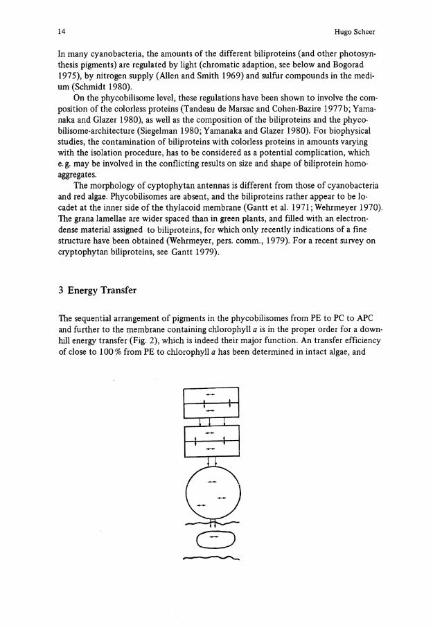
3 Energy Transfer
The sequential arrangement of pigments in the phycobilisomes from PE to PC to APC and further to the membrane containing Chlorophyll a is in the proper order for a down-hill energy transfer (Fig. 2), which is indeed their major function. An transfer efficiency of close to 100% from PE to Chlorophyll a has been determined in intact algae, and
C I D
Phycobiliproteins: Molecular Aspects of a Photosynthetic Antenna System 15
1.2
1.0
0.8-
0.6-
0A
0.2
C-PE
500 550
0.5
OA
0.3
1-0.2
0.1
0
0.1
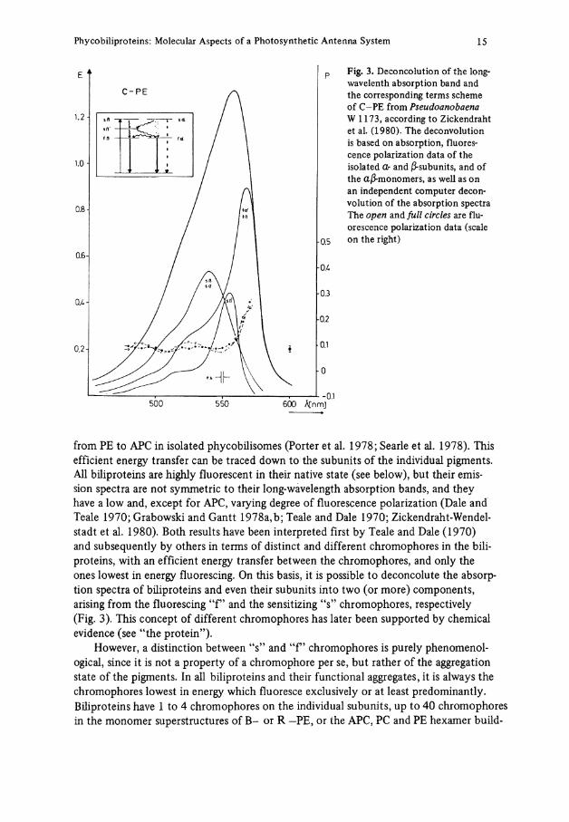
Fig. 3. Deconcolution of the long-wavelenth absorption band and the corresponding terms scheme of C-PE from Pseudoanobaena W 1173, according to Zickendraht et al. (1980). The deconvolution is based on absorption, fluorescence polarization data of the isolated <Z- and ß-subunits, and of the aß-monomers, as well as on an independent Computer deconvolution of the absorption spectra The open and füll circles are fluorescence polarization data (scale on the right)
600 ACnmj
from PE to APC in isolated phycobilisomes (Porter et al. 1978; Searle et al. 1978). This efficient energy transfer can be traced down to the subunits of the individual pigments. All biliproteins are highly fluorescent in their native State (see below), but their emis-sion spectra are not Symmetrie to their long-wavelength absorption bands, and they have a low and, except for APC, varying degree of fluorescence polarization (Dale and Teale 1970; Grabowski and Gantt 1978a,b; Teale and Dale 1970; Zickendraht-Wendelstadt et al. 1980). Both results have been interpreted first by Teale and Dale (1970) and subsequently by others in terms of distinet and different chromophores in the biliproteins, with an efficient energy transfer between the chromophores, and only the ones lowest in energy fluorescing. On this basis, it is possible to deconcolute the absorption spectra of biliproteins and even their subunits into two (or more) components, arising from the fluorescing "f" and the sensitizing "s" chromophores, respectively (Fig. 3). This coneept of different chromophores has later been supported by chemical evidence (see "the protein").
However, a distinetion between 4's" and " P chromophores is purely phenomenol-ogical, since it is not a property of a chromophore per se, but rather of the aggregation State of the pigments. In all biliproteins and their functional aggregates, it is always the chromophores lowest in energy which fluoresce exclusively or at least predominantly. Biliproteins have 1 to 4 chromophores on the individual subunits, up to 40 chromophores in the monomer superstruetures of B - or R -PE, or the APC, PC and PE hexamer build-
16 Hugo Scheer
ing blocs of phycobilisomes, and between 900 and 2500 chromophores in a?. c r u e n t u m phycobilisome, as estimated from the data of Wanner and Köst (1980) and Gantt (1976)3. In building up the latter, the percentage of the " P chromophores is constantly decreasing. Thus, the " P chromophore of isolated PE becomes an "s" chromophore in PE-PC heteroaggregates (Koller et al. 1978; Grabowski et al. 1980), and in the phycobilisomes it is only the few APC chromophores which fluorescence (Grabowski and Gantt 1978a,b; Porter et al. 1978; Searle et al. 1978).
The energy transfer of the individual subunits, the monomers and the various aggregates of the pigments has been analyzed in terms of a weak coupling (Förster) process. From the absorption and fluorescence spectra critical distances for non ordered orientations have been calculated (see Grabowski and Gantt, 1978 for further references), which are consider-ably larger than the diameters of the subunits, allowing an efficient transfer. The spatial distribution of the chromophores in different isolated phycobiliproteins has been estimated based on the Förster mechanism and the Jablonski "active shere" approximation. They indicated a surface distribution (Dale and Teale 1970; Zickendraht et al. 1980), in agreement with chemical evidence (see below). The observed energy transfer times within individual pigments (Kobayashi et al. 1979) and phycobilisomes (Searle et al. 1978) are also cinsistent with and analyzed on the basis of this transfer mechanism. For phycobilisomes of P. c r u e n t u m , hopping times of 280 ±40 ps, with an average of 28 jumps for the transfer from PE to PC, have been determined in agreement with the model described, when treated by the Pearlstein formalism (Grabowski and Gantt 1978b). The energy transfer has been investigated, too, for the individual subunits of a C-PE containing two and three chromophores, respectively, and the relative orientations of the dipoles determined (Zickendraht et al. 1980). The term scheme shown in Fig. 3 has been obtained from this work.
Strong coupling between chromophores seems to be less prominent in phycobiliproteins from cyanobacteria and red algae, and has in no case yet been shown conclu-sively. From CD data, exciton coupling has been implied for an APC (Gantt and Cana-ani 1980). The S-shaped CD bands for C-PC from Pseudoanabaena spec. W 1173 are indicative, too, of exciton Splitting, but here a definite decision is difficult in view of the five different chromophores present (Langer et al. 1980).
An intermediate coupling, has finally been suggested in APC to account for the pronounced red-shift upon trimer formation without an accompanying CD-effect (McColl et al. 1980). In view of the increase of the oscillator-strenght of the long-wave-length band, a chemical change (conformation, protonation) may be considered as well.
By contrast, strong coupling between chromophores has been deduced mainly from CD data for a cryptophytan PC (Jung et al. 1980). This is supported by a fast compo-nent (<8 ps) in the transient absorption of this pigment, assigned to an energy transfer process, as compared to 84 ps in a C-PC monomer (Kobayashi et al. 1979). This indicates again a certain Separation of the cryptophytan biliproteins, which is further evidenced by their spectral diversity and the occurence of special chromophores (see below, and Gantt 1979).
3 The "monomers" of B-PE, R-PE and possibly one of the APC have the rather complex a6 ß6y-structure, in which the 7- subunit fills the inner hole of the torus-shaped trimers and hexamers typical for most biliproteins (see below)
Phycobiliproteins: Molecular Aspects of a Photosynthetic Antenna System 17
From the morphological and energy transfer studies two different strategies for har-vesting green and orange light, their conversion to excitation energy corresponding to red light quanta, and their funneling to the reaction centers, seem to have been fol-lowed during the evolution of biliprotein antennas. One is exemplified in the cyanobacteria, where each of the pigments present Covers only a comparably narrow absorption ränge. To avoid gaps in the transfer chain and the absorption spectrum, the phycobiliproteins are arranged in intricate superstructures, the phycobilisomes. In the cryptophytan biliproteins, on the other hand, a miniature transfer chain has already evolved within each biliprotein, especially within the phycocyanins. Accordingly, a superstructure is not necessary, although the transfer from PE to Chi a would be facilitated by the Chi c's present in these organisms as intermediate carriers. In the red algae, both con-cepts have been united, since they have biliproteins covering a broader ränge of energies organized in phycobilisomes.
4 Chromophore Structure
4.1 Chromophores Cleaved from Biliproteins
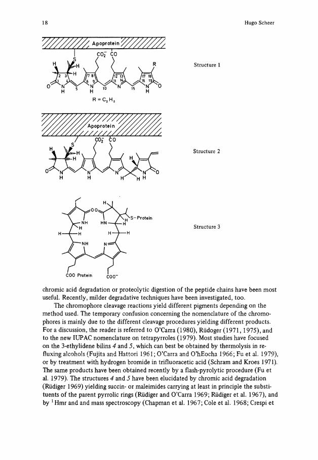
Only two chromophores are responsible for the broad ränge of absorption spectra of the majority of biliproteins (Table 2). Both are of the formerly4 so-called IX a Substitution type, characteristic for the mammalian bile pigments derivedfromheme cleavage at the methine bridge formerly designated "a", now C-5. Phycocyanobilin (1) is the blue chromophore of PC's and APC's. It is noteworthy that the same chromophore apears to be present in the phycochromes (Björn 1979; Ohad et al. 1979; Ohki and Fujita 1978; Scheibe 1972), and a very similar chromophore (18-vinyl instead of 18-ethyl) occurs in phytochrome(s) in the P T - form (Grombein et al. 1975; Rüdiger 1980). Phycoerythrobilin (2) is the red chromophore of PE's. Some of the biliproteins contain additional chromophores. R - and B-PE's carry phycourobilin chromophores, for which structure 3 has been proposed (O'Carra and O'hEocha 1976). Phycoerythrocya-nin has a red chromophore of unknown structure (Bryant et al. 1978), and at least two other chromophores have been proposed to occur in cryptophytan biliproteins (Glazer and Cohen-Bazire 1975; Jung et al. 1980; Mörschel and Wehrmeyer 1975). The evidence for these less-known chromophores comes from spectroscopic studies on the denatured pigments, which is useful for Screening. If done under carefully controled conditions, chromophores other than 1 or 2 can be easily recognized. It should be pointed out, however, that the structures 1 and 2 have been strictly proven only for a few biliproteins, and that alterations, e.g., of the side chains or especially the second protein bond, may remain unnoticed in such studies.
All chromophores are covalently bound to their respective apoproteins. This pre-vented a direct examination by the common analytical tools with the exception of uv-vis spectroscopic technics, and required initial degradative Steps. Chromophore cleavage,
4 A new IUPAC nomenclature of bile pigments has recently been agreed on (IUPAC 1979). For a survey of the older nomenclature Systems, see Bonnett (1978)
18 Hugo Scheer
Structure 1
Structure 2
Structure 3
chromic acid degradation or proteolytic digestion of the peptide chains have been most useful. Recently, milder degradative techniques have been investigated, too.
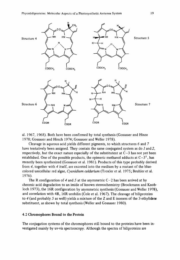
The chromophore cleavage reactions yield different pigments depending on the method used. The temporary confusion concerning the nomenclature of the chromophores is mainly due to the different cleavage procedures yielding different products. For a discussion, the reader is referred to O'Carra (1980), Rüdoger (1971,1975), and to the new IUPAC nomenclature on tetrapyrroles (1979). Most studies have focused on the 3-ethylidene bilins 4 and 5, which can best be obtained by thermolysis in re-fluxing alcohols (Fujita and Hattori 1961; O'Carra and O'hEocha 1966; Fu et al. 1979), or by treatment with hydrogen bromide in trifluoracetic acid (Schräm and Kroes 1971). The same products have been obtained recently by a flash-pyrolytic procedure (Fu et al. 1979). The structures 4 and 5 have been elucidated by chromic acid degradation (Rüdiger 1969) yielding succin- or maleimides carrying at least in principle the substi-tuents of the parent pyrrolic rings (Rüdiger and O'Carra 1969; Rüdiger et al. 1967), and by 1 Hmr and and mass spectroscopy (Chapman et al. 1967; Cole et al. 1968; Crespi et
Phycobiliproteins: Molecular Aspects of a Photosynthetic Antenna System 19
Structure 4
Structure 6
Structure 5
Structure 7
al. 1967, 1968). Both have been confirmed by total synthesis (Gossauer and Hinze 1978; Gossauer and Hirsch 1974; Gossauer and Weller 1978).
Cleavage in aqueous acid yields different pigments, to which structures 6 and 7 have tentatively been assigned. They contain the same conjugated System as do 1 and 2, respectively, but the exact nature especially of the substitutent at C—3 has not yet been established. One of the possible products, the epimeric methanol adducts at C - 3 1 , has recently been synthesized (Gossauer et al. 1981). Products of this type probably derived from 4, together with 4 itself, are excreted into the medium by a mutant of the blue-colored unicellular red algae, Cyanidium caldarium (Troxler et al. 1975; Beuhler et al. 1976).
The R configuration of 4 and 5 at the asymmetric C—2 has been arrived at by chromic acid degradation to an imide of known stereochemistry (Brockmann and Knobloch 1973), the 16R configuration by asymmetric synthesis (Gossauer and Weller 1978), and correlation with 4R, 16R urobilin (Cole et al. 1967). The cleavage of biliproteins to 4 (and probably 5 as well) yields a mixture of the Z and E isomers of the 3-ethylidene substituent, as shown by total synthesis (Weller and Gossauer 1980).
4.2 Chromophores Bound to the Protein
The conjugation Systems of the chromophores still bound to the proteins have been in-vestigated mainly by uv-vis spectroscopy. Although the spectra of biliproteins are
20 Hugo Scheer
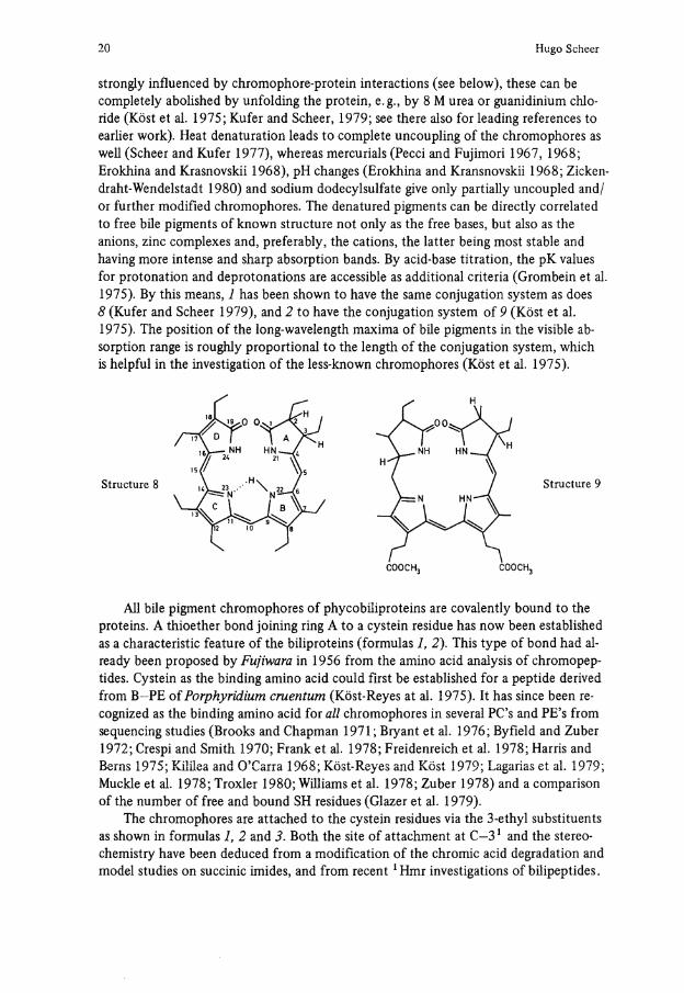
strongly influenced by chromophore-protein interactions (see below), these can be completely abolished by unfolding the protein, e.g., by 8 M urea or guanidinium Chloride (Köst et al. 1975; Kufer and Scheer, 1979; see there also for leading references to earlier work). Heat denaturation leads to complete uncoupling of the chromophores as well (Scheer and Kufer 1977), whereas mercurials (Pecci and Fujimori 1967, 1968; Erokhina and Krasnovskii 1968), pH changes (Erokhina and Kransnovskii 1968; Zickendraht-Wendelstadt 1980) and sodium dodecylsulfate give only partially uncoupled and/ or further modified chromophores. The denatured pigments can be directly correlated to free bile pigments of known structure not only as the free bases, but also as the anions, zinc complexes and, preferably, the cations, the latter being most stable and having more intense and sharp absorption bands. By acid-base titration, the pK values for protonation and deprotonations are accessible as additional criteria (Grombein et al. 1975). By this means, 1 has been shown to have the same conjugation System as does 8 (Kufer and Scheer 1979), and 2 to have the conjugation System of 9 (Köst et al. 1975). The position of the long-wavelength maxima of bile pigments in the visible absorption ränge is roughly proportional to the length of the conjugation system, which is helpful in the investigation of the less-known chromophores (Köst et al. 1975).
Structure 8 Structure 9
COOCH3 COOCH3
All bile pigment chromophores of phycobiliproteins are covalently bound to the proteins. A thioether bond joining ring A to a cystein residue has now been established as a characteristic feature of the biliproteins (formulas 1, 2). This type of bond had al-ready been proposed by Fujiwara in 1956 from the amino acid analysis of chromopep-tides. Cystein as the binding amino acid could first be established for a peptide derived from B-PE of Porphyridium c r u e n t u m (Köst-Reyes at al. 1975). It has since been re-cognized as the binding amino acid for all chromophores in several PC's and PE's from sequencing studies (Brooks and Chapman 1971; Bryant et al. 1976; Byfield and Zuber 1972; Crespi and Smith 1970; Frank et al. 1978; Freidenreich et al. 1978; Harris and Berns 1975; Kililea and O'Carra 1968; Köst-Reyes and Köst 1979; Lagarias et al. 1979; Mückle et al. 1978; Troxler 1980; Williams et al. 1978; Zuber 1978) and a comparison of the number of free and bound SH residues (Glazer et al. 1979).
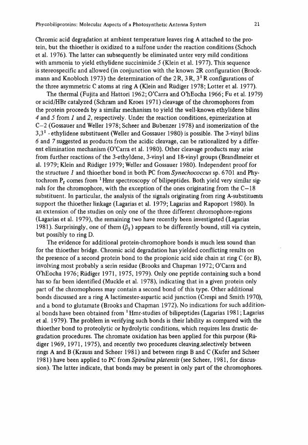
The chromophores are attached to the cystein residues via the 3-ethyl substituents as shown in formulas 1, 2 and 3. Both the site of attachment at C - 3 1 and the stereo-chemistry have been deduced from a modification of the chromic acid degradation and model studies on succinic imides, and from recent xHmr investigations of bilipeptides.
Phycobiliproteins: Molecular Aspects of a Photosynthetic Antenna System 21
Chromic acid degradation at ambient temperature leaves ring A attached to the protein, but the thioether is oxidized to a sulfone under the reaction conditions (Schoch et al. 1976). The latter can subsequently be eliminated unter very mild conditions with ammonia to yield ethylidene succinimide 5 (Klein et al. 1977). This sequence is stereospecific and allowed (in conjunction with the known 2R configuration (Brockmann and Knobloch 1973) the determination of the 2R, 3R, 3 1 R configurations of the three asymmetric C atoms at ring A (Klein and Rüdiger 1978; Lotter et al. 1977).
The thermal (Fujita and Hattori 1962; O'Carra and O'hEocha 1966; Fu et al. 1979) or acid/HBr catalyzed (Schräm and Kroes 1971) cleavage of the chromophores from the protein proceeds by a similar mechanism to yield the well-known ethylidene bilins 4 and 5 from 1 and 2, respectively. Under the reaction conditions, epimerization at C-2 (Gossauer and Weller 1978; Scheer and Bubenzer 1978) and isomerization of the 3,31 - ethylidene substituent (Weller and Gossauer 1980) is possible. The 3-vinyl büins 6 and 7 suggested as products from the acidic cleavage, can be rationalized by a different elimination mechanism (O'Carra et al. 1980). Other cleavage products may arise from further reactions of the 3-ethyldene, 3-vinyl and 18-vinyl groups (Brandlmeier et al. 1979; Klein and Rüdiger 1979; Weller and Gossauer 1980). Independent proof for the structure 1 and thioether bond in both PC from Synechococcus sp. 6701 and Phy-tochrom P r comes from 1 Hmr spectroscopy of bilipeptides. Both yield very similar Signals for the chromophore, with the exception of the ones originating from the C-18 substituent. In particular, the analysis of the Signals originating from ring A-substituents support the thioether linkage (Lagarias et al. 1979; Lagarias and Rapoport 1980). In an extension of the studies on only one of the three different chromophore-regions (Lagarias et al. 1979), the remaining two have recently been investigated (Lagarias 1981). Surprisingly, one of them (ß 2) appears to be differently bound, still via cystein, but possibly to ring D.
The evidence for additional protein-chromophore bonds is much less sound than for the thioether bridge. Chromic acid degradation has yielded conflicting results on the presence of a second protein bond to the propionic acid side chain at ring C (or B), involving most probably a serin residue (Brooks and Chapman 1972; O'Carra and O'hEocha 1976; Rüdiger 1971, 1975, 1979). Only one peptide containing such a bond has so far been identified (Mückle et al. 1978), indicating that in a given protein only part of the chromophores may contain a second bond of this type. Other additional bonds discussed are a ring A lactimester-aspartic acid junction (Crespi and Smith 1970), and a bond to glutamate (Brooks and Chapman 1972). No indications for such additional bonds have been obtained from 1 Hmr-studies of bilipeptides (Lagarias 1981; Lagarias et al. 1979). The problem in verifying such bonds is their lability as compared with the thioether bond to proteolytic or hydrolytic conditions, which requires less drastic degradation procedures. The Chromate oxidation has been applied for this purpose (Rüdiger 1969, 1971, 1975), and recently two procedures cleaving.selectively between rings A and B (Krauss and Scheer 1981) and between rings B and C (Kufer and Scheer 1981) have been applied to PC from Spirulina platensis (see Scheer, 1981, for discus-sion). The latter indicate, that bonds may be present in only part of the chromophores.
22 Hugo Scheer
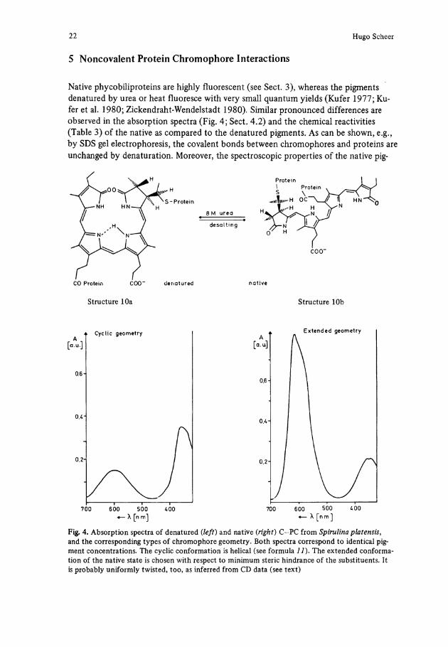
5 Noncovalent Protein Chromophore Interactions
Native phycobiliproteins are highly fluorescent (see Sect. 3), whereas the pigments denatured by urea or heat fluoresce with very small quantum yields (Kufer 1977; Kufer et al. 1980; Zickendraht-Wendelstadt 1980). Similar pronounced differences are observed in the absorption spectra (Fig. 4; Sect. 4.2) and the chemical reactivities (Table 3) of the native as compared to the denatured pigments. As can be shown, e.g., by SDS gel electrophoresis, the covalent bonds between chromophores and proteins are unchanged by denaturation. Moreover, the spectroscopic properties of the native pig-
CO Protein COO" denatured native
Structure 10a Structure 10b
1 1 1 ' I 1 r 1— 700 600 500 400 700 600 500 400
«— X [n m] «— X [n m ]
Fig. 4. Absorption spectra of denatured {left) and native (right) C-PC from Spirulina platensis, and the corresponding types of chromophore geometry. Both spectra correspond to identical pig-ment concentrations. The cyclic conformation is helical (see formula 1 1 ) . The extended conforma-tion of the native State is chosen with respect to minimum steric hindrance of the substituents. It is probably uniformly twisted, too, as inferred from CD data (see text)
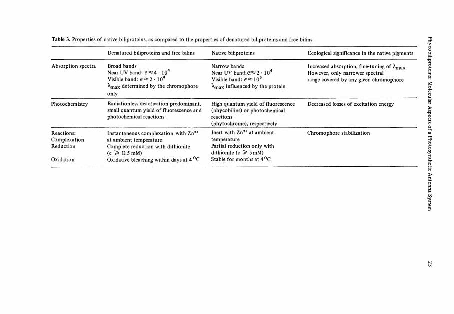
Table 3. Properties of native biliproteins, as compared to the properties of denatured biliproteins and free bilins
Denatured biliproteins and free bilins Native biliproteins Ecological significance in the native pigments
Absorption spectra Broad bands Near UV band: e^4- 104
Visible band: 6^2 • 104
X m a x determined by the chromophore only
Narrow bands Near UV b a n d : 2 - 104
Visible band: e « 1 0 5
Xmax influenced by the protein
Increased absorption, fine-tuning of Xmax However, only narrower spectral ränge covered by any given chromophore
Photochemistry Radiationless deactivation predominant, small quantum yield of fluorescence and photochemical reactions
High quantum yield of fluorescence (phycobilins) or photochemical reactions (phytochrome), respectively
Decreased losses of excitation energy
Reactions: Complexation Reduction
Oxidation
Instantaneous complexation with Zn 2 +
at ambient temperature Complete reduction with dithionite (c > 0.5 mM) Oxidative bleaching within days at 4 °C
Inert with Zn 2 + at ambient temperature Partial reduction only with dithionite (c ^ 5 mM) Stable for months at 4°C
Chromophore stabilization
24 Hugo Scheer
ments can be restored fully and in good yield (Scheer and Kufer 1977) within the fold-ing time of the protein (Bartholmes and Scheer 1980) if the concentration of the dena-turing agent or the temperature is decreased.
The spectroscopic properties and reactivities of the denatured pigments are identi-cal to those of free bile pigments of the proper structure, which has been commonly used in the structure analysis of biliproteins (see Sect. 4). This proves, on the other hand, that the properties of the denatured pigments can be accounted for completely by their proposed structures, and that the changes upon naturation are entirely due to noncovalent interactions between the chromophores and the proteins, and/or among the chromophores.
The ecological significance of these changes is obvious from Table 3. The oscillator strength, as a base for the efficiency of light absorption of biliprotein antenna Systems, is enhanced. Radiationless deexcitation, corresponding to a waste of the absorbed energy, is decreased, and at the same time chemical and photochemical side reactions leading to the destruction of the sensitive bile pigment chromophores is impeded. The biliproteins thus present an excellent example of molecular ecology, e.g., the adapta-tion of photoreceptor molecule as unfit as an A-dihydrobilindion like 1 or 4, to its function in an antenna pigment as efficient as phycocyanin. Recently, some progress has been made in understanding the principles involved, by a combination of chemical model studies, MO calculations, and denaturation-renaturation studies with the isolated pigments.
5.1 Topology of the Chromophore
The characteristics of the changes in the absorption spectra can be accounted for essen-tially by a change in the geometry of the chromophores. Bile pigments are at least prin-cipally flexible structures, and several research groups have treated theoretically the de-pendence of the absorption spectra on the molecular topology (Burke et al. 1971; Chae and Song 1975; Falk and Höllbacher 1978; Fuhrhop et al. 1974; Wagniere and Blauer 1976), and in some calculations also on a Charge at or close to the i t - System (Pasternak and Wagniere 1979; Sugimoto et al. 1976). The results agree in one fundamental aspect, that the ratio of the oscillator strengths of the two lowest electronic transitions,
Q = JYiS f near uv
is a rough measure of the shape of the molecule. Q is < 1 in cyclic porphyrin-type con-formations, but becomes > 1 when the molecule is stretched to an extended conforma-tion. On this basis, the spectroscopic properties of native and denatured phycocyanin can be rationalized by the former having an extended ( 1 0 b ) , the latter a cyclic conforma-tion ( 1 0 a , Fig. 4) (Scheer and Kufer 1977).
The theoretical calculations have been supported by the conformational analysis of bile pigments of the biliverdin type in Solution and in the crystal. A cyclic-helical conformation was proposed first for optically active urobilin on the basis of the large cotton effects observed, which are typical for inherently dissymmetric chromophores (Moscowitz et al. 1964). If the two asymmetric centers at C-4 and C-16 have the same
Phycobiliproteins: Molecular Aspects of a Photosynthetic Antenna System 25
configuration, one of the two helices of opposite chirality is strongly favored due to steric hindrance. The effect of asymmetric centers in shifting the equilibrium between the two helices is principally possible, too, in other helical bilins. For denatured PC, an energy difference of AH = 0.1-0.2 kcal/mol has been estimated from the steric hindrance arising from the asymmetric C—2, C-3, and C—3' (Scheer et al. 1979). In denatured PE, the contribution of C-16 is expected to counteract this effect. In agreement with this reasoning, denatured PC (Lehner and Scheer 1981), but not PE (Langer et al. 1980), has a pronounced optical activity.
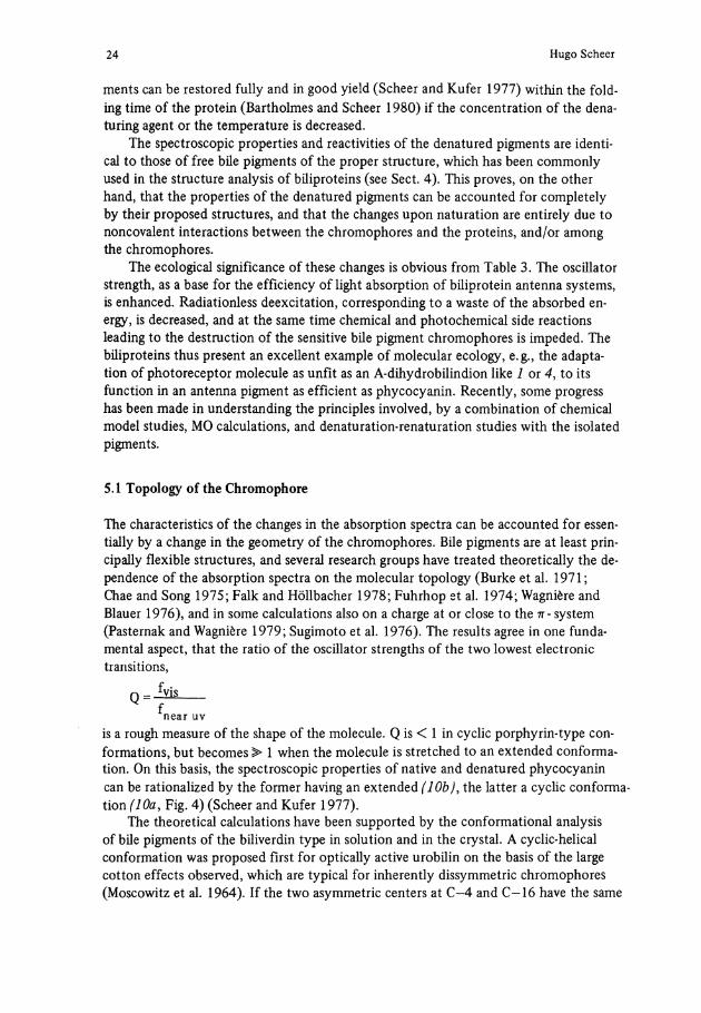
On the other hand, optical activity can be induced, too, by a chiral environment. The solvent-induced circular dichroism (SICD) seems to be a safe indicator of helical conformations (Lehner et al. 1978, 1981). All-syn, Z biliverdin-dimethylester ( 1 1 ) gives a strong SICD effect (Lehner et al. 1978), but neither the anti-E, syn-Z, Syn-Z isomer 12 (Gossauer et al. 1980) nor the formyltripyrrinone 13 (Lehner et al. 1981) do. In Solution 1 1 has a predominantly helical conformation (Lehner et al. 1978; Falk et al. 1978a), and a helical crystal structure (Lehner et al. 1978b; W.S. Sheldrick 1976). The energy barrier of 42 kJ/mol between the two helical forms allows a rapid intercon-version at ambient temperatures (Lehner et al. 1979). In a chiral solvent, e.g., lactate esters, the equilibrium between the two helical forms is shifted. As both forms are expected to be strongly optically active, a slight shift of the equilibrium from K = 1 is al-ready sufficient to give measurable CD Signals. In another way of reasoning, the pertur-bation by the chiral solvent is transmitted through the entire molecule only in a helical structure. 12 has a more extended conformation (according to MO studies), with no interaction between rings A and D (Falk et al. 1978b), while 13 is planar, as has been shown by the X-ray structure of an analog (Cullen et al. 1978), and thus also lacks the principal requirements for a strong SICD-effect. Falk et al. (1981) have recently car-ried out force field calculations on bile pigments. For the Z, Z, Z-bilindion, they con-verge at a structure very similar to the crystal structure of biliverdin-dimethylester (Sheldrick 1976).
Structure IIa Structure IIb
26 Hugo Scheer
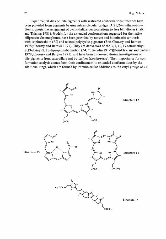
Experimental data on bile-pigments with restricted conformational freedom have been provided from pigments bearing intramolecular bridges. A 21,24-methano-bilin-dion Supports the assignment of cyclic-helical conformations to free bilindiones (Falk and Thirring 1981). Models for the extended conformations suggested for the native büiprotein-chromophores, have been provided by nature and biomimetic synthesis with isophorcabilin (15) and related polycyclic pigments (Bois-Choussy and Barbier 1978; Choussy and Barbier 1975). They are derivatives of the 3,7,12,17-tetramethyl-8,13-divinyl-2, 18-dipropionyl-bilindion ( 1 4 , "biliverdin IX 7")(Bois-Choussy and Barbier 1978; Choussy and Barbier 1975), and have been discovered during investigations on bile pigments from caterpillars and butterflies (Lepidoptera). Their importance for conformation analysis comes from their confinement to extended conformations by the additional rings, which are formed by intramolecular additions to the vinyl groups of 14.
Structure 13
Structure 12
Structure 14
Structure 15
C00CH 3
Phycobiliproteins: Molecular Aspects of a Photosynthetic Antenna System 27
The isophorcabilin 15 has an intense visible absorption band and a very weak near absorption (Bois-Choussy and Barbier 1978; Brandlmeier et al. 1981), thus supporting the theory. Its spectrum is very similar to that of native PC, for which an extended conformation is therefore highly likely, too (Fig. 4) (Scheer and Kufer 1977). This extended conformation has to be brought about by the protein, but the details of the process are not yet understood. It should be pointed out, that extended conformers of bile pigments like 1 and 12 are less stable than their cyclic conformers (Falk and Grubmayr 1979; Scheer et al. 1979). The energy for unfolding the chromophores would then have to be provided by the protein (Kufer and Scheer 1979). In agreement with this reasoning, the energies of denaturation of various PC's determined recently by Chen and Berns (1978) are conspicuously low as compared with the respective energies of globular proteins of similar size (Knapp and Pace 1974; Salahuddin and Tanford 1970).
5.2 Conformational Mobility
Another factor of the protein seems to be a restriction of conformational mobility to the chromophore. Bile pigments of the biliverdin type have broad absorption spectra, which even at low temperatures show only little fine structure (Chae and Song 1976; Friedrich et al. 1981a,b; Gautron et al. 1976; Holzwarth et al. 1978; Petrier et al. 1979, 1980; Scheer and Kufer 1977). This has been rationalized as a superposition of the spectra several conformers with slightly different absorptions, which are in rapid equilibrium with each other (Lehner et al. 1978a; Scheer et al. 1977; Scheer and Kufer 1978). This Interpretation has been substantiated recently by a careful fluorescence analysis. It could be shown that a Solution of 11 contains at least two species, their equilibrium being solvent-dependent (Braslavsky et al. 1980; Tegmo-Larsson et al. 1980a,b). From the Q values of the excitation spectra, a cyclic and a more extended conformation have been proposed, the latter being favored in rigid Solutions and especially in liposomes. Broad spectra are characteristic of most bile pigments, but a notable exception is the "purpurins", e.g., 1 3 . They have a double-peaked long-wavelength absorption, with each of the two components being narrow. 13 is planar (Cullen et al. 1978), and it has been suggested, that the two peaks arise from two comparably rigid, distinct forms, e.g., two tautomers, each fixed by a different type of intramolecular H-bond (Scheer et al. 1977).
Native biliproteins have broad absorption bands, too, in which some fine structure is generally obvious already at ambient temperature (see for examples: O'Carra and O'hEocha 1976) and becomes prominent at low temperatures (Frackowiak and Grabowski 1971; Frackowiak et al. 1975; Friedrich et al. 1980a,b; Gray et al. 1976; Scheer and Kufer, 1977; Zickendraht-Wendelstadt et al. 1980). Thus, their spectra are super-positions of different forms as well. There is, however, an important difference as compared with the denatured biliproteins or the free bile pigments with corresponding structures: The different forms are not in equilibrium, but rather correspond to different chromophores of the pigments in different environments well defined by the protein (see Scheer 1981; Zickendraht-Wendelstadt et al. 1980; Zuber 1978, for leading references). The Situation is reminiscent the purpurins, but obviously for different rea-sons. As an example, C-PC contains three chromophores. Its long-wavelength absorp-
28 Hugo Scheer
tion band is asymmetric with a distinct Shoulder at shorter wavelength already notice-able at room temperature, which is split into two narrow components at low temperatures (Frackowiak et al. 1975; Friedrich et al. 1980a,b; Scheer and Kufer 1977). The assignment to individual chromophores is yet unclear. The renatured a - and ß-subu-nits of C-PC from Synechococcus spec. (formerly A n a c y s t i s n i d u k n s ) absorb at 620 and 608 nm, respectively, indicating the a-subunit bearing the "f", the j3-subunit bear-ing two "s"-type chromophores (Glazer et al. 1973). The superposition of the spectra of the two subunits does not yield that of the native protein, however. Such an addi-tivity has been observed for a C-PE (Zickendraht et al. 1980). It is also indicated from the data of Binder et al. (1972) and unpublished data of the author for the renatured subunits of other C-PC's. In this case, the 0-subunit bearing two chromophores absorbs at longer wavelength than the a-subunit bearing a Single chromophore, which would correspond to the spectral shape of the native aß-monomer. Irrespective of this con-flicting Interpretation is the assignment of the two Compounds of the absorption spec-trum of C-PC to different chromophores supported by photochemical hole-burning experiments (Friedrich et al. 1980a,b), by partial denaturation (Scheer and Kufer 1977) and chemical modification, e.g., with sodium dithionite (Kufer and Scheer 1979). In each case, differential effects on the two absorption bands are observed.
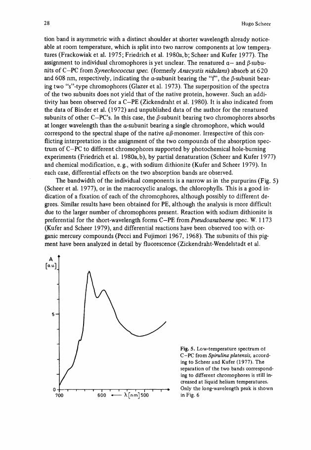
The bandwidth of the individual components is a narrow as in the purpurins (Fig. 5) (Scheer et al. 1977), or in the macrocyclic analogs, the Chlorophylls. This is a good in-dication of a fixation of each of the chromophores, although possibly to different de-grees. Similar results have been obtained for PE, although the analysis is more difficult due to the larger number of chromophores present. Reaction with sodium dithionite is preferential for the short-wavelength forms C-PE from Pseudoanabaena spec. W. 1173 (Kufer and Scheer 1979), and differential reactions have been observed too with or-ganic mercury Compounds (Pecci and Fujimori 1967, 1968). The subunits of this pigment have been analyzed in detail by fluorescence (Zickendraht-Wendelstadt et al.
A [Q.U.1
5 -
Fig. 5. Low-temperature spectrum oi C-PC from Spirulina platensis, according to Scheer and Kufer (1977). The Separation of the two bands correspond-ing to different chromophores is still in-creased at liquid helium temperatures.
0 700 600 * X[nm]500
Only the long-wavelength peak is shown in Fig. 6
Phycobiliproteins: Molecular Aspects of a Photosynthetic Antenna System 29
1980), and CD spectroscopy (Langer et al. 1980), and the results allow the distinction of all chromophores in the subunits, and of at least three of the five chromophores in the monomer.
The distinct environments of different chromophores in biliproteins is supported by analysis of the primary structure of chromopeptides and entire biliproteins. In all cases studied, there is a defined sequence to each chromophore, which is different for each chromophore in a given pigment, but similar for the corresponding chromophores in pigments from other organisms (Frank et al. 1978; Freidenreich et al. 1978; Otto et al. 1977; Troxler 1980; Zuber 1978).
A rigid fixation is necessary (although not sufficient, see below) to minimize radia-tionless decay of the excited states of biliproteins. This is accompanied by a narrowing of the absorption bands which is principally unfavorable to their antenna functions for two reasons: Only a narrow wavelength ränge is absorbed efficiently, and the overlap between emission bands of a sensitizing chromophore and the absorption of the next member of the Förster transfer chain becomes more crucial. Both effects are overcome by the development of chromophores absorbing at defined, closely spaced intervals, as realized most impressively in the PE's from red and cryptophytan algae, and in the phycobilisome superstructures (see Sect. 2).
Another necessary requirement for the suppression of radiationless processes is the suppression of photochemistry. This Channel is important in the phytochromes and phycochromes (Björn 1979; Rüdiger 1980; Scheer 1981). Especially the results obtained recently in the latter case (see Sect. 6) indicate that small changes in the protein structure are already sufficient to open the photochemical Channel, e.g., to increase radiationless transitions. Free bile pigments (Falk and Neufingerl 1979; Hudson and Smith 1975; Lightner 1977; Mac Donagh 1979; Manitto and Monti 1972), especially the A-Dihydrobilins (Kraus et al. 1979; Scheer and Krauss 1977; Scheer et al. 1977), are photolabile, but they react only with small quantum yields due to the competing radiationless deexcitation. It thus appears that the major effect of the protein is to de-crease the latter processes, wile the change from efficient fluorescence to efficient photochemistry requires comparatively small modifications in the protein environment.
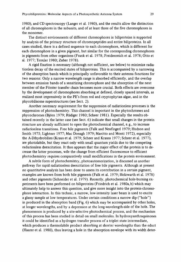
A subtle form of photochemistry, photoautomerization, is discussed as another pathway for rapid radiationless deexcitation of free bile pigments. Although at present no quantitative analysis has been done to assess its contribution in a certain pigment, examples are known from both bile pigments (Falk et al. 1979; Holzwarth et al. 1978) and other pigments (Schneider et al. 1979). Recently, photochemical hole-burning ex-periments have been performed on biliproteins (Friedrich et al. 1980a,b) which may ultimately help to answer this question, and give more insight into the protein-chromo-phore interaction. In this technic, a narrow, low-intensity laser beam is used to excite a glassy sample at low temperatures. Under certain conditions a narrow dip ("hole") in produced in the absorption band (Fig. 6) which may be accompanied by other holes, at longer wavelengths, and by a depression at the long-wavelength-side of the hole. The phenomenon is produced by a site-selective photochemical process, and the mechanism of this process has been studied in detail on small molecules. In hydroxyanthraquinones it could be identified as a hydrogen transfer process of a triplet State intermediate, which produces a thermolabile product absorbing at shorter wavelengths than the educt (Haarer et al. 1980), thus leaving a hole in the absorption envelope with its width deter-
30 Hugo Scheer
Temp: 1.8K
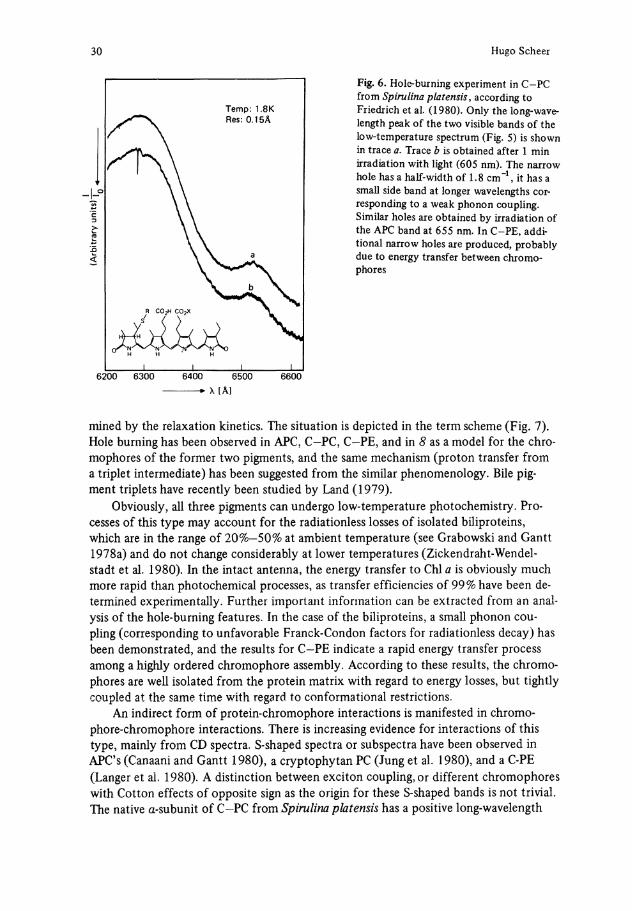
Fig. 6. Hole-burning experiment in C-PC from Spirulina platensis, according to Friedrich et al. (1980). Only the long-wavelength peak of the two visible bands of the low-temperature spectrum (Fig. 5) is shown in trace a . Trace b is obtained after 1 min irradiation with light (605 nm). The narrow hole has a half-width of 1.8 cm"1, it has a small side band at longer wavelengths cor-responding to a weak phonon coupling. Similar holes are obtained by irradiation of the APC band at 655 nm. In C-PE, additional narrow holes are produced, probably due to energy transfer between chromophores
H H H
6200 6300 6400 6500 6600
X [Ä ]
mined by the relaxation kinetics. The Situation is depicted in the term scheme (Fig. 7). Hole burning has been observed in APC, C-PC, C-PE, and in 8 as a model for the chromophores of the former two pigments, and the same mechanism (proton transfer from a triplet intermediate) has been suggested from the similar phenomenology. Bile pigment triplets have recently been studied by Land (1979).
Obviously, all three pigments can undergo low-temperature photochemistry. Processes of this type may account for the radiationless losses of isolated biliproteins, which are in the ränge of 20%—50% at ambient temperature (see Grabowski and Gantt 1978a) and do not change considerably at lower temperatures (Zickendraht-Wendelstadt et al. 1980). In the intact antenna, the energy transfer to Chi a is obviously much more rapid than photochemical processes, as transfer efficiencies of 99% have been determined experimentally. Further important Information can be extracted from an analysis of the hole-burning features. In the case of the biliproteins, a small phonon coupling (corresponding to unfavorable Franck-Condon factors for radiationless decay) has been demonstrated, and the results for C-PE indicate a rapid energy transfer process among a highly ordered chromophore assembly. According to these results, the chromophores are well isolated from the protein matrix with regard to energy losses, but tightly coupled at the same time with regard to conformational restrictions.
An indirect form of protein-chromophore interactions is manifested in chromo-phore-chromophore interactions. There is increasing evidence for interactions of this type, mainly from CD spectra. S-shaped spectra or subspectra have been observed in APC's (Canaani and Gantt 1980), a cryptophytan PC (Jung et al. 1980), and a C-PE (Langer et al. 1980). A distinction between exciton coupling, or different chromophores with Cotton effects of opposite sign as the origin for these S-shaped bands is not trivial. The native a-subunit of C—PC from Spirulina platensis has a positive long-wavelength
Phycobiliproteins: Molecular Aspects of a Photosynthetic Antenna System 31
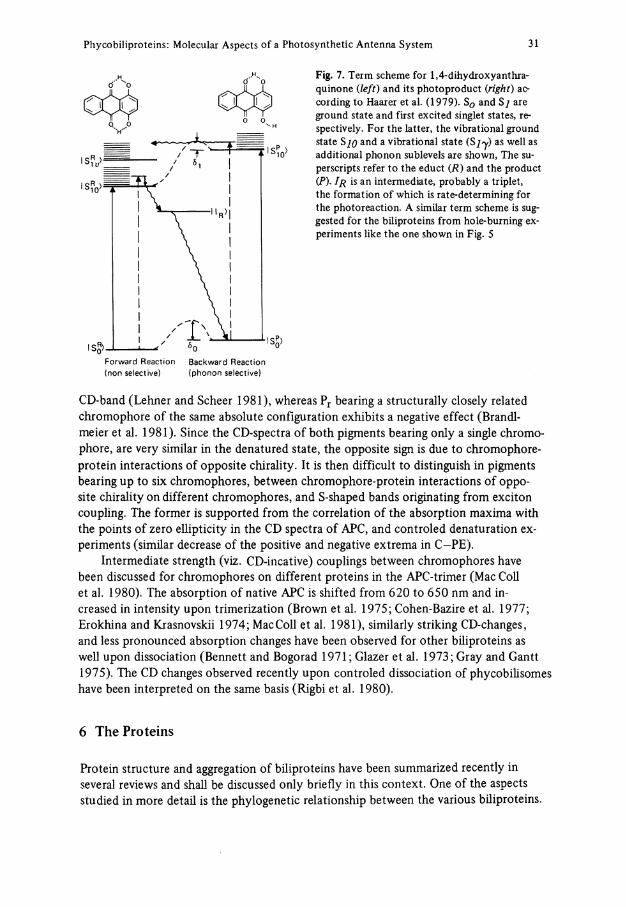
ö" *0 ö " H, Fig. 7. Term scheme for 1,4-dihydroxyanthra-
quinone (left) and its photoproduct (right) according to Haarer et al. (1979). S 0 and Sj are ground State and first excited singlet states, re-spectively. For the latter, the vibrational ground State SJQ and a vibrational State (S j y ) as well as additional phonon sublevels are shown, The su-perscripts refer to the educt ( R ) and the product (P). I R is an intermediate, probably a triplet, the formation of which is rate-determining for the photoreaction. A similar term scheme is suggested for the biliproteins from hole-burning ex-periments like the one shown in Fig. 5
Forward Reaction (non selective)
Backward Reaction {phonon selective)
CD-band (Lehner and Scheer 1981), whereas P r bearing a structurally closely related chromophore of the same absolute configuration exhibits a negative effect (Brandlmeier et al. 1981). Since the CD-spectra of both pigments bearing only a Single chromophore, are very similar in the denatured State, the opposite sign is due to chromophore-protein interactions of opposite chirality. It is then difficult to distinguish in pigments bearing up to six chromophores, between chromophore-protein interactions of opposite chirality on different chromophores, and S-shaped bands originating from exciton coupling. The former is supported from the correlation of the absorption maxima with the points of zero ellipticity in the CD spectra of APC, and controled denaturation ex-periments (similar decrease of the positive and negative extrema in C-PE).
Intermediate strength (viz. CD-incative) couplings between chromophores have been discussed for chromophores on different proteins in the APC-trimer (Mac Coli et al. 1980). The absorption of native APC is shifted from 620 to 650 nm and in-creased in intensity upon trimerization (Brown et al. 1975; Cohen-Bazire et al. 1977; Erokhina and Krasnovskii 1974; MacColl et al. 1981), similarly striking CD-changes, and less pronounced absorption changes have been observed for other biliproteins as well upon dissociation (Bennett and Bogorad 1971; Glazer et al. 1973; Gray and Gantt 1975). The CD changes observed recently upon controled dissociation of phycobilisomes have been interpreted on the same basis (Rigbi et al. 1980).
6 The Proteins
Protein structure and aggregation of biliproteins have been summarized recently in several reviews and shall be discussed only briefly in this context. One of the aspects studied in more detail is the phylogenetic relationship between the various biliproteins.
32 Hugo Scheer
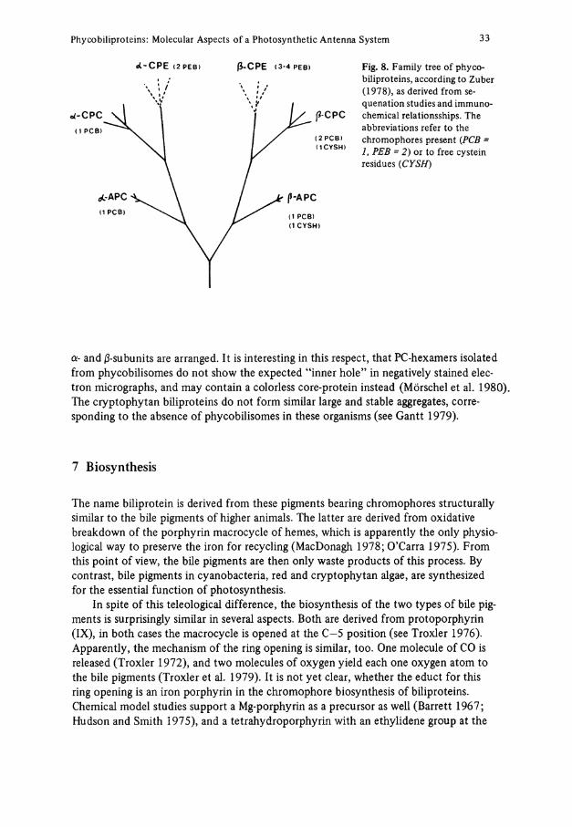
The family tree shown in Fig. 8 has been derived from sequenation studies and immunochemical investigations (Zuber 1978). The completed sequences of two C-PC's (Frank et al. 1978; Troxler 1980), an APC (Zuber et al. 1981) and of one subunit each of another C-PC (Freidenreich et al. 1978), principally support this picture, but there is evidence for very pronounced variations with sequenation studies of a marine cayno-bacterium A g m e n e l l u m quadruplicatum (Fox 1980). The completed sequences also in-dicate the presence of conservative as well as of more variable regions, the former being associated with the chromophore environments (see Mückle et al. 1977; Zuber 1980). This part may well be crucial for the efficiency and stability of the chromophores.
Due to conflicting earlier results (Berns 1967; Glazer et al. 1971), the cryptophytan biliproteins had not been included in Fig. 8. Recent immunological evidence Supports a relationship of both cryptophytan PC and PE with rhodophytan PE, but neither with rhodophytan PC nor cyanobacterial pigments (MacColl et al. 1976). This is yet another aspect of the somewhat special character of these pigments, since in both cyanobacterial and rhodophytan pigments the immunology parallels the spectroscopic Classification. But it also points to the increasing complexity of biliproteins emerging with an increasing number of pigments studied. Phycoerythrocyanin and R-PC are other examples of pigments with mixed chromophores, which could be correlated to the "common" pigments only by immunochemical methods (see Table 2 for references). Another example is the increasing number of biliproteins containing a 7-chain, which may have to be added to the family tree as another branch. Besides R— and B-PE, which form an a6ß6 7-structure, (Abad-Zapatero et al. 1977; Gantt and Lipschultz 1974; Sweet et al. 1977), APC—I with probably a3ß37-structure is the third example of this class (Zilinskas et al. 1978).
One of the most prominent properties of cyanobacterial and rhodophytan biliproteins is their pronounced aggregation. Especially the aggregation of PC's has been studied in great detail. Monomers, trimers, and hexamers are the most abundant species in Solutions of the purified pigments (MacColl et al. 1971), but dimers (Iso et al. 1977) and tetramers (Neufeld and Riggs 1969) have been observed, too. The influence of various parameters including concentration (Lee and Berns 1968; MacColl et al. 1974), ionic strength (MacColl et al. 1971a), pH (Lee and Berns 1968; Saito 1976), temperature (MacColl et al. 1971a), aromatic Compounds (MacColl and Berns 1973), Substitution of [ l H] by [2H] (Hattori et al. 1965; Lee and Berns 1968), inorganic and organic salts (Berns and Morgenstern 1968; MacColl et al. 1971b), biotope of the par-ent organisms (Chen and Berns 1979; Kao et al. 1973, 1975), and others (Chen and Berns 1978), have been investigated. A major contribution of hydrophobic interactions as the driving force for aggregation has been concluded.
The trimers and hexamers are the building blocs both of phycobilisomes (Gantt 1975; Glazer et al. 1979; Mörschel et al. 1980) and biliprotein crystals (Abad-Zapatero et ai. 1977; Bryant et al. 1976, Dobler et al. 1972; Sweet et al. 1977). Accordingly, higher aggregates (Berns 1971; MacColl and Berns 1979) as well as hetero-aggregates (Grabowski et al. 1980; Koller et al. 1978; Rigbi et al. 1980) can be obtained from controled dissociation of the former. In view of the architecture of the phycobilisomes, the 7-chain in R— and B—PE as well as in APC—I may then serve as a further means to stabilize the trimeric and hexameric building bloc, forming a core around which the
Phycobiliproteins: Molecular Aspects of a Photosynthetic Antenna System 33
«C-CPE ( 2 P E B ) ß-CPE ( 3 - 4 P E B ) Fig. 8. Family tree of phycobiliproteins, according to Zuber (1978), as derived from sequenation studies and immunochemical relationsships. The abbreviations refer to the chromophores present (PCB = 1, PEB = 2 ) or to free cystein residues (CYSH)
U-CPC (1 P C B )
( 2 P C B ) ( 1 C Y S H )
(3-CPC
a- and ß-subunits are arranged. It is interesting in this respect, that PC-hexamers isolated from phycobilisomes do not show the expected "inner hole" in negatively stained elec-tron micrographs, and may contain a colorless core-protein instead (Mörschel et al. 1980). The cryptophytan biliproteins do not form similar large and stable aggregates, corre-sponding to the absence of phycobilisomes in these organisms (see Gantt 1979).
7 Biosynthesis
The name biliprotein is derived from these pigments bearing chromophores structurally similar to the bile pigments of higher animals. The latter are derived from oxidative breakdown of the porphyrin macrocycle of hemes, which is apparently the only physio-logical way to preserve the iron for recycling (MacDonagh 1978; O'Carra 1975). From this point of view, the bile pigments are then only waste products of this process. By contrast, bile pigments in cyanobacteria, red and cryptophytan algae, are synthesized for the essential function of photosynthesis.
In spite of this teleological difference, the biosynthesis of the two types of bile pigments is surprisingly similar in several aspects. Both are derived from Protoporphyrin (IX), in both cases the macrocycle is opened at the C—5 position (see Troxler 1976). Apparently, the mechanism of the ring opening is similar, too. One molecule of CO is released (Troxler 1972), and two molecules of oxygen yield each one oxygen atom to the bile pigments (Troxler et al. 1979). It is not yet clear, whether the educt for this ring opening is an iron porphyrin in the chromophore biosynthesis of biliproteins. Chemical model studies support a Mg-porphyrin as a precursor as well (Barrett 1967; Hudson and Smith 1975), and a tetrahydroporphyrin with an ethylidene group at the
34 Hugo Scheer
proper position is known with bacteriochlorophyll b (Scheer et al. 1974). Brown et al. (1980) have, however, recently demonstrated the incorporation of exogenous hemin into phycobilins, but not Chlorophylls in Cyanidium caldarium, which strongly supports the "iron-pathway" in the former.
Nothing is yet known as to the point at which during biosynthesis the chromophore is linked to the protein. Facets to this problem are (1) the finding of various strains of Cyanidium caldarium which excrete after treatment with Ö-aminoevulinic acid (ALA), the ethylidene bilin 4 into the medium, together with products derived from addition reactions at the ethylidene function (Troxler and Bogorad 1966; Troxler et al. 1978); (2) the hitherto unsucessful search for the free apoprotein of any of these pigments; and (3) the demonstration of reversible addition reactions to the 3-ethylidene bond of 4 (Beuhler et al. 1976; Gossauer et al. 1980; Klein and Rüdiger 1978). Taken together, they indicate that the bilin 4 may be the biosynthetic precursor, which is attached to the protein in one of the last Steps.
The biosynthesis of biliproteins is effected by several nutritional factors, of which nitrogen (see Bogorad 1975), sulfur (Schmidt 1980) and especially light, are studied in detail. Light regulates the synthesis of both Chlorophylls and biliproteins. The tetra-pyrole skeletons of both pigments are derived from ALA and share a common part of their biosynthetic pathways, although possibly not their ALA pool. In a recent study, with mutants of Cyanidium caldarium defective in Chlorophyll or biliprotein biosynthesis, Schneider and Bogorad (1979) obtained evidence for two different, but strongly interacting photoregulation Systems. A protochlorophyll-type and a hemoprotein-type pigment, respectively, may be involved as photoreceptors. For leading references on light-dependent development in cyanobacteria and red algae the readerisreferredto: Björn 1979,1980;Bogorad 1975; Bogorad et al. 1975;Lazaroff 1973.
In many cyanobacteria and red algae, light-stimulated developmental responses have been observed, which suggest the presence of photoreversibly photochromic pigments as photoreceptors (see Björn 1979; Björn 1980; Bogorad 1975) (Table 4a). Chro-matic adaptation is a widespread response, by which (among other effects) the composi-tion of the biliprotein antenna is changed with the environmental light quality. The relative amount of green-light absorbing PE's is reversibly increased in green light and decreased in red light (Bogorad 1975; Lazaroff et al. 1973; Tandeau de Marsac 1977; Wagenmann 1977), with a concomitant change in phycobilisome composition (Siegel-man 1980). Another light-mediated response is the induction of filamentous growth, e.g., i n N o s t o c (Ginsburg and Lazaroff 1973). Corresponding to these effects, the terms adaptochromes and phycomorphochromes have been suggested (Bogorad 1975). In analogy to phytochrome, the photoreversible photochromic sensory pigment of higher plants (Rüdiger 1980), the term phycochromes is often used synonymously for these receptors.
It my be useful to reserve the term phycochromes to photoreversibly photochromic pigments irrespective of any proven function as a photoreceptor for the present time (see Björn 1979, for a discussion). This distinction arose from the attempts to isolate adaptochromes and phycomorphochromes, e.g., the genuine photoreceptors. During this search, Scheibe (1972) first isolated a biliprotein fraction from photo-bleached Tolypothrix t e n u i s , which gave photoreversible absorption differences remi-niscent of the action spectra for chromatic adaption in this species. The Björns have
Phycobiliproteins: Molecular Aspects of a Photosynthetic Antenna System 35
since characterized four different phycochromes (a, b, c, d) from various organisms, and purified to a different degree (Björn 1978; Björn and Björn 1976). All appear to be biliproteins, and thus require very tedious and extensive puriflcation from the bulk pigments, with isoelectric focusing as the key step. In only one of the photochromic frac-tions, the absorption differences exceed a few percent, although the quantum yields are reasonable. Surprisingly, only two of these four phycochromes (Table 4b) can presently be correlated spectroscopically to a known light response in the parent organisms, which led to the aforementioned distinction of phyochromes (pigments with photore-versible photochromic properties) on one hand, and adaptochromes and phycomorphochromes (as photoceptors for the respective processes) on the other. To link these chemical and functional properties to a certain pigment, it may be necessary to obtain further information besides the spectral data. As one such possibility, a differential temperature effect on the forward and back reaction of the photoreversion process has been suggested recently (Ohad et al. 1979).
The need for a distinction becomes even more obvious from recent results on the photochemical properties of phycobiliproteins partially denatured with low concentra-tions of guanidinium chloride (Ohki and Fujita 1979) or a decrease in pH (Ohad et al. 1979,1980) (Table 4c). By this treatment, APC and PC obtain photoreversible photochromic properties reminiscent of the phycochromes a and c respectively, of Björn and Björn (1976). According to all evidence, this is no artifact due to a co-isolated impurity. It rather appears that at least in these cases phycochromes are not separate pigments, but rather slightly denatured states of well-known biliprotein antenna pigments, or certain sub-populations there of. It is interesting in this respect that phycochrome d has been associated recently with another biliprotein, phycoerythrocyanin (Björn 1980). The denaturant-induced photochromicity of PC and APC decreases again at more severe denaturation conditions. Probably, the "tickling" of the protein looses the interactions with the protein sufficiently to open a photochemical Channel, while internal conversion and destructive photochemistry of the pigments are still inhibited. More severe uncoupling (see Sect. 5) then favors the latter processes. Stepwise denaturation has been observed with PC Spirulina platensis, suggesting a distinct intermediate in the unfolding process (Scheer and Kufer 1977). Similar intermediates may be produced in the pigments isolated from T. tenuis (Ohki and Fujita 1979) and F. diplosiphon (Ohad et al. 1979, 1980). MacColl et al. (1980b) recently pointed out, that similar difference spectra as in the phycochrome reaction, are observed in the reversible dissociation of trimeric (aß)3 APC II. It would then be crucial to link the dissociation-reassociation unequivocally to the light reaction rather than to an artefact, e.g., due to sample heating.
8 Concluding Remarks
This review on phycobiliproteins is far from complete. This is not only due to the wide-spread and still growing interest in these pigments, which is prohibitive to the citing of all pertinent work, but also to the deliberate attempt of the author, to focus mainly on the chromophore protein interactions. These interactions in the native biliproteins are crucial for their functions, and only recently some general principles have been recog-
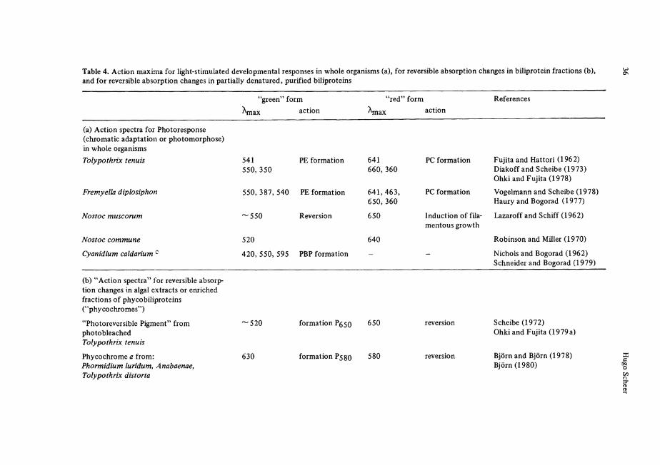
Table 4. Action maxima for light-stimulated developmental responses in whole organisms (a), for reversible absorption changes in biliprotein fractions (b), and for reversible absorption changes in partially denatured, purified biliproteins
"green" form Ajnax action
"red" form References
\na action
(a) Action spectra for Photoresponse (chromatic adaptation or photomorphose) in whole organisms Tolypothrix tenuis 541
550, 350 PE formation
Fremyella diplosiphon
Nostoc muscorum
Nostoc commune
Cyanidium caldarium c
550, 387, 540 PE formation
'550 Reversion
520
641 660, 360
641,463, 650,360
650
640
PC formation
PC formation
Induction of fila-mentous growth
420, 550, 595 PBP formation
Fujita and Hattori (1962) Diakoff and Scheibe (1973) Ohki and Fujita (1978)
Vogelmann and Scheibe (1978) Haury and Bogorad (1977)
Lazaroff and Schiff (1962)
Robinson and Miller (1970)
Nichols and Bogorad (1962) Schneider and Bogorad (1979)
(b) "Action spectra" for reversible absorption changes in algal extracts or enriched fractions of phycobiliproteins ("phycochromes")
"Photoreversible Pigment" from photobleached Tolypothrix tenuis
Phycochrome a from: P h o r m i d i u m l u r i d u m , Anabaenae, Tolypothrix distorta
'520
630
formation ?ß$0 650
formation P5gQ 580
Scheibe (1972) Ohki and Fujita (1979a)
Björn and Björn (1978) Björn (1980)
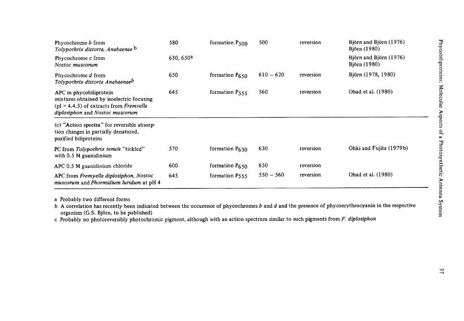
Phycochrome b from Tolypothrix distorta, Anabaenae^
580 formation P500 500 reversion Björn and Björn (1976) Björn (1980)
Phycochrome c from Nostoc muscorum
630,650a Björn and Björn (1976) Björn (1980)
Phycochrome d from Tolypothrix distorta Anabaenae^
650 formation ?S50 610-620 reversion Björn (1978, 1980)
APC in phycobiiiprotein mixtures obtained by isoelectric focusing (pl = 4.4.3) of extracts from Fremvella diplosiphon and Nostoc muscorum
645 formation P555 560 reversion Ohad et al. (1980)
(c) "Action spectra" for reversible absorption changes in partially denatured, purified biliproteins
PC from Tolypothrix tenuis "tickled" with 0.5 M guanidinium
570 formation Pg3Q 630 reversion Ohki and Fujita (1979b)
APC 0.5 M guanidinium Chloride 600 formation P650 650 reversion
APC from F r e m y e l l a diplosiphon, Nostoc muscorum and P h o r m i d i u m l u r i d u m at pH 4
645 formation P555 550 - 560 reversion Ohad et al. (1980)
a Probably two different forms b A correlation has recently been indicated between the occurence of phycochromes b and d and the presence of phycoerythrocyanin in the respective
organism (G.S. Björn, to be published) c Probably no photoreversibly photochromic pigment, although with an action spectrum similar to such pigments from F. diplosiphon
38 Hugo Scheer
nized. Most details are derived from spectroscopic and chemical studies of the chromophores. The protein environment is known only for a few pigments in its primary structure, and the amino acid residues responsible for the noncovalent interaction in the native pigments are still unknown. X-ray structure, as the most promising tool to study these interactions, has encountered unexpected difficulties, but may provide these data in the near future. Other major gaps in our present knowledge concern the last Steps in the biosynthesis of biliproteins, the processes of biliprotein aggregation to phycobilisomes, and the details about the phycobilisome-membrane junction critical for energy transfer to the reaction centers. It should also be borne in mind that biliproteins have been studied in detail only from a small number of species.
Phycobiliproteins are a good example of molecular ecology, and they may help not only in understanding better the processes of photosynthesis, but also the interactions between proteins and small molecules in general. This, together with the fun to work with these brilliantly colored pigments, is expected to fill at least part of the gaps in the near future.
Acknowledgement. The cited work of the author was supported by the Deutsche Forschungsgemeinschaft, Bonn-Bad Godesberg.
Notes Added in Proof
This manuscript contains references acessible to the author until mid 1981. Some recent progress is added below. The reader is also referred to a review on phycobilisomes and phycobiliproteins by R. MacColl to appear in Photochem. Photobiol.
Energy Transfer
The transfer efficiencies from R-PE to R-PC (93%), R-PC to "bulk" APC (98%) and "bulk" APC to the "terminal" APC (96%) have been determined for a rhodophytan phycobilisome [Bekasova et al.; Biofizika 26, 74 (1981)]. Several groups are investigating the energy transfer in biliproteins and phycobilisomes by picosecond time-resolved techniques. By comparison of low and high inten-sity excitation, 1 S- 1 S annihilation has been suggested as a complication both in isolated biliproteins [Wong et al.; Photochem. Photobiol. 33, 651 (1981); Doukas et al. ibid. 34, 505 (1981)] and in phycobilisomes (Holzwarth et al., unpubl.). Attempts to analyze the complex fluorescence or ground-state recovery are ambiguous, since good fits can be obtained using expotentials only, but also in-cluding nonlinear exponents (t"1/2) originating from a Förster mechanistic analysis (Holzwarth et al., unpublished; Hefferele, Nies, Wehrmeyer and Schneider, unpublished). Depolarization studies have been included in the latter study and also in denaturation work with C-PC in defined aggregation states (Hefferle. John: Scheer and Schneider, unpublished) to aid this analysis. Brody et al. (Biophys. J. 34, 439 (1981) reported the puzzling result, that the 715 nm fluorescence (generally associated with Chlorophyll) arose faster than that of biliproteins when exciting the latter, which may indicate alternative transfer schemes, e.g., via carotenoids (Szalontay and v.d. Ven, FEBS Lett. 131, 155 (1981).
Phycobiliproteins: Molecular Aspects of a Photosynthetic Antenna System 39
Structure of Phycobilisomes and Reconstitution of Functional Complexes
Kirilowski and Ohad (Proc. Natl. Acad. Sei. USA, in press) reported the reassembly of fully dissociated phycobilisome-membrane complexes, and Glick and Zilinskas (Plant Physiol., in press) demonstrated the importance of the structural peptides by reassembling phycobilisomes from the individual pigments in the presence of a colorless 29 kdalton peptide. The function and properties of the large structural peptide (75-90 kdalton) have been studied in some detail and the results seem to rational-ize earlier conflicting results. This colored peptide is readily susceptible to bleaching and proteo-lysis [Lundell and Glazer; J. Biol. Chem. 256, 12600 (1981); Lundell et al., J. Cell. Biol. 91, 315 (1981); Ruschkowski, Ph. D. Thesis, Rutgers University, 1981] and is probably the genuine 7-sub-unit of APC-I, serving as the terminal energy donor to Chlorophyll in these algae, and as (one of) the anchor-protein(s) of the phycobilisome to the membrane. The facile proteolysis of this peptide is reminiscent of other peripheral membrane proteins, e.g., some b-cytochromes. The functionality and structure of phycobilisomes during N-starvation has been studied by Yamanaka and Glazer [Aren. Microbiol. 124, 39 (1980)]. The APC core and adjacent PC hexamers essential for energy transfer are kept functional until a very late stage of starvation, whereas the phycobilisome peri-phery including specific structural peptides are digested. Nies and Wehrmeyer [Aren. Microbiol. 129, 374 (1981)] studied in some detail the phycobilisomes from M. laminosus and its dissociation products to arrive at a model very similar to that of the red algae, R . violacea shown in Fig. 1.
Isolated Biliproteins
The primary structure of a third C-PC (C caldarium) solved by Troxler et al. [J. Biol. Chem. 256, 12176 (1981)] and Offner et al. [ibid. p. 12167] supports the homology between pigments from different species. Murakami et al. [J. Biochem. 89, 79 (1981)] studied the reversible aggregation of APC under varying environmental conditions. By curve-resolution of the absorption band, they con-cluded that one of the two chromophores is unaffected by this process, whereas the second chromophore is blue-shifted and decreased in intensity in the monomer [Mimuro et al., Arch. Biochem. Bio-phys. (in press)]. This change is similar to the change observed upon deprotonation of bile-pigment cations to the free bases (see text). Yu et al. [Plant. Physiol. 68, 482 (1981)] found a Variation in the chromophore composition indicating an exchange of phycourobilin with phycoerythrobilin chromophores in R-PE of two closely related species, and also in a Single species by chromatic adaptation.
References
Abad-Zapatero, C , Fox, JL., Hackert, M.L.: Biochem. Biophys. Res. Comm. 78, 266 (1977) Abeliovich, A., Shilo, M.: Biochim. Biophys. Acta 283, 483-491 (1972) Adams, S.M., Kao O.H.W., Berns, D.S.: Plant Physiol. 64, 525-527 (1979) Alberte, R.S., Friedman, A.L., Gustafson, D.L., Rudnick, M.S., Lyman, H.: Biochim. Biophys.
Acta 635, 304-316 (1981) Allen, M.M., Smith, A.J., Arch. Mikrobiol. 69, 114-120 (1969) Barrett, J.: Nature 215, 733-35 (1967) Bartholmes, P., Scheer, H.: in preparation Bellin, J.S., Gergel, CA.: Photochem. Photobiol. 13, 399-409 (1971) Bennett, A., Bogorad, L.: Biochemistry 10, 3625 (1971) Bennett, A., Siegelman, H.W.: In: The Porphyrins; Dolphin, D. (ed.), pp. 493. New York: Academic
Press 1979 Berns, D.S.: In: Subunits in Biological Systems; Timasheff, S.N., Fasman, G.D. (eds.), Part A,
pp. 105, New York: Dekker 1971 Berns, D.S.: Photochem. Photobiol. 24, 117-139 (1976)
40 Hugo Scheer
Berns, D.S., Morgenstern, A.: Arch. Biochem. Biophys. 123, 640 (1980) Beuhler, R.J., Pierce, R.C., Friedman, L., Siegelman, H.W.: J. Biol. Chem. 251, 2405-2411 (1976) Binder, A., Wilson, K., Zuber, H.: FEBS Lett. 20, 111 (1972) Björn, G.S.: Physiol. Plant. 42, 321 (1978) Björn, G.S.: Ph. D. Thesis (Coden: Lunbds/CNBFB-1009)/l-28/ (1980), University of Lund 1980 Björn, G.S., Björn, L.O.: Physiol. Plant. 26, 297 (1976) tjörn, L.O.: Quart. Rev. Biophys. 12, 1-25 (1979) Blacha-Puller, M.: Dissertation, Technische Universität, Braunschweig 1979 Bogorad, L.: Annv. Rev. Plant Physiol. 26, 369-401 (1975) Bois-Choussy, M., Barbier, M.: Heterocycles 9, 677-690 (1978) Bonnett, R.: In: The Porphyrins; Dolphin, D. (ed.), Vol. I, p. 1, New York: Academic Press 1978 Boucher, L.J., Crespi, H.L., Katz, JJ.: Biochemistry 5, 3796-3802 (1966) Brandlmeier, T., Bios, I., Rüdiger, W.: In: "Photoreceptors and Plant Development"; de Greef, J.
(ed.), p. 47-54, Antwerpen University Press, 1980 Brandlmeier, T., Scheer, H., Rüdiger, W.: Z. Naturforsch. 36c, 431-439 (1981) Braslavsky, S.E., Holzwarth, A.R., Langer, E., Lehner, H., Matthews, J.J., Schaffner, K.: I sr. J.
Chem. 20,196-202 (1980) Brockmann, H. jr., Knobloch, G.: Chem. Ber. 106, 803 (1973) Brooks, C , Chapman, DJ.: Phytochemistry 11, 2663-2670 (1972) Brown, A.S., Foster, J.A., Voynow, P.V., Franzblau, C , Troxler, R.F.: Biochemistry 14, 3581
(1975) Brown, S.B., King, R.F.G.J.: Biochem. J. 170, 297-311 (1978) Bryant, D.A., Glazer, A.N., Eiserling, F.A.: Arch. Microbiol. 110, 61-75 (1976) Bryant, D.A., Hixson, CS., Glazer, A.N.: Biol. Chem. 253, 220-225 (1978) Burke, M.J., Pratt, D.C, Moscowitz, A.: Biochemistry 11, 4025 (1972) Byfield, P.G.H., Zuber, H.: FEBS Lett. 28, 36-40 (1972) Calzaferri, G., Gugger, H., Leutwyler, S.: Helv. Chim. Acta 59, 1969 (1976) Canaani, O.D., Gantt, E.: Biochemistry 19, 2950-2956 (1980) Canaani, O.D., Lipschultz, CA., Gantt, E.: FEBS Lett. 115, 225-229 (1980) Chae, Q., Song, P.S.: J. Am. Chem. Soc. 97, 4176 (1975) Chapman, DJ.: In: The Biology of Blue-Green Algae; Whitton, B.A. (ed.), p. 162. Berkeley: Uni
versity of California Press 1973 Chapman, DJ., Cole, W.J., Siegelman, H.W.: J. Am. Chem. Soc. 89, 5976 (1967) Chapman, DJ., Cole, W.J., Siegelman, H.W.: Biochem. J. 105, 903-905 (1967) Chen, C.H., Berns, D.S.: Biophys. Chem. 8, 191-202 (1978) Chen, C.H., Berns, D.S.: Phys. Chem. 82, 2781-2787 (1978a) Chen, S.S., Berns, D.S.: J. Membrane Biol. 47, 113-127 (1979) Choussy, M., Barbier, M.: Helv. Chim. Acta 58, 2651-2661 (1975) Clayton, R.K., Sistrom, W.R.: The Photosynthetic Bacteria. New York: Academic Press 1978 Cohen-Bazire, G., Be*guin, S., Rimon, S., Glazer, A.N., Brown, D.M.: Arch. Microbiol. III,
225-238 (1977) Cole, W.J., O'hEocha, C , Moscowitz, A., Krueger, W.R.: Eur. J. Biochem. 3, 202-207 (1967) Cole, W.J., Chapman, DJ., Siegelman, H.W.: Biochemistry 7, 2929 (1968) Crespi, H.L., Smith, U.H.: Phytochemistry 9, 205-212 (1970) Crespi, H.L., Boucher, L.J., Norman, G.D., Katz, JJ., Dougherty, R.C.: J. Am. Chem. Soc. 89, 3642 (1967) Crespi, H.L., Smith, U., Katz, JJ.: Biochemistry 7, 2232 (1968) Csatorday, K.: Biochim. Biophys. Acta 504, 341 (1978) Cuilen, D.L., Meyer, E.F. jr., Eivazi, F., Smith, K.M.: J. C. S. Perkin II 1978,259 Dale, R.E., Teale, F.W.J.: Photochem. Photobiol. 12, 99 (1970) Diakoff, S., Scheibe, J.: Plant Physiol. 51, 382 (1973) Dobler, S.D., Dover, K., Laves, K., Binder, A., Zuber, H.: J. Mol. Biol. 71, 785-787 (1972) Eder, J., Wagenmann, R., Rüdiger, W.: Immunochemistry 15, 315-321 (1978) Emerson, R.: Annu. Rev. Plant Physiol. 9, 1-24 (1958) Erokhina, L.G., Krasnovskii, A.A.: Biologya (Engl, transl.) 2, 442-450 (1968)
Phycobiliproteins: Molecular Aspects of a Photosynthetic Antenna System 41
Erokhina, L.G., Krasnovskii, A.A.: Molekulyarnaya Biologiya (Engl, transl.) 8, 517-523 (1974) Evstigneev, V.B., Bekasova, O.D.: Biofizika 17, 997-1006 (1972) Falk, H., Grubmayr, K.: Monatsh. Chem. 110, 1237 (1979) Falk, H., HöUbacher, G.: Monatsh. Chem. 109, 1429 (1978) Falk, H., Neufingerl, F.: Monatsh. Chem. 110, 987 (1979) Falk, H., Thirring, K.: Tetrahedron 37, 761-766 (1981) Falk, H., Grubmayr, K., Thirring, K.: Z. Naturforsch. 33b, 924 (1978a) Falk, H., Grubmayr, K., Haslinger, E., Schlederer, T., Thirring, K.: Monatsh. Chem. 109, 1451-
1473 (1978b) Falk, H., Grubmayr, K., Neufingerl, F.: Monatsh. Chem. 110, 1127 (1979) Falk, H„ HöUbacher, G., Hofer, O., Müller, N.: Monatsh. Chem. 112, 391-403 (1981) Frackowiak, D., Grabowski, J.: Photosynthetica 5, 146 (1971) Frackowiak, D., Skowron, A.: Photo synthetica 12, 76 (1978) Frackowiak, D., Fiksinski, K., Grabowski, J.: Photo synthetica 9, 185 (1975) Frackowiak, D., Grabowski, J., Manikowski, H.: Photosynthetica 10, 204-207 (1976) Frank, G., Sidler, W., Widmer, H., Zuber, H.: Hoppe-Seyler's Z. Physiol. Chem. 359, 1491 (1978)
-Freidenreich, P., Apell, G.S., Glazer, A.N.: J. Biol. Chem. 253, 212-219 (1978) Friedrich, J., Scheer, H., Zickendraht-Wendelstadt, B., Haarer, D.: J. Am. Chem. Soc. 103,
1030-1035 (1981a) Friedrich, J., Scheer, H., Zickendraht-Wendelstadt, B., Haarer, D.: J. Chem. Phys. 74, 2260-2266
(1981b) Fu, E., Friedman, L., Siegelman, H.W.: Biochem. J. 179, 1 (1979) Fuhrhop, J.-H., Wasser, P.K.W., Subramanian, J., Schräder, U.: Liebigs Ann. Chem. 1974,1450 Fujimori, E., Pecci, J.: Biochim. Biophys. Acta 221, 132 (1970) Fujita, Y., Hattori, A.: J. Biochem. (Tokyo) 51, 89 (1962) Fujita, Y., Hattori, A.: Plant Cell Physiol. 3, 209-220 (1962) Fujiwara, T.: Biochem. (Tokyo) 43, 195-203 (1956) Gantt, E.: Bio Science 25, 781-788 (1975) Gantt, E.: In: Biochemistry and Physiology of Protozoa; Levandowsky, M., Hutner, S.H. (eds.),
2nd ed., pp. 121-137. New York: Academic Press 1979 Gantt, E.: Ann. Rev. Plant Physiol. 32, 327-347 (1981) 'Gantt, E., Conti, S.F.: J. Cell. Biol. 29, 423 (1966) Gantt, E., Lipschultz, CA.: Biochemistry 13, 2960-66 (1974) Gantt, E., Lipschultz, CA.: J. Phycol. 13, 185-192 (1977) Gantt, E., Edwards, M.R., Provasoli, L.: J. CelL Biol. 48, 280-290 (1971) Gantt, E., Lipschultz, CA., Grabowski, J., Zimmermann, K.: Plant Physiol. 63, 615-620 (1979) Gautron, R., Jardon, P., Petrier, C , Choussy, M., Barbier, M., Vuillaume, M.: Experientia 32, 1100
(1976) Gendel, S., Ohad, I., Bogorad, L.: Plant Physiol. 64, 786-790 (1979) Ginsburg, R., Lazaroff, N.: J. Gen. Microbiol. 75, 1-9 (1973) Glazer, A.N.: Mol. Cell. Biochem. 18, 125-140 (1977) Glazer, A.N., Bryant, D.A.: Arch. Microbiol. 104, 15-22 (1975) Glazer, A.N., Cohen-Bazire, G.: Arch. Microbiol. 104, 29-32 (1975) Glazer, A.N., Hixson, CS.: J. Biol. Chem. 250, 5487 (1975) Glazer, A.N., Hixson, CS.: J. Biol. Chem. 252, 32-42 (1977) Glazer, A.N., Cohen-Bazire, G., Stanier, R.Y.: Arch. Microbiol. 80, 1 (1971a) Glazer, A.N., Cohen-Bazire, G., Stanier, R.Y.: Proc. Natl. Acad. Sei. USA 68, 3005 (1971b) Glazer, A.N., Fang, S., Brown, D.M.: J. Biol. Chem. 248, 5679-5685 (1973) Glazer, A.N., Hixson, CS., De Lange, R.J.: Anal. Biochem. 92, 489 (1979a) Glazer, A.N., Williams, R.C, Yamanaka, G., Schachman, H.K.: Proc. Natl. Acad. Sei. USA 76,
6161-6166 (1979b) Gossauer, A., Hinze, R.-P.: J. Org. Chem. 43, 283 (1978) Gossauer, A., Hirsch, W.: Liebigs Ann. Chem. 1974, 1496-1513 (1974) Gossauer, A., Weiler, J.-P.: J. Am. Chem. Soc. 100, 5928 (1978) Gossauer, A., Biacha-Puiler, M., Zeisberg, R., Wray, V.: Liebigs Ann. Chem. 1981, p. 342-346
42 Hugo Scheer
Gossauer, A., Hinze, R.P., Kutschan, R.: Chem. Ber. 114, 132-146 (1981) Govindjee: Bioenergetics of Photosynthesis. New York: Academic Press 1975 Grabowski, J., Gantt, E.: Photochem. Photobiol. 28, 39-45 (1978a) Grabowski, J., Gantt, E.: Photochem. Photobiol. 28, 47-54 (1978b) Grabowski, J., Lipschultz, CA., Gantt, E.: Plant Physiol., submitted for publication Gray, B.H., Gantt, E.: Photochem. Photobiol 21, 121-128 (1975) Gray, B.H., Lipschultz, CA., Gantt, E.: J. Bacteriol. 116, 471-478 (1973) Gray, B.H., Cosner, J., Gantt, E.: Photochem. Photobiol. 24, 299-302 (1976) Grombein, S., Rüdiger, W., Zimmermann, H.: Hoppe-Seyler's Z. Physiol. Chem. 356, 1709-1714
(1975) Gysi, J., Zuber, H.: FEBS Lett. 48, 209-213 (1974) Gysi, J., Zuber, H.: FEBS Lett. 68, 49-54 (1976) Hackert, M.L., Abad-Zapatero, C , Stevens, S.E. jr., Fox, J.L.: J. Mol. Biol. 111, 365-369 (1977) "Harnischfeger, G., Codd, G.A.: Biochim. Biophys. Acta 502, 507-513 (1978) Harris, J.U., Berns, D.S.: J. Mol. Evol. 5, 153-163 (1975) Hartmann, K.M., Haupt, W.: In: Biophysik; Hoppe, W., Lohmann, W., Markl, H., Ziegler, H. (eds.),
p. 449. Berlin-Heidelberg-New York: Springer 1978 Hattori, A., Crespi, H.L., Katz, J.J.: Biochemistry 4, 1225-1238 (1965) Haury, J.F., Bogorad, L.: Plant Physiol. 60, 835-839 (1977) Haxo, F.T.: In: Comparative Biochemistry of Photoreactive Systems; Allen, M.B. (ed.),
pp. 339-360. New York: Academic Press 1960 Holzwarth, A.R., Lehner, H., Braslavsky, S.E., Schaffner, K.: Liebigs Ann. Chem. 1978, 2002 Hudson, M.F., Smith, K.M.: Chem. Soc. Rev. 4, 363 (1975) Ilani, A., Berns, D.S.: Biochem. Biophys. Res. Comm. 45,1423-1430 (1971) Iso, N., Mizuno, H., Saito, T., Nitta, N., Yoshizaki, K.: Bull. Chem. Soc. Jap. 56, 2892 (1977) IUPAC: Nomenclature on Tetrapyrroles. Pure Appl. Chem. 51, 2251 (1979) Jung, J., Song, P.-S., Paxton, R.J., Edelstein, M.S., Swanson, R., Hazen, E.E. jr.: Biochemistry 19,
24-32 (1980) Kessel, M., MacColl. R., Berns,D.S., Edwards, M.R.: Can. J. Microbiol. 19, 831-836 (1973) Küilea, S.D., O'Carra, P.: Biochem. J. 110,14-15P (1968) Klein, G., Rüdiger, W.: Liebigs Ann. Chem. 1978, 267 Klein, G., Rüdiger, W.: Z. Naturforsch. 34c, 192-195 (1979) Klein, G., Grombein, S., Rüdiger, W.: Hoppe-Seyler's Z. Physiol. Chem. 358,1077-1079 (1977) Knapp, J.A., Pace, C.N.: Biochemistry 13, 1289-1294 (1974) Kobayashi, T., Degenkolb, E.O., Behrson, R., Rentzepis, P.M., MacColl, R., Berns, D.S.: Biochem
istry 18, 5073 (1979) Koller, K.P., Wehrmeyer, W., Moerschel, E.: Eur. J. Biochem. 91, 57 (1978) Köst, H.-P.: Private communication (1980) Köst, H.-P., Rüdiger, W., Chapman, DJ.: Liebigs Ann. Chem. 1975, 1582 (1975) Köst-Reyes, E., Köst, H.-P.: Eur. J. Biochem. 102, 83-91 (1979) Köst-Reyes, E., Köst, H.-P., Rügider, W.: Liebigs Ann. Chem. 1975, 1594 Kotera, A., Saito, T., Iso, N., Mizuno, H., Taki, N.: Bull. Chem. Soc. Jap. 48, 1176-1179 (1975) Krauss, C , Scheer, H.: Angew. Chem. 94, in press (1982) Krauss, C , Bubenzer, C , Scheer, H.: Photochem. Photobiol. 30, 473-477 (1979) Kufer, W.: Diplomarbeit, Universität München, 1977 Kufer, W., Scheer, H.: Hoppe-Seyler's Z. Physiol. Chem. 360, 935-956 (1979) Kufer, W., Scheer, H.: Angew. Chem. 94, in press (1982) Kufer, W., Holzwarth, A.R., Scheer: H.: to be published Kumbar, M., MacColl, R.: Res. Commun. Chem. Pathol. Pharmacol. 11, 627-637 (1975) Lagarias, J.C, Glazer, A.N., Rapoport, H.: J. Am. Chem. Soc. 101, 5030-5037 (1979) Lagarias, J.C, Rapoport, H.: J. Am. Chem. Soc. 102, 4821-4830 (1980) Lagarias, J.C: Private communication, in part presented at the Europ. Symp. on Photomorphoge-
nesis, Bischofsmais, Bavaria 1981 Land, E.J.: Photochem. Photobiol. 29, 483-487 (1979) Langer, E., Lehner, H., Rüdiger, W., Zickendraht-Wendelstadt, B.: Z. Naturforsch. 35c, 367-375
(1980)
Phycobiliproteins: Molecular Aspects of a Photosynthetic Antenna System 43
Lazaroff, N.: In: The Biology of Blue-Green Algae; Carr, N.G., Whitton, B.A. (eds.), pp. 279-319. Oxford: Blackwell 1973
Lazaroff, N., Schiff, J.: Science 137, 603-604 (1962) Lee, JJ., Berns, D.S.: Biochem. J. 110, 465 (1968) Lehner, H., Scheer, H.: Unpublished (1981) Lehner, H., Braslavsky, S.E., Schaffner, K.: Liebigs Ann. Chem. 1978,1990 (1978a) Lehner, H., Braslavsky, S.E., Schaffner, K.: Angew. Chem. 90, 1012-1013 (1978b) Lehner, H., Riemer, W., Schaffner, K.: Liebigs Ann. Chem. 1979, p. 1798 Lehner, H., Krauss, C , Scheer, H.: Z. Naturforsch. 36c, 735-738 (1981) Ley, A.C., Butler, W.L., Bryant, D.A., Glazer, A.N.: Plant Physiol. 59, 974 (1977) Lightner, D.A.: Photochem. Photobiol. 26, 427 (1977) Lipschultz, CA., Gantt, E.: Biolchemistry 20, 3371-3376 (1981) Lotter, H., Klein, G., Rüdiger, W., Scheer, H.: Tetrahedron Lett. 1977,2317 Lundell, DJ., Williams, R.C, Glazer, A,N.: J. Biol. Chem. 256, 3580-3592 (1981) MacColl, R., Berns, D.S.: Arch. Biochem. Biophys. 156, 161-167 (1973) MacColl, R., Berns, D.S.: Photochem. Photobiol. 27, 343-349 (1978) MacColl, R., Berns, D.S.: Trends Biochem. Sei. 4, 44-47 (1979) MacColl, R., Berns, D.S.: Biochem. Biophys. Res. Comm. 90, 849-855 (1979) MacColl, R., Berns, D.S., Koven, N.L.: Arch. Biochem. Biophys. 146, 477-482 (1971a) MacColl, R., Lee, J.J., Berns, D.S.: Biochem. J. 122, 421-426 (1971b) MacColl, R., Edwards, M.R., Mulks, M.H., Berns, D.S.: Biochem. J. 141, 419-425 (1974) MacColl, R., Berns, D.S., Gibbons, O.: Arch. Biochem. Biophys. 177, 265-275 (1976) MacColl, R., Csatorday, K., Berns, D.S., Traeger, E.: Biochemistry 19, 2817-2820 (1980) MacDonagh, A.F.: In: The Porphyrins; Dolphin, D. (ed.), Vol. VI, pp. 294-492. New York:
Academic Press 1979 Manitto, P., Monti, D.: Experientia 28, 379 (1972) Marme, D.: Annu. Rev. Plant Physiol. 28, 173 (1977) Mitrakos, K., Shropshire, W. jr.: Phytochrome. New York: Academic Press 1972 Mohr, H.: Lectures on Photomorphogenesis. Berlin-Heidelber-New York: Springer 1972
vMörschel, E., Wehrmeyer, W.: Arch. Mikrobiol. 105, 153-158 (1975) Mörschel, E., Wehrmeyer, W.: Arch. Microbiol. 113, 83-89 (1977)
^Mörschel, E., Koller, K.P., Wehrmeyer, W., Schneider, H.: Cytobiol. 16,118 (1977) Mörschel, E., Koller, K.P., Wehrmeyer, W.: Arch. Microbiol. 125, 43-52 (1980) Moskowitz, A., Krueger, W.L., Kay, LT., Skews, G., Bruckenstein, S.: Proc. Natl. Acad. Sci.v
USA 52,1190 (1964) Mückle, G., Rüdiger, W.: Z. Naturforsch. 32c, 957 (1977) Mückle, G., Otto, J., Rüdiger, W.: Hoppe-Seyler's Z. Physiol. Chem. 359, 345-355 (1978) Neufeld, G.J., Riggs, A.F.: Biochim. Biophys. Acta 181, 234 (1969) Nichols, K.E., Bogorad, L.: Bot. Gaz. 124, 85-93 (1962) O'Carra, P.: Biochem. J. 119, 2P-3P (1970) O'Carra, P.: In: Porphyrins and Metalloporphyrins; Smith, K.M. (ed.), pp. 123-157. Amsterdam:
Elsevier 1975 O'Carra, P., O'hEocha, C: Phytochemistry 5, 993 (1966) O'Carra, P., O'hEocha, C : In: Chemistry and Biochemistry of Plant Pigments; Goodwin, T.W.
(ed.), 2 nd, pp. 328-376. New York: Academic Press 1976 Oesterhelt, D.: In: Energy Transformation of Biological Systems. Ciba Found. Symp. (New Ser.)
31, 147-167 (1975) Ohad, I., Clayton, R.K., Bogorad, L.: Proc. Natl. Acad. Sei. USA 76, 5655-5659 (1979)
O Ohad, I., Schneider, H.-J.A.W., Gendel, S., Bogorad, L.: Plant Physiol. 65, 6-12 (1980) O'hEocha, C , O'Carra, P.: J. Am. Chem. Soc. 83, 1091-1093 (1961) Ohki, K., Fujita, Y.: Plant Cell Physiol. 19, 7 (1978) Ohki, K., Fujita, Y.: Plant Cell Physiol. 20, 1341-1348 (1979a)
O Ohki, K., Fujita, Y.: Plant Cell Physiol. 20, 483-490 (1979b) Pasternak, R., Wagniere, G.: J. Am. Chem. Soc. 101, 1662 (1979)
44 Hugo Scheer
Pecci, J., Fujimori, E.: Biochim. Biophys. Acta 131, 147-153 (1967) Pecci, J., Fujimori, E.: Biochim. Biophys. Acta 154, 332-341 (1968) Petrier, C , Jardon, P., Dupuy, C , Gautron, R.: J. Chim. Phys. 76, 97-103 (1979) Petrier, C , Dupuy, C , Jardon, P., Gautron, R.: Tetrahedron Lett. 22, 855-858 (1981) Petrier, C , Kufer, W., Scheer, H., Gautron, R.: To be published Porter, G., Tredwell, L.J., Searle, G.F.W., Barber, J.: Biochim. Biophys. Acta 501, 232 (1978) Pratt, L.H.: Photochem. Photobiol. 27, 81 (1978) Pullin, CA., Brown, R.G., Evans, E.H.: FEBS Lett. 101, 110-112 (1979) Raftery, M.A., O'hEocha, C : Biochem. J. 94, 166 (1965) Redlinger, T., Gantt, E.: Proc. 5 th. Int. Congr. Photosynth., Kassandra-Halkidiki, Greece Rigbi, M., Rosinski, J., Siegelman, H.W., Clark, J.: Proc. Natl. Acad. Sei. USA 77, 1961-1965
(1980) Robinson, B.L., Miller, J.H.: Physiol. Plant 23, 461-472 (1970) Rüdiger, W.: Hoppe-Seyler's Z. Physiol. Chem. 350,1921 (1969) Rüdiger, W.: Fortschr. Chem. Org. Naturst. 29, 60 (1971) Rüdiger, W.: Ber. Deutsch. Bot. Ges. 88, 125 (1975) Rüdiger, W.: In: Plant Growth and Light Perception; Deutsch, B., Deutsch, B.I., Gyldenholm, A.O.
(eds.), pp. 53-74. University of Aarhus 1978 Rüdiger, W.: Ber. Deutsch. Bot. Ges. 92, 413-426 (1979) Rüdiger, W.: Struct. Bond. 40, 101 (1980) Rüdiger, W., O'Carra, P.: Eur. J. Biochem. 7, 509 (1969) Rüdiger, W., O'Carra, P., O'hEocha, C : Nature 215, 5109 (1967) Rüdiger, W., Brandlmeier, T., Bios, I., Goassauer, A., Weller, J.P.: Naturforsch. 35c, 763-769
(1980) Saito, T., Iso, N., Mizuno, H., Kitamura, I.: Bull. Chem. Soc. Jap. 51, 3471-3474 (1976) Salahuddin, A., Tanford, C : Biochemistry 9, 1342-1347 (1970) Scheer, H.: In: Plant Growth and Light Perception; Deutsch, B., Deutsch, B.I., Gyldenholm, A.O.
(eds.), pp. 25-52. University of Aarhus 1978 Scheer, H.: Angew. Chem. 93, 230-250 (1981). - Angew. Chem. Int. Ed. (english) 20, 241-261
(1981) Scheer, H., Bubenzer, C: Unpublished Scheer, H., Krauss, C : Photochem. Photobiol. 25, 311 (1977) Scheer, H., Kufer, W.: Z. Naturforsch. 32c, 513 (1977) Scheer, H., Formanek, H., Rüdiger, W.: Z. Naturforsch. 34c, 1085-1093 (1979) Scheer, H., Svec, W.A., Cope, B.T., Studier, M.H., Scott, R.G., Katz, J.J.: J. Am. Chem. Soc.
96,3714(1974) Scheer, H., Linsenmeier, U., Krauss, C : Hoppe-Seyler's Z. Physiol. Chem. 358, 185 (1977) Scheibe, J.: Science 176, 1037-1039 (1972) Schmidt, A., MacDonagh, A.F.: In: The Porphyrins; Dolphin, D. (ed.), Vol. VI, p. 258. New York:
Academic Press 1978 Schmidt, A.: Private communication, in part presented at the Botaniker Tagung, Bochum 1980 Schneider, H.-J.A.W., Bogorad,X.: Z. Pflanzenphysiol. 94, 449-459 (1979) Schoch, S., Klein, G., Linsenmeier, U., Rüdiger, W.: Liebig Ann. Chem. 1976, 549 (1976) Schräm, B.L., Kroes, H.H.: Eur. J. Biochem. 19, 581 (1971) Searle, G.F.W., Barber, J., Porter, G., Tredwell, C.J.: Biochim. Biophys. Acta 501, 246 (1978) Sheldrick, W.S.: J.CS. Perlon II 1976, 1457-1462 Shimura, S., Fujita, Y.: Mar. Biol. 31, 121-128 (1975) Siegelman, H.W.: Private communication, 1980 Smith, H.: Phytochrome and Photomorphogenesis. London: McGraw Hill 1975 Smith, H., Kendrick, R.E.: In: Chemistry and Biochemistry of Plant Pigments; Goodwin, T.W.
/ed.), p. 378. New York: Academic Press 1976 Stoeckenius, W.: In: The Photosynthetic Bacteria; Clayton, R.R., Sistrom, W.R. (eds.),
pp. 571-592. New York: Acadecmic Press 1978 Struckmeier, G., Thewaldt, U., Fuhrhop, J.-H.: J. Am. Chem. Soc. 98, 278 (1976) Sugimoto, T., Ishikawa, K., Suzuki, H.: J. Phys. Soc. Jap. 40, 258 (1976) Sweet, R.M., Fuchs, H.E., Fisher, R.G., Glazer, A.N.: J. Biol. Chem. 252, 8258 (1977)
Phycobiliproteins: Molecular Aspects of a Photosynthetic Antenna System 45
Tandeau de Marsac, N.: J. Bacteriol. 130, 82 (1977a) Tandeau de Marsac, N., Cohen-Bazire, G.: Proc. Natl. Acad. Sei. USA 74, 1635 (1977b) Teale, F.W.J., Dale, R.E.: Biochem. J. 116, 161 (1970) Tegmo-Larsson, I.M., Braslavsky, S.E., Nicolau, C , Schaffner, K.: Submitted for publication
(1980a) Tegmo-Larsson, I.M., Braslavsky, S.E., Schaffner, K.: Private communication (1980b) Troxler, R.F.: Biochemistry 11, 4235-4242 (1972) Troxler, R.F.: In: Chemistry and Physiology of Bile Pigments; Berk,P.D., Berlin, N.I. (eds.),
pp. 431-454. Washington, DG: US Dept of Health DHEW Publ. No. (NIH) 77-1100, 1975 Troxler, R.: In preparation 1980 Troxler, R.: Private communication, 1980 Troxler, R.F., Bogorad, L.: Plant Physiol. 41, 491-499 (1966) Troxler, R.F., Brown, A.: Biochim. Biophys. Acta 215, 503-511 (1970) Troxler, R.F., Dokos, J.M.: Plant Physiol. 51, 72-75 (1973) Troxler, R.F., Brown, A.S., Köst, H.-P.: Eur. J. Biochem. 87,181 (1978) Troxler, R.F., Brown, A.S., Brown, S.B.: J. Biol. Chem. 254, 3411-3418 (1979)
- Velde, H.H. v.d.: Biochim. Biophys. Acta 303, 246 (1973) Vernotte, G : Photochem. Photobiol. 14, 163 (1971) Vogelmann, T.G, Scheibe, J.: Planta 143, 233-239 (1978) Wagenmann, R.: Dissertation, Universität München, 1977 Wageniere, G., Blauer, G.: J. Am. Chem. Soc. 98, 7806 (1976)
v'Wanner, G., Köst, H.-P.: Protoplasma 102, 97-109 (1980) Wehrmeyer, W.: Arch. Microbiol. 71, 367-383 (1970) Wehrmeyer, W.: Private communication, 1979 Wehrmeyer, W.: Private communication, 1980 Weller, J.-P., Gossauer, A.: Chem. Ber. 113, 1603-1611 (1980) Wildman, R.B., Bowen, C G : J. Bacteriol. 117, 866-881 (1974) Wüliams, V.P., Glazer, A.N.: J. Biol. Chem. 253, 202-211 (1978) Yamanaka, G., Glazer, A.N.: Arch. Microbiol. 124, 39-47 (1980) Yamanaka, G., Glazer, A.N., Williams, R.G: J. Biol. Chem. 253, 8303-8310 (1978) Zickendraht-Wendelstadt, B.: Dissertation, Universität München, 1980
v Zickendraht-Wendelstadt, B., Friedrich, J., Rüdiger, W.: Photochem. Photobiol. 31, 367-376 (1980)
Zilinskas, B.A., Zimmermann, B.K., Gantt, E.: Photochem. Photobiol. 27, 587-595 (1978) > Zuber, H.: Ber. Deutsch. Bot. Ges. 91, 459-474 (1978)