ligand-functionalized nanoparticles target endothelial ... · ligand-functionalized nanoparticles...
TRANSCRIPT
Ligand-functionalized nanoparticles target endothelialcells in retinal capillaries after systemic applicationKlaus Pollingera, Robert Henniga, Andreas Ohlmannb, Rudolf Fuchshoferb, Rebecca Wenzela, Miriam Breuniga,Joerg Tessmara, Ernst R. Tammb, and Achim Goepfericha,1
aDepartment of Pharmaceutical Technology, and bInstitute for Human Anatomy and Embryology, University of Regensburg, 93040 Regensburg, Germany
Edited by Robert Langer, Massachusetts Institute of Technology, Cambridge, MA, and approved March 4, 2013 (received for review November 21, 2012)
To date, diseases affecting vascular structures in the posterior eyeare mostly treated by laser photocoagulation and multiple intraoc-ular injections, procedures that destroy healthy tissue and can causevision-threatening complications. To overcome these drawbacks,weinvestigate the feasibility of receptor-mediated nanoparticle target-ing to capillary endothelial cells in the retina after i.v. application.Cell-binding studies using microvascular endothelial cells showedreceptor-specific binding and cellular uptake of cyclo(RGDfC)-modifiedquantumdots via theαvβ3 integrin receptor. Conversely,Mueller cellsand astrocytes, representing off-target cells located in the retina,revealed only negligible interactionwith nanoparticles. In vivo experi-ments, usingnudemiceas themodelorganism,demonstrateda strongbinding of the ligand-modified quantum dots in the choriocapillarisand intraretinal capillaries upon i.v. injection and 1-h circulation time.Nontargeted nanoparticles, in contrast, did not accumulate to a signif-icant amount in the target tissue. The presented strategy of targetingintegrin receptors in the retina could be of utmost value for futureintervention in pathologies of the posterior eye, which are to dateonly accessible with difficulty.
age-related macular degeneration | diabetic retinopathy | choroid |neovascularization | ligand receptor interaction
Age-related macular degeneration (AMD) and diabetic reti-nopathy (DR) are among the leading causes of blindness (1,
2). AMD currently affects ∼25 million (3) and DR 20–30 millionpeople worldwide (4, 5). These figures will increase tremendouslyin the future due to the aging population (3) and the significantincrease of diabetes patients (6), the majority of which are esti-mated to develop DR (7). Both AMD and DR cause a massivedeterioration of blood vessels in the posterior segment of the eye,which ultimately causes blindness due to massive damage of theretina. The current standard therapies to treat such capillary-associated pathologies are laser photocoagulation and repeatedintravitreal injections of antibodies against vascular endothelialgrowth factor (8–10). Both suffer from serious shortcomings: lasertreatment inevitably destroys retinal tissue (11), and intraocularinjections bear risks of endophthalmitis and retinal detachment,which both can lead to severe vision loss (12, 13).We hypothesize that a nonintraocular pharmacotherapeutic in-
tervention making use of receptor-mediated nanoparticle deliveryto retinal capillary endothelial cells could be a highly promisingalternative therapeutic option. Although preliminary results sug-gest that nanoparticles can be delivered to the αvβ3 integrin re-ceptor following laser-induced choroidal neovascularization (14,15), this disease model is not representative because it is typical ofend-stage retinal pathologies. Here, capillaries are highly dislo-cated, leaky, subject to major remodeling processes, and accom-panied by a massive αvβ3 integrin overexpression (16–18). Specificnanoparticle delivery, however, would be of utmost value in earlystages of retinal diseases, such as diabetes-associated degenerativediseases (19) or inflammatory processes like uveitis (20), in whichcapillaries are still close to their native state and αvβ3 integrin ex-pression is not substantially up-regulated (21, 22). Unfortunately,there is currently no evidence that i.v. injected nanoparticles can beaddressed to such endothelial cells in the retina. Therefore, we
address in the present study the question whether receptor-mediatednanoparticle delivery to endothelial cells of the retina is even pos-sible in the “normal” eye because this specific nanoparticle cellinteraction would open the door to preventive interventions inthe future.To this end, quantum dots (Qdots) with a fluorescence emission
maximum at 655 nmwere used as model nanoparticles because theiroptical properties make them ideal tools for analysis in biologicalmedia (23, 24). Cyclo(-Arg-Gly-Asp-D-Phe-Cys) [cyclo(RGDfC)]was immobilized on the particles as αvβ3 integrin-specific bindingligand (25). Following the fate of particles upon i.v. injection in mice,we explored whether nanoparticles can be delivered to retinalcapillaries via this route.
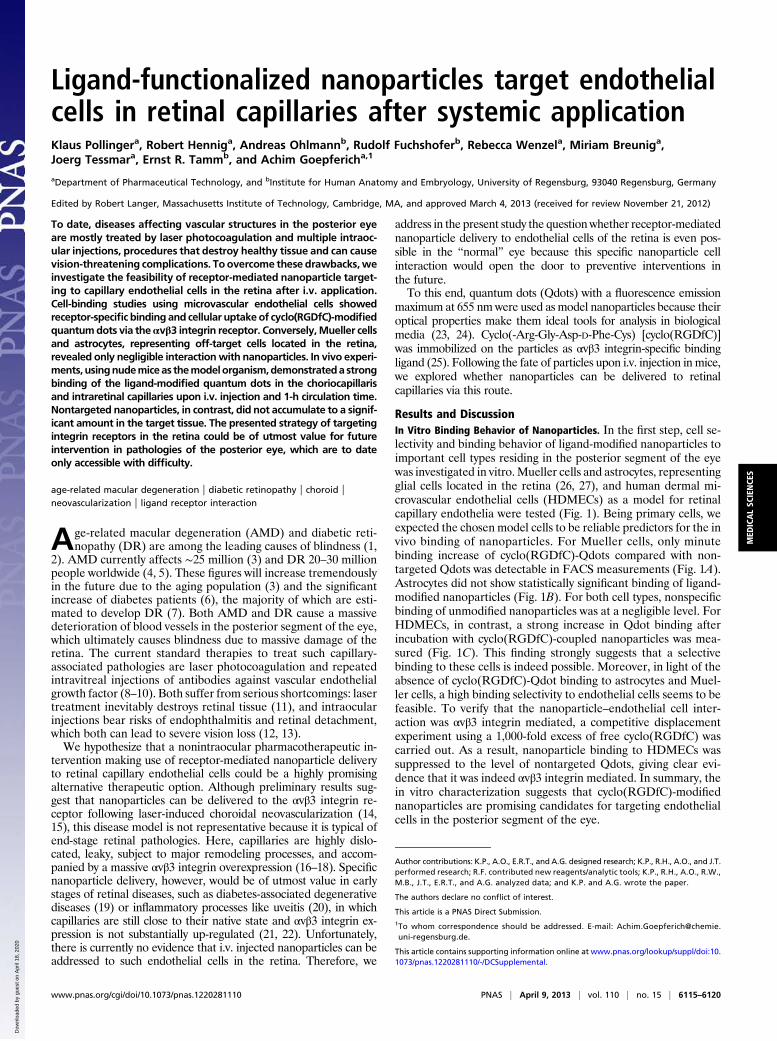
Results and DiscussionIn Vitro Binding Behavior of Nanoparticles. In the first step, cell se-lectivity and binding behavior of ligand-modified nanoparticles toimportant cell types residing in the posterior segment of the eyewas investigated in vitro.Mueller cells and astrocytes, representingglial cells located in the retina (26, 27), and human dermal mi-crovascular endothelial cells (HDMECs) as a model for retinalcapillary endothelia were tested (Fig. 1). Being primary cells, weexpected the chosen model cells to be reliable predictors for the invivo binding of nanoparticles. For Mueller cells, only minutebinding increase of cyclo(RGDfC)-Qdots compared with non-targeted Qdots was detectable in FACS measurements (Fig. 1A).Astrocytes did not show statistically significant binding of ligand-modified nanoparticles (Fig. 1B). For both cell types, nonspecificbinding of unmodified nanoparticles was at a negligible level. ForHDMECs, in contrast, a strong increase in Qdot binding afterincubation with cyclo(RGDfC)-coupled nanoparticles was mea-sured (Fig. 1C). This finding strongly suggests that a selectivebinding to these cells is indeed possible. Moreover, in light of theabsence of cyclo(RGDfC)-Qdot binding to astrocytes and Muel-ler cells, a high binding selectivity to endothelial cells seems to befeasible. To verify that the nanoparticle–endothelial cell inter-action was αvβ3 integrin mediated, a competitive displacementexperiment using a 1,000-fold excess of free cyclo(RGDfC) wascarried out. As a result, nanoparticle binding to HDMECs wassuppressed to the level of nontargeted Qdots, giving clear evi-dence that it was indeed αvβ3 integrin mediated. In summary, thein vitro characterization suggests that cyclo(RGDfC)-modifiednanoparticles are promising candidates for targeting endothelialcells in the posterior segment of the eye.
Author contributions: K.P., A.O., E.R.T., and A.G. designed research; K.P., R.H., A.O., and J.T.performed research; R.F. contributed new reagents/analytic tools; K.P., R.H., A.O., R.W.,M.B., J.T., E.R.T., and A.G. analyzed data; and K.P. and A.G. wrote the paper.
The authors declare no conflict of interest.
This article is a PNAS Direct Submission.1To whom correspondence should be addressed. E-mail: [email protected].
This article contains supporting information online at www.pnas.org/lookup/suppl/doi:10.1073/pnas.1220281110/-/DCSupplemental.
www.pnas.org/cgi/doi/10.1073/pnas.1220281110 PNAS | April 9, 2013 | vol. 110 | no. 15 | 6115–6120
MED
ICALSC
IENCE
S
Dow
nloa
ded
by g
uest
on
Apr
il 18
, 202
0
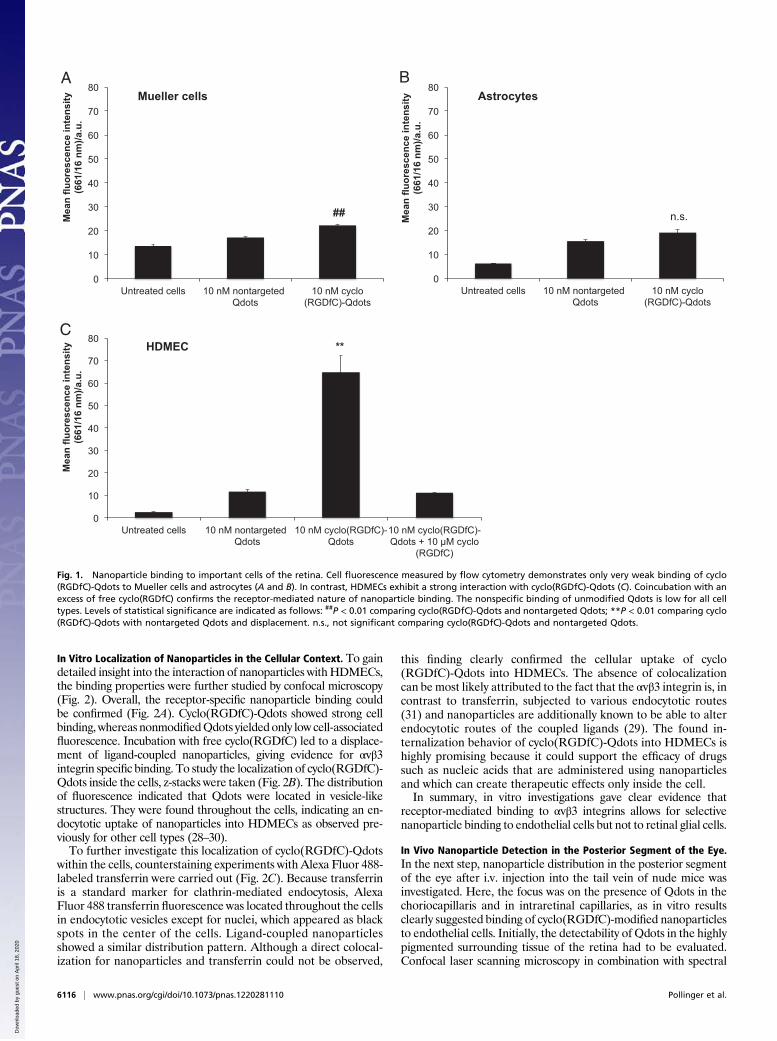
In Vitro Localization of Nanoparticles in the Cellular Context. To gaindetailed insight into the interaction of nanoparticles withHDMECs,the binding properties were further studied by confocal microscopy(Fig. 2). Overall, the receptor-specific nanoparticle binding couldbe confirmed (Fig. 2A). Cyclo(RGDfC)-Qdots showed strong cellbinding, whereas nonmodifiedQdots yielded only low cell-associatedfluorescence. Incubation with free cyclo(RGDfC) led to a displace-ment of ligand-coupled nanoparticles, giving evidence for αvβ3integrin specific binding. To study the localization of cyclo(RGDfC)-Qdots inside the cells, z-stacks were taken (Fig. 2B). The distributionof fluorescence indicated that Qdots were located in vesicle-likestructures. They were found throughout the cells, indicating an en-docytotic uptake of nanoparticles into HDMECs as observed pre-viously for other cell types (28–30).To further investigate this localization of cyclo(RGDfC)-Qdots
within the cells, counterstaining experiments with Alexa Fluor 488-labeled transferrin were carried out (Fig. 2C). Because transferrinis a standard marker for clathrin-mediated endocytosis, AlexaFluor 488 transferrin fluorescence was located throughout the cellsin endocytotic vesicles except for nuclei, which appeared as blackspots in the center of the cells. Ligand-coupled nanoparticlesshowed a similar distribution pattern. Although a direct colocal-ization for nanoparticles and transferrin could not be observed,
this finding clearly confirmed the cellular uptake of cyclo(RGDfC)-Qdots into HDMECs. The absence of colocalizationcan be most likely attributed to the fact that the αvβ3 integrin is, incontrast to transferrin, subjected to various endocytotic routes(31) and nanoparticles are additionally known to be able to alterendocytotic routes of the coupled ligands (29). The found in-ternalization behavior of cyclo(RGDfC)-Qdots into HDMECs ishighly promising because it could support the efficacy of drugssuch as nucleic acids that are administered using nanoparticlesand which can create therapeutic effects only inside the cell.In summary, in vitro investigations gave clear evidence that
receptor-mediated binding to αvβ3 integrins allows for selectivenanoparticle binding to endothelial cells but not to retinal glial cells.
In Vivo Nanoparticle Detection in the Posterior Segment of the Eye.In the next step, nanoparticle distribution in the posterior segmentof the eye after i.v. injection into the tail vein of nude mice wasinvestigated. Here, the focus was on the presence of Qdots in thechoriocapillaris and in intraretinal capillaries, as in vitro resultsclearly suggested binding of cyclo(RGDfC)-modified nanoparticlesto endothelial cells. Initially, the detectability of Qdots in the highlypigmented surrounding tissue of the retina had to be evaluated.Confocal laser scanning microscopy in combination with spectral
0
10
20
30
40
50
60
70
80
Untreated cells 10 nM nontargeted Qdots
10 nM cyclo(RGDfC)-Qdots
Me
an
flu
ore
sc
en
ce
in
te
ns
ity
(6
61
/1
6 n
m)/a
.u
.
Mueller cells
0
10
20
30
40
50
60
70
80
Untreated cells 10 nM nontargeted Qdots
10 nM cyclo(RGDfC)-Qdots
Me
an
flu
ore
sc
en
ce
in
te
ns
ity
(6
61
/1
6 n
m)/a
.u
.
Astrocytes
0
10
20
30
40
50
60
70
80
Untreated cells 10 nM nontargeted Qdots
10 nM cyclo(RGDfC)-Qdots
10 nM cyclo(RGDfC)-Qdots + 10 M cyclo
(RGDfC)
Me
an
flu
ore
sc
en
ce
in
te
ns
ity
(6
61
/1
6 n
m)/a
.u
.
HDMEC **
n.s. ##
A B
C
Fig. 1. Nanoparticle binding to important cells of the retina. Cell fluorescence measured by flow cytometry demonstrates only very weak binding of cyclo(RGDfC)-Qdots to Mueller cells and astrocytes (A and B). In contrast, HDMECs exhibit a strong interaction with cyclo(RGDfC)-Qdots (C). Coincubation with anexcess of free cyclo(RGDfC) confirms the receptor-mediated nature of nanoparticle binding. The nonspecific binding of unmodified Qdots is low for all celltypes. Levels of statistical significance are indicated as follows: ##P < 0.01 comparing cyclo(RGDfC)-Qdots and nontargeted Qdots; **P < 0.01 comparing cyclo(RGDfC)-Qdots with nontargeted Qdots and displacement. n.s., not significant comparing cyclo(RGDfC)-Qdots and nontargeted Qdots.
6116 | www.pnas.org/cgi/doi/10.1073/pnas.1220281110 Pollinger et al.
Dow
nloa
ded
by g
uest
on
Apr
il 18
, 202
0
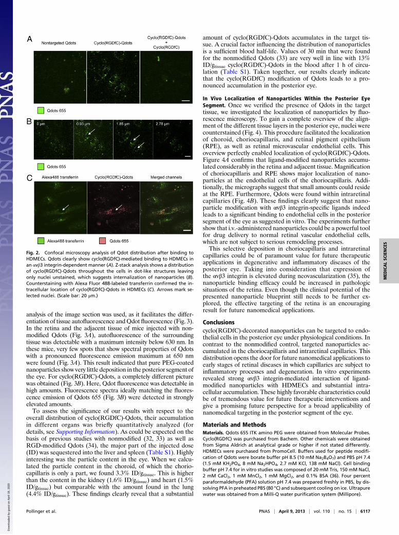
analysis of the image section was used, as it facilitates the differ-entiation of tissue autofluorescence andQdot fluorescence (Fig. 3).In the retina and the adjacent tissue of mice injected with non-modified Qdots (Fig. 3A), autofluorescence of the surroundingtissue was detectable with a maximum intensity below 630 nm. Inthese mice, very few spots that show spectral properties of Qdotswith a pronounced fluorescence emission maximum at 650 nmwere found (Fig. 3A). This result indicated that pure PEG-coatednanoparticles show very little deposition in the posterior segment ofthe eye. For cyclo(RGDfC)-Qdots, a completely different picturewas obtained (Fig. 3B). Here, Qdot fluorescence was detectable inhigh amounts. Fluorescence spectra ideally matching the fluores-cence emission of Qdots 655 (Fig. 3B) were detected in stronglyelevated amounts.To assess the significance of our results with respect to the
overall distribution of cyclo(RGDfC)-Qdots, their accumulationin different organs was briefly quantitatively analyzed (fordetails, see Supporting Information). As could be expected on thebasis of previous studies with nonmodified (32, 33) as well asRGD-modified Qdots (34), the major part of the injected dose(ID) was sequestered into the liver and spleen (Table S1). Highlyinteresting was the particle content in the eye. When we calcu-lated the particle content in the choroid, of which the chorio-capillaris is only a part, we found 3.3% ID/gtissue. This is higherthan the content in the kidney (1.6% ID/gtissue) and heart (1.5%ID/gtissue) but comparable with the amount found in the lung(4.4% ID/gtissue). These findings clearly reveal that a substantial
amount of cyclo(RGDfC)-Qdots accumulates in the target tis-sue. A crucial factor influencing the distribution of nanoparticlesis a sufficient blood half-life. Values of 30 min that were foundfor the nonmodified Qdots (33) are very well in line with 13%ID/gtissue cyclo(RGDfC)-Qdots in the blood after 1 h of circu-lation (Table S1). Taken together, our results clearly indicatethat the cyclo(RGDfC) modification of Qdots leads to a pro-nounced accumulation in the posterior eye.
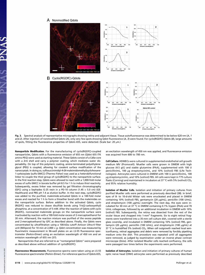
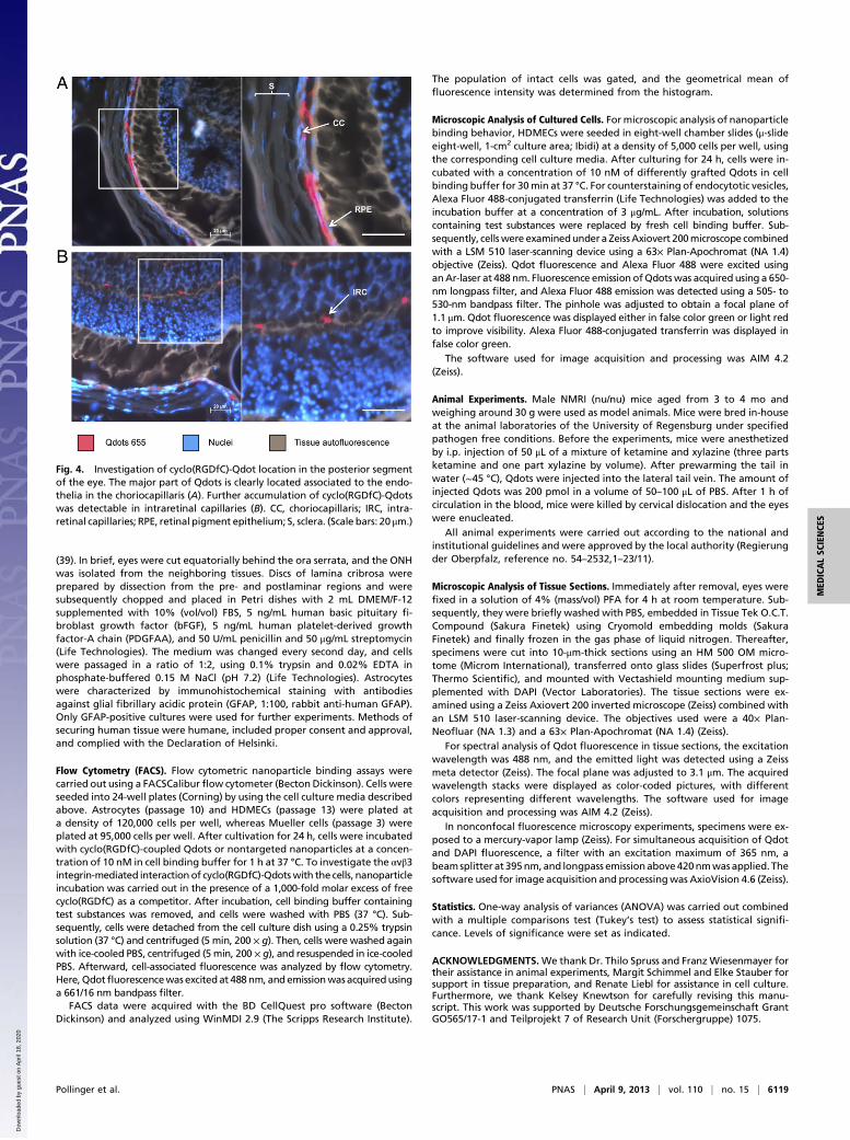
In Vivo Localization of Nanoparticles Within the Posterior EyeSegment. Once we verified the presence of Qdots in the targettissue, we investigated the localization of nanoparticles by fluo-rescence microscopy. To gain a complete overview of the align-ment of the different tissue layers in the posterior eye, nuclei werecounterstained (Fig. 4). This procedure facilitated the localizationof choroid, choriocapillaris, and retinal pigment epithelium(RPE), as well as retinal microvascular endothelial cells. Thisoverview perfectly enabled localization of cyclo(RGDfC)-Qdots.Figure 4A confirms that ligand-modified nanoparticles accumu-lated considerably in the retina and adjacent tissue. Magnificationof choriocapillaris and RPE shows major localization of nano-particles at the endothelial cells of the choriocapillaris. Addi-tionally, the micrographs suggest that small amounts could resideat the RPE. Furthermore, Qdots were found within intraretinalcapillaries (Fig. 4B). These findings clearly suggest that nano-particle modification with αvβ3 integrin-specific ligands indeedleads to a significant binding to endothelial cells in the posteriorsegment of the eye as suggested in vitro. The experiments furthershow that i.v.-administered nanoparticles could be a powerful toolfor drug delivery to normal retinal vascular endothelial cells,which are not subject to serious remodeling processes.This selective deposition in choriocapillaris and intraretinal
capillaries could be of paramount value for future therapeuticapplications in degenerative and inflammatory diseases of theposterior eye. Taking into consideration that expression ofthe αvβ3 integrin is elevated during neovascularization (35), thenanoparticle binding efficacy could be increased in pathologicsituations of the retina. Even though the clinical potential of thepresented nanoparticle blueprint still needs to be further ex-plored, the effective targeting of the retina is an encouragingresult for future nanomedical applications.
Conclusionscyclo(RGDfC)-decorated nanoparticles can be targeted to endo-thelial cells in the posterior eye under physiological conditions. Incontrast to the nonmodified control, targeted nanoparticles ac-cumulated in the choriocapillaris and intraretinal capillaries. Thisdistribution opens the door for future nanomedical applications toearly stages of retinal diseases in which capillaries are subject toinflammatory processes and degeneration. In vitro experimentsrevealed strong αvβ3 integrin-mediated interaction of ligand-modified nanoparticles with HDMECs and substantial intra-cellular accumulation. These highly favorable characteristics couldbe of tremendous value for future therapeutic interventions andgive a promising future perspective for a broad applicability ofnanomedical targeting in the posterior segment of the eye.
Materials and MethodsMaterials. Qdots 655 ITK amino PEG were obtained from Molecular Probes.Cyclo(RGDfC) was purchased from Bachem. Other chemicals were obtainedfrom Sigma Aldrich at analytical grade or higher if not stated differently.HDMECs were purchased from PromoCell. Buffers used for peptide modifi-cation of Qdots were borate buffer pH 8.5 (10 mM Na2B4O7) and PBS pH 7.4(1.5 mM KH2PO4, 8 mM Na2HPO4, 2.7 mM KCl, 138 mM NaCl). Cell bindingbuffer pH 7.4 for in vitro studies was composed of 20 mM Tris, 150 mM NaCl,2 mM CaCl2, 1 mM MnCl2, 1 mM MgCl2, and 0.1% BSA (36). Four percentparaformaldehyde (PFA) solution pH 7.4 was prepared freshly in PBS, by dis-solving PFA in preheated PBS (80 °C) and subsequent cooling on ice. Ultrapurewater was obtained from a Milli-Q water purification system (Millipore).
Fig. 2. Confocal microscopy analysis of Qdot distribution after binding toHDMECs. Qdots clearly show cyclo(RGDfC)-mediated binding to HDMECs inan αvβ3 integrin-dependent manner (A). Z-stack analysis shows a distributionof cyclo(RGDfC)-Qdots throughout the cells in dot-like structures leavingonly nuclei unstained, which suggests internalization of nanoparticles (B).Counterstaining with Alexa Fluor 488-labeled transferrin confirmed the in-tracellular location of cyclo(RGDfC)-Qdots in HDMECs (C). Arrows mark se-lected nuclei. (Scale bar: 20 μm.)
Pollinger et al. PNAS | April 9, 2013 | vol. 110 | no. 15 | 6117
MED
ICALSC
IENCE
S
Dow
nloa
ded
by g
uest
on
Apr
il 18
, 202
0
Nanoparticle Modification. For the manufacturing of cyclo(RGDfC)-couplednanoparticles, Qdots with a fluorescence emission of 655 nm (Qdot 655 ITKamino PEG) were used as startingmaterial. These Qdots consist of a CdSe corewith a ZnS shell and carry a polymer coating, which mediates water dis-persability. On top of this polymeric coating, amine-terminated polyethylene-glycol (PEG) is coupled, allowing for covalent surface modification of thenanocrystals. In doing so, sulfosuccinimidyl-4-(N-maleimidomethyl)cyclohexane-1-carboxylate (sulfo-SMCC) (Thermo Fisher) was used as a heterobifunctionallinker to couple the thiol group of cyclo(RGDfC) to the nanoparticle surface.In the first reaction step, Qdots were allowed to react with a 1,000-fold molarexcess of sulfo-SMCC in borate buffer pH 8.5 for 1 h to induce thiol reactivity.Subsequently, excess linker was removed by gel filtration chromatography(GFC) using a Sephadex G-25 resin in a PD-10 column (1.45 × 5.0 cm) (GE-Healthcare) and PBS pH 7.4 as elution buffer. In the next step, cyclo(RGDfC)was added to the purified, maleimide-activated Qdots in a 100-fold molarexcess and reacted for 1 h to form a thioether bond with the maleimides onthe nanoparticle surface. Before addition to the activated Qdots, cyclo(RGDfC) was reduced to cleave disulfide bonds using Tris(2-carboxyethyl)phosphine, at a concentration of 5 mM. After the reaction of Qdots with cyclo(RGDfC), nonreacted maleimide groups on the nanoparticle surface wereinactivated by reaction with a 100-fold molar excess of 2-mercaptoethanol for30 min. Afterward, the reaction mixture was purified of the excess peptideand 2-mercaptoethanol by GFC as described above. Finally, the Qdots wereconcentrated by ultrafiltration using a 100-kDa cutoff Amicon Ultra-4 filterunit (Milipore) for 10 min at 2,000 × g. Qdot concentration was measured byfluorimetric measurement in 96-well plates on an LS-55 fluorescence spec-trometer (Perkin-Elmer) using an excitation wavelength of 450 nm and anemission wavelength of 655 nm (37).
Nanoparticles that are referred to as “nontargeted Qdots”were preparedas described above without addition of cyclo(RGDfC).
Fluorescence Measurements. Fluorescence spectra were taken using an LS-55fluorescence spectrometer (Perkin-Elmer). For reference spectra of Qdots 655,
an excitation wavelength of 450 nm was applied, and fluorescence emissionwas acquired from 600 to 700 nm.
Cell Culture.HDMECs were cultured in supplemented endothelial cell growthmedium MV (Promocell). Mueller cells were grown in DMEM with highglucose (4.5 g/L) and stable glutamine (PAA), supplemented with 100 Upenicillin/mL, 100 μg streptomycin/mL, and 10% (vol/vol) FBS (Life Tech-nologies). Astrocytes were cultured in DMEM with 100 U penicillin/mL, 100μg streptomycin/mL, and 10% (vol/vol) FBS. All cells were kept in T75 cultureflasks (Corning) and maintained in incubators at 37 °C with 5% (vol/vol) CO2
and 95% relative humidity.
Isolation of Mueller Cells. Isolation and initiation of primary cultures frompurified Mueller cells were performed as previously described (38). In brief,eyes of 8- to 10-d-old Wistar rats were enucleated and placed in DMEMcontaining 10% (vol/vol) FBS, gentamycin (20 μg/mL), penicillin (100 U/mL),and streptomycin (100 μg/mL) overnight. The next day, the eyes were in-cubated for 30 min at 37 °C in DMEM containing 0.1% trypsin (PAA) and 70U/mL collagenase, followed by an additional incubation in DMEM with 10%(vol/vol) FBS. Subsequently, the retina was dissected from the rest of theocular tissue and chopped into 1-mm2 fragments. Six to eight retinal frag-ments were transferred into a 35-mm cell culture dish, covered with a sterileglass coverslip, and incubated in DMEM containing 10% (vol/vol) FBS, gen-tamycin (20 μg/mL), penicillin (100 U/mL), and streptomycin (100 μg/mL) at37 °C in humidified 5% (vol/vol) CO2. When cell outgrowth reached local sem-iconfluency, retinal aggregates and debris were removed by forcibly pipettingmedium onto the dish. This procedure was repeated until all aggregateswere removed, as judged by examination under an inverse phase contrastmicroscope (Zeiss). After isolated Mueller cells reached confluency, the cellswere passaged two times before the experiments were performed.
Isolation of Astrocytes. Isolation and initiation of primary cultures of humanoptic nerve head (ONH) astrocytes were performed as previously described
Fig. 3. Spectral analysis of representative micrographs showing retina and adjacent tissue. Tissue autofluorescence was determined to be below 620 nm (A, 1and 2). After injection of nonmodified Qdots (A), only very few spots showing Qdot fluorescence (A, 3) were found. For cyclo(RGDfC)-Qdots (B), large amountsof spots, fitting the fluorescence properties of Qdots 655, were detected. (Scale bar: 20 μm.)
6118 | www.pnas.org/cgi/doi/10.1073/pnas.1220281110 Pollinger et al.
Dow
nloa
ded
by g
uest
on
Apr
il 18
, 202
0
(39). In brief, eyes were cut equatorially behind the ora serrata, and the ONHwas isolated from the neighboring tissues. Discs of lamina cribrosa wereprepared by dissection from the pre- and postlaminar regions and weresubsequently chopped and placed in Petri dishes with 2 mL DMEM/F-12supplemented with 10% (vol/vol) FBS, 5 ng/mL human basic pituitary fi-broblast growth factor (bFGF), 5 ng/mL human platelet-derived growthfactor-A chain (PDGFAA), and 50 U/mL penicillin and 50 μg/mL streptomycin(Life Technologies). The medium was changed every second day, and cellswere passaged in a ratio of 1:2, using 0.1% trypsin and 0.02% EDTA inphosphate-buffered 0.15 M NaCl (pH 7.2) (Life Technologies). Astrocyteswere characterized by immunohistochemical staining with antibodiesagainst glial fibrillary acidic protein (GFAP, 1:100, rabbit anti-human GFAP).Only GFAP-positive cultures were used for further experiments. Methods ofsecuring human tissue were humane, included proper consent and approval,and complied with the Declaration of Helsinki.
Flow Cytometry (FACS). Flow cytometric nanoparticle binding assays werecarried out using a FACSCaliburflow cytometer (Becton Dickinson). Cells wereseeded into 24-well plates (Corning) by using the cell culture media describedabove. Astrocytes (passage 10) and HDMECs (passage 13) were plated ata density of 120,000 cells per well, whereas Mueller cells (passage 3) wereplated at 95,000 cells per well. After cultivation for 24 h, cells were incubatedwith cyclo(RGDfC)-coupled Qdots or nontargeted nanoparticles at a concen-tration of 10 nM in cell binding buffer for 1 h at 37 °C. To investigate the αvβ3integrin-mediated interaction of cyclo(RGDfC)-Qdotswith the cells, nanoparticleincubation was carried out in the presence of a 1,000-fold molar excess of freecyclo(RGDfC) as a competitor. After incubation, cell binding buffer containingtest substances was removed, and cells were washed with PBS (37 °C). Sub-sequently, cells were detached from the cell culture dish using a 0.25% trypsinsolution (37 °C) and centrifuged (5 min, 200 × g). Then, cells were washed againwith ice-cooled PBS, centrifuged (5 min, 200 × g), and resuspended in ice-cooledPBS. Afterward, cell-associated fluorescence was analyzed by flow cytometry.Here, Qdotfluorescencewasexcitedat 488nm, andemissionwasacquiredusinga 661/16 nm bandpass filter.
FACS data were acquired with the BD CellQuest pro software (BectonDickinson) and analyzed using WinMDI 2.9 (The Scripps Research Institute).
The population of intact cells was gated, and the geometrical mean offluorescence intensity was determined from the histogram.
Microscopic Analysis of Cultured Cells. Formicroscopic analysis of nanoparticlebinding behavior, HDMECs were seeded in eight-well chamber slides (μ-slideeight-well, 1-cm2 culture area; Ibidi) at a density of 5,000 cells per well, usingthe corresponding cell culture media. After culturing for 24 h, cells were in-cubated with a concentration of 10 nM of differently grafted Qdots in cellbinding buffer for 30min at 37 °C. For counterstaining of endocytotic vesicles,Alexa Fluor 488-conjugated transferrin (Life Technologies) was added to theincubation buffer at a concentration of 3 μg/mL. After incubation, solutionscontaining test substances were replaced by fresh cell binding buffer. Sub-sequently, cellswere examinedundera ZeissAxiovert 200microscope combinedwith a LSM 510 laser-scanning device using a 63× Plan-Apochromat (NA 1.4)objective (Zeiss). Qdot fluorescence and Alexa Fluor 488 were excited usinganAr-laser at 488 nm. Fluorescence emission ofQdotswas acquired using a 650-nm longpass filter, and Alexa Fluor 488 emission was detected using a 505- to530-nm bandpass filter. The pinhole was adjusted to obtain a focal plane of1.1 μm. Qdot fluorescence was displayed either in false color green or light redto improve visibility. Alexa Fluor 488-conjugated transferrin was displayed infalse color green.
The software used for image acquisition and processing was AIM 4.2(Zeiss).
Animal Experiments. Male NMRI (nu/nu) mice aged from 3 to 4 mo andweighing around 30 g were used as model animals. Mice were bred in-houseat the animal laboratories of the University of Regensburg under specifiedpathogen free conditions. Before the experiments, mice were anesthetizedby i.p. injection of 50 μL of a mixture of ketamine and xylazine (three partsketamine and one part xylazine by volume). After prewarming the tail inwater (∼45 °C), Qdots were injected into the lateral tail vein. The amount ofinjected Qdots was 200 pmol in a volume of 50–100 μL of PBS. After 1 h ofcirculation in the blood, mice were killed by cervical dislocation and the eyeswere enucleated.
All animal experiments were carried out according to the national andinstitutional guidelines and were approved by the local authority (Regierungder Oberpfalz, reference no. 54–2532,1–23/11).
Microscopic Analysis of Tissue Sections. Immediately after removal, eyes werefixed in a solution of 4% (mass/vol) PFA for 4 h at room temperature. Sub-sequently, they were briefly washed with PBS, embedded in Tissue Tek O.C.T.Compound (Sakura Finetek) using Cryomold embedding molds (SakuraFinetek) and finally frozen in the gas phase of liquid nitrogen. Thereafter,specimens were cut into 10-μm-thick sections using an HM 500 OM micro-tome (Microm International), transferred onto glass slides (Superfrost plus;Thermo Scientific), and mounted with Vectashield mounting medium sup-plemented with DAPI (Vector Laboratories). The tissue sections were ex-amined using a Zeiss Axiovert 200 inverted microscope (Zeiss) combined withan LSM 510 laser-scanning device. The objectives used were a 40× Plan-Neofluar (NA 1.3) and a 63× Plan-Apochromat (NA 1.4) (Zeiss).
For spectral analysis of Qdot fluorescence in tissue sections, the excitationwavelength was 488 nm, and the emitted light was detected using a Zeissmeta detector (Zeiss). The focal plane was adjusted to 3.1 μm. The acquiredwavelength stacks were displayed as color-coded pictures, with differentcolors representing different wavelengths. The software used for imageacquisition and processing was AIM 4.2 (Zeiss).
In nonconfocal fluorescence microscopy experiments, specimens were ex-posed to a mercury-vapor lamp (Zeiss). For simultaneous acquisition of Qdotand DAPI fluorescence, a filter with an excitation maximum of 365 nm, abeamsplitter at 395nm, and longpass emissionabove420nmwasapplied. Thesoftware used for image acquisition and processingwas AxioVision 4.6 (Zeiss).
Statistics. One-way analysis of variances (ANOVA) was carried out combinedwith a multiple comparisons test (Tukey’s test) to assess statistical signifi-cance. Levels of significance were set as indicated.
ACKNOWLEDGMENTS.We thank Dr. Thilo Spruss and FranzWiesenmayer fortheir assistance in animal experiments, Margit Schimmel and Elke Stauber forsupport in tissue preparation, and Renate Liebl for assistance in cell culture.Furthermore, we thank Kelsey Knewtson for carefully revising this manu-script. This work was supported by Deutsche Forschungsgemeinschaft GrantGO565/17-1 and Teilprojekt 7 of Research Unit (Forschergruppe) 1075.
Fig. 4. Investigation of cyclo(RGDfC)-Qdot location in the posterior segmentof the eye. The major part of Qdots is clearly located associated to the endo-thelia in the choriocapillaris (A). Further accumulation of cyclo(RGDfC)-Qdotswas detectable in intraretinal capillaries (B). CC, choriocapillaris; IRC, intra-retinal capillaries; RPE, retinal pigment epithelium; S, sclera. (Scale bars: 20 μm.)
Pollinger et al. PNAS | April 9, 2013 | vol. 110 | no. 15 | 6119
MED
ICALSC
IENCE
S
Dow
nloa
ded
by g
uest
on
Apr
il 18
, 202
0
1. Coorey NJ, Shen W, Chung SH, Zhu L, Gillies MC (2012) The role of glia in retinalvascular disease. Clin Exp Optom 95(3):266–281.
2. Lee J-H, Pidaparti RM, Atkinson GM, Moorthy RS (2012) Design of an implantabledevice for ocular drug delivery. J Drug Deliv 2012:527516.
3. Chopdar A, Chakravarthy U, Verma D (2003) Age related macular degeneration. BMJ326(7387):485–488.
4. Charbonnel B, Dormandy J, Erdmann E, Massi-Benedetti M, Skene A; PROactive StudyGroup (2004) The prospective pioglitazone clinical trial in macrovascular events(PROactive): Can pioglitazone reduce cardiovascular events in diabetes? Study designand baseline characteristics of 5238 patients. Diabetes Care 27(7):1647–1653.
5. Meleth AD, et al. (2005) Serum inflammatory markers in diabetic retinopathy. InvestOphthalmol Vis Sci 46(11):4295–4301.
6. Wild S, Roglic G, Green A, Sicree R, King H (2004) Global prevalence of diabetes: Es-timates for the year 2000 and projections for 2030. Diabetes Care 27(5):1047–1053.
7. Sivaprasad S, Gupta B, Crosby-Nwaobi R, Evans J (2012) Prevalence of diabetic reti-nopathy in various ethnic groups: A worldwide perspective. Surv Ophthalmol 57(4):347–370.
8. Giuliari GP (2012) Diabetic retinopathy: Current and new treatment options. CurrDiabetes Rev 8(1):32–41.
9. Kumar B, Gupta SK, Saxena R, Srivastava S (2012) Current trends in the pharmaco-therapy of diabetic retinopathy. J Postgrad Med 58(2):132–139.
10. Avery RL, et al. (2006) Intravitreal bevacizumab (Avastin) for neovascular age-relatedmacular degeneration. Ophthalmology 113(3):363–372, e5.
11. Maeshima K, Utsugi-Sutoh N, Otani T, Kishi S (2004) Progressive enlargement ofscattered photocoagulation scars in diabetic retinopathy. Retina 24(4):507–511.
12. Martidis A, et al. (2002) Intravitreal triamcinolone for refractory diabetic macularedema. Ophthalmology 109(5):920–927.
13. Moshfeghi DM, et al. (2003) Acute endophthalmitis following intravitreal tri-amcinolone acetonide injection. Am J Ophthalmol 136(5):791–796.
14. Singh SR, et al. (2009) Intravenous transferrin, RGD peptide and dual-targetednanoparticles enhance anti-VEGF intraceptor gene delivery to laser-induced CNV.Gene Ther 16(5):645–659.
15. Salehi-Had H, et al. (2011) Utilizing targeted gene therapy with nanoparticles bindingalpha v beta 3 for imaging and treating choroidal neovascularization. PLoS ONE 6(4):e18864.
16. Krzystolik MG, et al. (2002) Prevention of experimental choroidal neovascularizationwith intravitreal anti-vascular endothelial growth factor antibody fragment. ArchOphthalmol 120(3):338–346.
17. Spilsbury K, Garrett KL, Shen WY, Constable IJ, Rakoczy PE (2000) Overexpression ofvascular endothelial growth factor (VEGF) in the retinal pigment epithelium leads tothe development of choroidal neovascularization. Am J Pathol 157(1):135–144.
18. Yoshida T, Gong J, Xu Z, Wei Y, Duh EJ (2012) Inhibition of pathological retinal an-giogenesis by the integrin αvβ3 antagonist tetraiodothyroacetic acid (tetrac). Exp EyeRes 94(1):41–48.
19. Vincent JA, Mohr S (2007) Inhibition of caspase-1/interleukin-1beta signaling preventsdegeneration of retinal capillaries in diabetes and galactosemia. Diabetes 56(1):224–230.
20. Gomes Bittencourt M, et al. (2012) New treatment options for noninfectious uveitis.Dev Ophthalmol 51:134–161.
21. Hodivala-Dilke K (2008) alphavbeta3 integrin and angiogenesis: A moody integrin ina changing environment. Curr Opin Cell Biol 20(5):514–519.
22. Eliceiri BP, Cheresh DA (1999) The role of alphav integrins during angiogenesis: In-sights into potential mechanisms of action and clinical development. J Clin Invest103(9):1227–1230.
23. Hild WA, Breunig M, Goepferich A (2008) Quantum dots: Nano-sized probes for theexploration of cellular and intracellular targeting. Eur J Pharm Biopharm 68(2):153–168.
24. Walling MA, Novak JA, Shepard JRE (2009) Quantum dots for live cell and in vivoimaging. Int J Mol Sci 10(2):441–491.
25. Aumailley M, et al. (1991) Arg-Gly-Asp constrained within cyclic pentapeptides.Strong and selective inhibitors of cell adhesion to vitronectin and laminin fragmentP1. FEBS Lett 291(1):50–54.
26. Franze K, et al. (2007) Muller cells are living optical fibers in the vertebrate retina.Proc Natl Acad Sci USA 104(20):8287–8292.
27. Newman EA (2003) New roles for astrocytes: Regulation of synaptic transmission.Trends Neurosci 26(10):536–542.
28. Zhang LW, Monteiro-Riviere NA (2009) Mechanisms of quantum dot nanoparticlecellular uptake. Toxicol Sci 110(1):138–155.
29. Tekle C, Deurs Bv, Sandvig K, Iversen T-G (2008) Cellular trafficking of quantum dot-ligand bioconjugates and their induction of changes in normal routing of un-conjugated ligands. Nano Lett 8(7):1858–1865.
30. Ferreira L, et al. (2008) Human embryoid bodies containing nano- and micro-particulate delivery vehicles. Adv Mater (Deerfield Beach Fla) 20:2285–2291.
31. Ivaska J, Heino J (2011) Cooperation between integrins and growth factor receptorsin signaling and endocytosis. Annu Rev Cell Dev Biol 27:291–320.
32. Schipper ML, et al. (2007) microPET-based biodistribution of quantum dots in livingmice. J Nucl Med 48(9):1511–1518.
33. Praetner M, et al. (2010) The contribution of the capillary endothelium to bloodclearance and tissue deposition of anionic quantum dots in vivo. Biomaterials 31(26):6692–6700.
34. Cai W, et al. (2006) Peptide-labeled near-infrared quantum dots for imaging tumorvasculature in living subjects. Nano Lett 6(4):669–676.
35. Haubner R, et al. (2005) Noninvasive visualization of the activated alphavbeta3 in-tegrin in cancer patients by positron emission tomography and [18F]Galacto-RGD.PLoS Med 2(3):e70.
36. Shi W, Bartlett JS (2003) RGD inclusion in VP3 provides adeno-associated virus type 2(AAV2)-based vectors with a heparan sulfate-independent cell entry mechanism. MolTher 7(4):515–525.
37. Cai W, Chen X (2008) Preparation of peptide-conjugated quantum dots for tumorvasculature-targeted imaging. Nat Protoc 3(1):89–96.
38. Seitz R, Hackl S, Seibuchner T, Tamm ER, Ohlmann A (2010) Norrin mediates neuro-protective effects on retinal ganglion cells via activation of the Wnt/beta-cateninsignaling pathway and the induction of neuroprotective growth factors in Mullercells. J Neurosci 30(17):5998–6010.
39. Fuchshofer R, Birke M, Welge-Lussen U, Kook D, Lütjen-Drecoll E (2005) Transforminggrowth factor-beta 2 modulated extracellular matrix component expression in cul-tured human optic nerve head astrocytes. Invest Ophthalmol Vis Sci 46(2):568–578.
6120 | www.pnas.org/cgi/doi/10.1073/pnas.1220281110 Pollinger et al.
Dow
nloa
ded
by g
uest
on
Apr
il 18
, 202
0