leukocyte evaluation clinical textbook for veterinary technicians 4 th edition dennis m. mccurnin...
TRANSCRIPT
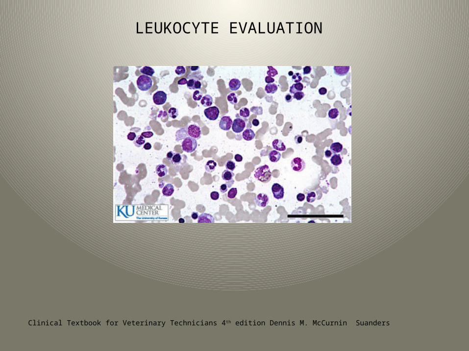
LEUKOCYTE EVALUATION
Clinical Textbook for Veterinary Technicians 4th edition Dennis M. McCurnin Suanders
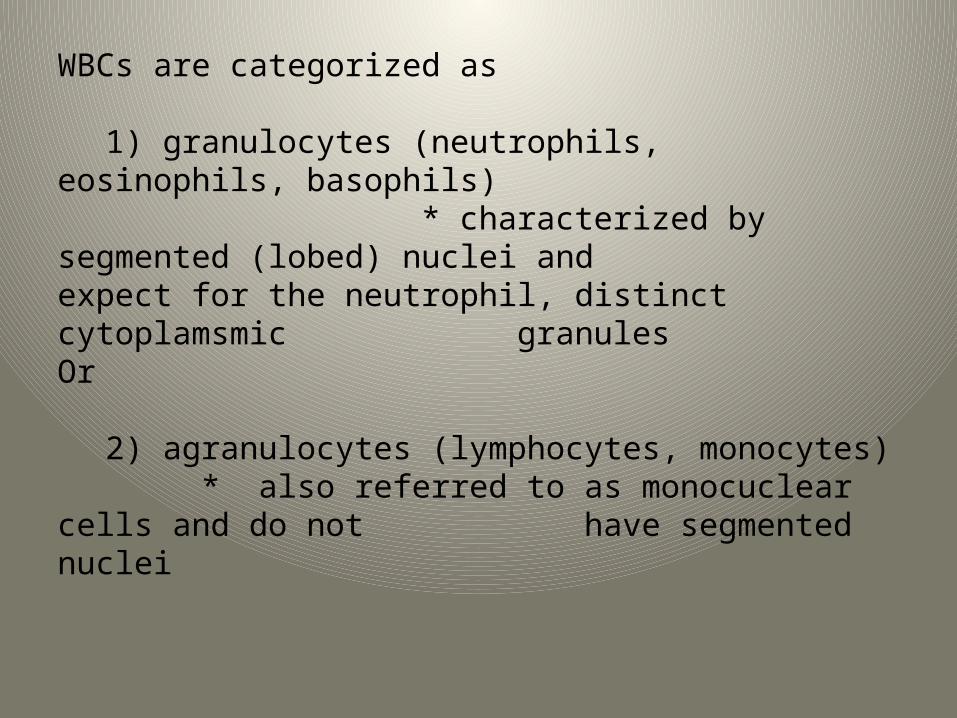
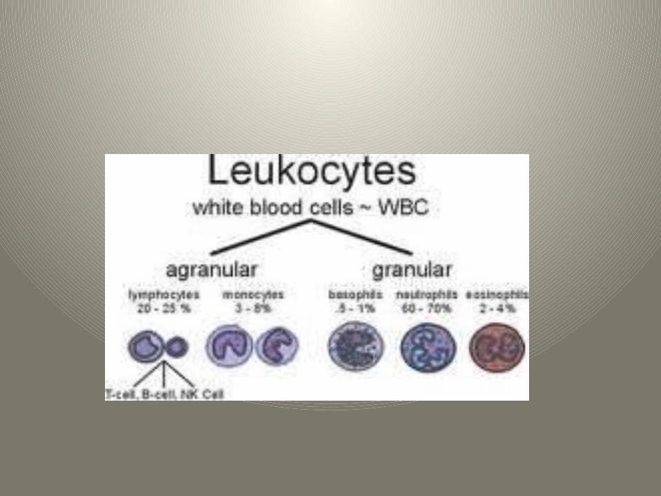
WBCs are categorized as
1) granulocytes (neutrophils, eosinophils, basophils) * characterized by segmented (lobed) nuclei and
expect for the neutrophil, distinct cytoplamsmic granules
Or
2) agranulocytes (lymphocytes, monocytes) * also referred to as monocuclear cells and do not have segmented nuclei
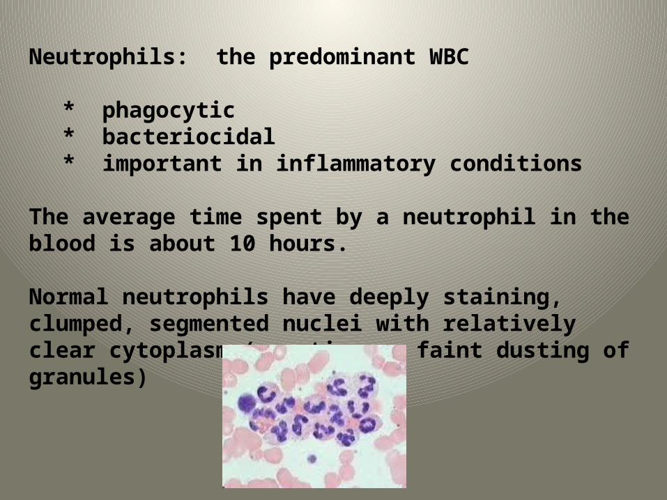
Neutrophils: the predominant WBC
* phagocytic * bacteriocidal* important in inflammatory conditions
The average time spent by a neutrophil in the blood is about 10 hours.
Normal neutrophils have deeply staining, clumped, segmented nuclei with relatively clear cytoplasm (sometimes a faint dusting of granules)
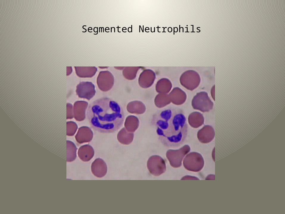
Segmented Neutrophils
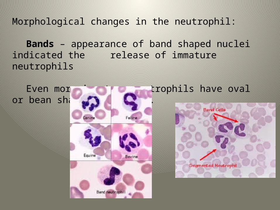
Morphological changes in the neutrophil:
Bands – appearance of band shaped nuclei indicated the release of immature neutrophils
Even more immature neutrophils have oval or bean shaped nuclei.
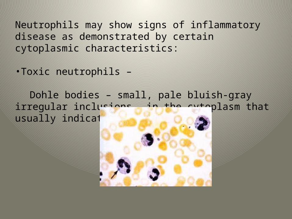
Neutrophils may show signs of inflammatory disease as demonstrated by certain cytoplasmic characteristics:
•Toxic neutrophils –
Dohle bodies – small, pale bluish-gray irregular inclusions in the cytoplasm that usually indicate mild toxemia.
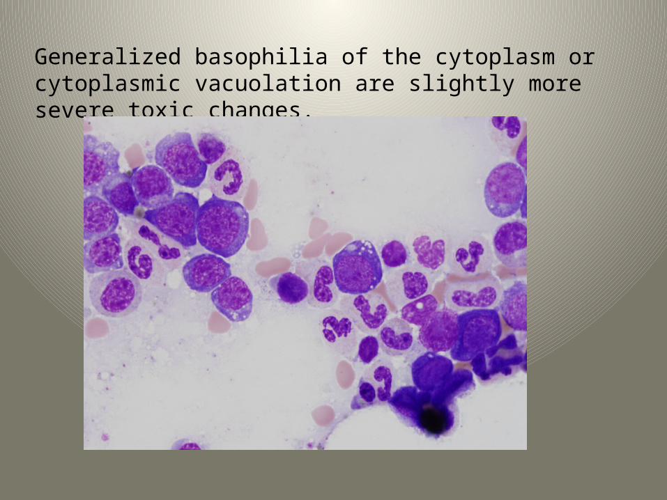
Generalized basophilia of the cytoplasm or cytoplasmic vacuolation are slightly more severe toxic changes.
Neutrophilia – increased number of total neutrophils.
Neutropenia – a decrease in circulating neutrophils. May occur when tissue demand is excessive as a result of severe inflammation exceeding the ability of the bone marrow to supply the cells.
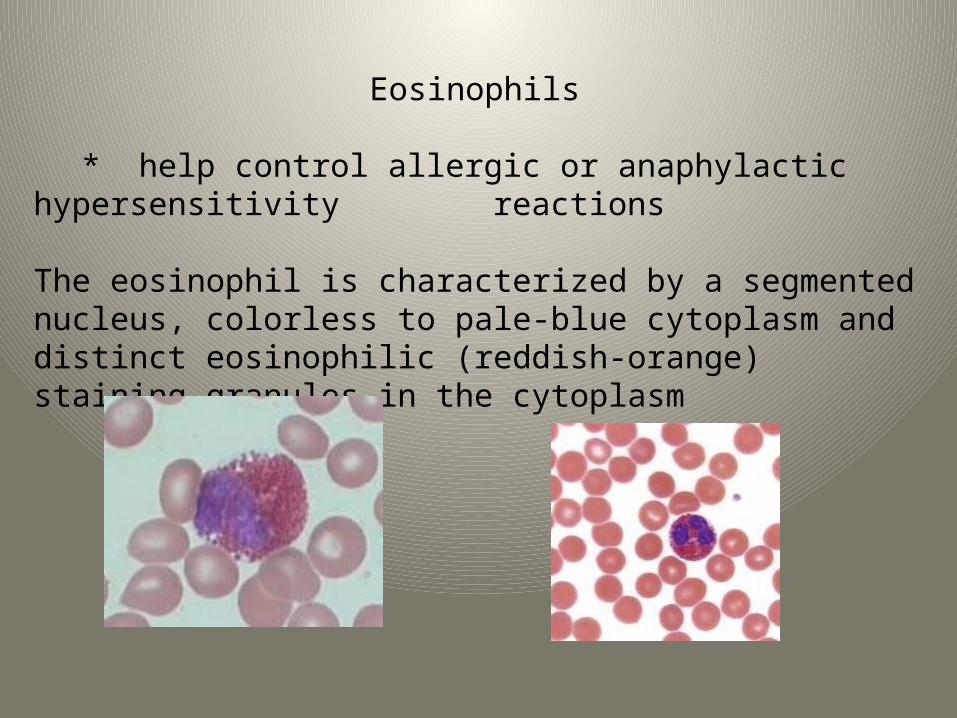
Eosinophils
* help control allergic or anaphylactic hypersensitivity reactions
The eosinophil is characterized by a segmented nucleus, colorless to pale-blue cytoplasm and distinct eosinophilic (reddish-orange) staining granules in the cytoplasm
Eosinophils Per species:
Cats – contain numerous tiny rod-shaped granules that may obscure the nucleus
Dogs – granules are less numerous and usually round but may vary considerably in size.
Horses – granules are extremely distinctive, being very large and round and a much brighter orange than smaller animals
Bovine – granules are also bright orange but are much smaller and more numerous than those of the horse.
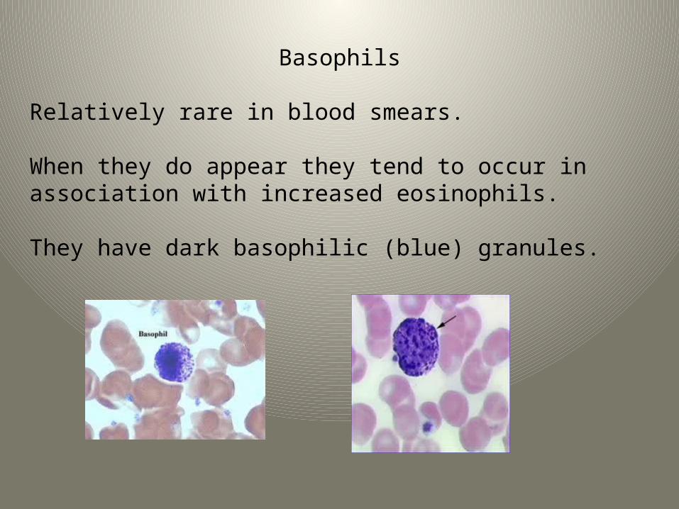
Basophils
Relatively rare in blood smears.
When they do appear they tend to occur in association with increased eosinophils.
They have dark basophilic (blue) granules.
Basophils per species:
Cats – granules tend to be light lavender to almost pink
Dogs – have few to no granules and must be differentiated from neutrophils.
Equine and Bovine – tend to have variable numbers of more typical dark granules
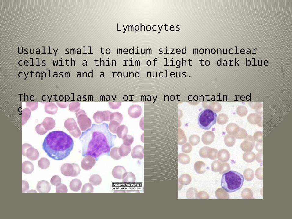
Lymphocytes
Usually small to medium sized mononuclear cells with a thin rim of light to dark-blue cytoplasm and a round nucleus.
The cytoplasm may or may not contain red granules.
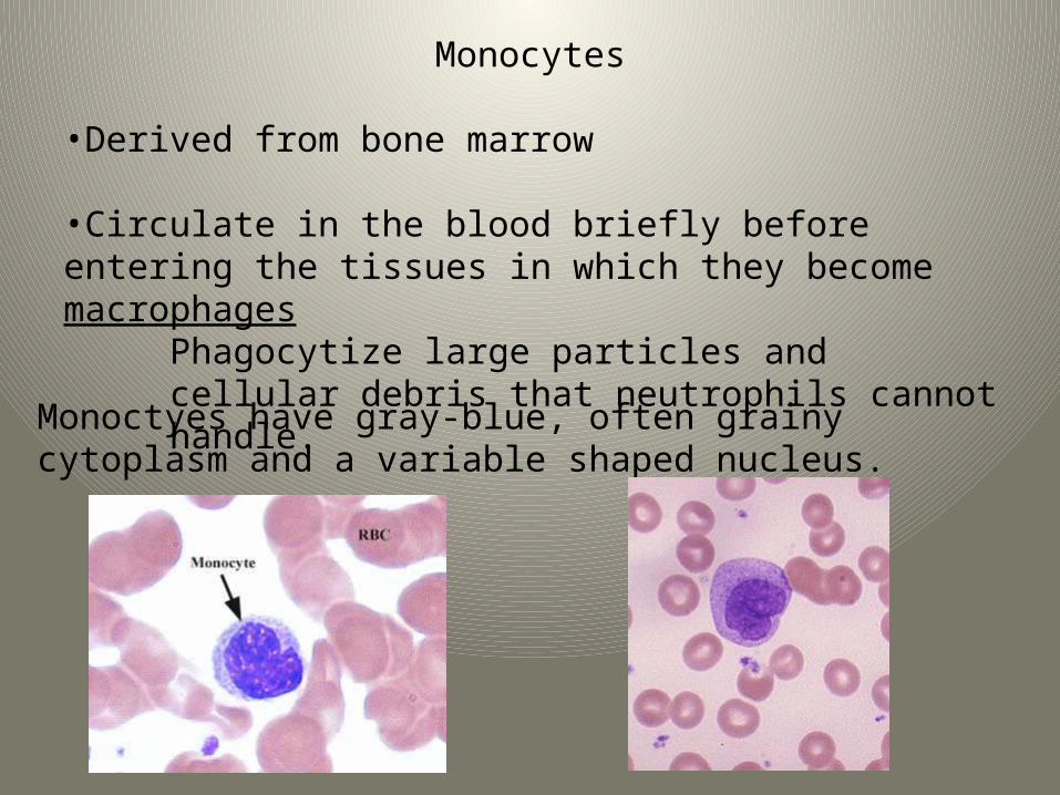
Monocytes
•Derived from bone marrow
•Circulate in the blood briefly before entering the tissues in which they become macrophages
Phagocytize large particles and cellular debris that neutrophils cannot handle.
Monoctyes have gray-blue, often grainy cytoplasm and a variable shaped nucleus.