lesoes cisticas hiv
TRANSCRIPT
8/3/2019 Lesoes Cisticas HIV
http://slidepdf.com/reader/full/lesoes-cisticas-hiv 1/5
BioMed Central
Page 1 of 5(page number not for citation purposes)
BMC Infectious Diseases
Open AccesCase report
Newly formed cystic lesions for the development of pneumomediastinum in Pneumocystis jirovecii pneumonia
Ju-Yeon Cho, Dong-Min Kim*, Yong Eun Kwon, Sung Ho Yoon andSeung Il Lee
Address: Department of Internal Medicine1, Chosun University, College of Medicine, Republic of Korea
Email: Ju-Yeon Cho - [email protected]; Dong-Min Kim* - [email protected]; Yong Eun Kwon - [email protected];Sung Ho Yoon - [email protected]; Seung Il Lee - [email protected]
* Corresponding author
Abstract
Background: Pneumocystis jirovecii, formerly named Pneumocystis carinii, is one of the most
common opportunistic infections in human immunodeficiency virus (HIV)-infected patients.
Case presentations: We encountered two cases of spontaneous pneumomediastinum withsubcutaneous emphysema in HIV-infected patients being treated for Pneumocystis jirovecii
pneumonia with trimethoprim/sulfamethoxazole.
Conclusion: Clinicians should be aware that cystic lesions and bronchiectasis can develop in spite
of trimethoprim/sulfamethoxazole treatment for P. jirovecii pneumonia. The newly formed
bronchiectasis and cyst formation that were noted in follow up high resolution computed
tomography (HRCT) but were not visible on HRCT at admission could be risk factors for the
development of pneumothorax or pneumomediastinum with subcutaneous emphysema in HIV-patients.
BackgroundPneumocystis jirovecii, formerly named Pneumocystis carinii,
is one of the most common opportunistic infections inhuman immunodeficiency virus (HIV)-infected patients[1,2]. Spontaneous pneumothorax has been recognized asa frequent complication in patients with P. jirovecii pneu-monia (PCP) since it was first described in 1984 [3], andpneumomediastinum is an uncommon complicationassociated with pneumothorax in the aforementionedpopulation. We report two cases of spontaneous pneumo-mediastinum with subcutaneous emphysema in HIV-infected patients being treated for P. jirovecii pneumonia
with trimethoprim/sulfamethoxazole.
Case presentationCase 1
A 33-year-old man presented with fever, dyspnea, andodynophagia. Five months prior to admission, the patient had been treated for dental caries at a local hospital, andat that time examination revealed seropositivity for human immunodeficiency virus. On admission, tempera-ture was 39.0'C, pulse 92 beats per minute, respiratory rate 20 breaths per minute and blood pressure 130/80mmHg. Physical examination revealed oral thrush, con-sistent with findings of extensive esophageal candidiasisin endoscopic gastroduodenscopy performed five daysbefore admission. Laboratory data on admission revealed
Published: 18 October 2009
BMC Infectious Diseases 2009, 9:171 doi:10.1186/1471-2334-9-171
Received: 29 January 2009Accepted: 18 October 2009
This article is available from: http://www.biomedcentral.com/1471-2334/9/171© 2009 Cho et al; licensee BioMed Central Ltd.This is an Open Access article distributed under the terms of the Creative Commons Attribution License (http://creativecommons.org/licenses/by/2.0),which permits unrestricted use, distribution, and reproduction in any medium, provided the original work is properly cited.
8/3/2019 Lesoes Cisticas HIV
http://slidepdf.com/reader/full/lesoes-cisticas-hiv 2/5
BMC Infectious Diseases 2009, 9:171 http://www.biomedcentral.com/1471-2334/9/171
Page 2 of 5(page number not for citation purposes)
a WBC count of 1,760/uL, Hb 10.9 g/dL, and platelet count of 297,000/uL. Arterial blood gas analysis whilebreathing room air revealed PaO2 of 48.0 mmHg, PaCO2
of 32.7 mmHg, and saturation of 88.5%, and the calcu-lated (A-a)DO2 was 53.7. CD4 count and HIV viral load
were 4/uL and 130,000 IU/mL, respectively. Diffuse bilat-eral infiltrates of both lung fields were noted, and nocystic lesions were observed on the chest X-ray and highresolution computed tomography (HRCT) taken onadmission. Bronchoscopic alveolar lavage for diagnosis of P. jirovecii was carried out, and microscopic examinationof the bronchoalveolar lavage fluid obtained showed P. jirovecii; no other microorganisms were detected by cul-ture. Treatment with trimethoprim/sulfamethoxazole,fluconazole and corticosteroids at standard dosages wasstarted. The patient had never been on HAART therapy prior to admission. HAART therapy was added to thetreatment on the 8th hospital day. During the treatment
with trimethoprim/sulfamethoxazole, pancytopenia worsened. Bone marrow biopsy revealed inflamed mar-row and partial necrosis. Granulocyte colony stimulating factor was used without avail. On the 22nd hospital day,the chest X-ray obtained as the patient's hypoxemia wors-ened revealed pneumomediastinum. HRCT showednewly formed cystic lesions in both lung fields. Pneumo-mediastinum was treated conservatively with high oxygensupply. CD4 cell count and HIV levels were not followedduring treatment. However, as the general condition of the patient deteriorated, the patient was started on intra-
venous pentamidine on the 23 rd hospital day. On the 25th
hospital day, his oxygen requirement increased. Without
intubation, as the patient and guardian refused the patient being put on a ventilator due to multiple economical andsociological reasons, the patient died on the 26th hospitalday.
Case 2
A 48-year-old man presented with insidious dyspnea that had developed over a period of 2 months. The patient hadbeen seropositive for human immunodeficiency virus in2005 in a routine physical examination for a job positionas a sailor. He was on zidovudine and didanosine for 8months but stopped taking these antiretroviral agents at another hospital for economic reasons. On admission,
temperature was 38.2'C, pulse 96 beats per minute, respi-ratory rate 22 breaths per minute, and blood pressure100/60 mmHg. Physical examination was normal except for decreased breathing sound in both lung fields. Labora-tory data showed a WBC count of 11,010/uL, Hb 13.6 g/dL, a platelet count of 425,000/uL, and LDH of 1,095 U/L. Arterial blood gas analysis in room air demonstratedPaO2 of 54.1 mmHg, PaCO2 of 34.4 mmHg, and satura-tion of 89.9%, and the calculated (A-a)DO2 was 45.5.CD4 count and HIV viral load were 21/uL and 260,000IU/mL, respectively. Chest X-ray on admission revealed
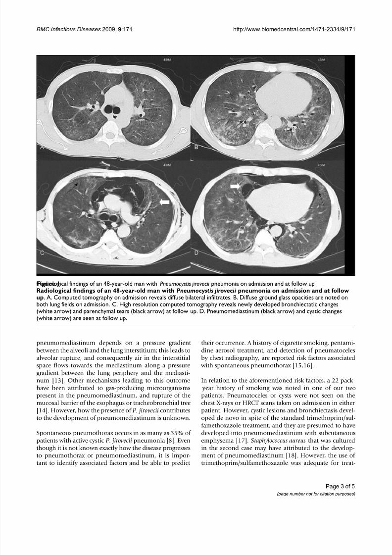
diffuse ground glass opacity in both lung fields. HRCT showed interlobular septal thickening of the bronchusand bronchioles without any cyst formation (Figure 1).Microscopic examination of bronchoalveolar lavagerevealed P. jiroveci, and Staphylococcus aureus was detected
by culture. The patient met the criteria of respiratory fail-ure [PaO2 less than 70 mmHg or (A-a)DO2 more than 35mmHg] and corticosteroids were co-administered withtrimethoprim/sulfamethoxazole. On hospital day 4 thepatient developed sudden chest pain radiating to theshoulder and neck. Chest X-ray, electrocardiography, andarterial blood gas analysis were performed. The chest X-ray revealed air lining the cardiac border, indicating devel-opment of pneumomediastinum. HRCT revealed newly developed cystic changes, bronchiectatic change, andparenchymal tear (Figure 1). The pneumomediastinum
was treated conservatively with administration of highoxygen supply without the need for invasive procedures.
On hospital day 10, HAART was started with lopinavir/ritonavir, lamivudine, and zidovudine. On hospital day 13, follow up HRCT revealed more aggravated pneumo-mediastinum, bronchiectasis and parenchymal tear in thelingular division of the left upper lobe. Trimethoprim/sul-famethoxazole was changed to intravenous pentamidine.
The patient experienced nausea, vomiting, and hypoglyc-emia on pentamidine, leading to a further change of anti-biotics to primaquine and clindamycin. The patient'sdyspnea improved and no particular complications wereobserved. The patient was discharged on the 42nd hospitalday and is being followed up in the outpatient clinic.
Discussion The overall incidence of P. jirovecii pneumonia hasdecreased with the use of highly active antiretroviral ther-apy [4]. However, approximately 85% of patients withadvanced HIV infections continue to experience P. jiroveciipneumonia in the course of their disease when manage-ment is inadequate [5]. The most common radiographic finding in P. jirovecii pneumonia is the presence of diffuse,bilateral perihilar interstitial infiltrates (ground-glassopacity) in both lungs [6]. Atypical radiographic manifes-tations of PCP include cystic spaces and bullae, adenopa-thy, pleural effusions and pneumothorax [7-10]. The exact mechanism behind the development of the pulmonary
cysts and P. jirovecii is not yet known. However, variousmechanisms have been proposed including direct lung destruction by P. jirovecii, over-distension of the lungscaused by obstructive bronchiolitis acting as a ball-valve(inflammatory exudates in the small bronchioles), inter-stitial emphysema and abnormal remodeling of pulmo-nary architecture due to interstitial fibrosis, and release of elastase and other proteolytic enzymes [5,10-12]. A reviewof the literature indicates that the development of sponta-neous pneumomediastinum with subcutaneous emphy-sema in HIV patients is rare. The pathophysiology of
8/3/2019 Lesoes Cisticas HIV
http://slidepdf.com/reader/full/lesoes-cisticas-hiv 3/5
BMC Infectious Diseases 2009, 9:171 http://www.biomedcentral.com/1471-2334/9/171
Page 3 of 5(page number not for citation purposes)
pneumomediastinum depends on a pressure gradient between the alveoli and the lung interstitium; this leads toalveolar rupture, and consequently air in the interstitialspace flows towards the mediastinum along a pressuregradient between the lung periphery and the mediasti-num [13]. Other mechanisms leading to this outcome
have been attributed to gas-producing microorganismspresent in the pneumomediastinum, and rupture of themucosal barrier of the esophagus or tracheobronchial tree[14]. However, how the presence of P. jirovecii contributesto the development of pneumomediastinum is unknown.
Spontaneous pneumothorax occurs in as many as 35% of patients with active cystic P. jirovecii pneumonia [8]. Eventhough it is not known exactly how the disease progressesto pneumothorax or pneumomediastinum, it is impor-tant to identify associated factors and be able to predict
their occurrence. A history of cigarette smoking, pentami-dine aerosol treatment, and detection of pneumatocelesby chest radiography, are reported risk factors associated
with spontaneous pneumothorax [15,16].
In relation to the aforementioned risk factors, a 22 pack-
year history of smoking was noted in one of our twopatients. Pneumatoceles or cysts were not seen on thechest X-rays or HRCT scans taken on admission in either patient. However, cystic lesions and bronchiectasis devel-oped de novo in spite of the standard trimethoprim/sul-famethoxazole treatment, and they are presumed to havedeveloped into pneumomediastinum with subcutaneousemphysema [17]. Staphylococcus aureus that was culturedin the second case may have attributed to the develop-ment of pneumomediastinum [18]. However, the use of trimethoprim/sulfamethoxazole was adequate for treat-
fRadiological findings of an 48-year-old man with Pneumocystis jirovecii pneumonia on admission and at follow upFigure 1
Radiological findings of an 48-year-old man with Pneumocystis jirovecii pneumonia on admission and at followup. A. Computed tomography on admission reveals diffuse bilateral infiltrates. B. Diffuse ground glass opacities are noted onboth lung fields on admission. C. High resolution computed tomography reveals newly developed bronchiectatic changes(white arrow) and parenchymal tears (black arrow) at follow up. D. Pneumomediastinum (black arrow) and cystic changes(white arrow) are seen at follow up.
8/3/2019 Lesoes Cisticas HIV
http://slidepdf.com/reader/full/lesoes-cisticas-hiv 4/5
BMC Infectious Diseases 2009, 9:171 http://www.biomedcentral.com/1471-2334/9/171
Page 4 of 5(page number not for citation purposes)
ing Staphylococcus aureus without the need for an addi-tional antibiotic [19]. Treatment of spontaneouspneumomediastinum is generally limited to observation
without the need for invasive measures [20]. However, inthe above patients, P. jirovecii pneumonia may have been
an underlying cause to the development of pneumomedi-astinum. Therefore, the standard trimethorpim/sulfame-thoxazole was analyzed as a treatment failure warranting a change of antibiotics to pentamidine.
There is no guideline regarding the treatment of the acutephase of P. jirovecii pneumonia in HIV-infected patients
with HAART. However, administration of HAART therapy early in the acute phase of P. jirovecii pneumonia was donein both patients as improved survival rates in HIV-infectedpatients with severe P. jirovecii pneumonia was associated
with HAART therapy [21,22]. Although the developement of pneumothorax was not anticipated in our patients
when HAART therapy was initiated, Morris et al reporteddecreased rates of pneumothorax development in P. jirovecii infected HIV-patients receiving HAART ther-apy[21]. The possible contribution of antiretroviral ther-apy to the clinical worsening of the patients wasconsidered. Wislez et al reported of acute respiratory fail-ure following HAART in P. jirovecii pneumonia due toimmune reconstitution inflammatory syndrome [23].HRCT of both patients in this study did not reveal any findings relevant to the development of acute respiratory failure.
The occurrence of newly formed cystic lesions or bron-
chiectasis despite treatment may be risk factors for thedevelopment of pneumothorax or pneumomediastinum
with subcutaneous emphysema in HIV-patients. There-fore close follow up with HRCT in HIV-patients with P. jirovecii pneumonia might assist in predicting the develop-ment of pneumothorax or pneumomediastinum. Our findings suggest that clinicians should be aware of theclinical importance of newly formed cystic lesions andbronchiectasis for the development of pneumomediasti-num and pneumothorax in P. jirovecii pneumonia.
ConclusionIn conclusion, clinicians should be aware that cystic
lesions and bronchiectasis can develop in spite of trimeth-oprim/sulfamethoxazole treatment for P. jirovecii pneu-monia. Newly formed bronchiectasis and cyst formationmay be risk factors for the development of pneumomedi-astinum with subcutaneous emphysema.
Competing interests The authors declare that they have no competing interests.
Authors' contributionsJu-Yeon Cho took care of the patient in the ICU and drew
up the first draft of the report, Yong Eun Kwon, Sung Ho Yoon, and Seung Il Lee, consultant pulmonologists, madea substantial contribution to draft the manuscript
and revised the draft all over the course of submission,
Dong-Min Kim conceived
of the study, participated in its design and coordinationand drafted the manuscript. All authors read andapproved the final manuscript.
AcknowledgementsWritten consent was obtained from the patient or their relative for publi-
cation of study.
References1. HIV/AIDS surveillance supplemental report. Centers for Dis-
ease Control and Prevention 2003:1-20.2. Stringer JR, Beard CB, Miller RF, Wakefield AE: A new name (Pneu-
mocystis jiroveci) for Pneumocystis from humans. Emerg infectDis 2002, 8:891-6.
3. Wollschlager CM, Khna FA, Chitkara RK, Shivaram U: Pulmonarymanifestations of the acquired immunodeficiency syndrome(AIDS). Chest 1984, 85:197-202.
4. Palella FJ Jr, Delaney KM, Moorman AC, et al.: Declining morbidityand mortality among patients with advanced human immu-nodeficiency virus infection. N Engl J Med 1998, 338:853-60.
5. Konishi M, Amimotom M, Yoshiomoto E, et al.: AIDS-related Pneu-mocystis carinii pneumonia with disappearance of cysticlesions after treatment. Intern Med 2002, 41:896-8.
6. Goodman PC, Gamsu G: Radiographic findings in the acquiredimmunodeficiency syndrome. Postgrad Radiol 1987, 7:3-15.
7. Kuhlman JE, Kavuru M, Fishman EK, Siegelman SS: Pneumocystiscarinii pneumonia: spectrum of parenchymal CT findings.Radiology 1990, 175:711-4.
8. Chow C, Templeton PA, White CS: Lung cysts associated withPneumocystis carinii pneumonia: radiographic characteris-tics, natural history, and complications. Am J Roentgenol 1993,161:527-31.
9. Pastores SM, Garay SM, Naidich DP, Rom WN: Review: pneumot-horax in patients with AIDS-related Pneumocystis cariniipneumonia. Am J Med Sci 1996, 312:229-34.
10. Sandhu JS, Goodman PC: Pulmonary cysts associated with Pneu-mocystis carinii pneumonia in patients with AIDS. Radiology 1989, 173:33-5.
11. Feurestein IM, Archer A, Pluda JM, et al.: Thin-walled cavities,cysts, and pneumothorax in Pneumocystis carinii pneumonia:further observations with histopathologic correlation. Radiol-ogy 1990, 174:697-702.
12. Eng RHK, Binshburg E, Smith SM: Evidence for destruction of lung tissues during Pneumocystis carinii infection. Arch Intern
Med 1987, 147:746-9.13. Macklin MT, Macklin CC: Malignant intersititial emphysema of
the lungs and mediastinum as an important occult complica-tion in many respiratory diseases and other conditions:
interpretation of the clinical literature in the light of labora-tory experiment. Medicine 1944, 23:281-358.14. Macia I, Moya J, Ramos R, et al.: Spontaneous pneumomediasti-
num: 41 cases. European journal of Cardio-thoracic surgery 2007,31:1110-4.
15. Metersky ML, Colt HG, Olson LK, Shanks TG: AIDS-related spon-taneous pneumothorax: risk factors and treatment. Chest1995, 108:946-51.
16. Gruden JF, Huang L, Turner J, et al.: HRCT in the evaluation of clinically suspected Pneumocystis carinii pneumonia in AIDSpatients with normal, equivocal, or nonspecific radiographicfindings. Am J Roentgenol 1997, 167:967-75.
17. The National Institutes of Health (NIH), the Centers for DiseaseControl and Prevention (CDC), and the HIV Medicine Association of the Infectious Disease Society of America (HIVMA/IDSA): Guidelinesfor Prevention and Treatment of Opportunistic Infections in HIV-infected
8/3/2019 Lesoes Cisticas HIV
http://slidepdf.com/reader/full/lesoes-cisticas-hiv 5/5
Publish with BioMed Central and everyscientist can read your work free of charge
"BioMed Central will be the most significant development for
disseminating the results of biomedical research in our lifetime."
Sir Paul Nurse, Cancer Research UK
Your research papers will be:
available free of charge to the entire biomedical community
peer reviewed and published immediately upon acceptance
cited in PubMed and archived on PubMed Central
yours — you keep the copyright
Submit your manuscript here:
http://www.biomedcentral.com/info/publishing_adv.asp
BioMedcentral
BMC Infectious Diseases 2009, 9:171 http://www.biomedcentral.com/1471-2334/9/171
Page 5 of 5(page number not for citation purposes)
Adults and Adolescents. MMWR 2009 [http://aidsinfo.nih.gov/contentfiles/Adult_OI.pdf ].
18. Macfarlane J, Rose D: Radiographic features of staphylococcalpneumonia in adults and children. Thorax 1996, 51:539-40.
19. Grim SA, Rapp RP, Martin CA,et al.: Trimethoprim-sulfamethox-azole as a viable treatment option for infections caused bymethicillin-resistant Staphylococcus aureus. Pharmacotherapy
2005, 25:253-64.20. Caceres M, Ali SZ, Braud R, et al.: Spontaneous pneumomediasti-num: A comparative study and review of the literature. AnnThorac Surg 2008, 86:962-6.
21. Morris A, Wachter RM, Luce J, et al.: Improved survival withhighly active antiretroviral therapy in HIV-infected patients with severe Pneumocystis carinii pneumonia. AIDS 2003,17:73-80.
22. Zolopa A, Andersen J, Komarow L, et al.: Immediate vs deferredART in the setting of acute AIDS-related opportunistic infec-tion: Final results of a randomized strategy trial, ACTGA5164. 15th Conference on Retroviruses Opportunistic Infection. 2008Feb 3-6. Boston. Oral abstract 142 .
23. Wislez M, Bergot E, Antoine M, et al.: Acute respiratory failurefollowing HAART introduction in patients treated for Pneu-mocystis carinii pneumonia. Am J Respir Crit Care Med 2001,164:847-51.
Pre-publication history The pre-publication history for this paper can be accessedhere:
http://www.biomedcentral.com/1471-2334/9/171/prepub