lesions of ventrolateral prefrontal or anterior orbitofrontal cortex in primates heighten negative...
TRANSCRIPT

Lesions of Ventrolateral Prefrontal or AnteriorOrbitofrontal Cortex in Primates Heighten NegativeEmotionCarmen Agustín-Pavón, Katrin Braesicke, Yoshiro Shiba, Andrea M. Santangelo, Yevheniia Mikheenko,Gemma Cockroft, Faaiza Asma, Hannah Clarke, Mei-See Man, and Angela C. Roberts
Background: Heightened fear and anxiety are core symptoms of a variety of neuropsychiatric disorders. They are associated with structuraland activity changes throughout neural circuitry that includes the ventral and medial prefrontal cortices (PFC), the amygdala, andhippocampus. Although the contributions of the medial PFC, amygdala, and hippocampus to fear and anxiety have been studied exten-sively with animal models, the selective roles of the ventral PFC—including the ventrolateral prefrontal cortex (vlPFC) and orbitofrontalcortex—are poorly understood.
Methods: We investigated the effects of selective excitotoxic lesions of either the vlPFC or anterior orbitofrontal cortex (antOFC) on anxiousbehavior and Pavlovian conditioned autonomic and behavioral fear responses in the New World primate, the common marmoset.
Results: Both vlPFC and antOFC lesions resulted in stronger, less adaptable conditioned fear responses. They also heightened the anxietyresponses of a marmoset to a human intruder. In contrast, only a lesion of the vlPFC affected the coping style that a marmoset displayed inthe presence of the human intruder, increasing the likelihood of proactive mobbing.
Conclusions: These results suggest that both the antOFC and vlPFC can downregulate fear and anxiety and, together, provide necessary
but independent contributions to the top-down control of negative emotion.c1itsocbdra
twttFaloaama
M
Kl
S
8m
Key Words: Anxiety, autonomic, emotion regulation, marmoset,negative emotion, prefrontal cortex
H eightened fear and anxiety are core symptoms of a numberof mood and anxiety disorders and are associated with dys-function in a variety of brain regions, including the ventral
and medial prefrontal cortices (PFC) (1– 4). Fear is viewed as a bio-logically adaptive response to actual or anticipated explicit threat,whereas anxiety involves uncertainty as to the expectancy of threatand, unlike fear, is often triggered by less explicit cues. Considerableinsight into the specific role of the medial PFC in the regulation offear and anxiety has been gained from experimental studies inanimals, in particular, rodents (for review see Sotres-Bayon andQuirk [5]). In contrast, far less is known of the specific role of theventral PFC, including ventrolateral (vlPFC) (areas 45, dorsal 47/12)and orbitofrontal regions (OFC) (areas 11, orbital 47/12, 13, 14), innegative emotion regulation.
The vlPFC is a well-characterized granular region of PFC in non-human primates (6,7), but there have been few studies investigat-ing the effects of its disruption on fear and anxiety (8,9). Whether ahomologous region exists in rodents is unclear. By contrast, contra-dictory results have arisen from rodent and primate studies of theOFC in fear and anxiety. Avoidance of mild, potentially threatening,unconditioned stimuli or contexts— often interpreted as anxiousbehavior—is unaffected in OFC-lesioned rats (10,11) but signifi-
From the Department of Physiology, Development and Neuroscience (CA-P,KB, YS, AMS, YM, FA, HC, M-SM, ACR); Behavioural and Clinical Neurosci-ence Institute (CA-P, KB, YS, AMS, YM, GC, FA, HC, M-SM, ACR); and theDepartment of Experimental Psychology (GC), University of Cambridge,Cambridge, United Kingdom.
Authors KB and YS contributed equally to this work.Address correspondence to Angela C. Roberts, Ph.D., Department of Physi-
ology, Development and Neuroscience, University of Cambridge, Down-ing Street, Cambridge, CB2 3DY UK; E-mail: [email protected].
tReceived Dec 22, 2011; revised and accepted Mar 8, 2012
0006-3223/$36.00doi:10.1016/j.biopsych.2012.03.007
antly reduced in monkeys after large (areas 11, orbital 12/47, 13,4) or restricted (areas 11, 13, 14) lesions of OFC (12–15) (but see
ncreased or no effects reported in [8,9,13,16]). In contrast, condi-ioned fear, which has been studied almost exclusively in rats (butee Machado et al. [16]), is enhanced after OFC lesions (10,17). Thesepposing effects might reflect the differential role of the OFC inonditioned fear and anxiety, because evidence is emerging thatrain circuits underlying fear and anxiety, although over-lapping,o show some divergence (18). Alternatively, the contradictory
esults might reflect differences in the extent and location of lesionscross studies or differences between species (rodent vs. primate).
Thus, the present study investigated the specific role(s) of ven-ral PFC in conditioned fear and anxiety in primates and determinedhether the vlPFC and OFC provided independent contributions to
he regulation of these negative emotions. New World monkeys,he common marmoset, received excitotoxic lesions (Figure 1 andigure S1 in Supplement 1) of either the vlPFC (area 12) or thenterior orbitofrontal cortex (antOFC) (areas 11 and anterior 13), the
atter having been shown previously to contribute to the regulationf positive emotional responding (19). Because emotional statesre composed of both behavioral and physiological components,utonomic and behavioral measures were recorded in these ani-als in a Pavlovian conditioned fear paradigm. In addition, their
nxious behavior was assessed in response to an unfamiliar human.
ethods and Materials
All procedures were conducted in accordance with the Unitedingdom 1986 Animals (Scientific Procedures) Act under project
icense PPL 80/2225.
ubjectsFifteen naïve common marmosets (Callithrix jacchus; 7 females,
males) were used in the lesion study. Additionally, 58 naïve mar-osets (26 females, 32 males) were tested on the human intruder
est only and used to establish the principal components underly-
BIOL PSYCHIATRY 2012;72:266–272© 2012 Society of Biological Psychiatry

wfiwd
S
epm
t(e1bnaabi
dod(b4fobl.csata
rbscr
nsdm
idclw
C
TSm
C. Agustín-Pavón et al. BIOL PSYCHIATRY 2012;72:266–272 267
ing the pattern of behaviors exhibited by marmosets on this test(see Results). For housing and diet information see Supplement 1.
Surgical ProceduresImplantation of Telemetric Devices. Blood pressure (BP)
transmitters were inserted into the abdominal cavity, and the probecatheter was implanted into the descending aorta after proceduresdescribed previously (20).
Excitotoxic Lesions of the antOFC and vlPFC. Details of sur-gical procedures are described in Supplement 1. The stereotaxiccoordinates used to lesion the antOFC (n � 5) and vlPFC (n � 5) (inaddition to the neurotoxin, infusion rate, and volumes) are de-scribed in Table S1 in Supplement 1. Sham-operated control ani-mals underwent the same surgical procedure as lesioned animals,except that they received infusions of sterile phosphate buffer ve-hicle, into the antOFC (n � 2) or vlPFC (n � 3).
Marmosets had a 2-week recovery period before testing. SeeSupplement 1 for detailed information about anesthesia and drugs.
Mild Aversive Pavlovian Discrimination ParadigmTest Apparatus. Behavioral testing took place within a sound-
attenuated box in a dark room. Each subject was transported to the
Figure 1. (A) Ventrolateral prefrontal cortex (vlPFC) and (B) anterior orbito-frontal cortex (antOFC) excitotoxic lesions. Schematic coronal sectionstaken through the frontal lobe (anterior–posterior) of the marmoset mon-key. The five decreasing shades of gray indicate regions that were lesionedin all five, four, three, two, or one monkey, respectively. The inset coronalsections illustrate OFC and vlPFC cytoarchitectonic regions identified withinprefrontal cortex, redrawn from Burman and Rosa (49). All animals in thevlPFC lesioned group sustained neuronal cell loss within the vlPFC that onlyencroached into the most lateral extent of antOFC unilaterally (right-handside). However, animals differed as to whether most cell loss in area 12/45was found more rostrally or caudally, a difference that did not correlate withoverall levels of fear and anxiety, thereby suggesting that damage to eithersection of 12/45 was sufficient to affect these negative emotions. In theantOFC lesion group most animals sustained marked neuronal lossthroughout area 11 (sparing its most lateral extent) and the anteromedialportion of area 13. Only in one animal was there significant neuronal loss,unilaterally, in area 14. Again, any individual variation in the extent ofantOFC damage did not correlate with overall fear and anxiety levels.
apparatus in a clear cuboidal Perspex box, which had one side that H
as removable, thus acting as the door. The entire box was thentted into the internal frame of the apparatus, an illustration ofhich can be found in Mikheenko et al. (21). See Supplement 1 foretails.
tudy DesignHabituation. All marmosets were habituated to the behavioral
quipment for between four to seven 10-min sessions after BProbe implantation. Once the heart rate (HR) was stable, they wereoved to the next stage.
Orienting. After habituation, marmosets were exposed, forwo sessions, to two novel sounds, a 4-kHz tone or a clicker, 70 dB,the “to-be” conditioned stimuli [CSs]), for a duration of 20 sec, onach of four separate occasions, with a variable interval of between20 and 180 sec. The aim of these sessions was to monitor theehavioral and autonomic reactions of the animals toward theovel stimuli. The stimulus that elicited the smaller behavioral andutonomic reaction was chosen as the CS� (to be followed by theversive stimulus), and the one that elicited the larger reactionecame the CS� (to be followed by a neutral stimulus), thus avoid-
ng any stimulus preparedness.Conditioning. Marmosets were exposed to a Pavlovian proce-
ure in which one of the sounds (CS�) was associated with a burstf mildly aversive loud noise (unconditioned stimulus [US]�, 120B, .3–.7 sec) and the other (CS�) with the offset of the houselight
US-, .5–1 sec). All training sessions started with a variable intervaletween 120 and 180 sec, followed by 4 presentations of CS� andpresentations of the CS�, played for 20 sec and immediately
ollowed by the US, in pseudorandom order with inter-trial periodsf between 120 and 180 sec. Marmosets were trained until bothehavioral and cardiovascular changes from the preceding base-
ine period (20 sec) elicited by the CS� differed significantly (p �05) from those by the CS�, across three consecutive sessions (dis-riminative criterion). Individual differences in learning rate andtrength of the HR and behavioral responses were counterbalancedcross groups (Figure S2 in Supplement 1 and Results). Animalshen were sham-operated or received an excitotoxic lesion of thentOFC or vlPFC.
Retention. After a postsurgery recovery period, animals firsteceived the same discrimination and stimulus contingencies asefore surgery, until they regained discriminative criterion. Ses-ions to criterion were analyzed along with the behavioral andardiovascular conditioned responses on the first three sessions ofetention as well as the final three discriminative criterion sessions.
Partial extinction. For one session only, 2 of the 4 CS� wereot followed by the presentation of the aversive noise. This probeession was included, because in an earlier pilot study we hadiscovered that such a manipulation in control monkeys had aarked impact on their subsequent conditioned responding.
Recovery. Immediately after the partial extinction session, an-mals were re-trained again as in Conditioning and Retention, untiliscriminative criterion was regained. As for retention, sessions toriterion were analyzed along with the behavioral and cardiovascu-
ar conditioned responses on the first three sessions of recovery asell as the final three discriminative criterion sessions.
ardiovascular and Behavioral AnalysisTo measure HR and BP changes remotely in animals, a PhysioTel
elemetry System (Data Sciences, St. Paul, Minnesota) was used.ystolic and diastolic BP events were extracted as local minima andaxima for each heartbeat cycle. See Supplement 1 for details. The
R was calculated from the time interval between systolic BPwww.sobp.org/journal

ccan
R
AR
stcfa
lnpbHwtsAttpmdwdgscipcpsg1FR(ebt
Ho
ihchetsc6S
268 BIOL PSYCHIATRY 2012;72:266–272 C. Agustín-Pavón et al.
events. Only HR and not BP showed reliable conditioning, consis-tent with our previous report (21).
All behavior was recorded on DVD and then subsequentlyscored by a person unaware of the experimental conditions. TheCS� related behaviors (typically displayed by marmosets in re-sponse to simple Pavlovian conditioning [21]) were treated as asingle measure of “vigilant scanning” and included attentive visualsearch of surroundings accompanied by tense postures marked byforward extension of body/head and rearing. See Supplement 1 forfurther details.
Human Intruder ParadigmAll lesioned and control animals took part in this paradigm,
except one antOFC-lesioned animal that—a short time after com-pleting the mild aversive Pavlovian discrimination paradigm— be-came unwell and was euthanized, and his brain was taken for post-mortem lesion placement analysis. Testing took place in the homecage with a procedure adapted from Carey et al. (22). The day beforethe test session animals were habituated to the testing procedure.They were separated from their cage mate and restricted to theupper right-hand quadrant of their home cage for 15 min. Duringthat time a video camera and microphone were placed just in frontof the cage. Twenty-four hours later they were again separatedfrom their cage mate (Separate condition), and after 8 min an un-known human entered the room (Intruder condition). The intruderstood 40 cm in front of the cage and stared at the marmoset,maintaining eye contact, where possible, for 2 min. Once the in-truder had left the room, recording went on for a further 5 min.Videotapes were scored by an experimenter blind to the experi-mental groups. Behavioral measures included vocalizations (tsik,egg, tsik-egg, and tse [tse and tse-egg]), overall distance from thecage front, jumps to the front, head and body bobbings, and loco-motion. See Supplement 1 for further details.
General Locomotor ActivityTo control for any effects of the surgery on general behavior, the
activities of animals in their home cage were recorded for 8 consec-utive days. Approximately 2 weeks after surgery, marmosets werefitted with an actiwatch (CamNtech, Cambridge, United Kingdom)attached to their identity neck chains. For detailed information, seeSupplement 1.
Histological AnalysisDetails of euthanasia and histological preparation are described
in Supplement 1. For verification of lesions, coronal sections wereviewed under a Leitz DMRD microscope (Leica Microsystems, Wet-zlar, Germany), and lesioned areas were defined by the presence ofmajor neuronal loss, often with marked gliosis. For each animal,areas with cell loss were schematized onto drawings of standardmarmoset coronal sections, and composite diagrams were thenmade to illustrate the extent of overlap between lesions (Figure 1).
Statistical AnalysisAll cardiovascular and behavioral data were analyzed with SPSS
software version 17.0.0 (SPSS, Chicago, Illinois). Lesion effects wereanalyzed with one-, two- and three-way analyses of variance(ANOVAs) as described in the results. Any significant interactionswere analyzed with simple main effects, with the mean square errorterm (SEM) from the original interaction where appropriate. Fisher’sprotected least square difference (LSD) test for three groups wasused for post hoc comparisons. Data that were skewed and violatedthe distribution requirement of ANOVA were transformed appro-priately (23,24). For the human intruder test, two principal compo-
nents were first derived from the eight distinct behaviors and vo- dwww.sobp.org/journal
alizations from a large cohort of 63 marmosets, with principalomponent analysis (25). The component scores for each controlnd lesioned animal were then obtained, by applying the compo-ent correlation coefficients to their standardized scores.
esults
n Enhanced and More Rigid Expression of Conditioned Fearesponses After Excitotoxic Lesions of Either vlPFC or antOFC
Because groups were counterbalanced for learning rate andtrength of conditioning (Figure S2 and Table S2 in Supplement 1),here were no group differences pre-operatively in acquiring dis-riminative Pavlovian conditioning (Figure 2A). One way ANOVAsor sessions to criterion and conditioned HR and vigilant scanningt criterion revealed no significant effects (F values � 1).
Postoperatively, all animals from control, vlPFC, and antOFCesioned groups (except one antOFC lesioned animal that sponta-eously reversed their conditioned responses: see Results in Sup-lement 1) regained discriminative criterion in an equivalent num-er of sessions (Figure 2B) (one-way ANOVA, Group, F � 1).owever, after exposure to a single session of partial extinction, inhich aversive loud noise did not accompany the offset of two of
he four CSs, antOFC and vlPFC lesioned animals required feweressions to regain discriminative criterion [Figure 2B: one-wayNOVA, Group, F (2,11) � 8.99 p � .005]. More specifically, rather
han regaining criterion, 4 of the 5 vlPFC lesioned animals and 2 ofhe 4 antOFC lesioned animals never displayed a decrement inerformance after partial extinction. Instead, they continued toaintain performance criterion (i.e., significant discriminative con-
itioning) across the subsequent three conditioning sessions. Thisas in marked contrast to the 5 control subjects, all of whom didisplay a decrement in conditioning. Thus, overall, the PFC lesionedroups displayed stronger conditioning than control subjects andeemed less sensitive to a transient change in the environmentalontingencies. In addition, vlPFC lesioned animals displayed signif-
cantly greater vigilant scanning across the retention and recoveryeriod [Figure 2C, three-way ANOVA (Group, Stage [Retention, Re-overy], Phase [session 1, Criterion]): Group, F (2,11) � 5.25 p � .025,ost hoc LSD, vlPFC: vs. control (p � .015), vs. antOFC (p � .021)] andignificantly enhanced conditioned HR after recovery from the sin-le session of partial extinction [Figure 2D: Table S2 in Supplement, three-way ANOVA (Group, Stage, Phase), Group � Stage,(2,11) � 7.44, p � .009; Simple main effects: Retention F � 1;ecovery: F (2,11) � 5.66, p � .02, post hoc LSD, vlPFC: vs. control
p � .006), vs. antOFC (not significant, p � .089)]. There were noffects of either lesion on HR, BP, or vigilant scanning during theaseline or during the CS� at either retention or after partial extinc-
ion (all F values � 1).
eightened Anxiety After Excitotoxic Lesions of Either vlPFCr antOFC
Consistent with their enhanced fear responsiveness on the Pavlov-an discrimination paradigm, both groups of animals also displayedeightened anxiety in the human intruder paradigm (Figure 3A). Toharacterize the pattern of behavior displayed in the presence of theuman intruder a principal component analysis was conducted onight behavioral variables with oblique rotation from 63 marmosets inhe colony (including the 5 sham control marmosets but not the le-ioned marmosets from the present study). It revealed two principalomponents with eigenvalues above 1 that, in combination, explained2.3% of the variance (see Methods and Materials and Table S3 inupplement 1). The behaviors that loaded on component 1 included
istance from the cage front, locomotor activity, numbers of jumps
sssmlt..n.gdtaHcS
D
csomaac
.01.
C. Agustín-Pavón et al. BIOL PSYCHIATRY 2012;72:266–272 269
toward the front, head bobbing, and egg, tse, and tsik-egg calls. Thosemarmosets with the highest component 1 score displayed reducedlocomotor activity; fewer jumps toward the front; increased head bob-bing; increased egg, tse, and tsik-egg calls; and also maintained agreater distance from the cage front, suggesting that this componentrepresents an overall level of emotionality/anxiety. The behaviors thatloaded on component 2 were primarily calls—that of tsik, tsik-egg, andegg—such that marmosets with the highest score made the greatestnumber of tsik and tsik-egg calls and the lowest number of egg calls.Tsik calls are mobbing calls, uttered toward conspecifics, potentialpredators, and unfamiliar humans and can be a major component of aproactive coping strategy to drive the threat away (26). They are in-versely related to egg calls; the latter, along with tse calls, primarilyassociated with vigilance behavior in potentially threatening contextsand more likely reflecting a reactive coping strategy (26). Thus, compo-nent 2 most likely represents the type of coping strategy adopted bythe marmoset in anxiety provoking situations (27,28).
Both antOFC and vlPFC lesioned groups displayed significantlyhigher component 1 scores in response to a human intruder (Fig-ures 3B and 3C). In particular, they displayed reduced locomotoractivity, increased head bobbing, and increased distance from the
Figure 2. Mild aversive Pavlovian discrimination paradigm. (A) Study deonditioning (acquisition, retention [Ret.], partial extinction, and recoverytimulus (CS) are depicted. (B) In the Recov. phase, both lesioned groups toof heart rate (HR) and vigilant scanning (VS) after partial extinction, comparmosets showed significantly greater discriminative conditioned VS resp
nd OFC lesioned animals. The y-axis reflects the difference score betweennimals also showed significantly stronger HR discriminative conditioned reontrol subjects, as reflected by the increased difference score between HR
intertrial interval; US, unconditioned stimulus; var., variable. *p � .05; **p �
cage front (Table S4 in Supplement 1). In contrast, although the h
trategy (component 2) scores of antOFC lesioned marmosets wereimilar to those of control marmosets, vlPFC lesioned animalshowed a significant increase. The vlPFC lesioned animals made
any more proactive tsik calls compared with control or antOFCesioned animals, the latter producing primarily vigilant calls (egg/se) [two-way ANOVA: Group � Component F (2,11) � 4.16, p �045; Simple Main effects, Component 1: Group F (2,11) � 9.03, p �005; LSD, control: vs. vlPFC, p � .002; vs. antOFC, p � .019; Compo-ent 2: Group F (2,11) � 5.55, p � .022; LSD, vlPFC: vs. control, p �
042; vs. antOFC, p � .008)]. It should be noted that the lesionedroups, compared with control subjects, also maintained a greateristance from the cage front and showed reduced locomotion in
he “separated” phase, having been separated from their partnernd confined to the top right hand quadrant of their home cage.owever, their overall, daily locomotor activity score in the homeage did not differ from control subjects (see Results and Table S4 inupplement 1).
iscussion
Abnormal fear and anxiety responses are characteristic of the
ndicating the sequence of training (habituation and orienting) and fearv.]). For illustrative purposes only two presentations of each conditioneder sessions to regain the criterion of significant discriminative conditioningwith control animals. (C). Ventrolateral prefrontal cortex (vlPFC) lesionedafter surgery (mean across Ret. and Recov. phases), compared with controlanges from baseline during the CS� and the CS�. (D). The vlPFC lesionedses in the Recov. phase, after the partial extinction session, compared withges during the CS� and the CS�. antOFC, anterior orbitofrontal cortex; ITI,
sign i[Recok fewared
onsesVS chsponchan
uman disorders of mood and anxiety and have been associated
www.sobp.org/journal

lticiTteiebbet
trahTwtbsidr
V
OftudottHlfifipeapsmdas
mpFaOaehlh
pffflctv
270 BIOL PSYCHIATRY 2012;72:266–272 C. Agustín-Pavón et al.
with alterations in activity and gray matter volume within the ven-tral PFC of patients suffering from these disorders (1–3). In thepresent study selective excitotoxic lesions of either vlPFC or antOFCin monkeys resulted in heightened anxiety and more rigid, lessadaptable conditioned fear responses, effects akin to those re-ported in anxiety disorders (29,30). These results provide empiricalevidence for the hypothesis that ventral PFC can downregulate fearand anxiety and demonstrate the necessary and independent con-tributions made by the vlPFC and antOFC to emotion regulation.
Ventral PFC and Conditioned FearMost experimental studies in animals have focused on the role
of the medial PFC of rats in the regulation of fear conditioning. Boththe prelimbic and infralimbic regions of medial PFC have beenshown to contribute to the regulation of fear responses throughtheir projections to the basal and intercalated cell groups of theamygdala (5). In contrast, few studies have investigated the contri-bution of the OFC, even though it also projects to the amygdala (31)and thus is also in a position to regulate fear responses. Even less isknown of the role of the vlPFC, because the effects of selectivelesions of this region on fear conditioning have not previously beenstudied in monkeys, and the rodent homologue of this prefrontalregion is unclear.
With respect to the OFC, ablation of orbital areas 11/13 in rhesusmonkeys has been shown to have no effect on the expression ofconditioned fear responses toward objects such as handling glovesand capture nets, responses acquired sometime in the monkeys’past, before the lesion (16). Moreover, large excitotoxic lesions ofthe agranular insula in rats had no effect on extinction of a condi-tioned fear response, although a temporary reduction in fear reac-
Figure 3. Human intruder paradigm. (A) Study design and apparatus. Forurposes of behavioral analysis the cage was divided up into sectors from
ront to back and from floor to ceiling. White italic lettering indicates cageurniture. (B) In the presence of the human intruder, both anterior orbito-rontal cortex (n � 4) and ventrolateral prefrontal cortex (vlPFC) (n � 5)esioned marmosets scored higher on Component 1 (C1), compared withontrol subjects (n � 5), whereas the vlPFC lesioned group scored higherhan both other groups on Component 2 (C2). (C) The scores of each indi-idual plotted for both C1 and C2. OFC, orbitofrontal cortex.
tivity to the context was reported (32). However, more restricted a
www.sobp.org/journal
esions of anterior agranular insula enhanced fear reactivity to bothhe context and the CS during acquisition and extinction (10). Sim-larly, a lesion of the neighboring ventral and ventrolateral orbitalortex produced generalized fear reactivity on a contextual discrim-
nation task and prolonged fear reactivity during extinction (17).hus, together, these findings do implicate the rodent OFC in theop-down regulation of conditioned fear responses, demonstratingnhanced/generalized fear reactivity and less flexible responding
n extinction after OFC dysfunction. The present study not onlyxtends these observations to the antOFC in nonhuman primatesut, importantly, shows that the altered fear reactivity is reflected inoth behavior and the underlying autonomic activity. Whether thisffect is specific to extinction or represents a more general disrup-ion of fear regulation cannot be determined in the present study.
The present study also reveals the equally important contribu-ion of the vlPFC, as well as the antOFC, to the regulation of feareactivity. Marmosets with vlPFC lesions displayed not only lessdaptable, more inflexible conditioned fear responses but alsoeightened behavioral and autonomic fear responses in general.hus, despite having an intact antOFC, these animals still displayedeakened fear regulation. This dual, parallel control of fear regula-
ion by the antOFC and vlPFC might explain why activations haveeen reported in both OFC (33,34) and vlPFC (35) in human imagingtudies, of fear conditioning specifically, and emotional regulationn general (36,37). However, unlike the antOFC, the vlPFC has lessirect projections to the amygdala (37,38), and so its effects on the
egulation of fear and anxiety responses might be indirect.
entral PFC and AnxietyThere have been a number of studies implicating the primate
FC in anxiety regulation, in contrast to conditioned fear. Unlikeear, anxiety is displayed in situations of uncertainty as to the expec-ancy of threat, and it has been proposed that the neural circuitrynderlying fear and anxiety—although overlapping—is somewhatistinct (18). Large lesions of the ventral PFC, including orbital partsf area 47/12, as well as areas 11, 13, and 14 in rhesus monkeys lead
o reduced freezing in the presence of a human intruder, an effecthat has been interpreted as a reduction in trait-like anxiety (15).owever, studies investigating the effect of more circumscribed
esions (areas 11/13/14 or 11/13) have not always replicated thisnding (12,13). Selective lesions of the OFC in rats have also failed tond an effect on classic tests of anxiety, including the elevatedlus-maze (10) and food neophobia (11) tests; although height-ned, anxiety-like defensive responding in mice to the presence ofrat did follow transient inactivation of the medial OFC (39). In theresent study, both selective lesions of the antOFC and vlPFC re-ulted in enhanced anxiety, with lesioned marmosets not only
aintaining a greater distance from the human intruder but alsoisplaying reduced locomotion and increased head bobbing. Suchpattern of anxious behavior in marmosets has been shown to be
ensitive to both anxiolytics and anxiogenics (22).There are at least two explanations for why OFC lesions in the
armoset enhance anxiety, whereas previous studies have re-orted reductions in anxiety after OFC ablations in rhesus monkeys.irst, it is possible that the reduction in anxiety in rhesus monkeysfter extensive OFC ablations is the result of damage to posteriorFC, including the entire area 13, a region of OFC with the greatestmygdala connectivity (31) but only partially lesioned in the pres-nt study. Given their reciprocal connectivity, posterior OFC mightave functions more similar to those of the amygdala, and certainly
esions of the amygdala cause a blunting of anxiety responses to auman intruder (40). An alternative explanation is that the blunted
nxiety responses in rhesus monkeys might be the result of the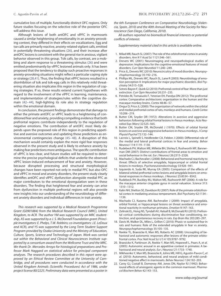
tcr
c
1
1
1
1
1
1
1
1
1
1
2
2
2
C. Agustín-Pavón et al. BIOL PSYCHIATRY 2012;72:266–272 271
cumulative loss of multiple, functionally distinct OFC regions. Onlyfuture studies focusing on the selective role of the posterior OFCwill address this issue.
Although lesions of both antOFC and vlPFC in marmosetscaused a similar heightening of emotionality in an anxiety-provok-ing context, they differed in their effects on vocalizations. Egg andtse calls are primarily reactive, anxiety-related vigilant calls, emittedin potentially threatening situations (26), and their increase afterantOFC lesions is consistent with the general rise in anxious, vigilantbehavior observed in this group. Tsik calls, by contrast, are a mob-bing and alarm response to a threatening stimulus (26) and wereemitted predominantly by vlPFC lesioned animals only. It has beenproposed that the tendency to display such proactive responses inanxiety-provoking situations might reflect a particular coping styleor strategy (28,41). Thus, the finding that vlPFC lesions resulted in adisinhibition of tsik and tsik-egg calls in this relatively mild threat-ening situation also implicates this region in the regulation of cop-ing strategies. If so, these results extend current hypotheses withregard to the involvement of the vlPFC in learning, maintenance,and implementation of strategies/rules within the cognitive do-main (42– 44), high-lighting its role also in strategy regulationwithin the emotional domain.
In conclusion, the present findings demonstrate that damage toeither the primate antOFC or vlPFC leads to a heightening of con-ditioned fear and anxiety, providing compelling evidence that bothprefrontal regions contribute independently to the regulation ofnegative emotions. The contribution of antOFC most likely de-pends upon the proposed role of this region in predicting appeti-tive and aversive outcomes and updating those predictions as en-vironmental contingencies change (45– 48). A disruption of thismechanism would certainly explain the more rigid fear responsesobserved in the present study and is likely to enhance anxiety bymaking fear predictions more ambiguous. The specific contributionof vlPFC is less clear, and future studies will be required to deter-mine the precise psychological deficits that underlie the observedvlPFC lesion-induced enhancement of fear and anxiety. However,because disrupted processing and structural morphologicalchanges have been reported not only in medial PFC but also OFCand vlPFC in mood and anxiety disorders, the present study clearlyidentifies antOFC and vlPFC dysfunction alongside medial PFC asmajor contributors to the emotional dysregulation seen in thesedisorders. The finding that heightened fear and anxiety can arisefrom dysfunction in multiple prefrontal regions will also providenew insights into our understanding of the neurobiology of differ-ent anxiety disorders and individual differences in trait anxiety.
This research was supported by a Medical Research ProgrammeGrant (G0901884) from the Medical Research Council (MRC), UnitedKingdom, to ACR. The author YM was supported by an MRC student-ship, AS was supported by a J. S. McDonnell Foundation grant (Princi-ple Investigators: E. Phelps, T.W. Robbins; co-investigators: J.E. LeDouxand ACR), and YS was supported by the Long Term Student SupportProgram provided by Osaka University and the Ministry of Education,Culture, Sports, Science and Technology of Japan. Work was carriedout within the Behavioral and Clinical Neurosciences Institute sup-ported by a consortium award from the Wellcome Trust and the MRC.We thank Dr. Mercedes Arroyo for histological preparations and Pro-fessor Mark Haggard on statistical advice for principal componentanalyses. The research procedures described in this report were ap-proved by an Ethical Review Committee at the University of Cam-bridge, and all procedures were conducted in accordance with theUnited Kingdom Animals (Scientific Procedures) Act of 1986, under
project license 80/2225. Preliminary data were presented as a poster inhe 6th European Conference on Comparative Neurobiology (Valen-ia, Spain, 2010) and the 40th Annual Meeting of the Society for Neu-oscience (San Diego, California, 2010).
All authors reported no biomedical financial interests or potentialonflicts of interest.
Supplementary material cited in this article is available online.
1. Milad MR, Rauch SL (2007): The role of the orbitofrontal cortex in anxietydisorders. Ann N Y Acad Sci 1121:546 –561.
2. Drevets WC (2001): Neuroimaging and neuropathological studies ofdepression: Implications for the cognitive-emotional features of mooddisorders. Curr Opin Neurobiol 11:240 –249.
3. Price JL, Drevets WC (2010): Neurocircuitry of mood disorders. Neuropsy-chopharmacology 35:192–216.
4. Phillips ML, Drevets WC, Rauch SL, Lane R (2003): Neurobiology of emo-tion perception II: Implications for major psychiatric disorders. Biol Psy-chiatry 54:515–528.
5. Sotres-Bayon F, Quirk GJ (2010): Prefrontal control of fear: More than justextinction. Curr Opin Neurobiol 20:231–235.
6. Petrides M, Tomaiuolo F, Yeterian EH, Pandya DN (2012): The prefrontalcortex: Comparative architectonic organization in the human and themacaque monkey brains. Cortex 48:46 –57.
7. Ongur D, Price JL (2000): The organization of networks within the orbitaland medial prefrontal cortex of rats, monkeys and humans. Cereb Cortex10:206 –219.
8. Butter CM, Snyder DR (1972): Alterations in aversive and aggressivebehaviors following orbital frontal lesions in rhesus monkeys. Acta Neu-robiol Exp (Wars) 32:525–565.
9. Butter CM, Snyder DR, McDonald J (1970): Effects of orbital frontallesions on aversive and aggressive behaviors in rhesus monkeys. J CompPhysiol Psychol 72:132–144.
0. Lacroix L, Spinelli S, Heidbreder CA, Feldon J (2000): Differential role ofthe medial and lateral prefrontal cortices in fear and anxiety. BehavNeurosci 114:1119 –1130.
1. Rudebeck PH, Walton ME, Millette BH, Shirley E, Rushworth MF, Banner-man DM (2007): Distinct contributions of frontal areas to emotion andsocial behaviour in the rat. Eur J Neurosci 26:2315–2326.
2. Machado CJ, Bachevalier J (2008): Behavioral and hormonal reactivity tothreat: Effects of selective amygdala, hippocampal or orbital frontallesions in monkeys. Psychoneuroendocrinology 33:926 –941.
3. Izquierdo A, Suda RK, Murray EA (2005): Comparison of the effects ofbilateral orbital prefrontal cortex lesions and amygdala lesions on emo-tional responses in rhesus monkeys. J Neurosci 25:8534 – 8542.
4. Rudebeck PH, Buckley MJ, Walton ME, Rushworth MF (2006): A role forthe macaque anterior cingulate gyrus in social valuation. Science 313:1310 –1312.
5. Kalin NH, Shelton SE, Davidson RJ (2007): Role of the primate orbitofron-tal cortex in mediating anxious temperament. Biol Psychiatry 62:1134 –1139.
6. Machado CJ, Kazama AM, Bachevalier J (2009): Impact of amygdala,orbital frontal, or hippocampal lesions on threat avoidance and emo-tional reactivity in nonhuman primates. Emotion 9:147–163.
7. Zelinski EL, Hong NS, Tyndall AV, Halsall B, McDonald RJ (2010): Prefron-tal cortical contributions during discriminative fear conditioning, ex-tinction, and spontaneous recovery in rats. Exp Brain Res 203:285–297.
8. Davis M, Walker DL, Miles L, Grillon C (2010): Phasic vs sustained fear inrats and humans: Role of the extended amygdala in fear vs anxiety.Neuropsychopharmacology 35:105–135.
9. Reekie YL, Braesicke K, Man MS, Roberts AC (2008): Uncoupling of be-havioral and autonomic responses after lesions of the primate orbito-frontal cortex. Proc Natl Acad Sci U S A 105:9787–9792.
0. Braesicke K, Parkinson JA, Reekie Y, Man MS, Hopewell L, Pears A, et al.(2005): Autonomic arousal in an appetitive context in primates: A be-havioural and neural analysis. Eur J Neurosci 21:1733–1740.
1. Mikheenko Y, Man MS, Braesicke K, Johns ME, Hill G, Agustin-Pavon C, etal. (2010): Autonomic, behavioral, and neural analyses of mild condi-tioned negative affect in marmosets. Behav Neurosci 124:192–203.
2. Carey GJ, Costall B, Domeney AM, Jones DN, Naylor RJ (1992): Behav-
ioural effects of anxiogenic agents in the common marmoset. Pharma-col Biochem Behav 42:143–153.www.sobp.org/journal

2
22
2
3
3
3
4
4
4
4
4
4
4
4
4
4
272 BIOL PSYCHIATRY 2012;72:266–272 C. Agustín-Pavón et al.
23. Howell DC (2002): Graduate-Level Statistics for Psychology and Neurosci-ence: ANOVA in Practice, and Complex ANOVA Designs. Belmont, CA: Dux-bury Press.
4. Cardinal R, Aitken MRF (2006): ANOVA for the Behavioural Sciences Re-searcher. Mahwah, NJ: Erlbaum Associates.
5. Field A (2009): Discovering Statistics Using SPSS. Los Angeles: SAGE.6. Bezerra B, Souto A (2008): Structure and usage of the vocal repertoire of
Callithrix jacchus. Int J Primatol 29:671–701.7. Koolhaas JM, Korte SM, De Boer SF, Van Der Vegt BJ, Van Reenen CG,
Hopster H, et al. (1999): Coping styles in animals: Current status inbehavior and stress-physiology. Neurosci Biobehav Rev 23:925–935.
28. Clara E, Tommasi L, Rogers LJ (2008): Social mobbing calls in commonmarmosets (Callithrix jacchus): Effects of experience and associatedcortisol levels. Anim Cogn 11:349 –358.
29. Grillon C, Lissek S, Rabin S, McDowell D, Dvir S, Pine DS (2008): Increasedanxiety during anticipation of unpredictable but not predictable aver-sive stimuli as a psychophysiologic marker of panic disorder. Am J Psy-chiatry 165:898 –904.
30. Grillon C, Pine DS, Lissek S, Rabin S, Bonne O, Vythilingam M (2009):Increased anxiety during anticipation of unpredictable aversive stimuliin posttraumatic stress disorder but not in generalized anxiety disorder.Biol Psychiatry 66:47–53.
31. Ghashghaei HT, Hilgetag CC, Barbas H (2007): Sequence of informationprocessing for emotions based on the anatomic dialogue betweenprefrontal cortex and amygdala. Neuroimage 34:905–923.
32. Morgan MA, LeDoux JE (1999): Contribution of ventrolateral prefrontalcortex to the acquisition and extinction of conditioned fear in rats.Neurobiol Learn Mem 72:244 –251.
33. Indovina I, Robbins TW, Nunez-Elizalde AO, Dunn BD, Bishop SJ (2011):Fear-conditioning mechanisms associated with trait vulnerability toanxiety in humans. Neuron 69:563–571.
34. Wager TD, Rilling JK, Smith EE, Sokolik A, Casey KL, Davidson RJ, et al.(2004): Placebo-induced changes in FMRI in the anticipation and expe-rience of pain. Science 303:1162–1167.
35. Drabant EM, Kuo JR, Ramel W, Blechert J, Edge MD, Cooper JR, et al.(2010): Experiential, autonomic, and neural responses during threatanticipation vary as a function of threat intensity and neuroticism. Neu-roimage 55:401– 410.
36. Ochsner KN, Ray RD, Cooper JC, Robertson ER, Chopra S, Gabrieli JD, et
al. (2004): For better or for worse: Neural systems supporting the cogni-www.sobp.org/journal
tive down- and up-regulation of negative emotion. Neuroimage23:483– 499.
7. Ray RD, Zald DH (2012): Anatomical insights into the interaction ofemotion and cognition in the prefrontal cortex. Neurosci Biobehav Rev36:479 –501.
8. Barbas H (2007): Flow of information for emotions through temporaland orbitofrontal pathways. J Anat 211:237–249.
9. Wall PM, Blanchard RJ, Yang M, Blanchard DC (2004): Differential effectsof infralimbic vs. ventromedial orbital PFC lidocaine infusions in CD-1mice on defensive responding in the mouse defense test battery and ratexposure test. Brain Res 1020:73– 85.
0. Kalin NH, Shelton SE, Davidson RJ (2004): The role of the central nucleusof the amygdala in mediating fear and anxiety in the primate. J Neurosci24:5506 –5515.
1. Coppens CM, de Boer SF, Koolhaas JM (2010): Coping styles and behav-ioural flexibility: Towards underlying mechanisms. Philos Trans R SocLond B Biol Sci 365:4021– 4028.
2. Bunge SA (2004): How we use rules to select actions: A review of evi-dence from cognitive neuroscience. Cogn Affect Behav Neurosci Review4:564 –579.
3. Dias R, Robbins TW, Roberts AC (1996): Dissociation in prefrontal cortexof affective and attentional shifts. Nature 380:69 –72.
4. Wallis JD, Dias R, Robbins TW, Roberts AC (2001): Dissociable contribu-tions of the orbitofrontal and lateral prefrontal cortex of the marmosetto performance on a detour reaching task. Eur J Neurosci 13:1797–1808.
5. Walton ME, Behrens TE, Buckley MJ, Rudebeck PH, Rushworth MF(2010): Separable learning systems in the macaque brain and the role oforbitofrontal cortex in contingent learning. Neuron 65:927–939.
6. Schoenbaum G, Takahashi Y, Liu TL, McDannald MA (2011): Does theorbitofrontal cortex signal value? Ann N Y Acad Sci 1239:87–99.
7. Murray EA, Izquierdo A (2007): Orbitofrontal cortex and amygdala con-tributions to affect and action in primates. Ann N Y Acad Sci 1121:273–296.
8. Morris JS, Dolan RJ (2004): Dissociable amygdala and orbitofrontal re-sponses during reversal fear conditioning. Neuroimage 22:372–380.
9. Burman KJ, Rosa MG (2009): Architectural subdivisions of medial andorbital frontal cortices in the marmoset monkey (Callithrix jacchus).
J Comp Neurol 514:11–29.