last of 2 articles on laparoscopic complications how to ... · adhesiolysis is a risky enterprise....
TRANSCRIPT
Variables that influence the risk of bowel injurypage 36
A review of the literature on intestinal complicationspage 38
How to protect the urinary tractpage 42
In thIs Article
34 OBG Management | October 2012 | Vol. 24 No. 10 obgmanagement.com
SurgicAl TecHniqueS
cASe Adhesions complicate multiple surgeriesIn early 2007, a 37-year-old woman with a his-
tory of hysterectomy, adhesiolysis, bilateral
partial salpingectomy, and cholecystectomy
underwent an attempted laparoscopic bilateral
salpingo-oophorectomy (BSO) for pelvic pain.
The operation was converted to laparotomy
because of severe adhesions and required
several hours to complete.
After the BSO, the patient developed
hydronephrosis in her left kidney second-
ary to an inflammatory cyst. In March 2007,
a urologist placed a ureteral stent to relieve
the obstruction. One month later, the patient
was referred to a gynecologic oncologist for
chronic pelvic pain.
On October 29, 2007, the patient under-
went operative laparoscopy for adhesiolysis
and appendectomy. No retroperitoneal explo-
ration was attempted at the time. According
to the operative note, the 10-mm port incision
was enlarged to 3 cm to enable the surgeon to
inspect the descending colon. Postoperatively,
the patient reported persistent abdominal pain
and fever and was admitted to the hospital for
observation. Although she had a documented
temperature of 102°F on October 31, with
tachypnea, tachycardia, and a white blood
cell (WBC) count of 2.9 x 10³/µL, she was dis-
charged home the same day.
The next morning, the patient returned
to the hospital’s emergency room (ER) report-
ing worsening abdominal pain and shortness
of breath. Her vital signs included a tempera-
ture of 95.8°F, heart rate of 135 bpm, respira-
tion of 32 breaths/min, and blood pressure
of 100/68 mm Hg. An examination revealed a
tender, distended abdomen, and the patient
exhibited guarding behavior upon palpation
in all quadrants. Bowel sounds were hypoac-
tive, and the WBC count was 4.2 x 10³/µL. No
differential count was ordered. A computed
tomography (CT) scan showed free air in the
abdomen, pneumomediastinum, and subcuta-
neous emphysema of the abdominal wall and
chest wall.
The next day, a differential WBC count
revealed bands elevated at a 25% level. A car-
diac consultant diagnosed heart failure and
Dr. Baggish practices Obstetrics and Gynecology at The Women’s Center at Saint Helena Hospital in Saint Helena, California. He also serves as Professor of Obstetrics and Gynecology at the University of California, San Francisco, and as Emeritus Chairman and Residency
Director, Department of Obstetrics and Gynecology, Good Samaritan Hospital, Cincinnati, Ohio.
Dr. Baggish reports no financial relationships relevant to this article.
lAST of 2 ArTicleS on lApAroScopic complicATionS
How to avoid intestinal and urinary tract injuries during gynecologic laparoscopy
By arming yourself with knowledge of the most common complications—and their causes—and employing well-chosen surgical strategies, you can lower the risk of laparoscopic-related morbidity and mortality
michael Baggish, mD
We lack definitive evidence that adhesions cause pelvic pain, or that adhesiolysis relieves such pain
OBG Management | October 2012 | Vol. 24 No. 1036 obgmanagement.com
SurgicAl TecHniqueS / gynecologic lApAroScopy
remarked that pneumomediastinum should
not occur after abdominal surgery. In the eve-
ning, the gynecologic oncologist performed a
laparotomy and observed enteric contents in
the abdominal cavity, as well as a defect of
approximately 2 mm in the lower portion of the
rectosigmoid colon. According to the opera-
tive note, the gynecologic oncologist stapled
off the area below the defect and performed a
descending loop colostomy.
Postoperatively, the patient remained
septic, and vegetable matter was recovered
from one of the drains, so a surgical consultant
was called. On November 9, a general surgeon
performed an exploratory laparotomy and
found necrosis, hemorrhage, acute inflamma-
tion of the colostomy, separation of the colos-
tomy from its sutured position on the anterior
abdominal wall, and mucosa at the end of the
Hartman pouch, necessitating resection of
this segment of the colon back to the rectum.
Numerous intra-abdominal abscesses were
also drained.
Two days later, the patient returned to the
OR for further abscess drainage and creation
of a left end colostomy. She was discharged
1 month later.
On January 4, 2008, she went to the ER
for nausea and abdominal pain. Five days
later, a plastic surgeon performed extensive
skin grafting on the chronically open abdomi-
nal wound. On March 12, the patient returned
to the ER because of abdominal pain and
was admitted for nasogastric drainage and
intravenous (IV) fluids. She returned to the ER
again on April 26, reporting pain. A CT scan
revealed a cystic mass in the pelvis, which was
drained under CT guidance. In June and July,
the patient was seen in the ER three times for
pain, nausea, and vomiting.
In January 2009, she underwent another
laparotomy for takedown of the colostomy,
lysis of adhesions, and excision of a left 4-cm
pelvic cyst (pathology later revealed the cyst to
be ovarian tissue). She also underwent a left-
sided myocutaneous flap reconstruction of an
abdominal wall defect, and a right-sided myo-
cutaneous flap with placement of a 16 x 20–cm
sheet of AlloDerm Tissue Matrix (LifeCell). She
continues to experience abdominal pain and
visits the ER for that reason. In March 2009,
she underwent repeat drainage of a pelvic col-
lection via CT imaging. No further follow-up is
available.
could this catastrophic course have been avoided? What might have prevented it?
Adhesions are likely after any abdominal procedure
The biggest risk factor for laparoscopy-related intestinal injury is the presence of pelvic or abdominal adhesions.1,2
Adhesions inevitably form after any intra-abdominal surgery, and new adhesions are likely with each successive intra-abdominal procedure. Even adhesiolysis leads to the formation of adhesions postoperatively.
Few reliable data suggest that adhe-sions cause pelvic pain, or that adhesiolysis relieves such pain.3 Furthermore, it may be impossible to predict with reasonable prob-ability where adhesions may be located pre-operatively or to know with certainty whether a portion of the intestine is adherent to the anterior abdominal wall directly below the usual subumbilical entry site. Because of the likelihood of adhesions in a patient who has undergone two or more laparotomies, it is risky to thrust a 10- to 12-mm trocar through the anterior abdominal wall below the navel.
A few variables influence the risk of injuryThe trocar used in laparoscopic procedures plays a role in the risk of bowel injury. For ex-ample, relatively dull reusable devices may push nonfixed intestine away rather than penetrate the viscus. In contrast, razor-sharp disposable devices are more likely to cut into the underlying bowel.
Body habitus is also important. The obese woman is at greater risk for entry in-juries, owing to physical aspects of the fatty anterior abdominal wall. When force is ap-plied to the wall, it moves inward, toward the posterior wall, trapping intestine. In a thin woman, the abdominal wall is less elastic, so there is less excursion upon trocar entry.
Read Dr. Baggish’s first article on laparoscopic complications
›› How to avoid major vessel injury during gynecologic laparoscopyMichael Baggish, MD (Surgical Techniques, August 2012)
intestinal status is another variable to consider. A collapsed bowel is unlikely to be perforated by an entry trocar, whereas a thin, distended bowel is vulnerable to injury. Bow-el status can be determined preoperatively using various modalities, including radio-graphic studies.
Careful surgical technique is impera-tive. Sharp dissection is always preferable to the blunt tearing of tissue, particularly in cases involving fibrous adhesions. Tear-ing a dense, unyielding adhesion is likely to remove a piece of intestinal wall because the tensile strength of the adhesion is typically greater than that of the viscus itself.
Thorough knowledge of pelvic anatomy is essential. It would be particularly egregious for a surgeon to mistake an adhesion for the normal peritoneal attachments of the left and sigmoid colon, or to resect the mesentery of the small bowel, believing it to be an adhesion.
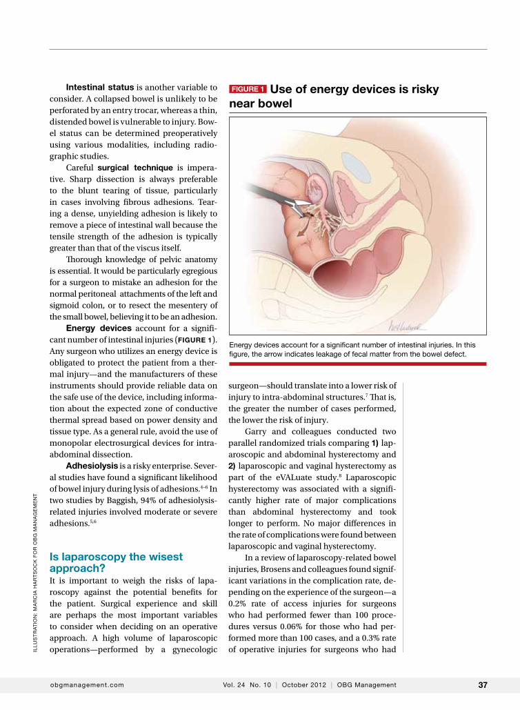
energy devices account for a signifi-cant number of intestinal injuries (figure 1). Any surgeon who utilizes an energy device is obligated to protect the patient from a ther-mal injury—and the manufacturers of these instruments should provide reliable data on the safe use of the device, including informa-tion about the expected zone of conductive thermal spread based on power density and tissue type. As a general rule, avoid the use of monopolar electrosurgical devices for intra-abdominal dissection.
Adhesiolysis is a risky enterprise. Sever-al studies have found a significant likelihood of bowel injury during lysis of adhesions.4–6 In two studies by Baggish, 94% of adhesiolysis-related injuries involved moderate or severe adhesions.5,6
is laparoscopy the wisest approach?It is important to weigh the risks of lapa-roscopy against the potential benefits for the patient. Surgical experience and skill are perhaps the most important variables to consider when deciding on an operative approach. A high volume of laparoscopic operations—performed by a gynecologic
surgeon—should translate into a lower risk of injury to intra-abdominal structures.7 That is, the greater the number of cases performed, the lower the risk of injury.
Garry and colleagues conducted two parallel randomized trials comparing 1) lap-aroscopic and abdominal hysterectomy and 2) laparoscopic and vaginal hysterectomy as part of the eVALuate study.8 Laparoscopic hysterectomy was associated with a signifi-cantly higher rate of major complications than abdominal hysterectomy and took longer to perform. No major differences in the rate of complications were found between laparoscopic and vaginal hysterectomy.
In a review of laparoscopy-related bowel injuries, Brosens and colleagues found signif-icant variations in the complication rate, de-pending on the experience of the surgeon—a 0.2% rate of access injuries for surgeons who had performed fewer than 100 proce-dures versus 0.06% for those who had per-formed more than 100 cases, and a 0.3% rate of operative injuries for surgeons who had
obgmanagement.com Vol. 24 No. 10 | October 2012 | OBG Management 37
Energy devices account for a significant number of intestinal injuries. In this figure, the arrow indicates leakage of fecal matter from the bowel defect.
figure 1 use of energy devices is risky near bowel
Ill
US
TR
AT
ION
: M
AR
CIA
HA
RT
SO
Ck
FO
R O
BG
MA
NA
GE
ME
NT
performed fewer than 100 procedures versus 0.04% for more experienced surgeons.7
A few precautions can improve the safety of laparoscopyIf adhesions are known or suspected, pri-mary laparoscopic entry should be planned for a site other than the infra-umbilical area. Options include:• entry via the left hypochondrium in the
midclavicular line• an open procedure.However, open laparoscopic entry does not always avert intestinal injury.9–11
If the anatomy is obscured once the ab-domen has been entered safely, retroperito-neal dissection may be useful, particularly for exposure of the left colon. When it is un-clear whether a structure to be incised is a loop of bowel or a distended, adherent ovi-duct, it is best to refrain from cutting it.
For adhesiolysis, traction and counter-traction are the techniques of choice. Dis-section of intestine should always be parallel to the axis of the viscus. Remember, too, that the blood supply enters via the mesenteric margin of the intestine.
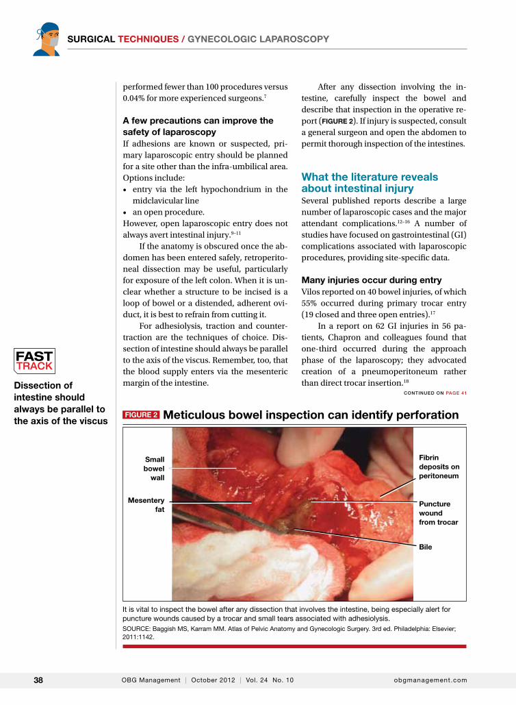
After any dissection involving the in-testine, carefully inspect the bowel and describe that inspection in the operative re-port ( figure 2). If injury is suspected, consult a general surgeon and open the abdomen to permit thorough inspection of the intestines.
What the literature reveals about intestinal injurySeveral published reports describe a large number of laparoscopic cases and the major attendant complications.12–16 A number of studies have focused on gastrointestinal (GI) complications associated with laparoscopic procedures, providing site-specific data.
many injuries occur during entryVilos reported on 40 bowel injuries, of which 55% occurred during primary trocar entry (19 closed and three open entries).17
In a report on 62 GI injuries in 56 pa-tients, Chapron and colleagues found that one-third occurred during the approach phase of the laparoscopy; they advocated creation of a pneumoperitoneum rather than direct trocar insertion.18
It is vital to inspect the bowel after any dissection that involves the intestine, being especially alert for puncture wounds caused by a trocar and small tears associated with adhesiolysis. SOURCE: Baggish MS, karram MM. Atlas of Pelvic Anatomy and Gynecologic Surgery. 3rd ed. Philadelphia: Elsevier; 2011:1142.
Dissection of intestine should always be parallel to the axis of the viscus
OBG Management | October 2012 | Vol. 24 No. 1038 obgmanagement.com
SurgicAl TecHniqueS / gynecologic lApAroScopy
figure 2 meticulous bowel inspection can identify perforation
Small bowel
wall
mesentery fat
Bile
fibrin deposits on peritoneum
puncture wound from trocar
CONTINuED ON PAgE 41
postoperative findings of tachycardia, tachypnea, elevated leukocyte count, and bandemia suggest sepsis syndrome
obgmanagement.com Vol. 24 No. 10 | October 2012 | OBG Management 41
SurgicAl TecHniqueS / gynecologic lApAroScopy
In a report from the Netherlands, 24 of 29 GI injuries occurred during the approach.2
In a review of 63 GI complications relat-ed to diagnostic and operative laparoscopy, 75% of injuries were associated with primary trocar insertion.19
Optical access trocars do not appear to be protective against bowel injury. One study of 79 complications associated with these devices found 24 bowel injuries.20
In addition, in two reports detailing 130 cases of small- and large-bowel perfo-rations associated with laparoscopic pro-cedures, Baggish found that 62 (77%) of small-bowel injuries and 20 (41%) of colonic injuries were entry-related.5,6
energy devices can be problematicIn the study by Chapron and colleagues of 62 GI injuries, six were secondary to the use of electrosurgical devices, four of them in-volving monopolar instruments.18
In a study from Scotland, 27 of 117 (23%) of bowel injuries during laparoscopic proce-dures were attributable to a thermal event.21
Baggish found that 43% of operative injuries among 130 intestinal perforations were energy-related.5,6
intraoperative diagnosis is optimalSoderstrom reviewed 66 cases of laparoscopy- related bowel injuries and found three deaths attributable to a delay in diagnosis exceeding 72 hours.4
In a study by Vilos, the mean time for di-agnosis of bowel injuries was 4 days (range, 0–23 days), with intraoperative diagnosis in only 35.7% of cases.17
In a Finnish nationwide analysis of lapa-roscopic complications, Harkki-Siren and Kurki found that small-bowel injuries were identified an average of 3.3 days after oc-currence; when electrosurgery was involved in the injury, the average time to diagnosis was 4.8 days.22 As for large-bowel injuries, 44% were identified intraoperatively. In the remainder of cases, the average time from injury to diagnosis was 10.4 days for electro-surgical injuries and 1.3 days for injuries re-lated to sharp dissection.
In the studies by Baggish, 82 of 130 (63%) intestinal injuries were diagnosed 48 hours or more after the operation.5,6
Baggish also made the following obser-vations:• The most common symptoms of in-
testinal injury were (in order of frequen-cy) abdominal pain, bloating, nausea and vomiting, and fever or chills (or both). The most common signs were abdominal tenderness, abdominal distension, dimin-ished bowel sounds, and elevated or sub-normal temperature.
• Sepsis was apparent (due to the onset of systemic inflammatory response syn-drome) in the majority of small-bowel perforations and virtually all colonic perforations. Findings of tachycardia, tachypnea, elevated leukocyte count, and bandemia suggested sepsis syndrome.
• radiologically observed free air was often misinterpreted by the radiologist as being consistent with residual gas from the initial laparoscopy. In reality, most—if not all—CO
2 gas is absorbed within
24 hours, particularly in obese women. Early CT imaging with oral contrast leads to the most expeditious, correct diagnosis, compared with flat and upright abdominal radiographs.
• obese women did not exhibit rebound tenderness even though subsequent op-erative findings revealed extensive and se-vere peritonitis.
• When infection occurred, it usually was polymicrobial in nature. The most frequently cultured organisms include Escherichia coli, Enterococcus, alpha and beta Streptococcus, Staphylococcus, and Bacteroides.
Baggish concluded that earlier diagno-sis could be achieved with careful inspection of the intestine at the conclusion of each op-erative procedure (figure 2, page 38).
Similarly, Chapron and colleagues recommended meticulous inspection of all areas where bowel lysis has been per-formed. “When there is the slightest doubt, carry out tests for leakage (transanal injec-tion of 200 mL methylene blue using a Foley
risk factors for urinary tract injury include previous cesarean delivery, multiple fibroids, and severe endometriosis
OBG Management | October 2012 | Vol. 24 No. 1042 obgmanagement.com
SurgicAl TecHniqueS / gynecologic lApAroScopy
catheter) in order not to overlook a rectosig-moid injury which would become apparent secondarily in a context of peritonitis,” they wrote. They also suggested that the patient be educated about the signs and symptoms of intestinal injury.18
Whenever a bowel injury is visualized intraoperatively, assume that it is transmural until it is proved otherwise.
How to avoid urinary tract injuries Along with major vessel injury and intestinal perforation, bladder and ureteral injuries are the most common complications of laparo-scopic surgery. Although urinary tract inju-ries are rarely fatal, they can cause a range of sequelae, including urinoma, vesicovagi-nal and ureterovaginal fistulas, hydroureter, hydronephrosis, renal damage, and kidney atrophy.
The incidence of ureteral injury dur-ing laparoscopy ranges from less than 0.1% to 1.0%, and the incidence of bladder injury ranges from less than 0.8% to 2.0%.23–26 Investigators in Singapore described eight urologic injuries among 485 laparoscopic hysterectomies and identified several risk factors:• previous cesarean delivery• multiple fibroids• severe endometriosis.27 Another set of investigators found a history of laparotomy to be a risk factor for bladder injury during laparoscopic hysterectomy.28
Rooney and colleagues studied the effect of previous cesarean delivery on the risk of injury during hysterectomy.29 Among 5,092 hysterectomies—including 433 laparoscopic-assisted vaginal hysterec-tomies, 3,140 abdominal procedures, and 1,539 vaginal operations—the rate of blad-der injury varied by approach. Cystotomy was observed in 0.76% of abdominal hyster-ectomies (33% had a previous cesarean de-livery), 1.3% of vaginal procedures (21% had a previous cesarean), and 1.8% of laparo-scopic operations (62.5% had a previous ce-sarean). The odds ratio for cystotomy during
hysterectomy among women with a previ-ous cesarean delivery was 1.26 for the ab-dominal approach, 3.00 for the vaginal route, and 7.50 for laparoscopic-assisted vaginal hysterectomy.29
Two studies highlight common aspects of injuryIn a recent report of 75 urinary tract injuries associated with laparoscopic surgery, Bag-gish identified a total of 33 injuries involving the bladder and 42 of ureteral origin. Twelve of the bladder injuries were associated with the approach, and 21 were related to the sur-gery. In contrast, only one of the 42 ureteral injuries was related to the approach.30
Baggish also found that just under 50% of urinary tract injuries were related to the use of thermal energy, including all three vesicovaginal fistulas. Fourteen bladder lac-erations occurred during separation of the bladder from the uterus during laparoscopic hysterectomy.30
Common sites of injury were at the in-fundibulopelvic ligament, between the in-fundibulopelvic ligament and the uterine vessels, and at or below the uterine vessels.30
None of the 42 ureteral injuries were di-agnosed intraoperatively. In fact, 37 of these injuries were not correctly diagnosed until more than 48 hours after surgery. Two utero-vaginal fistulas were also diagnosed in the late postoperative period.30
Bladder injuries were identified via cys-toscopy or cystometrogram or by the instilla-tion of methylene blue into the bladder, with observation from above for leakage. Ureteral injuries were identified by IV pyelogram, retrograde pyelogram, or attempted pas-sage of a stent. Every ureteral injury showed up as hydroureter and hydronephrosis via pyelography.30
Grainger and colleagues reported five ureteral injuries associated with laparoscop-ic procedures.31 The principal symptoms were low back pain, abdominal pain, leuko-cytosis, and peritonitis. All five injuries were associated with endometriosis surgery, most commonly near the uterosacral ligaments.
Grainger and colleagues cited eight
peristalsis is not an indication of ureteral integrity. in fact, an obstructed ureter will pulsate more vigorously than a normal one.
obgmanagement.com Vol. 24 No. 10 | October 2012 | OBG Management 43
additional cases of injury. Three patients among the 13 total cases lost renal function, and two eventually required nephrectomy.31
How to prevent, identify, and manage urinary tract injuriesThorough knowledge of anatomy and me-ticulous technique are imperative to prevent urinary tract injuries. Strategies include:• Use sharp rather than blunt dissection.• Know the risk factors for urinary tract inju-
ry, which include previous cesarean deliv-ery or intra-abdominal surgery, presence of adhesions, and deep endometriosis.
• Be aware of the dangers posed by energy devices when they are used near the blad-der and ureter. Even bipolar devices can cause thermal injury.
• Employ hydrodissection when there are bladder adhesions, and work nearer the uterus or vagina than the bladder, leaving a margin of tissue.
• When the ureter’s location is unclear rela-tive to the operative site, do not hesitate to open the retroperitoneal space to observe the ureter. If necessary, dissect the ureter distally.
• Perform cystoscopy with IV indigo car-mine injection at the conclusion of surgery to ensure that the ureter is not occluded.
• Be aware that peristalsis is not an indi-cation of ureteral integrity. In fact, an obstructed ureter will pulsate more vigor-ously than a normal one.
• Consider preoperative ureteral catheter-ization, which may avert injury without increasing operative time, blood loss, and hospital stay,32 although the data are not definitive.33
• Be vigilant. Early identification of inju-ries reduces morbidity. In the case of ureteral obstruction, immediate stenting will usually obviate the need for ureteral implantation and nephrostomy if the ob-struction is not complete.
• Intervene early to cut an obstructing su-ture or relieve ureteral bowing. Doing so may eliminate the obstruction altogether in many cases.
• If a laceration is found in the bladder
trigone or its vicinity, always perform ure-teral catheterization to help prevent the inadvertent suturing of the intravesical ureter into the repair.
• After repair of a bladder laceration, per-form cystoscopy with IV injection of in-digo carmine to ensure ureteral integrity.
• Use only absorbable suture in bladder re-pairs. I recommend 2-0 chromic catgut for the first layer, which should encompass muscularis and mucosa. Place a second layer of sutures using 3-0 polyglactin 910 (Vicryl), imbricating the first layer.
• After completion of a bladder repair, in-still a solution of diluted methylene blue (1 part methylene blue to 100 parts sterile water or saline) to distend the bladder, and carefully inspect the closure to ensure that it is watertight. Then place a Foley catheter for a minimum of 2 weeks. Four to 6 weeks after repair, perform a cystogram to ensure that healing is complete, with no leakage.
• Call a urologist if you are not well-versed in bladder repair, or if the ureter is injured (or injury is suspected).
• Watch for fistula formation, an inevitable outcome of untreated bladder and ureteral injury, which may occur early or late in the postoperative course.
choose an approach wiselyLaparoscopy is a learned skill. Supervised practice generally leads to greater levels of proficiency, and repetition of the same operations improves dexterity and execution. However, laparoscopy is also an art—some people have the touch and some do not.
Although laparoscopic techniques offer many advantages, they also have shortcom-ings. The complications described here, and the strategies I have offered for preventing and managing them, should help gyneco-logic surgeons determine whether laparos-copy is the optimal route of operation, based on surgical experience, characteristics of the individual patient, and other variables.
references1. Brill AW, Nezhat F, Nezhat CH, et al. The incidence of
adhesions after prior laparotomy: a laparoscopic appraisal. CONTINuED ON PAgE 44
OBG Management | October 2012 | Vol. 24 No. 1044 obgmanagement.com
SurgicAl TecHniqueS / gynecologic lApAroScopy
Obstet Gynecol. 1995;85(2):269–279.2. Jansen FW, Kapiteyn K, Trimbos-Kemper T, et al.
Complications of laparoscopy: a prospective multicenter observational study. Br J Obstet Gynecol. 1997;104(5):595–600.
3. Hammoud A, Gago A, Diamond M. Adhesions in patients with chronic pelvic pain: a role for adhesiolysis? Fertil Steril. 2004;82(6):1483–1491.
4. Soderstrom RM. Bowel injury litigation after laparoscopy. J Am Assoc Gynecol Laparosc. 1993;1(1):74–73.
5. Baggish MS. How to avoid injury to bowel during laparoscopy. OBG Manage. 2008;20(7):47–60.
6. Baggish MS. One hundred and thirty small- and large-bowel injuries associated with gynecologic laparoscopic operations. J Gynecol Surg. 2007;23(3):83–95.
7. Brosens I, Gordon A, Campo R, et al. Bowel injury in gynecologic laparoscopy. J Am Assoc Gynecol Laparosc. 2003;10(1):9–13.
8. Garry R, Fountain J, Mason S, et al. The eVALuate study: two parallel randomized trials, one comparing laparoscopic with abdominal hysterectomy, the other comparing laparoscopic with vaginal hysterectomy. BMJ. 2004; 328(7432):129.
9. Chapron C, Querleu D, Bruhat MA, et al. Surgical complications of diagnostic and operative gynecological laparoscopy: a series of 29966 cases. Hum Reprod. 1998;13(4):867–872.
10. Jansen FW, Kolkman W, Bakkum EA, et al. Complications of laparoscopy: an inquiry about closed versus open-entry technique. Am J Obstet Gynecol. 2004;190(3):634–638.
11. Shirk GJ, Johns A, Redwine DB. Complications of laparoscopic surgery: how to avoid them and how to repair them. J Minim Invasive Surg. 2006;13(4):352–359.
12. Fuller J, Binita AS, Carey-Corrado J. Trocar-associated injuries and fatalities: an analysis of 1399 reports to the FDA. J Min Invasive Gynecol. 2005;12(4):302–307.
13. Saidi MH, Vancaille TG, White J, et al. Complications of major operative laparoscopy. Obstet Gynecol Surv. 1996;51(11):661–662.
14. Makinen J, Johansson J, Tomas C, et al. Morbidity of 10,110 hysterectomies by type of approach. Hum Reprod. 2001;16(7):1473–1478.
15. Bhoyrul S, Vierra MA, Nezhat CR, et al. Trocar injuries in laparoscopic surgery. J Am Coll Surg. 2001;192(6):677–683.
16. Bateman BG, Kolp LA, Hoeger K. Complications of laparoscopy—operative and diagnostic. Fertil Steril. 1996;66(1):30–35.
17. Vilos GA. Laparoscopic bowel injuries: forty litigated gynecological cases in Canada. J Obstet Gynaecol Canada. 2002;24(3):224–230.
18. Chapron C, Harchaoui Y, Lacroix S, et al. Gastrointestinal injuries during gynecological laparoscopy. Hum Reprod. 1999;14(2):333–337.
19. Champault G, Cazacu F, Taffinder N. Serious trocar accidents in laparoscopic surgery: a French survey of 103,852 operations. Surg Laparosc Endosc. 1996;6(5):367–370.
20. Sharp HT, Dodson MK, Draper ML, et al. Complications associated with optical-access laparoscopic trocars. Obstet Gynecol. 2002;99(4):553–555.
21. Brown CJA, Chamberlain GVP, Jordan JA, et al. Gynecological laparoscopy: the report of the Working Party of the Confidential Enquiry into Gynecological Laparoscopy. Br J Obstet Gynaecol. 1978;85:401–403.
22. Harkki-Siren P, Kurki T. A nationwide analysis of laparoscopic complications. Obstet Gynecol. 1997;89(1):108–112.
23. Tamussino KF, Lang PF, Breinl E. Ureteral complications with operative gynecologic laparoscopy. Am J Obstet Gynecol. 1998;178(5):967–970.
24. Aslan P, Brooks A, Drummond M, et al. Incidence and management of gynecological related ureteric injuries. Aust N Z J Obstet Gynecol. 1999;39(2):178–181.
25. Wang PH, Lee WL, Yuan CC, et al. Major complications of operative and diagnostic laparoscopy for gynecologic disease. J Am Assoc Gynecol Laparosc, 2001;8(1):68–73.
26. Oh BR, Kwon DD, Park KS, et al. Late presentation of ureteral injury after laparoscopic surgery. Obstet Gynecol. 2000;95(3):337–339.
27. Siow A, Nikam YA, Ng C. et al. Urological complications of laparoscopic hysterectomy: a four year review at KK women’s and children’s hospital, Singapore. Singapore Med J. 2007;48(3):217–221.
28. Lafay PMC, Leonard F, Chopin N, et al. Incidence and risk factors of bladder injuries during laparoscopic hysterectomy indicated for benign pathologies: a 14.5 years experience in a continuous series of 1501 procedures. Hum Reprod. 2009;24(4):842–849.
29. Rooney CM, Crawford AT, Vassallo BJ, et al. Is previous cesarean section a risk for incidental cystotomy at the time of hysterectomy? A case-controlled study. Am J Obstet Gynecol. 2005;193(6):2041–2044.
30. Baggish MS. Urinary tract injuries secondary to gynecologic laparoscopic surgery: analysis of seventy-five cases. J Gynecol Surg. 2010;26(2):79–92.
31. Grainger DA, Soderstrom RM, Schiff SF, et al. Ureteral injuries at laparoscopy: insights into diagnosis, management, and prevention. Obstet Gynecol. 1990;75(5):839–843.
32. Chapron C, Dubuisson JB, Ansquer Y, et al. Bladder injuries during total laparoscopic hysterectomy: diagnosis, management and prevention. J Gynecol Surg. 1995; 11(2):95–98.
33. Kuno K, Menzin A, Kauder HH, et al. Prophylactic ureteral catheterization in gynecologic surgery. Urology. 1998;52(6):1004–1008.
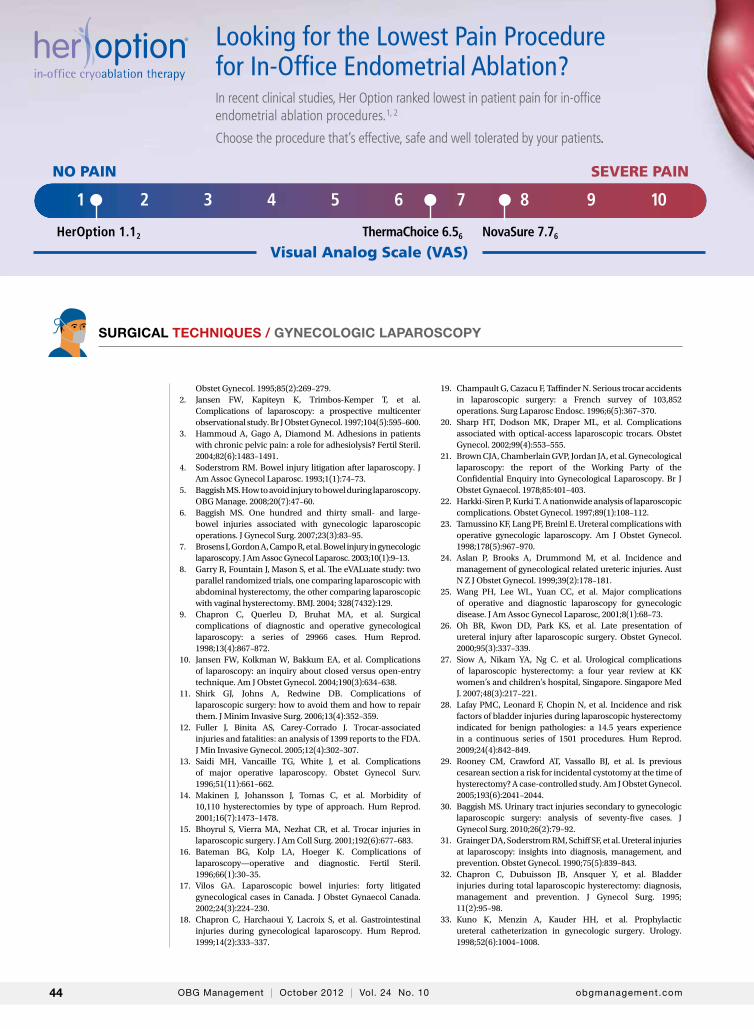
Looking for the Lowest Pain Procedure for In-Office Endometrial Ablation?In recent clinical studies, Her Option ranked lowest in patient pain for in-office endometrial ablation procedures.1, 2
Choose the procedure that’s effective, safe and well tolerated by your patients.
Visual Analog Scale (VAS)HerOption 1.12 ThermaChoice 6.56 NovaSure 7.76
NO PAIN SEVERE PAIN
1 2 3 4 5 6 7 8 9 10
HerOption1/3pg:Layout 1 12/21/11 11:03 AM Page 1
Maximum Patient Comfort1,2
Lowest Complication Rate3
Shown Actual Size Ultra-Slim 5.5 mm Probe
The Best Choice for In-Office Endometrial Ablation Procedures• Exceptional patient outcomes...high patient satisfaction4
• Short and efficient total treatment time from pre-procedure to recovery
• Sub-zero temperature provides a natural analgesic effect5
• No intravenous sedation required
To find out how Her Option can benefit your practice...and your patients, call 800.243.2974 or visit www.HerOption.com
82075 Rev. 12/11© 2011 CooperSurgical, Inc.
Scan. Watch. Learn.about the Her Option Procedure
1, 2, 3, 4, 5 For reference details see http://www.coopersurgical.com/Documents/HerOptionBrochure.pdf6 Clark et al; Bipolar Radiofrequency Compaired with Thermal Balloon Endometrial Ablation in the Office; Obstetrics & Gynecology; Jan 2011
81788HerOptionOBGM.qrk:Layout 1 12/21/11 11:01 AM Page 1