large femoral-neck cysts in association with femoroacetabular impingement by klaus-peter günther,...
TRANSCRIPT
Large Femoral-Neck Cysts in Association with Femoroacetabular Impingement
by Klaus-Peter Günther, Albrecht Hartmann, Peter Aikele, Daniela Aust, and Jörg Ziegler
J Bone Joint Surg AmVolume 89(4):863-870
April 1, 2007
©2007 by The Journal of Bone and Joint Surgery, Inc.

Figs. 1-A, 1-B, and 1-C A thirteen-year-old boy with unilateral stable slipped capital femoral epiphysis of the right hip had been treated with in-situ screw fixation at another institution in
1983.
Klaus-Peter Günther et al. J Bone Joint Surg Am 2007;89:863-870
©2007 by The Journal of Bone and Joint Surgery, Inc.
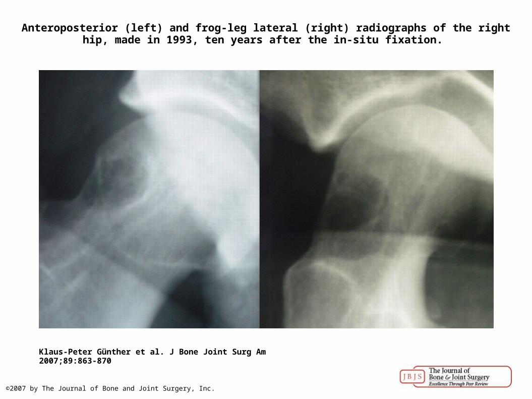
Anteroposterior (left) and frog-leg lateral (right) radiographs of the right hip, made in 1993, ten years after the in-situ fixation.
Klaus-Peter Günther et al. J Bone Joint Surg Am 2007;89:863-870
©2007 by The Journal of Bone and Joint Surgery, Inc.
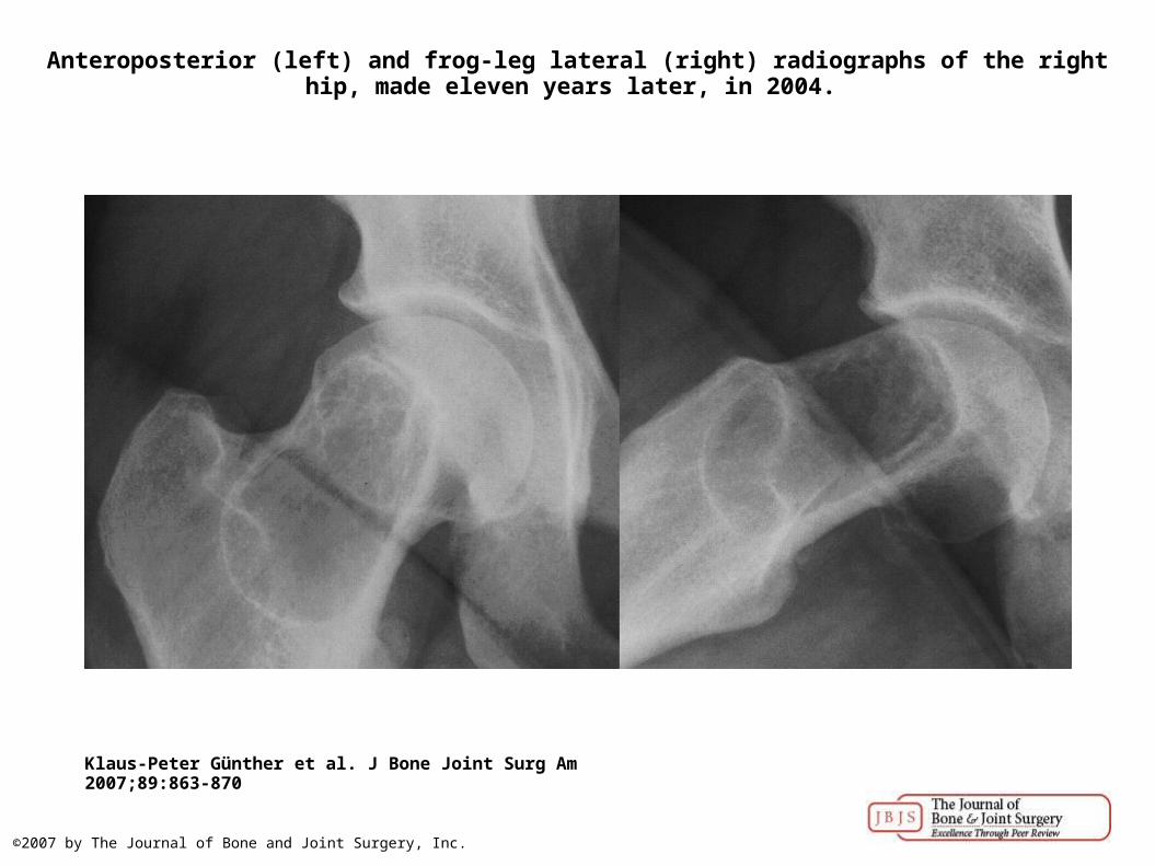
Anteroposterior (left) and frog-leg lateral (right) radiographs of the right hip, made eleven years later, in 2004.
Klaus-Peter Günther et al. J Bone Joint Surg Am 2007;89:863-870
©2007 by The Journal of Bone and Joint Surgery, Inc.
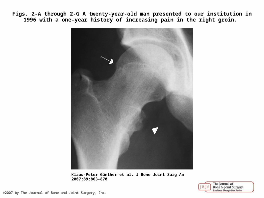
Figs. 2-A through 2-G A twenty-year-old man presented to our institution in 1996 with a one-year history of increasing pain in the right groin.
Klaus-Peter Günther et al. J Bone Joint Surg Am 2007;89:863-870
©2007 by The Journal of Bone and Joint Surgery, Inc.
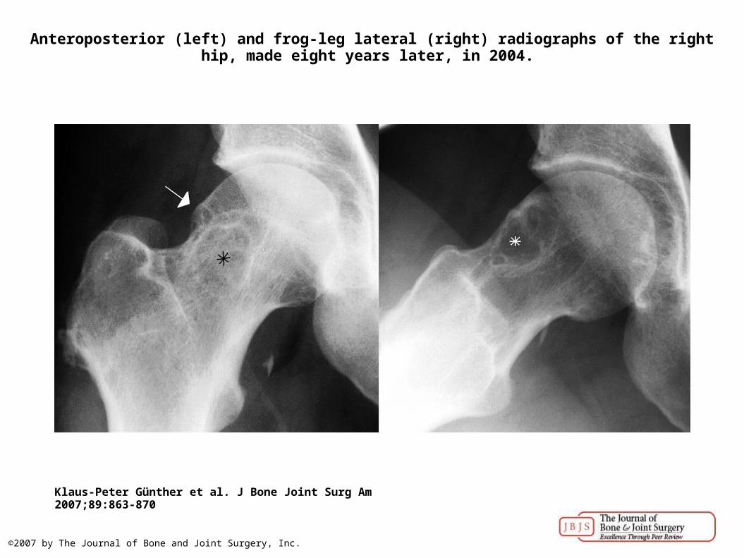
Anteroposterior (left) and frog-leg lateral (right) radiographs of the right hip, made eight years later, in 2004.
Klaus-Peter Günther et al. J Bone Joint Surg Am 2007;89:863-870
©2007 by The Journal of Bone and Joint Surgery, Inc.
Klaus-Peter Günther et al. J Bone Joint Surg Am 2007;89:863-870
©2007 by The Journal of Bone and Joint Surgery, Inc.
Klaus-Peter Günther et al. J Bone Joint Surg Am 2007;89:863-870
©2007 by The Journal of Bone and Joint Surgery, Inc.
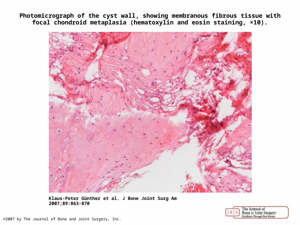
Photomicrograph of the cyst wall, showing membranous fibrous tissue with focal chondroid metaplasia (hematoxylin and eosin staining, ×10).
Klaus-Peter Günther et al. J Bone Joint Surg Am 2007;89:863-870
©2007 by The Journal of Bone and Joint Surgery, Inc.
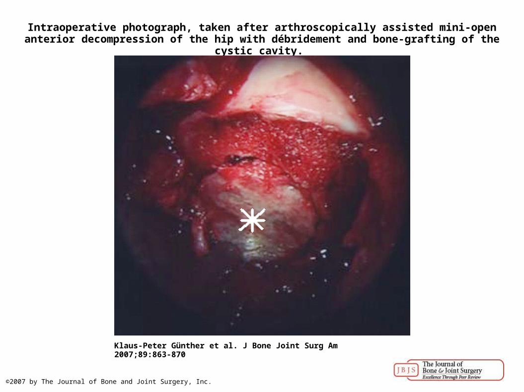
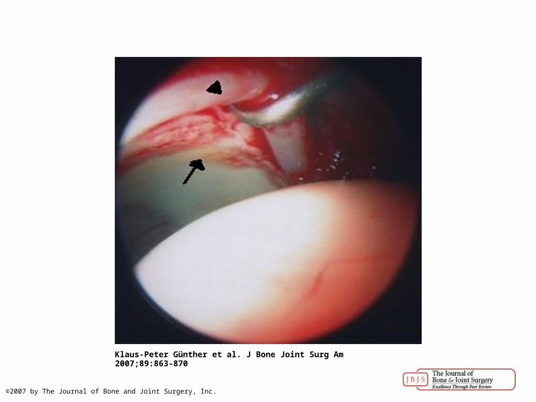
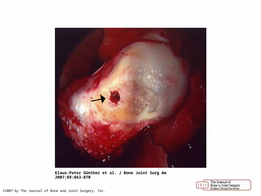
Intraoperative photograph, taken after arthroscopically assisted mini-open anterior decompression of the hip with débridement and bone-grafting of the cystic cavity.
Klaus-Peter Günther et al. J Bone Joint Surg Am 2007;89:863-870
©2007 by The Journal of Bone and Joint Surgery, Inc.
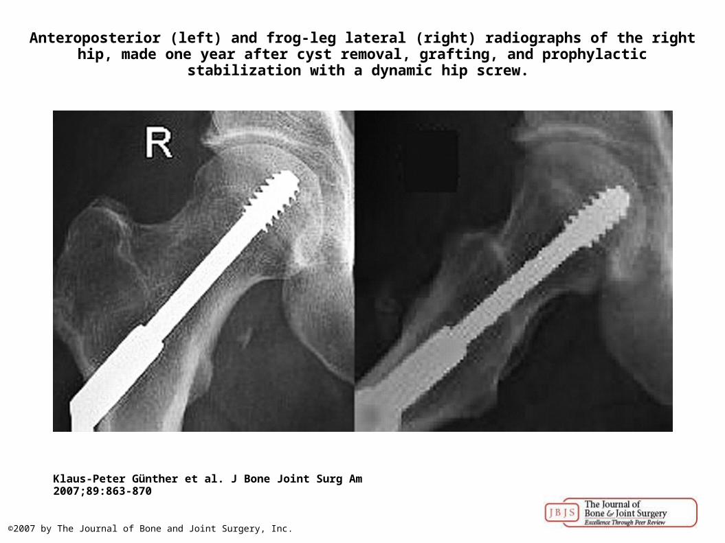
Anteroposterior (left) and frog-leg lateral (right) radiographs of the right hip, made one year after cyst removal, grafting, and prophylactic stabilization with a dynamic hip screw.
Klaus-Peter Günther et al. J Bone Joint Surg Am 2007;89:863-870
©2007 by The Journal of Bone and Joint Surgery, Inc.
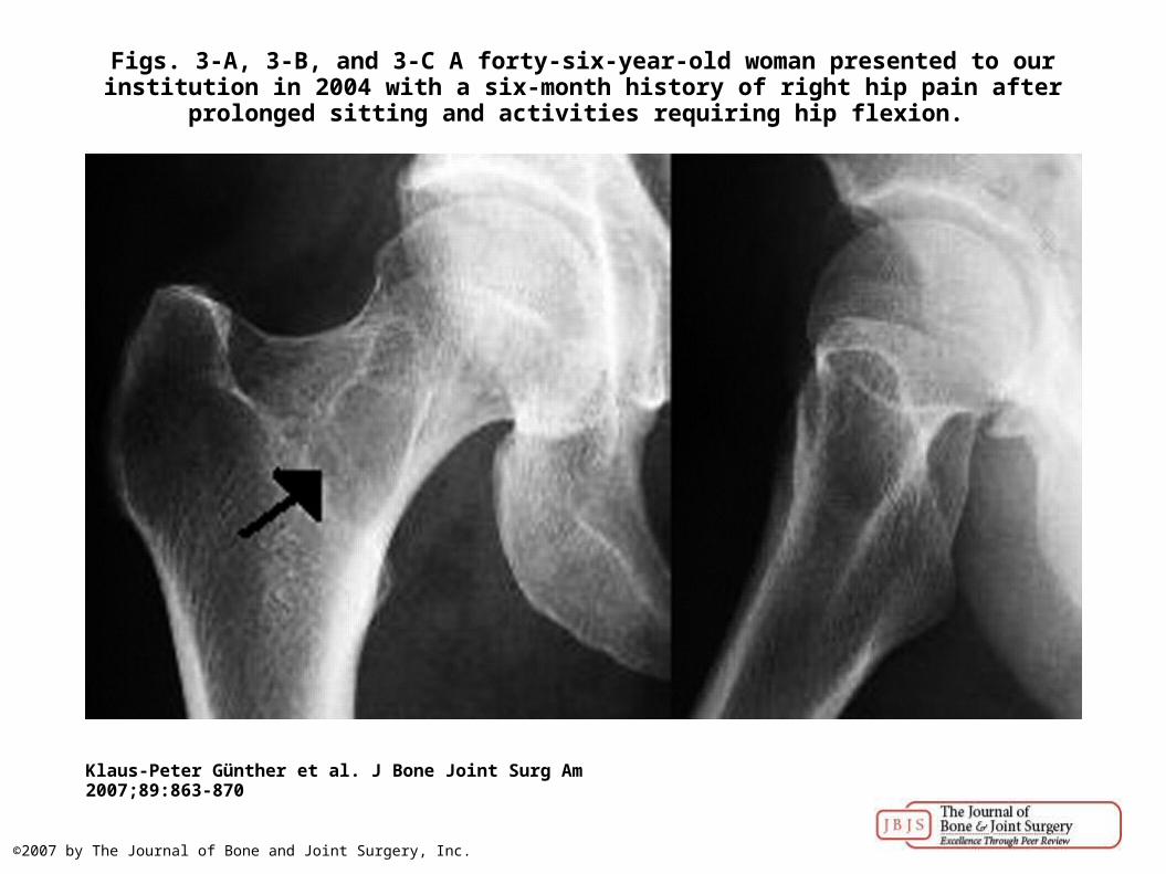
Figs. 3-A, 3-B, and 3-C A forty-six-year-old woman presented to our institution in 2004 with a six-month history of right hip pain after prolonged sitting and activities requiring hip flexion.
Klaus-Peter Günther et al. J Bone Joint Surg Am 2007;89:863-870
©2007 by The Journal of Bone and Joint Surgery, Inc.
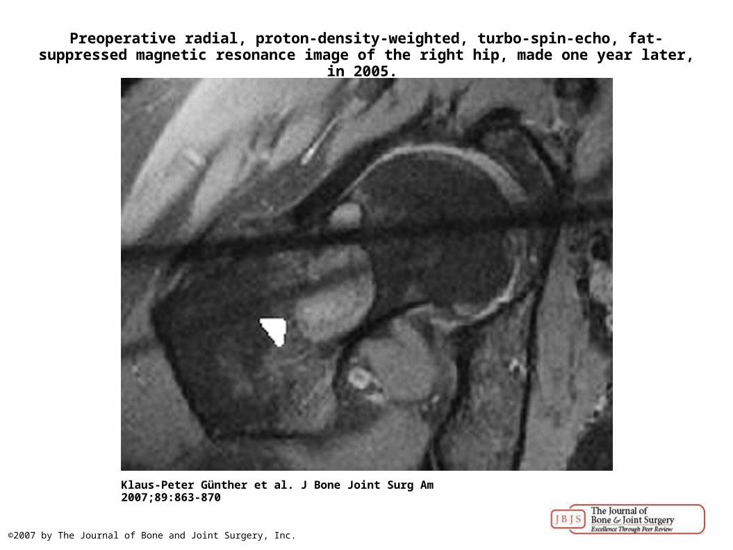
Preoperative radial, proton-density-weighted, turbo-spin-echo, fat-suppressed magnetic resonance image of the right hip, made one year later, in 2005.
Klaus-Peter Günther et al. J Bone Joint Surg Am 2007;89:863-870
©2007 by The Journal of Bone and Joint Surgery, Inc.
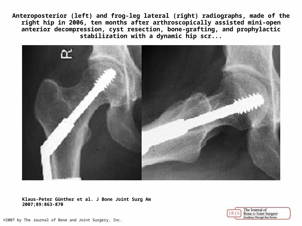
Anteroposterior (left) and frog-leg lateral (right) radiographs, made of the right hip in 2006, ten months after arthroscopically assisted mini-open anterior decompression, cyst resection, bone-
grafting, and prophylactic stabilization with a dynamic hip scr...
Klaus-Peter Günther et al. J Bone Joint Surg Am 2007;89:863-870
©2007 by The Journal of Bone and Joint Surgery, Inc.