klasični način opisa proteina - unizg.hr1 makromolekule biološki (prirodni) polimeri fizikalna...
TRANSCRIPT

1
makromolekule
biološki (prirodni) polimeri
fizikalna kemija makromolekula
sintetski polimeri
fizikalna kemija proteina
Edwin J. Cohn
Simoni R D et al. J. Biol. Chem. 2002;277
©2002 by American Society for Biochemistry and Molecular Biology
Cohn, E. J., “The Properties and Functions of Plasma Proteins with consideration of the Methods for their Separation and Purification”, Chem. Rev. 28 (1941) 395.
Topljivost i kiselo-bazna svojstva proteina
Cohn, E. J., Hendry, J. L., and Prentiss, A. M.,“Studies in the Physical Chemistry of the Proteins. V. Molecular Weights of the Proteins”,J. Biol. Chem. 63 (1925) 721-766.
Klasični način opisa proteina:
• Primarna struktura - redoslijed (sekvencija) aminokiselina
•Sekundarna struktura –konformacija peptidnih lanaca
•Tercijarna struktura
•Kvaterna struktura
Razine strukture proteina
Spektroskopsko istraživanje sekundarne strukture proteina
• Cirkularni dikroizam (Circular dichroism,CD)
• Infracrvena (IR) i Raman spektroskopija
• Nuklearna magnetska rezonancija (NMR)
Fizikalni principi CD-a
• Kiralne ili asimetrične molekule daju CD spektar zato jer različito apsorbirajulijevo i desno polariziranu svjetlost i zato se smatraju "optički aktivnim"
2
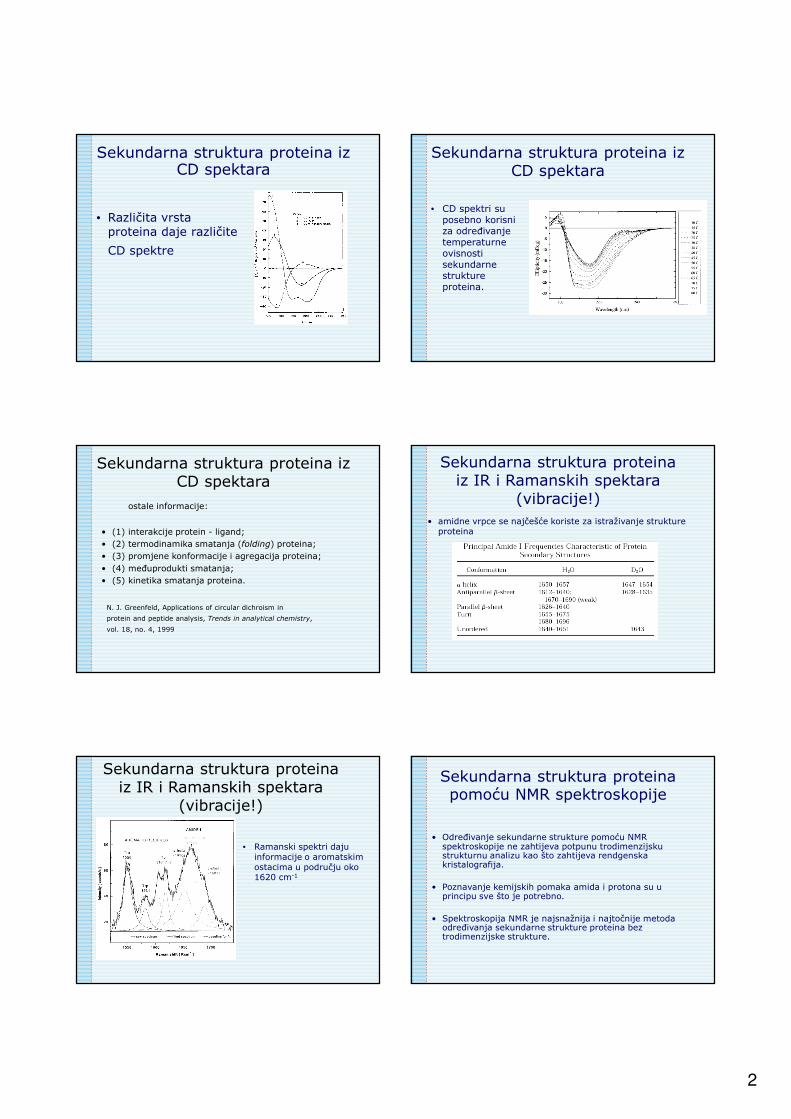
• Različita vrsta proteina daje različite
CD spektre
Sekundarna struktura proteina izCD spektara
Sekundarna struktura proteina izCD spektara
• CD spektri su posebno korisniza određivanjetemperaturneovisnostisekundarne strukture proteina.
ostale informacije:
• (1) interakcije protein - ligand;
• (2) termodinamika smatanja (folding) proteina;
• (3) promjene konformacije i agregacija proteina;
• (4) međuprodukti smatanja;
• (5) kinetika smatanja proteina.
Sekundarna struktura proteina izCD spektara
N. J. Greenfeld, Applications of circular dichroism in
protein and peptide analysis, Trends in analytical chemistry,
vol. 18, no. 4, 1999
Sekundarna struktura proteinaiz IR i Ramanskih spektara
(vibracije!)
• amidne vrpce se najčešće koriste za istraživanje struktureproteina
• Ramanski spektri dajuinformacije o aromatskimostacima u području oko1620 cm-1
Sekundarna struktura proteinaiz IR i Ramanskih spektara
(vibracije!)
Sekundarna struktura proteinapomoću NMR spektroskopije
• Određivanje sekundarne strukture pomoću NMR spektroskopije ne zahtijeva potpunu trodimenzijsku strukturnu analizu kao što zahtijeva rendgenska kristalografija.
• Poznavanje kemijskih pomaka amida i protona su u principu sve što je potrebno.
• Spektroskopija NMR je najsnažnija i najtočnije metoda određivanja sekundarne strukture proteina bez trodimenzijske strukture.

3
Interakcija proteina sa...
• ... polielektrolitima
• ... polisaharidima
• ... DNA
• ... itd
• Primjena!!
Kompleksi protein-polielektrolit
Karakterizacija polielektrolitno-proteinskih višeslojeva pomoću optičke reflektometrije
0
0.4
0.8
1.2
0 20 40 60 80 100
t / min
∆∆ ∆∆S
+
+
+
-
--
PVP+/BSA
PAMA/BSA
0.0
0.4
0.8
1.2
1.6
0 40 80 120 160 200
t / min
ΔS
pH = 9
pH = 7
pH = 4
+
+
++
--
-
-
potenciometrijska titracija
3
5
7
9
11
0 0.5 1
V (NaOH) / cm3
pHBSA
BSA + PAMA
BSA + PAH
J. Mathew et al., Fabrication of switchable protein resistant and adhesive multilayer membranes, Colloids and Surfaces B:
Biointerfaces 94 (2012) 118– 124
• Fabrication of protein adhesive and resistant surfaces based on chitosan/polystyrene sulfonate (CHI/PSS) multilayer membranes is presented. Adsorption behavior of bovine serum albumin (BSA) and lysozyme to CHI/PSS multilayer was studied.

4
Shematski prikaz adsorpcije lizozima(a) pri pH = 8,8 (b) desorpcija, reverzna migracija na površinu.
AFM iprikazi adsorpcije lizozima na CHI/PSS višesloj (10 slojeva)(a) pH = 8,8 (b) pH = 10,6
SEM prikazi adsorpcije BSA (pH 4.8)(a) 6 CHI/PSS slojeva (b) 14 CHI/PSS slojeva.
“Scanning Probe Microscopy” techniques
Atomic Force Microscopy (AFM)
Contact modeTapping mode
Atomic Force Microscopy (AFM)

5
D. Störkle, S. Duschner, N. Heimann, M. Maskos, and M. Schmidt, Complex Formation of DNA with Oppositely Charged Polyelectrolytes of Different Chain Topology: Cylindrical Brushes and Dendrimers, Macromolecules, 2007, 40 (22), pp 7998–8006.
Polyelectrolyte – DNA complexes
Block copolymer micelles for gene therapyTransfection of plasmid DNA using diblock copolymer. DNA is released
inside the cytosol and appears in the nucleus to express a desired protein.
Forster and M. Konrad, J. Mater. Chem., 2003
Sample configurations of a simulation of the adsorption of a 20 monomer PE chain on a charged surface, yielding a surface monomer density of 2 monomers/nm2. The bulk electrolyte concentration was 0.010 M. Green: polyelectrolyte monomers. Red: negative ions. Blue: positive ions.
CH2 CH
SO O
O-
n
Molecular dynamics simulation of the structure of sodium poly(styrenesulfonate) (t = 5 ns)
Molecular dynamics simulation of the structure of
poly(allylammonium perchlorate) (t = 5 ns)
CH2 CH
CH2 NH3+
n
Illustration of the mapping procedure of sodium polystyrene sulfonate (NaPSS) in water. The repeat units of NaPSS are replaced by spherical monomers connected by springs and the solvent molecules are represented by a continuum with dielectricconstant e. Counterions are represented by spherical beads.
6
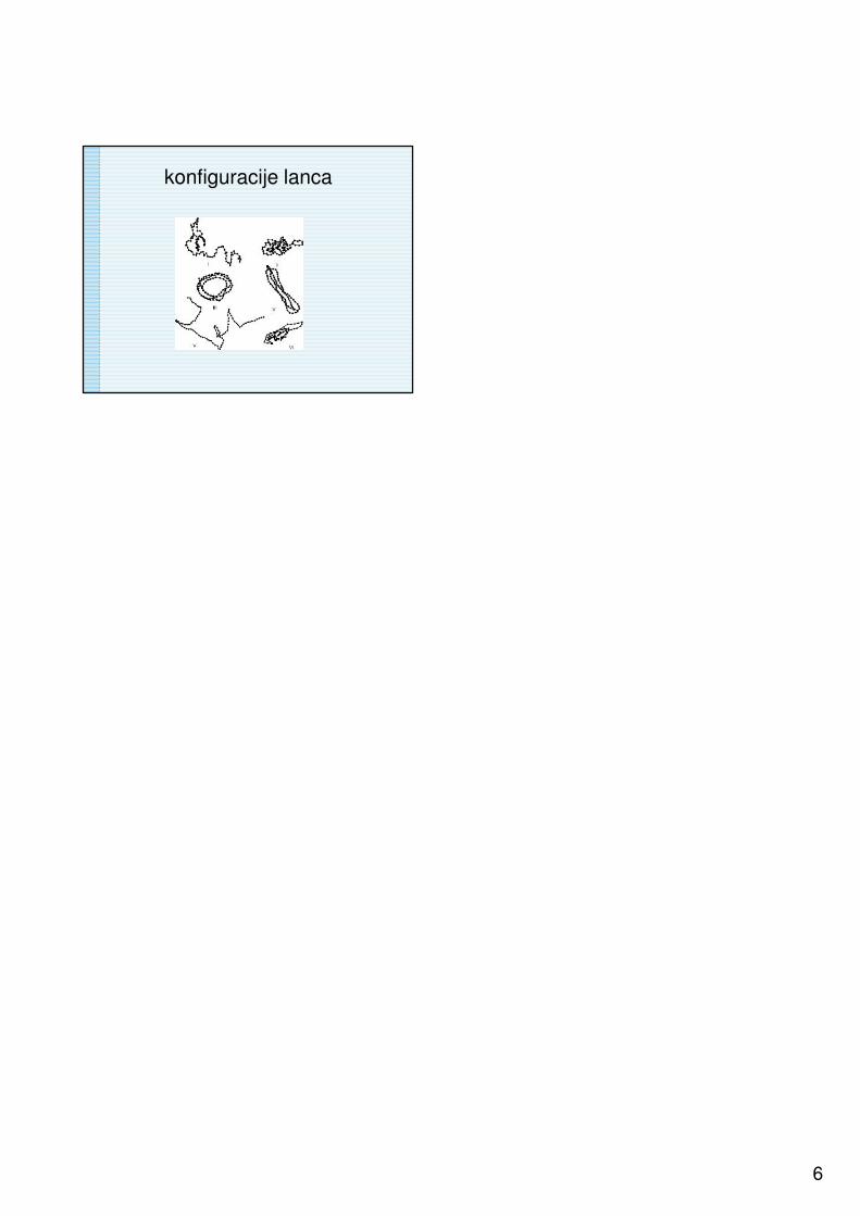
konfiguracije lanca