juvenile neuronal ceroid...
TRANSCRIPT
JUVENILE NEURONAL CEROID LIPOFUSCINOSIS;
BRAIN-RELATED SYMPTOMS AND THEIR TREATMENT
Laura Åberg
Hospital for Children and Adolescents, Department of Child Neurology
and Department of Radiology
University of Helsinki
Academic dissertation
To be publicly discussed with the permission of the Faculty of Medicine of the University of Helsinki, in the Niilo Hallman Hall of the Hospital for
Children and Adolescents, Stenbäckinkatu 11, Helsinki, on September 7th 2001 at 12 noon
Helsinki 2001
Supervised by Professor Pirkko Santavuori, M.D. Hospital for Children and Adolescents
Department of Child Neurology University of Helsinki, Helsinki, Finland and Docent Taina Autti, M.D. Department of Radiology University of Helsinki, Helsinki, Finland Reviewed by Docent Leena Vainionpää, M.D.
Department of Pediatrics University of Oulu, Oulu, Finland
and
Docent Jouko Isojärvi, M.D. Department of Neurology University of Oulu, Oulu, Finland and Docent Aapo Ahonen, M.D. Laboratory Department, Division of Nuclear Medicine University of Helsinki, Helsinki, Finland
Opponent Professor Paul Uvebrant, M.D. Department of Pediatrics Östra Hospital, Gothenburg, Sweden ISBN 952-91-3800-8 ISBN 952-10-0123-2 Yliopistopaino Helsinki 2001

To Klaus and Wilhelm
4
CONTENTS LIST OF ORIGINAL PUBLICATIONS......................................................... 6 ABBREVIATIONS ........................................................................................ 7 SUMMARY .................................................................................................... 8 INTRODUCTION .......................................................................................... 9 REVIEW OF THE LITERATURE.................................................................. 10 1. Neuronal ceroid lipofuscinoses ................................................................. 10 1.1. History ................................................................................................ 10
1.2. Classification and different NCL subtypes ........................................ 11 1.3. Epidemiology...................................................................................... 16
1.3.1. Epidemiology in Finland ......................................................... 18 1.4. Neuropathological findings ................................................................ 18 1.5. Neurophysiological findings .............................................................. 19 1.6. Neuroimaging studies ........................................................................ 21
2. Clinical picture in JNCL ........................................................................... 23 2.1. Visual failure ...................................................................................... 23 2.2. Mental decline .................................................................................... 23 2.3. Epilepsy .............................................................................................. 23 2.4. Extrapyramidal symptoms .................................................................. 24 2.5. Hormonal changes in girls with JNCL ............................................. 24 2.6. Psychiatric disturbances and sleep disturbances ................................ 25 2.7. Other symptoms ................................................................................. 25
3. Diagnostics of JNCL ................................................................................. 26 4. Treatment of JNCL ................................................................................... 26
4.1. Antiepileptic drugs ............................................................................. 26 4.2. Treatment of extrapyramidal signs ..................................................... 27 4.3. Hormonal interventions ...................................................................... 28 4.4. Psychotropic treatment ....................................................................... 28 4.5. Other treatment modalities .................................................................. 28
AIMS OF THE PRESENT STUDY ............................................................... 30 MATERIALS AND METHODS .................................................................... 31 1. Patients ..................................................................................................... 31 2. Neuropsychological test battery ................................................................ 31 3. Data collection on epilepsy and AEDs ...................................................... 36 4. Evaluation of extrapyramidal symptoms .................................................. 37 5. Magnetic resonance imaging ..................................................................... 41 6. Therapy with antiparkinsonian drugs......................................................... 42 7. Statistical methods ..................................................................................... 43
5
RESULTS ....................................................................................................... 44 1. Neuropsychological decline ...................................................................... 44 2. Epilepsy and antiepileptic drug therapy .................................................... 46 3. Extrapyramidal signs and ß-CIT-SPECT .................................................. 48 4. Magnetic resonance imaging ..................................................................... 49 5. Treatment of extrapyramidal signs ........................................................... 51 DISCUSSION ................................................................................................. 52 1. Neuropsychological decline ...................................................................... 52 2. Epilepsy and antiepileptic drug therapy .................................................... 53 3. Evaluation of extrapyramidal signs ........................................................... 54 4. Antiparkinsonian drugs ............................................................................. 57 5. Polypharmacy in JNCL ............................................................................. 58 6. Future prospects ........................................................................................ 59 CONCLUSIONS.............................................................................................. 60 ACKNOWLEDGEMENTS ............................................................................ 61 REFERENCES ................................................................................................ 63 ORIGINAL PUBLICATIONS ........................................................................ 78
6
LIST OF ORIGINAL PUBLICATIONS This thesis is based on the following original articles, which are referred to in the text by Roman numerals. I Lamminranta S, Åberg LE, Autti T, Moren R, Laine T, Kaukoranta
J, Santavuori P. Neuropsychological test battery in the follow-up of patients with juvenile neuronal ceroid lipofuscinosis. J Intellect Disabil Res 2001; 45: 8-17.
II Åberg L, Kirveskari E, Santavuori P. Lamotrigine therapy in
juvenile neuronal ceroid lipofuscinosis. Epilepsia 1999; 40: 796-799.
III Åberg LE, Bäckman M, Kirveskari E, Santavuori P. Epilepsy and
antiepileptic drug therapy in juvenile neuronal ceroid lipofuscinosis. Epilepsia 2000; 41: 1296-1302.
IV Åberg L, Liewendahl K, Nikkinen P, Autti T, Rinne JO, Santavuori
P. Decreased striatal dopamine transporter density in JNCL patients with extrapyramidal signs. Neurology 2000; 54: 1069-1074.
V Åberg LE, Rinne JO, Rajantie I, Santavuori P. A favorable response
to antiparkinsonian treatment in juvenile neuronal ceroid lipofuscinosis. Neurology 2001; 56: 1236-1239.
7
ABBREVIATIONS AED(s) antiepileptic drug(s) AFI amaurotic familial idiocy ANCL adult onset neuronal ceroid lipofuscinosis BMI body mass index CIT 2β-carbomethoxy-3β-(4-iodophenyl) tropane cLINCL classical late infantile neuronal ceroid lipofuscinosis CNS central nervous system CZP clonazepam DAT dopamine transporter EEG electroencephalogram EM electron microscopy ERG electroretinogram FDG 2-deoxy-2[18F]fluoro-D-glucose GROD granular osmiophilic deposit INCL infantile neuronal ceroid lipofuscinosis IQ intelligence quotient JNCL juvenile neuronal ceroid lipofuscinosis LINCL late infantile neuronal ceroid lipofuscinosis LTG lamotrigine MAO monoamine oxidase MRI magnetic resonance imaging NCL neuronal ceroid lipofuscinoses PB phenobarbitone PCO polycystic ovaries PET positron emission tomography PPT palmitoyl protein thioesterase SAP sphingolipid activator protein (saposin) SCMAS subunit c of mitochondrial ATP synthase SEP somatosensory evoked potential SERT serotonin transporter SPECT single photon emission computerized tomography SSRI selective serotonergic inhibitor TPP tripeptidyl peptidase UPDRS unified Parkinson’s disease rating scale VEP visual evoked potential vLINCL variant late infantile neuronal ceroid lipofuscinosis VPA valproate WISC-R Wechsler Intelligence Scale for Children - Revised
8
SUMMARY The neuronal ceroid lipofuscinoses (NCL) are inherited, progressive neurodegenerative disorders, with onset mostly in early childhood. They are characterised by intracellular accumulation of autofluorescent storage material, seen both in neurons and other cells. Three main types with childhood onset are recognised, infantile (INCL), late infantile (LINCL), and juvenile NCL (JNCL). The features common to these NCL types include visual failure, mental decline, epilepsy, and motor impairment. Thus far, no curative treatment is available and therapy is limited to symptomatic drug treatment and rehabilitation. In JNCL, the leading sign of the disease is visual failure, noticed around the age of 4-7 years. Later, mental decline becomes evident. To estimate the neuropsychological profile of blind patients with JNCL, a special neuropsychological test battery was developed. The test battery revealed uneven cognitive impairment. During the first 5 years after the diagnosis the ability to recognise similarities remains fairly good, while the digit memory span declines more quickly than the other sub-items. The special neuropsychological profile provides important guidelines for rehabilitation. The first epileptic seizure is noticed at a mean age of 10 years. The most common seizure types at the onset are generalised tonic-clonic seizures and complex partial seizures, whereas myoclonia are seen in the final stages of the disease. As the first antiepileptic drugs (AEDs), valproate and lamotrigine are equally effective, but adverse effects are less common with lamotrigine than with valproate therapy. Lamotrigine also seems to have a favourable effect on the general well-being. Clonazepam is found to be favourable as an add-on AED. ß-CIT-SPECT revealed decreased density of dopamine transporters, reflecting nigrostriatal dysfunction. The degree of dopaminergic hypofunction was related to the severity of the extrapyramidal symptoms. This justified a trial of dopaminergic drugs, with either levodopa or selegiline. In 1 year, the extrapyramidal symptoms decreased significantly in the patients on levodopa, as compared with the controls. However, there was considerable variation in this response. In the patients receiving selegiline, the response was less marked. As the thalamus and basal ganglia are also impaired by the disease process, the response to therapy is milder and probably also of shorter duration than that seen in Parkinson’s disease.
9
INTRODUCTION The neuronal ceroid lipofuscinoses (NCL) are the most common group of progressive, neurodegenerative disorders of childhood. These diseases are assumed to occur world-wide, though their existence in the developing countries has not been documented. The childhood forms are inherited recessively, a gene defect having now been localised for each main type (Mole 1999). These disorders are characterised by intracellular accumulation of fluorescent ceroid and lipofuscin-like storage material. This storage material can be found both in neural and in extra-neural cells, although the symptoms mainly reflect the disease of the central nervous system (CNS). There is widespread loss of nerve cells, which in the cerebral cortex, is pronounced. The NCL disorders are classified as lysosomal diseases. In the main NCL types, the gene defect results in depletion of a protein, which either resides in a lysosome or is an unknown transmembrane protein. However, the precise pathogenesis still remains unknown. The clinical symptoms common to all the different childhood NCL types are impaired vision, mental deterioration, epilepsy, psychiatric problems, and extrapyramidal and pyramidal symptoms. In the different types, these symptoms may develop at different age and in a different order. The child’s condition gradually deteriorates and the disease eventually leads to premature death. As yet, there is no curative treatment. However, symptomatic drug treatment brings about some relief in most patients, and is widely used. Furthermore, as these diseases are characterised by multiple symptoms, polytherapy is often needed, though it may give rise to problems in the form of adverse effects and unwanted interactions. Therefore, a systematic evaluation of the symptoms and their current treatment was considered necessary. Although the drug treatment is important and brings about relief of the symptoms, the most important thing to keep in mind, in the treatment of these patients, is support for their families. Rehabilitation, in the form of adjustment courses, physical therapy, occupational therapy and speech therapy, forms an important part of this support.
10
REVIEW OF THE LITERATURE 1. NEURONAL CEROID LIPOFUSCINOSES (NCL) 1.1. History In 1826 Otto Christian Stengel described four siblings with loss of vision and mental deterioration, these symptoms being noticed after 6 years of age (Goebel 1995). The siblings lived in a Norwegian mining town, and at first the surroundings were blamed for the disease. In the hope of preventing further loss of abilities, the younger siblings were removed from the area, but nevertheless the disease progressed. This is probably the first description of NCL disorders in the literature. Around the turn of the century, several descriptions of the disease were made. In 1896, Sachs introduced the term “amaurotic familial idiocy” (AFI), a group of diseases with infantile onset, characterised by blindness, psychomotor deterioration, and early death. In 1903, Batten described patients suffering from “family cerebral degeneration with macular change” with a later onset and separated these patients clinically from those with infantile onset AFI. In 1905, this distinction was confirmed by Spielmeyer, who published his findings on siblings suffering from “a special form of AFI”, and by Vogt, who described patients suffering from “juvenile AFI”. Although clinical differences were noticed between the different forms of AFI, they were still regarded as clinical variants of a uniform disease entity. Indeed, Schaffer’s pathogenetic studies (1905), in which accumulating lipid material was found in swollen nerve cells in all these diseases, supported this view of a uniform disease entity. In the reports of Batten (1903, 1914), late infantile and juvenile onset AFI were not separated either. In 1908 Jansky described a special form of AFI with cerebellar atrophy. In a paper published 1 year later, Vogt observed that siblings suffering from late onset AFI usually manifested the symptoms of the disease in the same chronological order, but that marked differences in the course of the disease existed between different families. Furthermore, in 1913 Bielchowsky reported his clinical and pathological findings in three siblings with seizures manifesting at the age of 3.5 years and followed by rapid deterioration
11
characterised by dementia and blindness. He proposed that this was a late infantile AFI, separating it from the juvenile AFI. Patients with infantile onset AFI were long included in the group of patients with AFI, and it was not until 1931 that Sjögren, on clinical grounds, clearly separated the juvenile type of AFI from the infantile type. All of the 115 patients described by Sjögren had AFI with the juvenile onset. Separation of the infantile onset AFI from forms of AFI with the later onset was supported by the isolation of ganglioside from the brains of patients with infantile AFI, presently known as Tay-Sachs disease (Klenk 1939). In 1963, Zeman and Alpert reported on autofluorescent lipopigments in patients with late onset AFI, which finally distinguished patients with AFI from patients with other storage disorders, including Tay-Sachs disease. Subsequently, Zeman and Dyken formed the concept of “neuronal ceroid lipofuscinoses”, based on the storage material resembling ceroid and lipofuscin, and separated the age-dependent types of the disease (Zeman et al. 1969, 1970). 1.2. Classification and different NCL types The late infantile and juvenile NCL types were thus described in the beginning of the 20th century, first on the basis of the clinical course, and later also on the electronmicroscopic and neurophysiologic findings. Infantile NCL, different from the infantile onset AFI, was described in 1973 by Santavuori et al. These are three main childhood onset NCL types. Thereafter, several variants of the three main types were described, but these were not confirmed to be independent diseases until the 1990s, when the gene loci were mapped (Table 1). In addition to the NCL types, several subtypes have also been described; these have the same gene defect as the main NCL type, but the clinical course differs. Thus, localisation of these genes explained the varying clinical course, but, on the other hand, also showed that, in clinically similar diseases, the gene locus can be in different chromosomes. Furthermore, even if the gene defect is the same, the clinical phenotype may vary, as in other genetic diseases. Along with identification of the genes causing the diseases, the molecular background is gradually starting to emerge, and lysosomal degradation appears to be implicated in the main childhood types of NCL. CLN1 and
12
CLN2 both encode lysosomal enzymes, whereas CLN3 and CLN5 encode membrane proteins of unknown function. _____________________________________________________________ Table 1. The gene loci of the neuronal ceroid lipofuscinoses (NCL) __________________________________________________________________ Gene Disease Locus Reference _____________________________________________________________ ? Congenital NCL ? Martin et al. 1999 CLN1 Infantile NCL 1p32 Järvelä et al. 1991 CLN2 Classical LINCL 11p15 Sharp et al. 1997 CLN3 Juvenile NCL 16p12 Eiberg et al. 1989 CLN4 Adult onset NCL ? Berkovic et al. 1988 CLN5 Finnish vLINCL 13q22 Savukoski et al. 1994 CLN6 Variant LINCL 15q22 Sharp et al. 1997 CLN7 Turkish vLINCL ? Williams et al. 1999 CLN8 Northern epilepsy 8p23 Tahvanainen et al. 1994 _____________________________________________________________
? = not known, LINCL = late infantile neuronal ceroid lipofuscinosis, vLINCL = variant
late infantile neuronal ceroid lipofuscinosis Congenital neuronal ceroid lipofuscinosis A rare congenital form of AFI was described by Norman and Wood in 1941. This NCL type, not found in Finland, is characterised by severe, generalised neonatal convulsions. The progression of the disease is rapid, leading to death before the age of 7 weeks, though usually within the first days of life. Microcephaly is prominent (Kohlschutter and Lake 1999), and, using ultrasound, can be noticed already during pregnancy. Infantile neuronal ceroid lipofuscinosis (CLN1) Infantile NCL (INCL), also called Santavuori-Haltia disease, was first described by Santavuori et al. in 1973. The first symptoms manifest around the age of 1 year as muscular hypotonia, delayed motor and psychomotor development, and progressive microcephaly. Thereafter, retardation of all
13
skills sets in. Ataxia, irritability and sleep disorders are also common signs in the early phase. Visual failure is noticed between the ages of 12 and 22 months and rapidly leads to blindness. Epileptic seizures and myoclonic jerks are prominent. The patient’s condition rapidly deteriorates, and by the age of 3 years, all cognitive and motor skills are lost. Death usually occurs between 8 and 11 years of age. The CLN1 gene codes for the palmitoyl protein thioesterase (PPT) (Järvelä et al. 1991, Vesa et al. 1995), a lysosomal enzyme with the ability to remove fatty acids from palmitoylated proteins in vivo (Camp and Hofmann 1993). In the literature, late infantile onset (Wisniewski et al. 1997, Das et. al. 1998) and juvenile onset subtypes of INCL (Lake et al. 1996, Crow et al. 1997, Mitchinson et al. 1998) have also been described. In these variant forms, the gene locus has been confirmed to be the INCL locus and the electronmicroscopic findings are identical to those of the classical INCL, but the onset of the disease is later. Classic late infantile neuronal ceroid lipofuscinosis (CLN2) Classic late infantile NCL (cLINCL, Jansky-Bielchowsky disease) was originally described by Jansky (1908) and Bielchowsky (1913), although they were unable to separate this type from the forms with later onset. The onset of the disease is noticed between the ages of 2 and 4 years, the first sign usually being epilepsy. Sometimes delayed speech may precede epilepsy. Additional symptoms include dementia, ataxia, and myoclonic jerks. Visual failure leads to blindness usually by 5 or 6 years of age. Death usually occurs between 6 and 15 years of age. Although the disease is rare in Finland, it is one of the most common NCL types in the United States and Canada. The CLN2 gene codes for a lysosomal enzyme tripeptidyl peptidase 1 (TPP1), which removes the terminal amino acids from proteins undergoing lysosomal degradation (Sleat et al. 1997¸ Vines and Warburton 1999). A variant subtype, late onset LINCL, has been recognised in LINCL. In this subtype, the gene locus and the enzyme defects are identical to that of classical LINCL, but the onset of the disease occurs later and the clinical course is slower (Sleat et al. 1999).
14
Juvenile neuronal ceroid lipofuscinosis (CLN3) The juvenile type of NCL (JNCL) can be recognised in the descriptions of Stengel (1826), Batten (1903), Spielmeyer (1905), Vogt (1909), and Sjögren (1931). Thus, the disease is also known as Spielmeyer-Vogt-Sjögren’s disease, and in the American literature as Batten’s disease, although the latter name is sometimes used to refer all the childhood onset types of NCL. Children with JNCL are born healthy and, at first, the developmental milestones are reached normally. However, around the age of 5 years, visual failure is noticed. In the early school years, gradual psychomotor deterioration becomes evident, and around the age of 10 years, the first epileptic seizures occur. Extrapyramidal signs start to develop in adolescence, leading to impaired ability to move around. In girls with JNCL, common findings are acne, obesity, and hirsutism, and in both sexes psychiatric symptoms and sleep disturbances are seen. The disease leads to premature death between the ages of 16 and 35 years. (Santavuori 1988, Hofman et al. 1999). The CLN3 gene was localised to chromosome 16p12 (Eiberg et al. 1989, Gardiner et al. 1990). It encodes for a previously unknown transmembrane protein, localised to the lysosomes (The International Batten Disease Consortium 1995, Järvelä et al. 1999). World-wide, 81% of the chromosomes carry the major mutation, a 1.02 kb deletion (Järvelä et al. 1996). Patients with the major mutation on both chromosomes are homozygotes for the major mutation. On the other hand, those patients in whom the major mutation is found in one of the chromosomes and another mutation in the other, are compound heterozygotes. In Finland, the major mutation is further enriched, and 90% of the Finnish CLN3 chromosomes carry the major deletion (Järvelä et al. 1996). Although the majority of the patients have the major mutation in at least one of the chromosomes, one patient was recently found to have a rare mutation on both chromosomes (Santavuori, personal communication). In JNCL also, variants of the classical phenotype have been described. These consist mainly of patients with a protracted clinical course, the leading sign usually being visual failure (Goebel 1993, Åberg et al. 1998, Lauronen et al. 1999).
15
Adult onset neuronal ceroid lipofuscinosis (CLN4) The adult form of NCL (ANCL) was first described by Kufs in 1925 as a late form of amaurotic idiocy. ANCL is considered to be rare, although it is possible that not all the cases have been diagnosed. On the other hand, a considerable proportion of the reported cases of ANCL are probably not true cases, but may represent one of the other forms of lipidosis (Berkovic et al. 1988). Two separate forms of ANCL have been described (Berkovic et al. 1988). The clinical phenotype A (Kufs’ disease) is characterised by progressive myoclonic epilepsy, dementia, ataxia, and pyramidal and extrapyramidal symptoms. Vision is normal, but retinal degeneration may be noticed. In the clinical phenotype B (Parry disease, CLN4), on the other hand, behavioural changes, dementia, ataxia, facial dyskinesia and extrapyramidal signs are seen. Epileptic seizures may be noticed, though they are usually non-progressive. Visual failure has not been reported in this subtype. In Kufs’ disease, autosomal recessive inheritance is seen, but in the Parry disease, autosomal dominant inheritance has been described, in contrast to all the other NCL types. However, the gene locus and the function of the mutated genes are still unknown. Finnish variant late infantile neuronal ceroid lipofuscinosis (CLN5) The Finnish variant type of late infantile NCL (vLINCLFinn) is seen almost exclusively in Finland. The first manifestations of the disease occur between the ages of 4 and 7 years. These manifestations usually are attention deficit, visuomotor difficulties, and motor clumsiness, and they are followed by mental retardation and visual failure (Santavuori et al. 1999). Other symptoms include epilepsy, myoclonic jerks, and ataxia. Death usually occurs between the ages of 14 and 36 years. The CLN5 codes for a 407 aminoacid transmembrane protein, with an unknown function. Variant late infantile NCL (CLN6) Variant late infantile NCL (vLINCL), sometimes also called early juvenile NCL was first described by Lake and Cavanagh in 1978. This subtype also includes the so-called Czech variant LINCL (Elleder et al. 1997). The clinical course varies, but the first symptoms usually include motor delay,
16
ataxia, regression, dysarthria, seizures and myoclonia. Visual failure may occur early. The disease leads to early death between the ages of 5 and 12 years. Turkish variant late infantile NCL (CLN7) In the process of characterising the genes responsible for the different types of LINCL, all the families of Turkish origin were found to have gene loci differing from the known NCL loci (Williams et al. 1999). This subtype was therefore named the Turkish variant LINCL (vLINCLTurk). The onset of symptoms accords with the other types of LINCL, the early features of the disease being seizures and motor problems. Visual failure has a variable onset, and in some cases may be the leading sign. Cognitive impairment follows the first symptoms. The exact gene locus has not been found yet. Northern Epilepsy (CLN8) Northern Epilepsy, also known as progressive epilepsy with mental retardation (EPMR), was first described by Hirvasniemi et al. in 1994 in patients from the Kainuu region in Finland. On the basis of the ultrastructure of the storage material and the immunocytochemistry, the disease was later found to be one of the NCL disorders (Herva et al. 2000). The clinical signs appear between the ages of 5 and 10 years as epileptic seizures and learning disability (Hirvasniemi et al. 1995). Seizure frequency increases towards puberty, but then gradually decreases again. Motor clumsiness becomes evident in early adulthood. In contrast to the other NCL disorders with childhood onset, visual loss does not occur in the early stage of the disease (Herva et al. 2000). The CLN8 gene codes for a 286 aminoacid transmembrane protein with an unknown function (Ranta et al. 1999). 1.3. Epidemiology The NCL diseases have been reported in several western countries. Along with the mapping of the main NCL subtypes, the common disease haplotypes were revealed. The mutational analysis of these genes confirmed the existence of the so-called founder effect. This refers to random variation in the frequency of rare genes during the formation of a new subpopulation of limited size. Thus, if the founders forming the subpopulation included a member with a rare recessive allele, the frequency of this allele will be much higher in the subpopulation than outside it.

17
In Finland, the predominant NCL genes are CLN1, CLN3 and CLN5. Altogether 95% and 89% of the Finnish patients with CLN1 and CLN5, respectively, carry the major mutation on both chromosomes (Järvelä et al 1991, Das et al. 1998, Savukoski et al. 1998). As regards CLN3, the major mutation (a 1.02 kb deletion) is also enriched in the Finnish population, and around 80% of the Finnish patients with CLN3 carry the major mutation on both chromosomes. Thus, around 90% of the chromosomes carry this mutation. World-wide, around 80% of the chromosomes of patients with JNCL carry this mutation. (The International Batten Disease Consortium 1995, Munroe et al. 1997, Zhong et al. 1998). The reported incidence and prevalence rates are given in Table 2. _____________________________________________________________ Table 2. Incidence (a) and prevalence (b) rates for the main NCL types, given as per 100,000 births (a) or 1,000,000 inhabitants (b) _____________________________________________________________ INCL LINCL1 JNCL (a) (b) (a) (b) (a) (b) _________________________________________________________________________ Finland2 5.0 5.4 ? 3.2 4.8 12.2 Sweden2 0.6 0.7 ? 0.5 2.2 4.6 Norway2 ? 0.2 ? 0.7 3.7 6.5 Denmark2 ? ? ? ? 2.0 3.1 Iceland2 ? ? ? 3.8 7.0 11.0 Germany3 ? ? 0.5 ? 0.7 ? Italy4 ? ? 0.4 ? 0.2 ? Netherlands5 ? ? 0.5 ? 1.5 ?
Canada6 ? ? 2.2 ? 0.6 ? _____________________________________________________________ 1Incidence and prevalence rates for LINCL are given for the group comprising both classic and the variant form of LINCL. 2Uvebrant et al. 1997, 3Claussen et al. 1992, 4Cardona and Rosati 1995, 5Hofman and Taschner 1999, 6MacLeod et al. 1976. ? = not known, NCL = neuronal ceroid lipofuscinosis, INCL = infantile neuronal ceroid lipofuscinosis, LINCL = late infantile neuronal ceroid lipofuscinosis, JNCL = juvenile neuronal ceroid lipofuscinosis.
18
1.3.1. Epidemiology in Finland In Finland, up to June 2001, altogether 411 patients with NCL have been diagnosed. This includes 163 patients with CLN1, 188 patients with CLN3, 26 patients with CLN8, 28 patients with CLN5, 5 patients with CLN2, and 1 patient with CLN4. No patients with congenital NCL, CLN6 or CLN7 have been diagnosed. From the incidence rates, it appears that two or three new patients with INCL and JNCL are born every year, whereas patients with LINCL are diagnosed more seldom. 1.4. Neuropathological findings In every NCL type, there is a uniform accumulation of intralysosomal autofluorescent lipopigments. These pigments have been found to consist mainly of the saposins (SAPs) (Tyynelä et al. 1993) or subunit c of mitochondrial ATP synthase (SCMAS) (Hall et al. 1991, Palmer et al. 1992). Saposins are found in the congenital form of NCL and in INCL, whereas SCMAS is found in the other types. SCMAS is also known to accumulate in some other lysosomal diseases, including mucopolysaccharidosis, mucolipidosis and gangliosidosis. Ultrastructurally, the storage material is characterised by granular, curvilinear, rectilinear, or fingerprint patterns (Table 3). The accumulation of storage material results in ballooning of the neuronal perikarya, a phenomenon called the Schaffer-Spielmeyer process (Goebel 1995). In addition, there is loss of nerve cells, the degree of loss depending on the NCL subtype and on the age of the patient (Goebel 1995). The cells most easily obtained for microscopic examinations are the lymphocytes in the peripheral blood. Vacuolisation of the lymphocytes, observable with the light microscope, can be used as a screening method for JNCL. In the other types of NCL, vacuolisation is not seen with the light microscope, and inclusions must be searched for with the electronmicroscope (EM). However, inclusions are present only in a minority of the lymphocytes, which makes the EM an unreliable diagnostic tool, when using peripheral lymphocytes. Thus, if it is necessary to confirm the diagnosis with EM, either a skin biopsy including the cells of the sweat glands (Carpenter et al. 1977) or a rectal biopsy including the submucosal
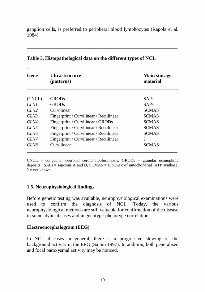
19
ganglion cells, is preferred to peripheral blood lymphocytes (Rapola et al. 1984). _____________________________________________________________ Table 3. Histopathological data on the different types of NCL _____________________________________________________________ Gene Ultrastructure Main storage (patterns) material ___________________________________________________________________ (CNCL) GRODs SAPs CLN1 GRODs SAPs CLN2 Curvilinear SCMAS CLN3 Fingerprint / Curvilinear / Rectilinear SCMAS CLN4 Fingerprint / Curvilinear / GRODs SCMAS CLN5 Fingerprint / Curvilinear / Rectilinear SCMAS CLN6 Fingerprint / Curvilinear / Rectilinear SCMAS CLN7 Fingerprint / Curvilinear / Rectilinear ? CLN8 Curvilinear SCMAS _____________________________________________________________ CNCL = congenital neuronal ceroid lipofuscinosis, GRODs = granular osmiophilic deposits, SAPs = saposins A and D, SCMAS = subunit c of mitochondrial ATP synthase. ? = not known. 1.5. Neurophysiological findings Before genetic testing was available, neurophysiological examinations were used to confirm the diagnosis of NCL. Today, the various neurophysiological methods are still valuable for confirmation of the disease in some atypical cases and in genotype-phenotype correlation. Electroencephalogram (EEG) In NCL diseases in general, there is a progressive slowing of the background activity in the EEG (Sainio 1997). In addition, both generalised and focal paroxysmal activity may be noticed.
20
In INCL, the first abnormalities noted in the EEG are lack of attenuation to eye opening, and disappearance of sleep spindles (Vanhanen 1997). Thereafter, attenuation of the EEG gradually increases, leading to a totally inactive EEG by the age of 2-3 years. In LINCL, the specific EEG features include posterior spikes to low-frequency photic stimulation. These are noticed around the age of 3 years in cLINCL and between the ages of 7 and 11 years in vLINCLFinn
(Pampiglione and Harden 1977, Santavuori et al. 1991). In JNCL, the EEG reveals runs of high-amplitude delta waves, intermingled with spikes and/or sharp waves (Lagenstein et al. 1978, Westmoreland et al. 1979). Until the age of 9, the EEG is usually normal. Thereafter, a progressive background abnormality and an increase in paroxysmal activity are seen (Larsen et al. 2001) When using quantitative analysis, the EEGs were found to be significantly slower than those of age-matched controls. The peak and mean frequencies and the fast/slow ratio were significantly lower, and the total power and the percentage of theta significantly higher than those of the controls. With age, the increase in the fast/slow ratio was significantly lower in the patients than in the controls. Electroretinogram (ERG) and visual evoked potential (VEP) Retinal degeneration leads to attenuation of ERG amplitude in all the main childhood-onset NCL types. Furthermore, in both INCL and JNCL, there is progressive attenuation of the potential of VEP and prolongation of the latency in VEP. In patients with LINCL, however, giant VEPs are seen. In patients with INCL, the ERG ceases to be recordable between the ages of 1 and 4 years (Harden et al. 1973, Bischof et al. 1983). The visual evoked potentials (VEPs) are extinguished after the ages of 2 to 5 years (Harden et al. 1973, Bischof et al. 1983, Vanhanen et al. 1997). In patients with cLINCL, the ERG is extinguished at an early stage, but a giant VEP can be elicited by flash stimulation (Harden et al. 1973). This finding coincides with posterior spikes in the photic stimulation. The abnormal VEP may be seen until the late stages of the disease, when it diminishes. Similar kinds of ERG and VEP are found in patients with vLINCLFinn (Santavuori et al. 1999).
21
In patients with JNCL, the leading sign is visual failure. VEP and ERG are abolished early (Raitta & Santavuori 1981). Even prior to the onset of the disease, depressed VEPs and subnormal ERGs may be seen (Raitta and Santavuori 1981, Horiguchi and Miyake 1992). Somatosensory evoked potential (SEP) and somatosensory evoked magnetic fields (SEF) In INCL, the cortical SEP amplitudes are progressively attenuated. However, in patients with the variant and classic types of LINCL, a giant SEP is a typical finding (Santavuori et al. 1991, Schmitt et al. 1994). In JNCL, both normal (Harden & Pampiglione 1982) and enhanced (Schmitt et al. 1994) SEP findings have been described. In MEG recordings, the amplitude of the SEF is increased (Lauronen et al. 1997). 1.6. Neuroimaging studies Non-invasive and rapid neuroimaging techniques are perhaps not diagnostic for the NCL disorders as such, but taken together with the clinical symptoms, they become highly diagnostic. Indeed, imaging methods can be considered as “windows to the brain”. Furthermore, they offer valuable information on the progress of the disease. Thus, they can also be used in monitoring the effects of any new therapies. Magnetic resonance imaging (MRI) In patients with INCL pathological MRI findings can be seen from the age of 7-11 months (Vanhanen et al. 1995). On T2-weighted images, the thalami are hypointense compared to the basal ganglia, and rims of high signal intensity may surround the lateral ventricles. Atrophy develops a few months later, cerebral atrophy outweighing cerebellar atrophy. In patients with cLINCL and vLINCL, prominent cerebellar atrophy is an early finding, followed by cerebral atrophy (Autti et al. 1997). High-signal rims in the white matter surround the ventricles (Autti et al. 1992). In addition, at least in the Finnish variant LINCL, the signal intensity of the thalami is lower than that of the basal ganglia (Autti et al. 1992). In patients with JNCL, MRI is usually normal in the early stages of the disease (Autti et al. 1996). Cerebral and later also cerebellar atrophy may be
22
seen after the age of 14 years. The signal intensity of the white matter is increased, especially in the periventricular area. However, these changes are not so pronounced as in INCL and LINCL. Slightly lowered signal intensities of the thalami may be seen after the age of 11 years. Spectroscopy In patients with INCL, there is almost complete loss of N-acetylaspartate, signifying a loss of neurons. Creatine and choline-containing compounds are markedly decreased as a sign of reduced glial membrane turnover. Myoinositol and lactate are elevated (Brockmann et al. 1996). In patients with LINCL, N-acetylaspartate is decreased, whereas lactate, myoinositol, creatine and choline-containing compounds are increased (Brockmann et al. 1996, Seitz et al. 1998). In patients with JNCL the spectroscopy is normal at the early stage, but later on, reduced N-acetylaspartate and creatine are noticed in the grey matter (Brockmann et al. 1996). Single photon emission computerised tomography (SPECT) In INCL non-specific cortical hypoperfusion was noticed in brain perfusion SPECT at an early stage, whereas reduction in cerebellar perfusion appeared later (Liewendahl et al. 1997). Perfusion of the basal ganglia and thalami was relatively well preserved up to the terminal stage. Patients with vLINCLFinn had bilateral supra- and infratentorial hypoperfusion even at an early stage of the disease, and cerebellar hypoperfusion was a characteristic finding (Autti et al. 1992). In JNCL, on the other hand, hypoperfusion was pronounced in the temporal lobes and mild in the parietal and occipital lobes as well as in the cerebellum (Launes et al. 1996). Positron emission tomography (PET) Using 2-deoxy-2[18F]fluoro-D-glucose (FDG) as a tracer for PET studies, age-dependent, progressive hypometabolism was found in patients with JNCL (Philippart et al. 1994). The hypometabolism started in the calcarine area and spread rostrally to the entire cortex. However, the basal ganglia and brainstem were relatively spared, and the uptake of FDG in these areas was considered normal.
23
2. CLINICAL PICTURE IN JNCL 2.1. Visual failure In JNCL, the first sign is almost invariably visual failure, noticed around the age of 4 to 7 years. Presumably retinal, cortical, and optic nerve degeneration all account for the declining vision. The loss of photoreceptors in the retina gradually spreads from the retinal periphery towards the macula (Goebel 1995). In the ophthalmological examination the typical findings include macular degeneration, optic atrophy, thinning of the vessels, and accumulation of pigment in the peripheral retina (Spalton et al. 1980). These specific findings are suggestive of the diagnosis of an NCL disorder. At the start of the visual failure, paradoxically, patients often seem to use their peripheral retina, giving an impression of “overlooking” with their heads turned to the other side (Hofman et al. 1999). The rapid decline of the vision usually leads to blindness within 2 to 6 years. However, light perception may be preserved for years. Moreover, even after the patients are blind, vivid visual hallucinations may sometimes occur (Boustany 1992). 2.2. Mental decline The most difficult symptom for parents with JNCL children to accept is probably dementia (Boustany and Filipek 1993). A slight mental decline sets in already at an early stage of the disease (Lou and Kristensen 1973, Santavuori and Moren 1977, Kristiansen 1987). In the study by Lou and Kristensen, involving 28 patients aged 6–28 years, only one had an IQ of over 90. In the early school years, gradual psychomotor deterioration becomes evident, and the children have difficulties in following the normal educational program, partly also because of their visual failure. Lou and Kristensen (1973) found a low digit memory span in the majority of their patients and assumed that digit memory span is one of the first functions to be impaired in JNCL. However, they found no marked tendency for the digit memory span to decline with age. Kristiansen (1987), on the other hand, found a low and declining digit memory span at an early stage of the disease.
24
2.3. Epilepsy The predominant type of seizure is a generalised tonic-clonic seizure, but complex partial seizures are also noticed (Hofman et al. 1999). Absence seizures have not been observed. Furthermore, in EEG recordings, the typical 3 Hz spike and slow wave discharges of absences have not been found. The epilepsy in JNCL is regarded as myoclonic (Berkovic 1986), and the enhanced somatosensory evoked magnetic fields (Lauronen et al. 1997) support the assumption that these patients have a myoclonic component, although myoclonia is not always observed. With age, the seizures tend to increase in frequency and in severity and, especially at puberty, an increase in the seizure frequency may be observed (Boustany and Filipek 1993). However, there is great individual variability, and in patients with early onset and poor seizure control, the clinical course tends to be malignant (Boustany 1992, Kohlschutter et al. 1988). 2.4. Extrapyramidal symptoms Parkinsonian signs are noted in about half the patients between the ages of 12 and 14 years (Järvelä et al. 1997). In the rest, these symptoms occur later. The most common extrapyramidal symptoms in patients with JNCL include impaired balance, rigidity, hypokinesia, stooped posture and shuffling gait (Hofman et al. 1999). In a PET study of nine patients with JNCL, a correlation was found between extrapyramidal symptoms and a decline in [18F]fluorodopa uptake in the putamen (Ruottinen et al. 1997), indicating nigrostriatal dysfunction. 2.5. Hormonal changes in girls with JNCL Girls with JNCL have an early menarche (Lou and Kristensen 1973, Åberg et al., submitted). Irregular cycles are common, and in girls with regular cycles the cycle length is short. Acne, obesity, and hirsutism, reflecting hyperandrogenism, are often seen. These may be due to the use of valproate (VPA), previously found to cause hyperandrogenism and polycystic ovaries (PCO), especially in young women (Isojärvi et al. 1993, Vainionpää et al. 1999). Indeed, 80% of the JNCL patients with hyperandrogenism had present or previous therapy with VPA. On the other hand, PCO were found in only 2 out of 8 girls with spontaneous cycles, a frequency within the
25
normal range at which PCO are found in young women (Michelmore et al. 1999). However, both girls with PCO had a full-blown polycystic ovary syndrome (PCOS), whereas usually only a minority of patients with PCO manifest this syndrome (Knochenhauer et al. 1998, Michelmore et al. 1999). 2.6. Psychiatric disturbances and sleep disturbances In patients with JNCL, severe psychiatric symptoms are common (Hofman 1999, Santavuori et al. 1993, Boustany 1992). These include anxiety, depression, and psychotic symptoms, and they greatly influence the well-being of the patients and their families. During the first few years after the diagnosis, the behavioural symptoms prevail. Depression is sometimes interpreted as a normal reaction to the deteriorating condition, but it may also assume a more serious connotation (Boustany and Filipek 1993). Depression may be manifested not only as sorrow, but also as unrest, aggressive outbursts, autoaggressive behaviour, and anxiety (Santavuori et al. 1993). Sleep problems are common in patients with JNCL, and may have a considerable effect on the well-being of the patients and their families. Sleep disorders, including settling problems, nocturnal awakenings, and nightmares had occurred in more than half of the patients, becoming evident at a mean age of 11 years (Santavuori et al. 1993). These problems seemed to increase during psychotic and restless periods as well as during periods of poor seizure control and tension (Santavuori et al. 1993, Kirveskari 2000). In the later stages of the disease, psychotic symptoms become more common, and may be observed in more than 75% of the patients (Sørensen and Parnas 1979). These psychotic incidents may manifest as hallucinations or delusions, characterised by strong anxiety, motor restlessness, or agitation. The visual hallucinations are often frightening, although sometimes these may also be experienced as colourful and harmless patterns. 2.7. Other symptoms Although, in JNCL, the symptoms mainly reflect the disease of the CNS, cardiac problems are also seen. Electrocardiographic abnormalities are frequent in the later stages of the disease, and include ST depression and
26
negative T waves (Hofman et al. 1999, Hofman et al. 2001). In addition, arrhythmias are common (Hofman et al. 2001), and in some patients these have led to installation of a pace-maker (Santavuori, personal communication). In the morphological evaluation, prominent involvement of the heart is seen; storage material is found not only in the myocardium, but also in the valvules and the conduction system. Storage is associated with hypertrophy, dilatation of the ventricles, degenerative myocardial changes, interstitial fibrosis and fatty replacement (Hofman et al. 2001). 3. DIAGNOSTICS OF JNCL The first sign of JNCL is visual impairment, appearing between the ages of 4 and 8 years. This usually leads to an ophthalmological examination, at which typical findings are made (chapter 2.1.). The ophthalmological findings, along with the age of the patient, are suggestive of JNCL. Furthermore, if vacuolated lymphocytes are seen with the light microscope, the diagnosis is even more likely. However, the diagnosis is usually confirmed by gene testing, which can be used even prenatally (Munroe et al. 1996). This possibility may be offered to families with children already diagnosed with JNCL. If gene testing is not available, or if it appears negative, as in patients with a rare mutation on both chromosomes, electron microscopy of a biopsy sample is needed. In Finland, a rectal biopsy is preferred, whereas in some other countries, a skin biopsy is taken. Thus, if characteristic ultrastructure is seen in the ganglion cells of the submucosa of a rectal biopsy specimen or in the sweat glands of a skin biopsy specimen, the diagnosis is confirmed. 4. TREATMENT OF JNCL Antiepileptic drugs The combination of partial and generalised seizures with a myoclonic component makes AED therapy in JNCL demanding. Previously, several AEDs, including phenytoin, carbamazepine, phenobarbitone, and ethosuximide, have been tried in patients with JNCL (Boustany and Kolodny 1989). However, in myoclonic epilepsies, phenytoin and
27
carbamazepine are contraindicated. In fact, phenytoin was found to be unfavourable in patients with JNCL (Viukari 1969). Furthermore, behavioural problems, including aggression and depression, which are frequent in patients with JNCL, contraindicate the use of phenobarbitone (Committee on Drugs 1985). Ethosuximide has not been used widely in JNCL, for the most common seizure types are generalised tonic-clonic seizures, and absences have not been observed. In earlier studies, VPA and CZP were considered favourable (Boustany and Kolodny 1989). However, the hormonal side effects associated with valproate (Isojärvi et al. 1993) may aggravate the problems in blind girls with JNCL, who have difficulties in moving around due to motor impairment. LTG has previously been found to be effective in both partial and generalised seizures (Besag et al. 1995), but in myoclonic epilepsies, the results have been contradictory (Wallace 1990, Gibbs et al. 1992, Timmings and Richens 1993, Schlumberger et al. 1994). LTG has also been well tolerated in children with severe developmental abnormalities, and may have resulted in improvements in concentration, learning, and behaviour, independently of seizure control (Hosking et al. 1993, Uvebrant and Bauziene 1994). Treatment of extrapyramidal signs Only one controlled study has been reported of the antiparkinsonian treatment on patients with JNCL (Zweije-Hofman et al. 1982). In this study, eight patients with JNCL were included, and these patients were divided into two groups of four patients, one group receiving antiparkinsonian drugs and the other receiving placebo on a double-blind basis. As antiparkinsonian drugs, levodopa, amantadine and orfenadrine were used for a period of 12-16 weeks. After this, two new groups were randomly selected. Three patients did not complete the 1-year-long trial. As regards the rest, no effects of the treatment were found, but the duration of treatment was short and only four of the patients received levodopa. However, encouraging results were also reported on the antiparkinsonian treatment of patients with JNCL, although these results were gained in an uncontrolled, unsystematic fashion (Zeman 1970).
28
Hormonal interventions In the 1960s, as the progress of the disease was observed to increase at the time of the puberty, and as there were difficulties in maintaining hygiene, oophorectomy was performed in some female patients with JNCL (Järvinen et al. 1996). As new hormonal therapies became available, it became possible to abolish menstruation, if this was considered necessary by the families. Psychotropic treatment Until recent years, the psychotropic drugs most commonly used in patients with JNCL were the conventional antipsychotics, such as levomepromazine, and benzodiazepines (Boustany 1992, Santavuori 1993). However, there is now an increasing tendency to use atypical antipsychotics and serotonin-selective reuptake inhibitors even in children with intellectual disability (Santosh and Baird 1999). In a recent study on patients with JNCL, citalopram and new atypical antipsychotics were tried and found beneficial (Bäckman et al. 2001). Other drug treatment modalities Based on the theory suggesting that NCLs are due to a defect in lipid peroxidation, antioxidant treatment has been tried in patients with JNCL, in the hope of preventing rapid deterioration (Santavuori et al. 1988). To date, however, no definite benefits of this therapy have been confirmed, but because of the favourable effect of the antioxidant treatment on the secondary impairment of peroxidation and apoptosis (Kieseier et al. 1997), therapy with antioxidants has been continued in most patients. As polyunsaturated fatty acids were observed to reverse the accumulation of storage material in cultured lymphoblasts from patients with JNCL (Bennett et al. 1994), they were tried in MND mice, but with disappointing results. Therefore, polyunsaturated fatty acids have not been tried in patients with JNCL or in any other patients with NCL either. In some lysosomal disorders, bone marrow transplantation has been successful (Kaye 1995). The idea is that the donor’s macrophages crossing the blood brain barrier would supply the deficient enzyme. However, unless

29
the infiltration is part of the natural clinical course, these macrophages will not supply adequate amounts of the deficient enzyme. Indeed, in a study on bone marrow transplantation on English setters, serving as an animal model for the disease, the results were not encouraging (Deeg et al. 1990). Bone marrow and stem cell transplantation have been tried, nevertheless, in several patients with INCL (Vanhanen et al. 1997, Lake et al. 1997) and in one patient with JNCL (Lake et al. 1997). In INCL, stem cell transplantation seemed initially to retard the progression of the disease, but no long-term benefit was observed (Lönnqvist et al., in press). In a patient with JNCL, even a temporary effect of the stem cell transplantation would be more difficult to evaluate, as this disease progresses more slowly. Furthermore, the defect in INCL being an intralysosomal protein and in JNCL a lysosomal transmembrane protein, the potential of a transplantation seems limited in JNCL. Thus, stem cell transplantation has not been tried in other patients with JNCL. Furthermore, transplantation should be performed at a very early stage of the disease, as considerable nerve cell damage has already taken place when the first signs appear.
30
AIMS OF THE PRESENT STUDY The two main goals of the present study were to define the clinical picture and to optimise the treatment of JNCL. To achieve the main goals, the study focused on the following aspects: 1. To develop a neuropsychological test battery for patients with JNCL and
to clarify the neuropsychological profile of the patients with JNCL. 2. To characterise the type of epilepsy in patients with JNCL and to find
determine the optimal antiepileptic drugs for use in these patients. 3. To evaluate the mechanism underlying the motor impairment in JNCL
and to test the effect of antiparkinsonian treatment on these patients.
31
MATERIALS AND METHODS 1. PATIENTS Sixty of the 61 patients with JNCL alive in Finland during the years 1996-2000 were included in the study. The characteristics of the patients and their medication are listed in Table 4. This table provides information as to the studies in which the patients were included. In all cases the diagnosis of JNCL, based on the clinical picture and electronmicroscopic findings on a rectal biopsy, had earlier been confirmed at the Hospital for Children and Adolescents, University of Helsinki. In addition, DNA testing was used to confirm the disease in all patients except one. The majority of the patients (n=40) were regularly followed-up at the Hospital for Children and Adolescents. In some cases, the patients were treated in local hospitals, but contact was kept by telephone or by visits to the local hospitals. As regards genotype, 48 patients were homozygous for the major mutation, a 1.02 kb deletion, 11 were compound heterozygotes, and in one case the genotype was not tested. All the patients were visually impaired, and most of them also had varying degrees of neurological impairment. Most patients were on antiepileptic medication, while some also had psychotropic medication, antiparkinsonian treatment and/or hormonal therapy. The studies were accepted by the Ethical Committee of the Hospital for Children and Adolescents, University of Helsinki, and all the patients were examined with the approval of their parents. 2. NEUROPSYCHOLOGICAL TEST BATTERY Fourteen patients diagnosed at the Hospital for Children and Adolescents consecutively during the years 1988–1990 were included in the prospective neuropsychological follow-up. They were five boys and nine girls. Their ages at confirmation of the diagnosis ranged from 5 to 10 years (mean 8.2 years). The first neuropsychological examination was carried out within a
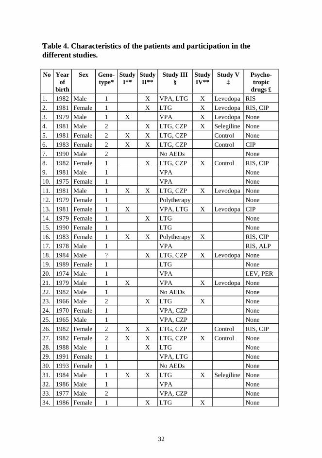
32
Table 4. Characteristics of the patients and participation in the different studies. No Year
of birth
Sex Geno-type*
Study I**
Study II**
Study III §
Study IV**
Study V ‡
Psycho-tropic
drugs £ 1. 1982 Male 1 X VPA, LTG X Levodopa RIS
2. 1981 Female 1 X LTG X Levodopa RIS, CIP
3. 1979 Male 1 X VPA X Levodopa None
4. 1981 Male 2 X LTG, CZP X Selegiline None
5. 1981 Female 2 X X LTG, CZP Control None
6. 1983 Female 2 X X LTG, CZP Control CIP
7. 1990 Male 2 No AEDs None
8. 1982 Female 1 X LTG, CZP X Control RIS, CIP
9. 1981 Male 1 VPA None
10. 1975 Female 1 VPA None
11. 1981 Male 1 X X LTG, CZP X Levodopa None
12. 1979 Female 1 Polytherapy None
13. 1981 Female 1 X VPA, LTG X Levodopa CIP
14. 1979 Female 1 X LTG None
15. 1990 Female 1 LTG None
16. 1983 Female 1 X X Polytherapy X RIS, CIP
17. 1978 Male 1 VPA RIS, ALP
18. 1984 Male ? X LTG, CZP X Levodopa None
19. 1989 Female 1 LTG None
20. 1974 Male 1 VPA LEV, PER
21. 1979 Male 1 X VPA X Levodopa None
22. 1982 Male 1 No AEDs None
23. 1966 Male 2 X LTG X None
24. 1970 Female 1 VPA, CZP None
25. 1965 Male 1 VPA, CZP None
26. 1982 Female 2 X X LTG, CZP Control RIS, CIP
27. 1982 Female 2 X X LTG, CZP X Control None
28. 1988 Male 1 X LTG None
29. 1991 Female 1 VPA, LTG None
30. 1993 Female 1 No AEDs None
31. 1984 Male 1 X X LTG X Selegiline None
32. 1986 Male 1 VPA None
33. 1977 Male 2 VPA, CZP None
34. 1986 Female 1 X LTG X None
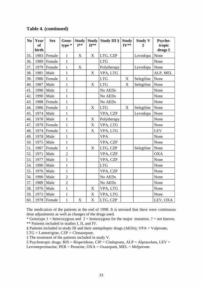
33
Table 4. (continued) No Year
of birth
Sex Geno-type *
Study I**
Study II**
Study III § Study IV**
Study V ‡
Psycho-tropic
drugs £ 35. 1983 Female 1 X X LTG, CZP Levodopa None
36. 1989 Female 1 LTG None
37. 1979 Female 1 X Polytherapy Levodopa None
38. 1981 Male 1 X VPA, LTG ALP, MEL
39. 1986 Female 1 LTG X Selegiline None
40. 1987 Male 1 X LTG X Selegiline None
41. 1990 Male 1 No AEDs None
42. 1990 Male 1 No AEDs None
43. 1988 Female 1 No AEDs None
44. 1986 Female 1 X LTG X Selegiline None
45. 1974 Male 1 VPA, CZP Levodopa None
46. 1978 Male 1 X Polytherapy None
47. 1979 Female 1 X VPA, LTG None
48. 1974 Female 1 X VPA, LTG LEV
49. 1978 Male 1 VPA None
50. 1975 Male 1 VPA, CZP None
51. 1987 Female 1 X LTG, CZP Selegiline None
52. 1971 Male 2 VPA, CZP OXA
53. 1977 Male 1 VPA, CZP None
54. 1990 Male 1 LTG None
55. 1976 Male 1 VPA, CZP None
56. 1990 Male 2 No AEDs None
57. 1989 Male 2 No AEDs None
58. 1976 Male 1 X VPA, LTG None
59. 1973 Male 1 X VPA, LTG None
60. 1978 Female 1 X X LTG, CZP LEV, OXA The medication of the patients at the end of 1998. It is stressed that there were continuous dose adjustments as well as changes of the drugs used. * Genotype 1 = heterozygous and 2 = homozygous for the major mutation. ? = not known. ** Patients included in studies I, II, and IV. § Patients included in study III and their antiepileptic drugs (AEDs); VPA = Valproate, LTG = Lamotrigine, CZP = Clonazepam. ‡ The treatment of the patients included in study V. £ Psychotropic drugs: RIS = Risperidone, CIP = Citalopram, ALP = Alprazolam, LEV = Levomepromazine, PER = Perazine, OXA = Oxazepam, MEL = Melperone.
34
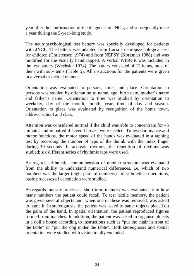
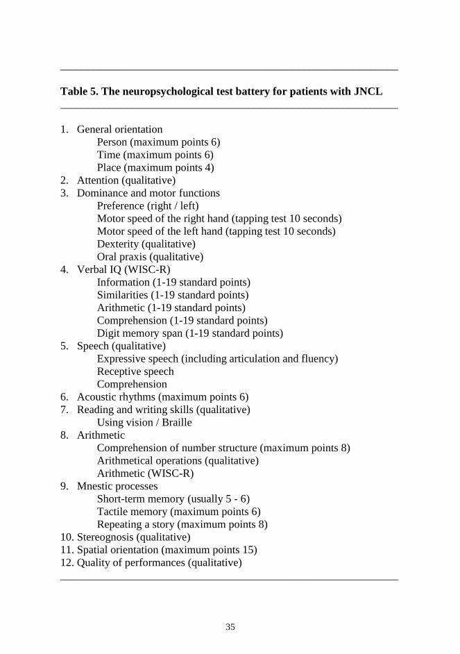
year after the confirmation of the diagnosis of JNCL, and subsequently once a year during the 5-year-long study. The neuropsychological test battery was specially developed for patients with JNCL. The battery was adapted from Luria’s neuropsychological test for children (Christensen 1974) and from NEPSY (Korkman 1988) and was modified for the visually handicapped. A verbal WISC-R was included in the test battery (Wechsler 1974). The battery consisted of 12 items, most of them with sub-items (Table 5). All instructions for the patients were given in a verbal or tactual manner. Orientation was evaluated to persons, time, and place. Orientation to persons was studied by orientation to name, age, birth date, mother’s name and father’s name. Orientation to time was studied by orientation to weekday, day of the month, month, year, time of day and season. Orientation to place was evaluated by recognition of the home town, address, school and class. Attention was considered normal if the child was able to concentrate for 45 minutes and impaired if several breaks were needed. To test dominance and motor functions, the motor speed of the hands was evaluated in a tapping test by recording the number of taps of the thumb with the index finger during 10 seconds. In acoustic rhythms, the repetition of rhythms was studied; six different series of rhythmic taps were used. As regards arithmetic, comprehension of number structure was evaluated from the ability to understand numerical differences, i.e. which of two numbers was the larger (eight pairs of numbers). In arithmetical operations, basic processes of calculation were studied. As regards mnestic processes, short-term memory was evaluated from how many numbers the patient could recall. To test tactile memory, the patient was given several objects and, when one of these was removed, was asked to name it. In stereognosis, the patient was asked to name objects placed on the palm of the hand. In spatial orientation, the patient reproduced figures formed from matches. In addition, the patient was asked to organise objects in a doll’s house according to instructions such as “put the chair in front of the table” or “put the dog under the table”. Both stereognosis and spatial orientation were studied with vision totally excluded.
35
_____________________________________________________________ Table 5. The neuropsychological test battery for patients with JNCL _____________________________________________________________ 1. General orientation
Person (maximum points 6) Time (maximum points 6) Place (maximum points 4)
2. Attention (qualitative) 3. Dominance and motor functions
Preference (right / left) Motor speed of the right hand (tapping test 10 seconds) Motor speed of the left hand (tapping test 10 seconds) Dexterity (qualitative) Oral praxis (qualitative)
4. Verbal IQ (WISC-R) Information (1-19 standard points) Similarities (1-19 standard points) Arithmetic (1-19 standard points) Comprehension (1-19 standard points) Digit memory span (1-19 standard points)
5. Speech (qualitative) Expressive speech (including articulation and fluency) Receptive speech Comprehension
6. Acoustic rhythms (maximum points 6) 7. Reading and writing skills (qualitative)
Using vision / Braille 8. Arithmetic
Comprehension of number structure (maximum points 8) Arithmetical operations (qualitative) Arithmetic (WISC-R)
9. Mnestic processes Short-term memory (usually 5 - 6) Tactile memory (maximum points 6) Repeating a story (maximum points 8)
10. Stereognosis (qualitative) 11. Spatial orientation (maximum points 15) 12. Quality of performances (qualitative) _____________________________________________________________
36
Two of the patients were not included in the analysis of changes in IQ; these patients were excluded as outliers. One was a female, homozygous for the major mutation, who was already mentally retarded when first seen at the age of 10. The other was a compound heterozygous male with an increase in IQ during the study period . Fifteen healthy children served as control subjects in the tapping test. Otherwise, no control patients were used, since both the WISC-R and the NEPSY have previously been standardised in healthy children. 3. DATA COLLECTION ON EPILEPSY AND AEDs Sixty patients were included in this retrospective and cross-sectional study. Data on seizures and AEDs were obtained from the medical records, by interviewing parents, and by contacting the personnel of the local hospitals or institutions where some of the patients were followed up. The age at onset of epilepsy and the seizure types were recorded. Seizures were classified according to the Proposal for revised clinical and electroencephalographic classification of the International League Against Epilepsy (1981). The seizure frequencies were taken as the total numbers of seizures during one year, and were divided into three categories; none, one to six seizures, and seven or more seizures a year. Even the mean duration of the seizures was checked. Seizure control was defined as good in the absence of seizures, and satisfactory if the frequency was six seizures or fewer a year. Poor seizure control was defined as a seizure frequency of more than six a year. As regards seizure duration, seizure control was defined as poor if there were any prolonged seizures (more than 20 minutes) during the study year. AED treatment had been initiated as monotherapy according to the common practice, and the dose was titrated upward until a seizure-free condition was reached or the seizures decreased. If there were severe side effects, an alternative AED was tried. However, if seizure control was insufficient despite the use of the maximal dose tolerated, combination therapy was initiated. In cases of refractory epilepsy, polytherapy was used. The effect of the first AED used was determined retrospectively. The seizure frequencies at baseline (2 months) and after 1 year were compared; a
37
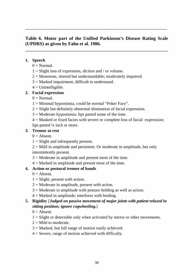
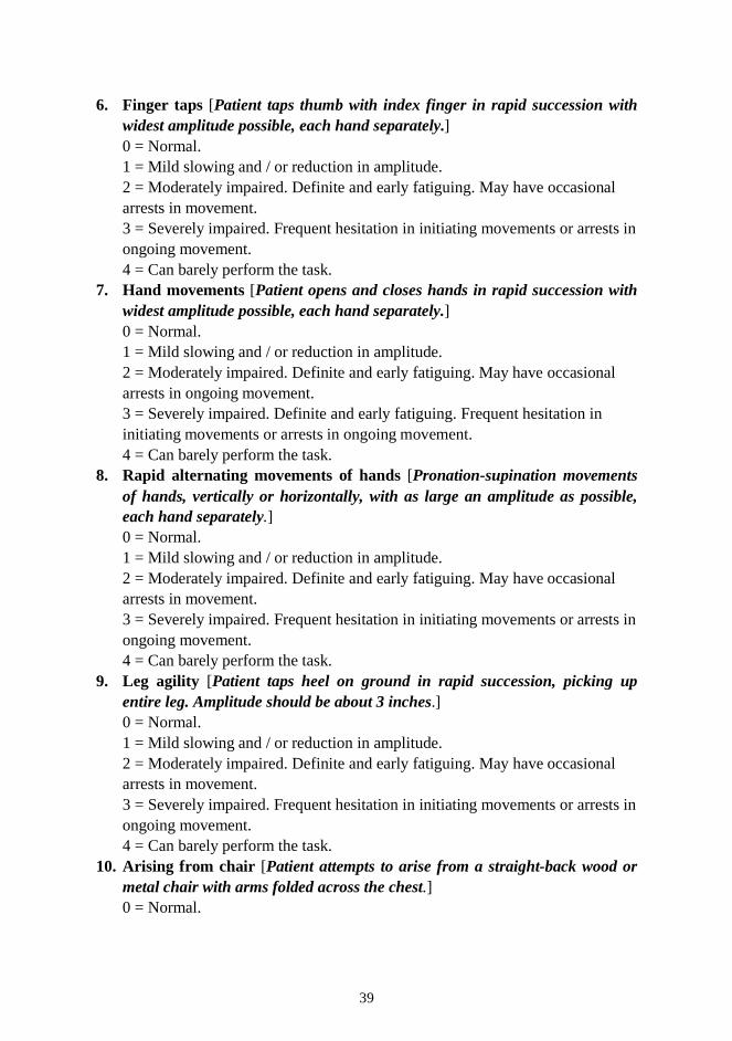
decrease in seizure frequency of over 50% was denoted as a decrease in the seizure frequency. In addition, the severity of the seizures and the general well-being of patients on LTG therapy (n=28) were compared. A decrease of over 50% in the length of the seizure and/or the post-ictal phase was denoted as a decrease in the severity of the seizures. A change in well-being was based on the general agreement of both doctors and parents. An increase in well-being included at least one of the following items: improvement in the quality of sleep with fewer settling problems and awakenings, in attention, and in daytime activity, and a decrease in irritation. The AED treatment of patients during the study year of 1998 was recorded and the patients were grouped on the basis of this treatment. For each group, the mean seizure frequency and the mean age of the patients were calculated, and the percentages of patients with good, satisfactory and poor seizure control were determined. The daily dosage of AEDs and the serum concentrations of VPA were checked. The AEDs used before the study year were studied as well. The side effects of these AEDs and the reasons for any withdrawal were clarified. In addition, any beneficial effects of these drugs, apart from seizure control, were assessed. 4. EVALUATION OF EXTRAPYRAMIDAL SYMPTOMS A ß-CIT-SPECT study was performed to assess the role of the dopamine transporters (DATs) and possible advantages of the dopaminergic drugs on the extrapyramidal symptoms in JNCL. Patients followed up in the Hospital for Children and Adolescents, 10 years of age or older, and with no previous antiparkinsonian treatment were invited. Four of the patients refused and 17 signed an informed consent. The mean age of the patients included was 15 years (range 10–31). For clinical evaluation of the extrapyramidal signs, the motor part of the Unified Parkinson´s Disease Rating Scale (Fahn et al. 1987) was applied. With this method, the more severe the patient’s symptoms, the higher is the score (Table 6). Scoring was performed systematically by the same person (L.Å.).
38
_____________________________________________________________ Table 6. Motor part of the Unified Parkinson’s Disease Rating Scale (UPDRS) as given by Fahn et al. 1986. ___________________________________________________________________ 1. Speech
0 = Normal. 1 = Slight loss of expression, diction and / or volume. 2 = Monotone, slurred but understandable; moderately impaired. 3 = Marked impairment, difficult to understand. 4 = Unintelligible.
2. Facial expression 0 = Normal. 1 = Minimal hypomimia, could be normal “Poker Face”. 2 = Slight but definitely abnormal diminution of facial expression. 3 = Moderate hypomimia; lips parted some of the time. 4 = Masked or fixed facies with severe or complete loss of facial expression; lips parted ¼ inch or more.
3. Tremor at rest 0 = Absent. 1 = Slight and infrequently present. 2 = Mild in amplitude and persistent. Or moderate in amplitude, but only intermittently present. 3 = Moderate in amplitude and present most of the time. 4 = Marked in amplitude and present most of the time.
4. Action or postural tremor of hands 0 = Absent. 1 = Slight; present with action. 2 = Moderate in amplitude, present with action. 3 = Moderate in amplitude with posture holding as well as action. 4 = Marked in amplitude; interferes with feeding.
5. Rigidity [Judged on passive movement of major joints with patient relaxed in sitting position; ignore cogwheeling.] 0 = Absent. 1 = Slight or detectable only when activated by mirror or other movements. 2 = Mild to moderate. 3 = Marked, but full range of motion easily achieved. 4 = Severe, range of motion achieved with difficulty.
39
6. Finger taps [Patient taps thumb with index finger in rapid succession with widest amplitude possible, each hand separately.] 0 = Normal. 1 = Mild slowing and / or reduction in amplitude. 2 = Moderately impaired. Definite and early fatiguing. May have occasional arrests in movement. 3 = Severely impaired. Frequent hesitation in initiating movements or arrests in ongoing movement. 4 = Can barely perform the task.
7. Hand movements [Patient opens and closes hands in rapid succession with widest amplitude possible, each hand separately.] 0 = Normal. 1 = Mild slowing and / or reduction in amplitude. 2 = Moderately impaired. Definite and early fatiguing. May have occasional arrests in ongoing movement. 3 = Severely impaired. Definite and early fatiguing. Frequent hesitation in initiating movements or arrests in ongoing movement. 4 = Can barely perform the task.
8. Rapid alternating movements of hands [Pronation-supination movements of hands, vertically or horizontally, with as large an amplitude as possible, each hand separately.] 0 = Normal. 1 = Mild slowing and / or reduction in amplitude. 2 = Moderately impaired. Definite and early fatiguing. May have occasional arrests in movement. 3 = Severely impaired. Frequent hesitation in initiating movements or arrests in ongoing movement. 4 = Can barely perform the task.
9. Leg agility [Patient taps heel on ground in rapid succession, picking up entire leg. Amplitude should be about 3 inches.] 0 = Normal. 1 = Mild slowing and / or reduction in amplitude. 2 = Moderately impaired. Definite and early fatiguing. May have occasional arrests in movement. 3 = Severely impaired. Frequent hesitation in initiating movements or arrests in ongoing movement. 4 = Can barely perform the task.
10. Arising from chair [Patient attempts to arise from a straight-back wood or metal chair with arms folded across the chest.] 0 = Normal.
40
1 = Slow; or may need more than one attempt. 2 = Pushes self up from arms of seat. 3 = Tends to fall back and may have to try more than one time, but can get up without help. 4 = Unable to arise without help.
11. Posture 0 = Normal erect. 1 = Not quite erect, slightly stooped posture; could be normal for older person. 2 = Moderately stooped posture, definitely abnormal; can be slightly leaning to one side. 3 = Severely stooped posture with kyphosis; can be moderately leaning to one side. 4 = Marked flexion with extreme abnormality of posture.
12. Gait 0 = Normal. 1 = Walks slowly, may shuffle with short steps, but no festination (hastening steps) or propulsion. 2 = Walks with difficulty, but requires little or no assistance; may have some festination, short steps or propulsion. 3 = Severe disturbance of gait, requiring assistance. 4 = Cannot walk at all, even with assistance.
13. Postural stability [Response to sudden, strong posterior displacement produced by pull on shoulders while patient erect with eyes open and feet slightly apart. Patient is prepared, and can have had some practice runs.] 0 = Normal. 1 = Retropulsion, but recovers unaided. 2 = Absence of postural response; would fall if not caught by examiner. 3 = Very unstable, tends to lose balance spontaneously. 4 = Unable to stand without assistance.
14. Body bradykinesia and hypokinesia [Combining slowness, hesitancy, decreased armswing, small amplitude, and poverty of movements in general.] 0 = None. 1 = Minimal slowness, giving movement a deliberate character; could be normal for some persons. Possibly reduced amplitude. 2 = Mild degree of slowness and poverty of movement which is definitely abnormal. Alternatively, some reduced amplitude. 3 = Moderate slowness, poverty or small amplitude of movement. 4 = Marked slowness, poverty or small amplitude of movement.
___________________________________________________________________
41
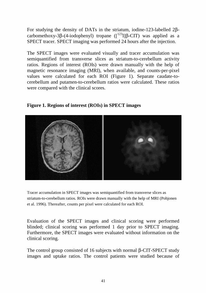
For studying the density of DATs in the striatum, iodine-123-labelled 2β-carbomethoxy-3β-(4-iodophenyl) tropane ([123I]β-CIT) was applied as a SPECT tracer. SPECT imaging was performed 24 hours after the injection. The SPECT images were evaluated visually and tracer accumulation was semiquantified from transverse slices as striatum-to-cerebellum activity ratios. Regions of interest (ROIs) were drawn manually with the help of magnetic resonance imaging (MRI), when available, and counts-per-pixel values were calculated for each ROI (Figure 1). Separate caudate-to-cerebellum and putamen-to-cerebellum ratios were calculated. These ratios were compared with the clinical scores. Figure 1. Regions of interest (ROIs) in SPECT images Tracer accumulation in SPECT images was semiquantified from transverse slices as
striatum-to-cerebellum ratios. ROIs were drawn manually with the help of MRI (Pohjonen
et al. 1996). Thereafter, counts per pixel were calculated for each ROI. Evaluation of the SPECT images and clinical scoring were performed blinded; clinical scoring was performed 1 day prior to SPECT imaging. Furthermore, the SPECT images were evaluated without information on the clinical scoring. The control group consisted of 16 subjects with normal β-CIT-SPECT study images and uptake ratios. The control patients were studied because of
42
dystonia or suspected parkinsonism. The mean age of this control group was 43 years (range 14-56). For ethical reasons, age-matched controls were not available.
5. MAGNETIC RESONANCE IMAGING All 14 patients participating in the neuropsychological test battery were examined with a 1.0 T MRI at the beginning of the study. After a mean follow-up period of 5 years, 11 patients were re-examined with either a 1.0 or a 1.5 T MRI. After T1-weighted sagittal images, T2-weighted axial slices were obtained and the changes in atrophy and signal intensity were graded as previously described (Autti et al. 1996). Of the 17 patients participating in the SPECT study, 16 underwent a 1.5 T MRI 1 day prior to SPECT imaging. This MRI study was unlinked to the previous MRI study. One patient participating in the SPECT study, did not consent to undergo the MRI study. Axial T2-weighted and sagittal three-dimensional T1-weighted images were obtained. The signal intensities of the striatum and thalami were measured from T2-weighted images as previously described (Autti et al. 1994) and the ratios between the signal intensities of the striatum and thalami were calculated. Cerebral and cerebellar atrophy were classified as mild, moderate, or severe (Autti et al. 1996). As a control group for this MRI study, we used 16 age-matched healthy volunteers. 6. THERAPY WITH ANTIPARKINSONIAN DRUGS After the ß-CIT-SPECT and MRI studies had been performed, the patients included in the study participated in an open study on the effects of the antiparkinsonian treatment. In addition, other patients, including those who did not give their permission for the SPECT study, were included in the study on the antiparkinsonian treatment. The mean age of the patients at the start of the study was 15 years (range 10–23). On the basis of the UPDRS score, treatment was initiated with either selegiline or levodopa. When the UPDRS score exceeded 10, therapy was initiated with selegiline (n=6). If the UPDRS score at the first evaluation was already 20 or more, treatment was initiated with levodopa (n=10). Five
43
patients with no parkinsonian therapy served as control subjects. These included patients with severe psychotic behaviour, contraindicating antiparkinsonian treatment. In addition, the control group included patients who did not give permission for drug treatment of parkinsonism. Selegiline was initiated at 2.5 mg a day and after 2 weeks the dose was increased to 5 mg a day. The dose of selegiline was then increased in steps up to 7.5–10 mg a day. Levodopa was used in combination with carbidopa (100/25) to prevent peripheral decarboxylation. Levodopa therapy was initiated with a dose of 50 mg a day and the dose increased by 50 mg every 2 weeks until a maintenance dose of 300-600 mg a day was achieved. Throughout the therapy, all the patients were followed up regularly for dose adjustments and for possible side effects. During visits to out-patient departments, patients were evaluated by a physician and a physiotherapist. Rehabilitation was kept constant during the follow-up. 7. STATISTICAL METHODS In study II, Fisher’s exact test was used to examine the associations between seizure control and sex, genotype, and AED group. Regression analysis was used to test whether seizure frequency was correlated with age or with onset of epilepsy. In study IV, SPECT uptake ratios and MRI signal intensity ratios were compared in patients with extrapyramidal signs and in controls, using the Mann-Whitney U test. Multiple regression analysis was used to test for correlations between UPDRS, the striatal dopamine uptake ratios in SPECT, and the signal intensity ratios in MRI. In study V, the UPDRS score after 1 year was compared with the score at the start of the study, and the changes in the scores were studied in the different groups, using ANOVA.
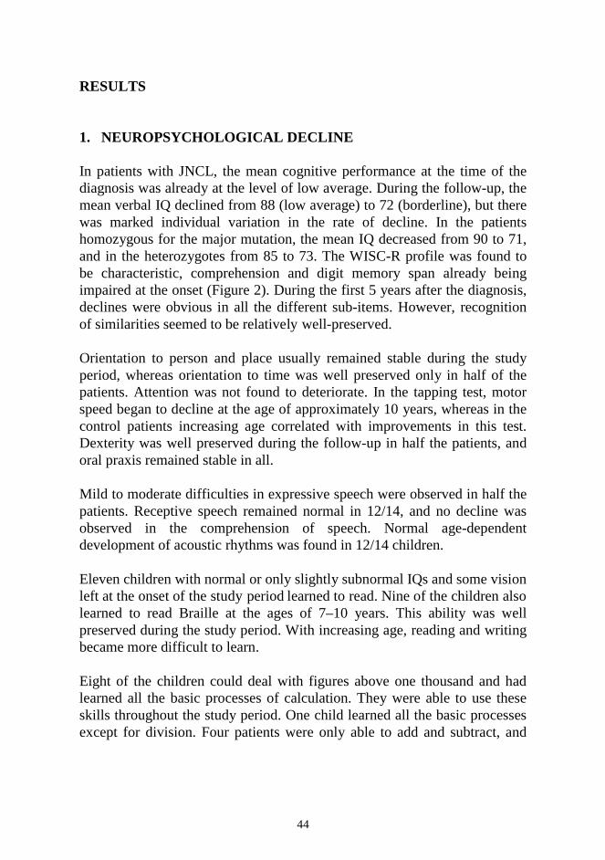
44
RESULTS 1. NEUROPSYCHOLOGICAL DECLINE In patients with JNCL, the mean cognitive performance at the time of the diagnosis was already at the level of low average. During the follow-up, the mean verbal IQ declined from 88 (low average) to 72 (borderline), but there was marked individual variation in the rate of decline. In the patients homozygous for the major mutation, the mean IQ decreased from 90 to 71, and in the heterozygotes from 85 to 73. The WISC-R profile was found to be characteristic, comprehension and digit memory span already being impaired at the onset (Figure 2). During the first 5 years after the diagnosis, declines were obvious in all the different sub-items. However, recognition of similarities seemed to be relatively well-preserved. Orientation to person and place usually remained stable during the study period, whereas orientation to time was well preserved only in half of the patients. Attention was not found to deteriorate. In the tapping test, motor speed began to decline at the age of approximately 10 years, whereas in the control patients increasing age correlated with improvements in this test. Dexterity was well preserved during the follow-up in half the patients, and oral praxis remained stable in all. Mild to moderate difficulties in expressive speech were observed in half the patients. Receptive speech remained normal in 12/14, and no decline was observed in the comprehension of speech. Normal age-dependent development of acoustic rhythms was found in 12/14 children. Eleven children with normal or only slightly subnormal IQs and some vision left at the onset of the study period learned to read. Nine of the children also learned to read Braille at the ages of 7–10 years. This ability was well preserved during the study period. With increasing age, reading and writing became more difficult to learn. Eight of the children could deal with figures above one thousand and had learned all the basic processes of calculation. They were able to use these skills throughout the study period. One child learned all the basic processes except for division. Four patients were only able to add and subtract, and
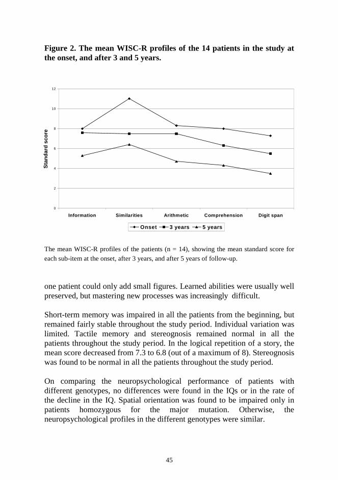
45
Figure 2. The mean WISC-R profiles of the 14 patients in the study at the onset, and after 3 and 5 years.
The mean WISC-R profiles of the patients (n = 14), showing the mean standard score for
each sub-item at the onset, after 3 years, and after 5 years of follow-up. one patient could only add small figures. Learned abilities were usually well preserved, but mastering new processes was increasingly difficult. Short-term memory was impaired in all the patients from the beginning, but remained fairly stable throughout the study period. Individual variation was limited. Tactile memory and stereognosis remained normal in all the patients throughout the study period. In the logical repetition of a story, the mean score decreased from 7.3 to 6.8 (out of a maximum of 8). Stereognosis was found to be normal in all the patients throughout the study period. On comparing the neuropsychological performance of patients with different genotypes, no differences were found in the IQs or in the rate of the decline in the IQ. Spatial orientation was found to be impaired only in patients homozygous for the major mutation. Otherwise, the neuropsychological profiles in the different genotypes were similar.
0
2
4
6
8
10
12
Information Similarities Arithmetic Comprehension Digit span
Sta
nd
ard
sco
re
Onset 3 years 5 years
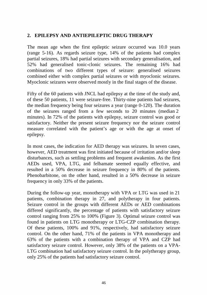
46
2. EPILEPSY AND ANTIEPILEPTIC DRUG THERAPY The mean age when the first epileptic seizure occurred was 10.0 years (range 5-16). As regards seizure type, 14% of the patients had complex partial seizures, 18% had partial seizures with secondary generalisation, and 52% had generalised tonic-clonic seizures. The remaining 16% had combinations of two different types of seizure: generalised seizures combined either with complex partial seizures or with myoclonic seizures. Myoclonic seizures were observed mostly in the final stages of the disease. Fifty of the 60 patients with JNCL had epilepsy at the time of the study and, of these 50 patients, 11 were seizure-free. Thirty-nine patients had seizures, the median frequency being four seizures a year (range 0-120). The duration of the seizures ranged from a few seconds to 20 minutes (median 2 minutes). In 72% of the patients with epilepsy, seizure control was good or satisfactory. Neither the present seizure frequency nor the seizure control measure correlated with the patient’s age or with the age at onset of epilepsy. In most cases, the indication for AED therapy was seizures. In seven cases, however, AED treatment was first initiated because of irritation and/or sleep disturbances, such as settling problems and frequent awakenins. As the first AEDs used, VPA, LTG, and felbamate seemed equally effective, and resulted in a 50% decrease in seizure frequency in 80% of the patients. Phenobarbitone, on the other hand, resulted in a 50% decrease in seizure frequency in only 33% of the patients. During the follow-up year, monotherapy with VPA or LTG was used in 21 patients, combination therapy in 27, and polytherapy in four patients. Seizure control in the groups with different AEDs or AED combinations differed significantly, the percentage of patients with satisfactory seizure control ranging from 25% to 100% (Figure 3). Optimal seizure control was found in patients on LTG monotherapy or LTG-CZP combination therapy. Of these patients, 100% and 91%, respectively, had satisfactory seizure control. On the other hand, 71% of the patients in VPA monotherapy and 63% of the patients with a combination therapy of VPA and CZP had satisfactory seizure control. However, only 38% of the patients on a VPA-LTG combination had satisfactory seizure control. In the polytherapy group, only 25% of the patients had satisfactory seizure control.
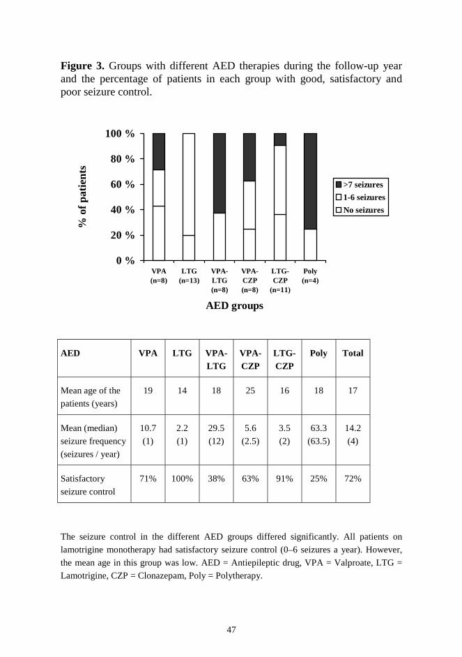
47
Figure 3. Groups with different AED therapies during the follow-up year and the percentage of patients in each group with good, satisfactory and poor seizure control.
AED VPA LTG VPA- LTG
VPA-CZP
LTG-CZP
Poly Total
Mean age of the
patients (years)
19 14 18 25 16 18 17
Mean (median)
seizure frequency
(seizures / year)
10.7
(1)
2.2
(1)
29.5
(12)
5.6
(2.5)
3.5
(2)
63.3
(63.5)
14.2
(4)
Satisfactory
seizure control
71% 100% 38% 63% 91% 25% 72%
The seizure control in the different AED groups differed significantly. All patients on
lamotrigine monotherapy had satisfactory seizure control (0–6 seizures a year). However,
the mean age in this group was low. AED = Antiepileptic drug, VPA = Valproate, LTG =
Lamotrigine, CZP = Clonazepam, Poly = Polytherapy.
0 %
20 %
40 %
60 %
80 %
100 %
VPA(n=8)
LTG(n=13)
VPA-LTG(n=8)
VPA-CZP(n=8)
LTG-CZP
(n=11)
Poly(n=4)
AED groups
% o
f pa
tien
ts
>7 seizures
1-6 seizures
No seizures
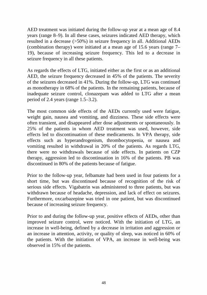
48
AED treatment was initiated during the follow-up year at a mean age of 8.4 years (range 8–9). In all these cases, seizures indicated AED therapy, which resulted in a decrease (>50%) in seizure frequency in all. Additional AEDs (combination therapy) were initiated at a mean age of 15.6 years (range 7–19), because of increasing seizure frequency. This led to a decrease in seizure frequency in all these patients. As regards the effects of LTG, initiated either as the first or as an additional AED, the seizure frequency decreased in 45% of the patients. The severity of the seizures decreased in 41%. During the follow-up, LTG was continued as monotherapy in 68% of the patients. In the remaining patients, because of inadequate seizure control, clonazepam was added to LTG after a mean period of 2.4 years (range 1.5–3.2). The most common side effects of the AEDs currently used were fatigue, weight gain, nausea and vomiting, and dizziness. These side effects were often transient, and disappeared after dose adjustments or spontaneously. In 25% of the patients in whom AED treatment was used, however, side effects led to discontinuation of these medicaments. In VPA therapy, side effects such as hyperandrogenism, thrombocytopenia, or nausea and vomiting resulted in withdrawal in 20% of the patients. As regards LTG, there were no withdrawals because of side effects. In patients on CZP therapy, aggression led to discontinuation in 16% of the patients. PB was discontinued in 80% of the patients because of fatigue. Prior to the follow-up year, felbamate had been used in four patients for a short time, but was discontinued because of recognition of the risk of serious side effects. Vigabatrin was administered to three patients, but was withdrawn because of headache, depression, and lack of effect on seizures. Furthermore, oxcarbazepine was tried in one patient, but was discontinued because of increasing seizure frequency. Prior to and during the follow-up year, positive effects of AEDs, other than improved seizure control, were noticed. With the initiation of LTG, an increase in well-being, defined by a decrease in irritation and aggression or an increase in attention, activity, or quality of sleep, was noticed in 60% of the patients. With the initiation of VPA, an increase in well-being was observed in 15% of the patients.
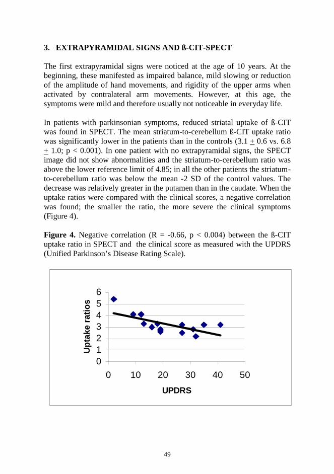
49
3. EXTRAPYRAMIDAL SIGNS AND ß-CIT-SPECT The first extrapyramidal signs were noticed at the age of 10 years. At the beginning, these manifested as impaired balance, mild slowing or reduction of the amplitude of hand movements, and rigidity of the upper arms when activated by contralateral arm movements. However, at this age, the symptoms were mild and therefore usually not noticeable in everyday life. In patients with parkinsonian symptoms, reduced striatal uptake of ß-CIT was found in SPECT. The mean striatum-to-cerebellum ß-CIT uptake ratio was significantly lower in the patients than in the controls (3.1 + 0.6 vs. 6.8 + 1.0; p < 0.001). In one patient with no extrapyramidal signs, the SPECT image did not show abnormalities and the striatum-to-cerebellum ratio was above the lower reference limit of 4.85; in all the other patients the striatum-to-cerebellum ratio was below the mean -2 SD of the control values. The decrease was relatively greater in the putamen than in the caudate. When the uptake ratios were compared with the clinical scores, a negative correlation was found; the smaller the ratio, the more severe the clinical symptoms (Figure 4). Figure 4. Negative correlation (R = -0.66, p < 0.004) between the ß-CIT uptake ratio in SPECT and the clinical score as measured with the UPDRS (Unified Parkinson’s Disease Rating Scale).
0123456
0 10 20 30 40 50
UPDRS
Up
take
rat
ios
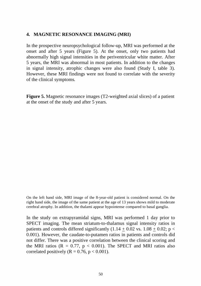
50
4. MAGNETIC RESONANCE IMAGING (MRI) In the prospective neuropsychological follow-up, MRI was performed at the onset and after 5 years (Figure 5). At the onset, only two patients had abnormally high signal intensities in the periventricular white matter. After 5 years, the MRI was abnormal in most patients. In addition to the changes in signal intensity, atrophic changes were also found (Study I, table 3). However, these MRI findings were not found to correlate with the severity of the clinical symptoms. Figure 5. Magnetic resonance images (T2-weighted axial slices) of a patient at the onset of the study and after 5 years. On the left hand side, MRI image of the 8-year-old patient is considered normal. On the right hand side, the image of the same patient at the age of 13 years shows mild to moderate cerebral atrophy. In addition, the thalami appear hypointense compared to basal ganglia. In the study on extrapyramidal signs, MRI was performed 1 day prior to SPECT imaging. The mean striatum-to-thalamus signal intensity ratios in patients and controls differed significantly (1.14 + 0.02 vs. 1.08 + 0.02; p < 0.001). However, the caudate-to-putamen ratios in patients and controls did not differ. There was a positive correlation between the clinical scoring and the MRI ratios (R = 0.77, p < 0.001). The SPECT and MRI ratios also correlated positively (R = 0.76, p < 0.001).
51
5. TREATMENT OF EXTRAPYRAMIDAL SIGNS Ten patients had UPDRS scores of 20 or more at the onset, and were started on levodopa therapy. Their mean age at the initiation of the treatment was 16 years (range 13–23). In 1 year, the mean UPDRS decreased from 29 to 26. However, there was quite wide variation in the response to levodopa, and in four cases the UPDRS score increased during the follow-up, while in three cases the decrease in the UPDRS score was 10 or more. Even in three of the four patients showing increased UPDRS scores at 1 year, a short-term benefit of levodopa was noticed, for at the 6-month visit the UPDRS score was below the baseline. Six patients had UPDRS scores of 10–19 and started therapy with selegiline; their mean age was 13 years (range 11–17). In the group with selegiline treatment, the mean UPDRS score at the onset was 13 and after 1 year on therapy 16. Individually, only two of the patients had a slight (2 and 3 points) decrease in the UPDRS score. Five patients with a mean age of 15 years (range 14–16) had no therapy for their extrapyramidal symptoms. One of these patients received levodopa (50–100 mg/day) for one month, but the therapy was discontinued because of side effects. None of the other patients in this group received levodopa or selegiline on any occasion. In this group, the mean UPDRS score was 16 at the onset and after 1 year of follow-up, it was 26. In all but one patient the individual UPDRS score increased during the follow-up. When comparing the groups on different treatments, the change in the UPDRS score in patients on levodopa therapy was found to differ from that in the patients with no antiparkinsonian therapy (p=0.01). The change in the UPDRS score in the patients on selegiline treatment, on the other hand, did not differ from that in the patients on levodopa therapy (p=0.20) or in the patients with no antiparkinsonian treatment (p=0.16). In one case, therapy with levodopa was discontinued after 1 month while still in the titration phase, because of restlessness, agitation and frightening visual hallucinations. Otherwise, no side effects of levodopa were observed. As regards the patients on selegiline therapy, one patient had nausea at the initiation of the treatment.
52
DISCUSSION Juvenile neuronal ceroid lipofuscinosis is a progressive, neurodegenerative disease with multiple symptoms originating from the CNS, including mental decline, epilepsy and parkinsonian symptoms. Thus far, no curative treatment is available. However, we found that symptomatic drug treatment may bring about a relief in the everyday life of the patients, and so improve the quality of life. From a psychological point of view, despite the devastating disease process, even minor improvements are important. 1. NEUROPSYCHOLOGICAL DECLINE The neuropsychological test battery used in the follow-up of patients with JNCL was found to be reliable, quick and easy to use. Furthermore, the children enjoyed the test. The first abnormal findings in the WISC-R were already noticed during the first years of the follow-up. Of the 14 patients included in study I, only four had an IQ of over 100 at the first evaluation. At this early stage, however, the decline in cognitive abilities was not noticeable in everyday life. Later on, the test results became increasingly abnormal, characterising the decline in the different cognitive functions. The best sub-item throughout the study was similarities, whereas digit memory span was found to decline most rapidly. This is in accord with an earlier study, in which the digit memory span was one of the first functions to become impaired (Lou and Kristensen 1973) Except for one compound heterozygous male, only a few differences in the neuropsychological test battery were marked between homozygous and compound heterozygous patients. In light of the earlier reports, this was surprising (Järvelä et al. 1997). However, spatial orientation was impaired only in the homozygous patients. The increasing neurological dysfunction in patients with JNCL may result from accumulation of intracellular storage material. However, the precise pathogenesis is still unknown. Furthermore, other factors, including epileptic activity, may contribute to the mental and motor deterioration in JNCL. In MRI, progression of the disease was reflected as signal intensity changes and dilatation of the CSF spaces. However, the degree of atrophy
53
did not correlate with the severity of the clinical symptoms. Indeed, the neuropsychological findings seemed to reflect the progression of the disease better than the conventional MRI studies. Thus, functional impairment apparently precedes the structural abnormalities. The early mental impairment in JNCL emphasises the importance of early rehabilitation. One of the most important points is to teach Braille reading at an early stage, even if there is still enough vision for normal reading or reading with the help of visual aids. After the age of 10, learning Braille becomes difficult or even impossible, because of the short digit memory span, declining IQ, and motor impairment of the hands. 2. EPILEPSY AND ANTIEPILEPTIC DRUG THERAPY The progressive myoclonic epilepsy in JNCL may sometimes be refractory. In the cross-sectional study of the epilepsy in JNCL, however, seizure control was satisfactory in most of the patients. The first epileptic seizure was noticed at the mean age of 10, the most common seizure types being generalised tonic-clonic seizures. No tendency was found for increasing seizure control with age, although this has been reported earlier (Boustany 1992). AED therapy was first initiated as monotherapy, and the dose was titrated upwards until seizures decreased or side effects were observed. As the first AEDs, VPA, LTG, and FBM were found to be equally effective, when studied retrospectively. After LTG was proved valuable in JNCL, it was preferred as the first AED. Prior to this, valproate was used as the first-line AED, and was continued unless severe side effects developed. If seizure control was insufficient in monotherapy, despite the maximal doses tolerated, combination therapy was initiated. When choosing the additional AED, no specific strategy was applied. Thus, the groups on combination therapy in the cross-sectional study may be considered comparable. However, the size of each group was small, which may have influenced the results. Seizure control in patients with LTG monotherapy or LTG-CZP combination therapy was satisfactory in almost all the patients. However, the mean ages of these groups were low and thus progression of the disease
54
was still limited, which may have affected the results, although, in general, seizure frequency was not found to correlate with age. In VPA monotherapy or VPA-CZP combination therapy seizure control was satisfactory in most of the patients. Surprisingly, the majority of patients receiving combination of VPA and LTG had poor seizure control, despite the reported synergistic effect of these AEDs (Brodie and Yuen 1997) and the previous observation that this combination was effective in infantile NCL (Åberg et al. 1997). When examining the effects on the epilepsy of LTG, used either as monotherapy or in combination therapy, a decrease in the frequency or severity of seizures was found in about half of the patients and an increase in well-being in more than half. The different types of seizure responded to LTG treatment almost without exception, but severe myoclonia did not decrease or increase. This characterises the variable effect of LTG on myoclonia (Wallace 1990, Gibbs et al. 1992, Timmings and Richens 1993, Schlumberger et al. 1994). In two girls, in whom LTG was initiated because of the side effects of VPA, the seizure frequency increased during LTG monotherapy. Thus, in these cases, VPA was possibly more effective as monotherapy. In two other girls, in whom VPA was replaced by LTG, no change in the seizure frequency was observed. Side effects resulting in withdrawal of an AED were observed in a quarter of the patients, mostly in patients on PB therapy, but also in patients on VPA or CZP therapy. However, in LTG therapy there were no withdrawals because of side effects. In addition to absence of severe side effects, LTG seemed to have favourable cognitive and behavioural effects in these patients, as also noticed in other studies on patients with intellectual disability (Besag 1999). 3. EVALUATION OF EXTRAPYRAMIDAL SIGNS Patients with JNCL have gradually deteriorating motor impairment. Underlying the motor impairment, both pyramidal and extrapyramidal disturbances are seen (Hofman et al. 1999). However, the pyramidal signs, if noticed at all, are usually less marked. In all the patients with elevated UPDRS scores a reduced DAT density was found in the striatum, indicating nigrostriatal dopamine deficiency. The reduced DAT density was more pronounced in the putamen than in the
55
caudate, also a common finding in patients with idiopathic PD. A negative correlation was found between the UPDRS score and the DAT density in SPECT, which accords with the findings in patients with Parkinson’s disease (Seibyl et al. 1995, Rinne et al. 1995). This also accords with the PET study on nine patients with JNCL, in whom a correlation was found between extrapyramidal symptoms and a decline in [18F]fluorodopa uptake in the putamen (Ruottinen et al. 1997). In patients with Alzheimer´s disease, reduced [11C]β-CFT uptake was found as well, reduction of the uptake correlating with the severity of the extrapyramidal symptoms (Rinne et al. 1998). However, the putamen and the caudate nucleus were equally affected, in contrast to patients with JNCL. Therefore, the SPECT findings in patients with JNCL resemble those found in patients with Parkinson’s disease. The control subjects in the SPECT study were older than the patients with JNCL. As the transporter density is known to decline with age (Van Dyck et al. 1995), the decrease in striatal DATs in JNCL was presumably underestimated. However, as there are no β-CIT studies on healthy children under 18 years of age, it is not known whether the transporter ratio in children can be accurately extrapolated from studies on adults. In the present study, the striatal uptake of β-CIT was reduced even in patients 10-11 years of age. In the clinical evaluation, these patients usually had mild extrapyramidal symptoms, including impaired balance, slowing or reduction of the amplitude of hand movements and rigidity of the upper arms when activated by other movements. Thus, extrapyramidal symptoms seem to develop earlier than was previously reported by us (Järvelä et al. 1997), although, at this age, the symptoms may not have a noticeable effect on everyday life. The accumulations of β-CIT were calculated as striatum-to-cerebellum ratios, since accumulation in the cerebellum is considered non-specific. In JNCL, mild or moderate cerebellar atrophy usually appears after 14 years of age (Autti et al. 1996). Cerebellar changes might therefore influence the uptake ratios, although a marked effect is unlikely. In the brain, ß-CIT traces DATs, serotonin transporters (SERTs), and to some extent also noradrenaline transporters (Laruelle et al. 1994). Whereas DATs are situated predominantly in the basal ganglia, SERTs are found in the hypothalamus and midbrain (Ciliax et al. 1995, Jagust et al. 1996).
56
Indeed, ß-CIT binding in the striatum can be displaced by a DAT inhibitor but not by a SERT inhibitor (Laruelle et al. 1993). In the diencephalon and brainstem, on the other hand, the ß-CIT binding can be displaced by the SERT inhibitor, but not be the DAT inhibitor. Furthermore, the peak activity of ß-CIT in the hypothalamus-midbrain area is observed at 4 hours, whereas the striatal activity peaks at around 20 hours (Brucke et al. 1993). Therefore, ß-CIT binding to these two transporters can be differentiated by region of interest and by timing of imaging. In the present study, imaging was performed 24 hours after the injection and the regions of interest were drawn to the striatum, and thus, ß-CIT binding to SERTs can be considered minimal. Although first used to reveal the DATs in Parkinson’s disease, ß-CIT has also been employed to trace the SERTs. In patients with impulsive aggression (Tiihonen et al. 1997) and in alcoholics (Heinz et al. 1998), reduced SERT availability was found in the hypothalamus and midbrain, whereas DAT availability did not differ from controls. Surprisingly, in a ß-CIT study on depressive children and adolescents, increased SERT availability was found, as compared with non-depressive children (Dahlström et al. 2000). Again, the striatal DAT availability in depressive and non-depressive children did not differ. In adult patients with major depression, however, increased striatal DAT density was found (Laasonen-Balk et al. 1999). In patients with JNCL, who often suffer from depression, SERT availability has not been studied thus far. Using MRI, increased striatum-to-thalamus signal intensity ratios were observed on the T2-weighted images. It is evident that the pathological process, also found in the thalamus (Autti et al. 1997), may interfere with this ratio. However, in a MRI study on patients with parkinsonism, it was impossible to distinguish the patients with non-idiopathic parkinsonism from those with idiopathic parkinsonism on the basis of signal intensity measurements, although the “signal intensity index” of the putamen was lower in the patients with secondary parkinsonism (Schwarz et al. 1999). The MRI signal intensity ratios showed a positive correlation with the clinical scoring and a negative correlation with the striatum-to-cerebellum ratios in SPECT. Thus, the more symptoms the patient had, the darker the thalami. These findings provide evidence for the contribution of thalamic dysfunction, in addition to nigrostriatal and striatal dysfunction, as the pathomechanism for the parkinsonian symptoms in JNCL.
57
4. ANTIPARKINSONIAN DRUGS The decrease in striatal dopamine transporter density offered a rational basis for a trial of dopaminergic drugs in the treatment of JNCL. In the open study on the effects of antiparkinsonian treatment on patients with JNCL, a favourable response to levodopa therapy in JNCL was observed, although there was great variation in this response. The design was open, since only limited data are available on the effect of levodopa and there are no data on the effect of selegiline in patients with JNCL. The patients for the different treatment groups were not selected randomly. The antiparkinsonian treatment depended on the UPDRS score. When the UPDRS score exceeded 10, selegiline was used, and when the score exceeded 20, the patient was given levodopa. Thus, in the levodopa group, the baseline UPDRS score was higher, reflecting further progression of the disease. In addition, the groups differed in genotype. In the control group, compound heterozygous patients were over-represented, whereas in the levodopa group, all the patients were homozygotes. As compound heterozygous patients may have a slower progression of extrapyramidal symptoms (Järvelä et al. 1997), the positive effect of levodopa treatment found in the present study is further emphasised. In patients without any treatment for their parkinsonian symptoms, the mean UPDRS score increased more than in the group on selegiline, but this difference was not statistically significant. Thus, it is difficult to judge whether selegiline has an effect in JNCL or not. At least in Parkinson’s disease, selegiline has a favourable effect, which may be either symptomatic, neuroprotective, or both (Olanow et al. 1995, Schulzer et al. 1992). Both levodopa and selegiline were generally well tolerated by patients with JNCL. However, in one patient on levodopa therapy, visual hallucinations, agitation, and anxiety occurred one month after initiation of levodopa. This patient had had psychotic behaviour even prior to the treatment, but since levodopa is known to induce psychosis, the therapy was discontinued and treatment was initiated with risperidone.
58
There were also two other patients with previous psychotic symptoms having levodopa treatment. However, when on continuous risperidone therapy, these patients had no aggravation of the psychotic symptoms during the levodopa therapy. These patients also responded to levodopa treatment, as did the patients without antipsychotic treatment. Although risperidone has an affinity for dopamine (D2) receptors, and it thus has the potential to induce extrapyramidal symptoms, the affinity is not of the same extent as in the case of older antipsychotics (Schotte et al. 1996). The results of the present study indicate a favourable effect of levodopa treatment in patients with JNCL, at least in the short term. This positive effect of dopaminergic drugs is in accord with the decreased striatal dopaminergic transporter density in JNCL. Since in JNCL there is also pathology in the thalamus and in the basal ganglia (Autti et al. 1996), it is understandable that the response to levodopa is milder and probably also of shorter duration than that seen in Parkinson’s disease. Therefore, in the future, long-term controlled trials are indicated to confirm the present results. 5. POLYPHARMACY IN JNCL Because of the complicated clinical phenotype in JNCL, several CNS drugs are used simultaneously. This polypharmacy may give rise to unwanted interactions. In the use of AEDs, metabolic interactions are often seen. Some of the AEDs, including phenobarbitone, phenytoin, and carbamazepine, result in induction of hepatic enzymes. These AEDs are no longer used in Finnish patients with JNCL. Of the AEDs used at present, VPA has potential for inhibiting hepatic enzymes (Brodie 1992). Thus, in the concomitant use of VPA and LTG, the half-life of LTG is extended, and the doses must be adjusted (Yuen et al. 1992). On the other hand, LTG, which was found to be favourable in patients with JNCL, does not alter the metabolism of the other AEDs in general (Eriksson et al. 1996). A rare side effect seen in conjunction with the psychotropic polypharmacy, is the serotonergic syndrome (Bodner et al. 1995). This results from imbalance of the neurotransmitters, mainly the high serotonin, in the CNS. With the use of selective serotonergic inhibitors (SSRIs) alone or in
59
combination with monoamine oxidase (MAO) inhibitors, the serotonergic syndrome may occur. In some of the patients of the present series, citalopram was simultaneously used with selegiline, which is a MAO-B inhibitor. This did not result in any adverse effects. The concomitant use of citalopram and selegiline has also been well tolerated in healthy volunteers (Laine et al. 1997). Several of the CNS drugs, including antipsychotics, have been observed to induce electrocardiographic abnormalities, prolonging the QT time (Reilly et al. 2000). Citalopram may also induce unfavourable cardiac effects by impairing conduction and by shortening the repolarization time (Pacher et al. 1999). Electrocardiographic abnormalities are frequently found even in JNCL patients not taking drugs (Hofman et al. 1999, Hofman et al. 2001), and therefore, the role of the CNS drugs in producing electrocardiographic abnormalities in these patients remains unknown. In addition to unwanted interactions, the treatment of one symptom may have unfavourable effects on the other symptoms of JNCL. For instance, the psychotropic drugs may increase the extrapyramidal signs by blocking the dopamine receptors. Correspondingly, by increasing the dopamine levels necessary for alleviating extrapyramidal symptoms, the antiparkinsonian drugs may induce psychosis (Young et al. 1997). In one of the patients in our study, severe psychotic behaviour was observed 1 month after initiation of levodopa. Although the patient had had psychotic symptoms prior to the initiation of levodopa, the increased availability of the dopamine probably caused worsening of the psychotic symptoms. 6. FUTURE PROSPECTS The ultimate goals in NCL research are to unravel the complete pathogenesis of these diseases and to find a curative treatment that would reverse the disease process. Meanwhile, in the near future, in order to avoid unnecessary polypharmacy, double-blind studies on the effects of drug therapy are indicated. This applies to both antiepileptic, antiparkinsonian, and psychotropic drug treatment. As the disease process is highly variable, and as any kind of medical intervention may result in temporary improvements, this affords a great challenge.
60
CONCLUSIONS 1. The neuropsychological test battery developed for patients with JNCL
was found to be reliable and easy to use, and offered valuable information on the progress of the disease. It also provided important guidelines for rehabilitation.
2. The first epileptic seizure was noticed at a mean age of 10 years, the
most common seizure types being generalised tonic-clonic seizures and complex partial seizures. As first line AEDs, VPA and LTG were found to be equally effective. However, adverse effects were less common with the use of LTG. Thus, AED treatment is based on LTG or VPA. In combination therapy, CZP is a valuable add-on AED.
3. Extrapyramidal signs were already noticed at the age of 10 years. In ß-
CIT-SPECT, the mean striatum-to-cerebellum uptake ratio was significantly lower in patients than in controls, the decrease being more pronounced in the putamen than in the caudate nucleus. There was a negative correlation between the clinical scoring and the uptake ratios in SPECT. These findings indicate nigrostriatal dopaminergic hypofunction underlying the extrapyramidal symptoms.
4. Levodopa treatment had a favourable effect on the extrapyramidal
symptoms in patients with JNCL. Furthermore, levodopa was well tolerated. On selegiline therapy, the symptoms neither decreased nor increased. In patients with no treatment for the parkinsonism, the symptoms progressed. Thus, levodopa may be indicated in the treatment of extrapyramidal signs in patients with JNCL, but further studies are needed to confirm its definitive role in the treatment.
61
5. ACKNOWLEDGEMENTS The present work was carried out during the years 1996-2001 in the Hospital for Children and Adolescents and the Department of Radiology, University of Helsinki. I wish to thank Professors Matti Iivanainen and Helena Pihko, the Heads of the Department of Child Neurology, and Professor Carl-Gustaf Standertskjöld-Nordenstam, the Head of the Department for Radiology, for kindly placing the facilities of their institutes at my disposal. I want to express my deepest gratitude to Professor Pirkko Santavuori and Docent Taina Autti, my supervisors, for introducing the world of science. Along with this project, both Pirkko and Taina have become important friends. Their warm but persistent guidance have undoubtedly been the driving force for completion of this work. Docent Leena Vainionpää, Docent Jouko Isojärvi and Docent Aapo Ahonen, the referees appointed by the Medical Faculty of the University of Helsinki, are gratefully acknowledged for carefully reviewing the manuscript and for their constructive criticism. I also wish to thank Jean Margaret Perttunen, B. Sc. Hons., for the revision of the English language of the papers of this thesis and the manuscript. I am most grateful to all of the patients with JNCL and their families for their positive attitude in this study and for participating. All the volunteers in this work are also warmly acknowledged. I wish to thank my research colleagues Docent Hannu Heiskala, Dr. Tuula Lönnqvist, Dr. Sanna-Leena Vanhanen, Dr. Mirja Hämäläinen, Dr. Erika Kirveskari, Dr. Taina Hätönen, Dr. Leena Lauronen and Ms. Maria Bäckman for their support and collaboration. Their companionship has been most delightful, not the least during the congress trips. Professor Kristian Liewendahl, Dr. Päivi Nikkinen, and Dr. Juha Rinne, are warmly acknowledged for their expertise in the fields of nuclear medicine and treatment of parkinsonism as well as for their brilliant collaboration. I also wish to express my gratitude to Irmeli Rajantie, PT, and Sirkka-Liisa Lamminranta, M. A. Psych., for their collaboration, which was definitely pleasant.
62
During the last year, I’ve been privileged to have been able to work in the Jorvi Hospital, Department of Pediatrics. I wish to thank Docent Jukka Rajantie, Head of the Department of Pediatrics, and all the colleagues in the Department for their kindness and helpfulness. Finally, I owe my sincere thanks to my family for the support and love that was so important. I also want to thank all my friends outside this project for balancing the research work with great moments of leisure. The advice and wisdom have been tried to be taken into account. This study was financially supported by the Rinnekoti Foundation, the Batten Disease Support and Research Foundation, the Farmos and Orion Research Foundation, and the Finnish Medical Foundation. Helsinki, August 2001 Laura Åberg
63
REFERENCES Autti T, Raininko R, Launes J, Nuutila A, Santavuori P. Jansky-Bielschowsky variant disease: CT, MRI, and SPECT findings. Pediatr Neurol 1992; 8: 121-126. Autti T, Raininko R, Vanhanen SL, Kallio M, Santavuori P. MRI of the normal brain from early childhood to middle age. I. Appearances on T2- and proton density-weighted images and occurrence of incidental high-signal foci. Neuroradiology 1994; 36: 644-648. Autti T, Raininko R, Vanhanen SL, Santavuori P. MRI of neuronal ceroid lipofuscinosis. I. Cranial MRI of 30 patients with juvenile neuronal ceroid lipofuscinosis. Neuroradiology 1996; 38: 476-482. Autti T, Raininko R, Santavuori P, Vanhanen SL, Poutanen VP, Haltia M. MRI of neuronal ceroid lipofuscinosis. II. Post-mortem MRI and histopathological study of the brain in 16 cases of neuronal ceroid lipofuscinosis of juvenile or late infantile type. Neuroradiology 1997; 39: 371-377. Autti T, Raininko R, Vanhanen S-L, Santavuori P. Magnetic resonance techniques in neuronal ceroid lipofuscinoses and some other lysosomal diseases affecting the brain. Curr Opin Neurol 1997; 10: 519-524. Batten FE. Cerebral degeneration with symmetrical changes in the maculae in two members of a family. Trans Ophthal Soc U.K. 1903; 23: 386-390. Batten FE. Family cerebral degeneration with macular change (so-called juvenile form of family amaurotic idiocy. Quart J Med 1914; 7: 444-454. Bennett MJ, Gayton AR, Rittey CDC, Hosking GP. Juvenile neuronal ceroid lipofuscinosis: developmental progress after supplementation with polyunsaturated fatty acids. Dev Med Child Neurol 1994; 36: 630-638. Berkovic SF, Andermann F, Carpenter S, Wolfe L. Progressive myoclonus epilepsies: Specific causes and diagnosis. N Engl J Med 1986; 315: 296-305. Berkovic SF, Carpenter S, Andermann F, Andermann E, Wolfe LS. Kuf’s disease: a critical reappraisal. Brain 1988; 111: 27-62. Besag FMC, Wallace SJ, Dulac O, Spencer SC, Hosking G. Lamotrigine in the treatment of epilepsy in childhood. J Pediatr 1995; 127: 991-997. Besag FMC. Lamotrigine in the treatment of epilepsy in people with intellectual disability. J Intellect Disabil Res 1999; 42: 50-56.
64
Bischof G, Hammerstein W, Goebel HH. Fundusdystrophie und Ceroid-Lipofuscinose. Fortschr Ophthalmol 1983; 80: 97-99. Bielchowsky M. Über spät-infantile familiäre amaurotische idiotie mit kleinhirnsymptomen. Dtsch Z Nervenheilk 1913; 50: 7-29. Bodner RA, Lynch T, Lewis L, Kahn D. Serotonin syndrome. Neurology 1995; 45: 219-223. Boustany R-M, Kolodny EH. The neuronal ceroid-lipofuscinoses: a review. Rev Neurol 1989; 145: 105-110. Boustany R-M. Neurology of the neuronal ceroid-lipofuscinosis: Late infantile and juvenile types. Am J Med Genet 1992; 42: 533-535. Boustany R-M and Filipek P. Seizures, depression and dementia in teenagers with Batten disease. J Inherit Metab Dis 1993; 16: 252-255. Brockmann K, Pouwels, PJ, Christen HJ, Frahm J, Hanefeld F. Localised proton magnetic resonance spectroscopy of cerebral metabolic disturbances in children with neuronal ceroid lipofuscinosis. Neuropediatrics 1996; 27: 242-248. Brodie MJ. Drug interactions in epilepsy. Epilepsia 1992; 33: 13-22. Brodie MJ, Yuen AW. Lamotrigine substitution study: evidence for synergism with sodium valproate? 105 Study Group. Epilepsy Res 1997; 26: 423-32. Brucke T, Kornhuber J, Angelberger P, Assenbaum S, Frassine H, Podreka I. SPECT imaging of dopamine and serotonin transporters with [123C]ß-CIT. Binding kinetics in human brain. J Neural Transm 1993; 94: 137-146. Bäckman ML, Åberg LE, Aronen ET, Santavuori PR. New antidepressive and antipsychotic drugs in JNCL - a pilot study. Eur J of Paed Neurol 2001; 5: 163-166. Camp LA, Hofmann SL. Purification and properties of a palmitoyl-protein thioesterase that cleaves palmitate from H-Ras, J Biol Chem 1993; 268: 22566-22574. Cardona F, Rosati E. Neuronal ceroid lipofuscinoses in Italy: An epidemiological study. Am J Med Genet 1995; 57: 142-143.
65
Carpenter S, Karpati G, Andermann F, Jacob JC, Andermann E. The ultrastructural characteristics of the abnormal cytosomes in Batten-Kufs’ disease. Brain 1977; 100: 137-156. Christensen AL. Luria’s neuropsychological investigations. Munksgaard. P.J. Schmidts Bogtrykkeri, Copenhagen, 1974. Ciliax BJ, Heilman C, Demchyshyn LL, Pristupa ZB, Ince E, Hersch SM et al. The dopamine transporter: immunochemical characterisation and localisation in brain. J Neurosci 1995; 15: 1714-1723. Claussen M, Heim P, Knispel J, Goebel HH, Kohlschutter A. Incidence of neuronal ceroid-lipofuscinoses in West Germany: Variation of a method for studying autosomal recessive disorders. Am J Med Genet 1992; 42: 536-538. Committee on Drugs, 1984-1985. Behavioural and cognitive effects of anticonvulsant therapy. Pediatrics 1985; 76: 644-647. Crow YJ, Tolmie JL, Howatson AG, Patrick WJA, Stephenson JBP. Batten disease in the West of Scotland 1974-1995 including five cases of the juvenile form with granular osmiophilic deposits. Neuropediatrics 1997; 28: 140-144. Dahlström M, Ahonen A, Ebeling H, Torniainen P, Heikkilä J, Moilanen I. Elevated hypothalamic/midbrain serotonin (monoamine) transporter availability in depressive drug-naive children and adolescents. Mol Psychiatry 2000; 5: 514-522. Das AK, Becerra CHR, Yi W, Lu J-L, Siakotos AN, Wisniewski KE, Hofmann SL. Molecular genetics of palmitoyl-protein thioesterase deficiency in the US. J Clin Invest 1998; 102: 361-370. Deeg HJ, Shulman HM, Albrechtsen D, Graham T, Storb R, Koppang N. Batten’s disease: Failure of allogeneic bone marrow transplantation to arrest disease progression in a canine model. Clin Genet 1990; 37: 264-270. Eiberg H, Gardiner RM, Mohr J. Batten disease (Spielmeyer-Sjögren disease) and haptoglobins (HP): Indication of linkage and assignment to chromosome 16. Clin Genet 1989; 36: 217-218. Elleder M, Franc J, Kruas J, Nevsimalova S, Sixtova K, Zeman J. Neuronal ceroid lipofuscinosis in the Chech Republic: analysis of 57 cases. Report of the “Prague NCL group”. Eur J Paed Neurol 1997; 4: 109-114.
66
Eriksson A-S, Hoppu K, Nergårdh A, Boreus L. Pharmacokinetic interactions between lamotrigine and other antiepileptic drugs in children with intractable epilepsy. Epilepsia 1996; 37: 769-773. Fahn S, Elton RL, members of the UPDRS Development Committee. Unified Parkinson´s Disease Rating Scale. In: Fahn S, Marsden CD, Goldstein M, Calne DB (eds). Recent Developments in Parkinson´s Disease. Vol II. McMillan Healthcare Information, Florham Park, NJ, 1987: 153-163. Gardiner RM, Sanford A, Deadman M, Poulton M, Cookson W, Reeders S et al.: Batten disease (Spielmeyer-Vogt; juvenile onset neuronal ceroid lipofuscinosis) maps to human chromosome 16. Genomics 1990; 8: 387-390. Gibbs J, Appleton RE, Rosenbloom L, Yuen WC. Lamotrigine for intractable childhood epilepsy: a preliminary communication. Dev Med Child Neurol 1992; 34: 369-371. Goebel HH. Protracted juvenile neuronal ceroid-lipofuscinosis. J Inherit Metab Dis 1993; 16: 233-236. Goebel HH. The neuronal ceroid-lipofuscinoses. J Child Neurol 1995; 10: 424-437. Hall NA , Lake BD, Dewji NN, Patrick AD. Lysosomal storage of subunit c of mitochondrial ATP synthase in Batten’s disease (ceroid-lipofuscinosis). Biochem J 1991; 275: 269-272. Harden A, Pampiglione G, Picton-Robinson N. Electroretinogram and visual evoked response in a form of “neuronal lipidosis” with diagnostic EEG features. J Neurol Neurosurg Psychiatry 1973; 36: 61-67. Harden A, Pampiglione G. Neurophysiological studies (EEG/ERG/VEP/SEP) in 88 children with so-called neuronal ceroid lipofuscinosis. In: Armstrong D, Koppang N, Rider JA (eds). Ceroid-lipofuscinosis (Batten’s diseases). Elsevier Biomedical Press, Amsterdam 1982: 61-68. Heinz A, Ragan P, Jones DW, Hommer D, Williams W, Knable MB et al. Reduced central serotonin transporters in alcoholism. Am J Psychiatry 1998; 155: 1544-1549. Herva R, Tyynelä J, Hirvasniemi A, Syrjäkallio-Ylitalo M, Haltia M. Northern Epilepsy: A novel form of neuronal ceroid-lipofuscinosis. Brain Pathol 2000; 10: 215-222.
67
Hirvasniemi A, Lang H, Lehesjoki A-E, Leisti J. Northern epilepsy syndrome: an inherited childhood onset epilepsy with associated mental retardation. J Med Genet 1994; 31: 177-182. Hirvasniemi A, Herrala P, Leisti J. Northern epilepsy syndrome: Clinical course and the effect of medication on seizures. Epilepsia 1995; 36: 792-797. Hofman I, Kohlschutter A, Santavuori P, Gottlob I, Goebel HH, Lake BD et al. CLN3, Juvenile NCL. In: Goebel HH, Mole SE, Lake BD (eds). The Neuronal Ceroid Lipofuscinoses (Batten Disease). IOS Press, Amsterdam 1999: 55-76. Hofman I, Taschner PEM. NCL in different European countries. The Netherlands. In: Goebel HH, Mole SE, Lake BD (eds). The Neuronal Ceroid Lipofuscinoses (Batten Disease). IOS Press, Amsterdam 1999: 139. Hofman I, Van der Wal AC, Dingemans KP, Becker AE. Cardiac pathology in neuronal ceroid lipofuscinoses – a clinicopathologic correlation in three patients. Eur J Paed Neurol 2001; 5: 213-217. Horiguchi M, Miyake Y. Batten disease – deteriorating course of ocular findings. Jpn J Ophthalmol 1992; 36: 91-96. Hosking G, Spencer S, Yuen A. Lamotrigine in children with severe developmental abnormalities in a paediatric population with refractory seizures. Epilepsia 1993; 34: 42. Isojärvi J, Laatikainen T, Pakarinen A, Juntunen K, Myllylä VV. Polycystic ovaries and hyperandrogenism in women taking valproate for epilepsy. N Engl J Med 1993; 329: 1383-1388. Jagust WJ, Eberling JL, Biegon A, Taylor SE, VanBrocklin HF, Jordan S et al. Iodine-123-5-iodo-6-nitroquipazine: SPECT radiotracer to image the serotonin transporter. J Nucl Med 1996; 37: 1207-1214. Jansky J. Dosud nepopsaný pripad familiarni amaurotické idiotie komplikované s hypoplasii mozeckovou. Sb Lek 1908; 13: 165-196. Järvelä I, Schleutker J, Haataja L, Santavuori P, Puhakka L, Manninen T et al. Infantile neuronal ceroid lipofuscinosis (CLN1) maps to the short arm of chromosome 1. Genomics 1991; 9: 170-173. Järvelä I., Mitchinson H.M., Munroe P.B., O’Rawe A.M., Mole S.E., Syvänen A.C. Rapid diagnostic test for the major mutation underlying Batten disease. J Med Genet 1996; 33: 1041-1042.
68
Järvelä I, Autti T, Lamminranta S, Åberg L, Raininko R, Santavuori P. Clinical and magnetic resonance imaging findings in Batten disease: analysis of the major mutation (1.02 kb deletion). Ann Neurol 1997; 42: 799-802. Järvelä I, Lehtovirta M, Tikkanen R, Kyttälä A, Jalanko A. Defective intracellular transport of CLN3 is the molecular basis of Batten disease (JNCL). Hum Mol Genet 1999; 8: 1091-1098. Järvinen O, Santavuori P. The polycystic ovaries of JNCL patients: are they side effects of valproic acid or associated with the disease? Abstracts from the Sixth International Congress on Neuronal Ceroid-lipofuscinoses, June 8-11, 1996. Gustavelund, Finland: 86. Kaye EM. Therapeutic approaches to lysosomal storage diseases. Curr Opin Pediatr 1995; 7: 650-654. Kieseier BC, Wisniewski KE, Park E, Schuller-Levis G, Mehta PD, Goebel HH- Leukocytes in neuronal ceroid lipofuscinosis. Brain Dev 1997; 19: 317-322. Kirveskari E. Circadian rhythms and sleep in neuronal ceroid lipofuscinoses. Thesis. University of Helsinki 2000. Klenk E. Beiträge zur chemie der lipoidosen, Niemann-Picksche krankheit und amaurotische idiotie. Hoppe-Seylers Z Physiol Chem 1939; 262: 128-143. Knochenhauer ES, Key TJ, Kahsar-Miller M, Waggoner W, Boots LR, Azziz R. Prevalence of the polycystic ovary syndrome in unselected black and white women of the southeastern United States: a prospective study. J Clin Endocrinol Metab 1998; 83: 3078-3082. Kohlschutter A, Laabs R, Albani M. Juvenile neuronal ceroid lipofuscinosis (JNCL): Quantitative description of its clinical variability. Acta Pediatr Scand 1988; 77: 867-872. Kohlschutter A, Lake BD. NCL variants without genetic assignment. In: The neuronal ceroid lipofuscinosis (Batten disease). Goebel HH, Mole SE, Lake BD (eds). IOS Press, Amsterdam 1999: 125-126. Korkman M. NEPSY. A proposed neuropsychological test battery for young developmentally disabled children. Theory and evaluation. Thesis. University of Helsinki, 1988.
69
Kristiansen K. Spielmeyer-Vogt disease. In: Proceedings of the Nordic meeting on Spielmeyer-Vogt disease. Uvebrant P (ed). Allmåsa, 1987: 29. Kufs H. Über eine spätform der amaurotischen idiotie und ihre heredofamiliären grundlagen. Z Ges Neurol Psychiatr 1925; 95: 168-188. Laasonen-Balk T, Kuikka J, Viinamäki H, Husso-Saastamoinen M, Lehtonen J, Tiihonen J. Striatal dopamine transporter density in major depression. Psychopharmacology 1999; 144: 282-285. Lagenstein I, Sternowsky HJ, Koepp P, Leiber U. Myoklonisch-astatische Anfälle (Lennox-syndrom) im Verlauf der juvenilen neuronalen Ceroidlipofuscinose (M. Batten-Spielmeyer-Vogt). Klin Padiatr 1978; 190: 507-511. Laine K, Anttila M, Heinonen E, Helminen A, Huupponen R, Mäki-Ilola O et al. Lack of adverse interactions between concomitantly administered selegiline and citalopram. Clin Neuropharmacol 1997; 20: 419-433. Lake BD, Cavanagh NPC. Early juvenile Batten’s disease – a recognizable subgroup distinct from other forms of Batten’s disease. J Neurol Sci 1978; 36: 265-271. Lake BD, Brett EM, Boyd SG. A form of juvenile Batten disease with granular osmiophilic deposits. Neuropediatrics 1996; 27: 265-269. Lake BD, Steward CG, Oakhill A, Wilson J, Perham TGM. Bone marrow transplantation in late infantile Batten disease and juvenile Batten disease. Neuropediatrics 1997; 28: 80-81. Larsen A, Sainio K, Åberg L, Santavuori P. Electroencephalography in juvenile neuronal ceroid lipofuscinosis: visual and quantitative analysis. Eur J Paed Neurol 2001; 5: 179-183. Laruelle M, Baldwin RM, Malison RT, Zea-Ponce Y, Zoghbi SS, al-Tikriti MS. SPECT imaging of dopamine and serotonin transporters with 123I-ß-CIT: pharmacological characterization of brain uptake in nonhuman primates. Synapse 1993; 13: 295-309. Laruelle M, Wallace A, Seibyl JP, Baldwin RM, Zea-Ponce Y, Zoghbi SS et al. Graphical, kinetic, and equilibrium analyses in vivo 123I-ß-CIT binding to dopamine transporters in healthy human subjects. J Cereb Blood Flow Metab 1994; 14: 982-994.
70
Launes J, Heiskala H, Nikkinen P & Santavuori P. Brain perfusion SPECT in juvenile neuronal ceroid lipofuscinosis. Neuropediatrics 1996; 27: 84-87. Lauronen L, Heikkilä E, Autti T, Sainio K, Huttunen J, Aronen HJ et al. Somatosensory evoked magnetic fields from the primary sensorimotor cortex in patients with juvenile neuronal ceroid lipofuscinosis. J Child Neurol 1997; 12: 355-360. Lauronen L, Munroe PB, Järvelä I, Autti T, Mitchinson HM, O’Rawe AM et al. Delayed classic and protracted phenotypes of compound heterozygous juvenile neuronal ceroid lipofuscinosis. Neurology 1999; 52: 360-365. Liewendahl K, Vanhanen SL, Heiskala H, Raininko R, Nikkinen P, Launes J, Santavuori P. Brain perfusion SPECT abnormalities in neuronal ceroid lipofuscinoses. Neuropediatrics 1997; 28: 71-73. Lou HC, Kristensen K. A clinical and psychological investigation into juvenile amaurotic idiocy in Denmark. Dev Med Child Neurol 1973; 15: 313-323. Lönnqvist T, Vanhanen SL, Vettenranta K, Autti T, Rapola J, Santavuori P, Saarinen-Pihkala U. Hematopoietic stem cell transplantation in infantile neuronal ceroid lipofuscinosis. Neurology, in press. Martin J-J, Gottlob I, Goebel HH, Mole SE. CLN4, adult NCL. In: The neuronal ceroid lipofuscinoses (Batten disease). Goebel HH, Mole SE, Lake BD (eds). IOS Press, Amsterdam 1999: 77-90. MacLeod PM, Dolman CL, Chang E, Applegarth DA, Bryant B. The neuronal ceroid lipofuscinoses in British Columbia: A clinical, epidemiologic and ultrastructural study. Birth Defects 1976; 12: 289-296. Michelmore KF, Balen AH, Dunger DB, Vessey MP. Polycystic ovaries and associated clinical and biochemical features in young women. Clin Endocrinol 1999; 51: 779-786. Mitchinson HM, Hofmann SL, Becerra CHR, Munroe PB, Lake BD, Crow YJ et al. Mutations in the palmitoyl-protein thioesterase gene (PPT; CLN1) causing juvenile neuronal ceroid lipofuscinosis with granular osmiophilic deposits. Hum Mol Genet 1998; 7: 291-297. Mole SE. Batten’s disease: eight genes and still counting. Lancet 1999; 354: 443-445.
71
Munroe PB, Rapola J, Mitchinson HM, Mustonen A, Mole SE, Gardiner RM, Järvelä I. Prenatal diagnosis of Batten’s disease. Lancet 1996; 347: 1014-1015. Munroe PB, Mitchinson HM, O’Rawe AM, Anderson JW, Boustany R-M, Lerner TJ et al. Spectrum of mutations in the Batten disease gene, CLN3. Am J Hum Genet 1997; 61: 310-316. Norman RM, Wood N. Congenital form of amaurotic family idiocy. J Neurol Psychiat 1941; 4: 175-190. Olanow CW, Hauser RA, Gauger L, Malapira T, Koller W, Hubble J et al. The effect of deprenyl and levodopa on the progression of Parkinson´s disease. Ann Neurol 1995; 38: 771-777. Pacher U, Ungvari Z, Nanasi PP, Furst S, Kecskemeti V. Speculations on difference between tricyclic and selective serotonin reuptake inhibitor antidepressants on their cardiac effects. Is there any ? Curr Med Chem 1999; 6: 469-480. Palmer D, Fearnley I, Walker J, Hall N, Lake B, Wolfe L et al. Mitochondrial ATP synthase subunit c storage in the Ceroid-Lipofuscinoses (Batten disease). Am J Med Genet 1992; 42: 561-567. Pampiglione G, Harden A. So-called neuronal ceroid lipofuscinosis. neurophysiological studies in 60 children. J Neurol Neurosurg Psychiatry 1977; 40: 323-330. Philippart M, Messa C, Chugani HT. Spielmeyer-Vogt (Batten, Spielmeyer-Sjögren) disease. Distinctive patterns of cerebral glucose utilization. Brain 1994; 117: 1085-1092. Pohjonen H, Nikkinen P, Sipilä O, Launes J, Salli E, Salonen O, Karp P, Ylä-Jääski J, Katila T, Liewendahl K. Registration and display of brain SPECT and MRI using external markers. Neuroradiology 1996; 38: 108-114. Proposal for revised clinical and electroencephalographic classification of epileptic seizures: from the Commission on Classification and Terminology of the International League Against Epilepsy. Epilepsia 1981; 22: 489-501. Raitta K, Santavuori P. Ophthalmological findings and main clinical characteristics in childhood types of neuronal ceroid lipofuscinoses. In: Neurogenetics and Neuro-ophthalmology. Huber A, Klein D (eds.). Elsevier North Holland Biomedical Press 1981: 307-316.
72
Ranta S, Zhang Y, Ross B, Lonka L, Takkunen E, Messer A et al. The neuronal ceroid lipofuscinoses in human EPMR and mnd-mutant mice are associated with mutations in CLN8. Nat Genet 1999; 23: 233-237. Rapola J, Santavuori P, Savilahti E. Suction biopsy of rectal mucosa in the diagnosis of infantile and juvenile types of neuronal ceroid lipofuscinosis. Hum Pathol 1984; 15: 352-360. Reilly JG, Ayis SA, Ferrier IN, Jones SJ, Thomas SH. QTc-interval abnormalities and psychotropic drug therapy in psychiatric patients. Lancet 2000; 355: 1048-1052. Rinne JO, Kuikka JT, Bergström KA, Rinne UK. Striatal dopamine transporter in different disability stages of Parkinson’s disease studied with [123I]ß-CIT SPECT. Parkinsonism & Related Disorders 1995; 1: 47-51. Rinne JO, Sahlberg N, Ruottinen H, Nåegren K, Lehikoinen P. Striatal uptake of the dopamine reuptake ligand [11C]ß-CFT is reduced in Alzheimer´s disease assessed by positron emission tomography. Neurology 1998; 50: 152-156. Ruottinen HM, Rinne JO, Haaparanta M, Solin O, Bergman J, Oikonen VJ et al. [18F]Fluorodopa PET shows striatal dopaminergic dysfunction in juvenile neuronal ceroid lipofuscinosis. J Neurol Neurosurg Psychiatry 1997; 62: 622-625. Sachs B. A family form of idiocy, generally fatal and associated with early blindness. NY Med J 1896; 63: 697-703. Sainio K. Neurophysiological findings in neuronal ceroid lipofuscinoses. Neuropediatrics 1997; 28: 70. Santavuori P, Haltia M, Rapola J, Raitta C. Infantile type of so-called neuronal ceroid-lipofuscinosis. I. A clinical study of 15 cases. J Neurol Sci 1973; 18: 257-267. Santavuori P. Neuronal ceroid lipofuscinoses in childhood. Brain Dev 1988; 10: 80-83. Santavuori P, Heiskala H, Westermarck T, Sainio K, Moren R. Experience over 17 years with antioxidant treatment in Spielmeyer-Sjögren disease. Am J Med Genet 1988; 5: 265-274. Santavuori P, Rapola J, Nuutila A, Raininko R, Lappi M, Launes J et al. The spectrum of Jansky-Bielschowsky disease. Neuropediatrics 1991; 22: 92-96.
73
Santavuori P, Linnankivi T, Jaeken J, Vanhanen SL, Telakivi T, Heiskala H. Psychological symptoms and sleep disturbances in neuronal ceroid-lipofuscinoses. J Inherit Metab Dis 1993; 16: 245-248. Santavuori P, Rapola J, Haltia M, Tyynelä J, Peltonen L, Mole SE. CLN5, Finnish variant late infantile NCL. In: The neuronal ceroid lipofuscinoses (Batten disease). Goebel HH, Mole SE, Lake BD (eds). IOS Press, Amsterdam 1999: 91-101. Santosh PJ, Baird G. Psychopharmacotherapy in children and adults with intellectual disability. Lancet 1999; 354: 233–242. Savukoski M, Kestila M, Williams R, Järvelä I, Sharp J, Harris J et al. Defined chromosomal assignment of CLN5 demonstrates that at least four genetic loci are involved in the pathogenesis of human ceroid lipofuscinoses. Am J Hum Genet 1994; 55: 695-701. Savukoski M, Klockars T, Holmberg V, Santavuori P, Lander ES, Peltonen L. CLN5, a novel gene encoding a putative transmembrane protein mutated in Finnish variant late infantile neuronal ceroid lipofuscinosis. Nat Genet 1998; 19: 286-288. Schaffer K. Zur pathogenese der Tay-Sachsschen amaurotischen idiotie. Neurol Cbl 1905; 24: 386-392 and 437-448. Schlumberger E, Chavetz F, Palacios L, Rey E, Pajot N, Dulac O. Lamotrigine in treatment of 120 children with epilepsy. Epilepsia 1994; 35: 359-367. Schmitt B, Thun-Hohenstein L, Molinari L, Superti-Furga A, Boltshauser E. Somatosensory evoked potentials with high cortical amplitudes: Clinical data in 31 children. Neuropediatrics 1994; 25: 78-84. Schotte A, Janssen PE, Gommeren W, Luyten WH, Van Bompel P, Lesage LS et al. Risperidone compared with new and reference antipsychotic drugs; in vitro and in vivo receptor binding. Psychopharmacology 1996; 124: 57-73. Schulzer M, Mak E, Calne DB. The antiparkinsonian efficacy of deprenyl derives from transient improvement that is likely to be symptomatic. Ann Neurol 1992; 32: 795-798. Schwarz J, Kraft E, Vogl T, Arnold G, Tatsch K, Oertel WH. Relative quantification of signal on T2-weighted images in the basal ganglia: limited value in differential diagnosis of patients with parkinsonism. Neuroradiology 1999; 41: 124-128.
74
Seibyl JP, Marek KL, Quinlan D, Sheff K, Zoghbi S, Zea-Ponce Y et al. Decreased single-photon emission computed tomographic [123I]ß-CIT striatal uptakes correlates with symptom severity in Parkinson’s disease. Ann Neurol 1995; 38: 589-598. Seitz D, Grodd W, Schwab A, Seeger U, Klose U, Nagele T. MR imaging and localised MR spectroscopy in late infantile neuronal ceroid lipofuscinosis. AJNR 1998; 19: 1373-1377. Sharp JD, Wheeler RB, Lake BD, Savukoski M, Järvelä IE, Peltonen L et al. Loci for classical and a variant late infantile neuronal ceroid lipofuscinoses map to chromosomes 11015 and 15q21-23. Hum Mol Genet 1997; 6: 591-596. Sjögren T: Die juvenile amaurotische Idiotie. Klinische und erblichkeitsmedizinsche Untersuchungen. Hereditas, Lund 1931; 14: 197-426. Sleat DE, Donnelly RJ, Lackland H, Liu C-G, Sohar I, Pullarkat R, Lobel P. Association of mutations in a lysosomal protein with classical late-infantile neuronal ceroid lipofuscinosis. Science 1997; 277: 1802-1805. Sleat DE, Gin RM, Sohar I, Wisniewski K, Sklower-Brooks S, Pullarkat RK et al. Mutational analysis of the defective protease in classic late-infantile neuronal ceroid lipofuscinosis, a neurodegenerative lysosomal storage disorder. Am J Hum Genet 1999; 64: 1511-1523. Sørensen JB and Parnas J. A clinical study of 44 patients with juvenile amaurotic family idiocy. Acta Psychiatr Scand 1979; 59: 449–461. Spalton DJ, Taylor DS, Sanders MD. Juvenile Batten’s disease: an ophthalmological assessment of 26 patients. Br J Ophthalmol 1980; 64: 726-732. Spielmeyer W. Über familiären amaurotische idiotie. Neurol Cbl 1905; 24: 620-621. Stengel E. Beretning om et maerkeligt sygdomstilfoelde hos fire sødskende i naerheden af Røraas. Eyr et medicinsk Tidskrift 1826; 1: 347-352. Tahvanainen E, Ranta S, Hirvasniemi A, Karila E, Leisti J, Sistonen P et al. The gene for recessively inherited human childhood progressive epilepsy with mental retardation maps to the distal short arm of chromosome 8. Proc Natl Acad Sci 1994; 91: 7267-7270. The International Batten Disease Consortium. Isolation of a novel gene underlying Batten disease (CLN3). Cell 1995; 82: 949-957.
75
Tiihonen J, Kuikka JT, Bergström KA, Karhu J, Viinamäki H, Lehtonen J et al. Single-photon emission tomography imaging of monoamine transporters in impulsive violent behaviour. Eur J Nucl Med 1997; 24: 1253-1260. Timmings PL, Richens A. Efficacy of lamotrigine as monotherapy for juvenile myoclonic epilepsy: Pilot study of results. Epilepsia 1993; 34: 160. Tyynelä J, Palmer DN, Baumann M, Haltia M. Storage of saposin A and D in neuronal ceroid lipofuscinosis. FEBS Lett 1993; 330: 8-12. Uvebrant P, Bauziene R. Intractable epilepsy in children. The efficacy of lamotrigine treatment, including non-seizure-related benefits. Neuropediatrics 1994; 25: 284-289. Uvebrant P, Hagberg B. Neuronal ceroid lipofuscinoses in Scandinavia. Epidemiology and clinical pictures. Neuropediatrics 1997; 28: 6-8. Vainionpää L, Rättyä J, Knip M, Tapanainen JS, Pakarinen AJ, Lanning P et al. Valproate-induced hyperandrogenism during pubertal maturation in girls with epilepsy. Ann Neurol 1999; 45: 444-450. Van Dyck CH, Seibyl JP, Malison RT, Laruelle M, Wallace E, Zoghbi SS et al. Age-related decline in striatal dopamine transporter binding with iodine-123-ß-CIT-SPECT. J Nucl Med 1995; 36: 1175-1181. Vanhanen SL, Raininko R, Autti T, Santavuori P. MRI evaluation of the brain in infantile neuronal ceroid-lipofuscinosis. Part 2: MRI findings in 21 patients. J Child Neurol 1995; 10: 444-450. Vanhanen SL, Sainio K, Lappi M, Santavuori P. EEG and evoked potentials in infantile neuronal ceroid lipofuscinosis. Dev Med Child Neurol 1997; 39: 456-463. Vesa J, Hellsten E, Verkruyse LA, Camp LA, Rapola J, Santavuori P et al. Mutations in the palmitoyl protein thioesterase gene causing infantile neuronal ceroid lipofuscinosis. Nature 1995; 376: 584-587. Vines DJ, Warburton MJ. Classical late infantile neuronal ceroid lipofuscinosis fibroblast are deficient in lysosomal tripeptidyl peptidase I. FEBS Lett 1999; 443: 131-135. Viukari NMA. Intolerance to diphenylhydantoin in five cases of Vogt-Spielmeyer disease. Journal of Mental Deficiency Research 1969; 13: 245-248.
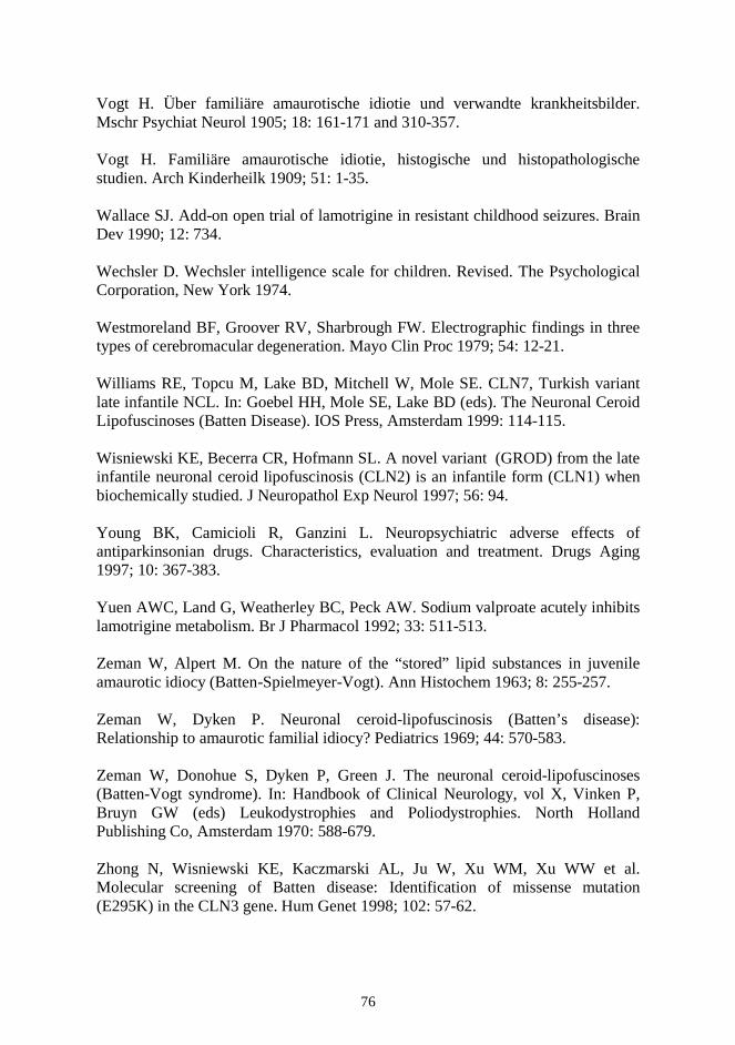
76
Vogt H. Über familiäre amaurotische idiotie und verwandte krankheitsbilder. Mschr Psychiat Neurol 1905; 18: 161-171 and 310-357. Vogt H. Familiäre amaurotische idiotie, histogische und histopathologische studien. Arch Kinderheilk 1909; 51: 1-35. Wallace SJ. Add-on open trial of lamotrigine in resistant childhood seizures. Brain Dev 1990; 12: 734. Wechsler D. Wechsler intelligence scale for children. Revised. The Psychological Corporation, New York 1974. Westmoreland BF, Groover RV, Sharbrough FW. Electrographic findings in three types of cerebromacular degeneration. Mayo Clin Proc 1979; 54: 12-21. Williams RE, Topcu M, Lake BD, Mitchell W, Mole SE. CLN7, Turkish variant late infantile NCL. In: Goebel HH, Mole SE, Lake BD (eds). The Neuronal Ceroid Lipofuscinoses (Batten Disease). IOS Press, Amsterdam 1999: 114-115. Wisniewski KE, Becerra CR, Hofmann SL. A novel variant (GROD) from the late infantile neuronal ceroid lipofuscinosis (CLN2) is an infantile form (CLN1) when biochemically studied. J Neuropathol Exp Neurol 1997; 56: 94. Young BK, Camicioli R, Ganzini L. Neuropsychiatric adverse effects of antiparkinsonian drugs. Characteristics, evaluation and treatment. Drugs Aging 1997; 10: 367-383. Yuen AWC, Land G, Weatherley BC, Peck AW. Sodium valproate acutely inhibits lamotrigine metabolism. Br J Pharmacol 1992; 33: 511-513. Zeman W, Alpert M. On the nature of the “stored” lipid substances in juvenile amaurotic idiocy (Batten-Spielmeyer-Vogt). Ann Histochem 1963; 8: 255-257. Zeman W, Dyken P. Neuronal ceroid-lipofuscinosis (Batten’s disease): Relationship to amaurotic familial idiocy? Pediatrics 1969; 44: 570-583. Zeman W, Donohue S, Dyken P, Green J. The neuronal ceroid-lipofuscinoses (Batten-Vogt syndrome). In: Handbook of Clinical Neurology, vol X, Vinken P, Bruyn GW (eds) Leukodystrophies and Poliodystrophies. North Holland Publishing Co, Amsterdam 1970: 588-679. Zhong N, Wisniewski KE, Kaczmarski AL, Ju W, Xu WM, Xu WW et al. Molecular screening of Batten disease: Identification of missense mutation (E295K) in the CLN3 gene. Hum Genet 1998; 102: 57-62.

77
Zweije-Hofman IL, van der Zee HJ, van Nieuwenhuizen O. Anti-parkinson drugs in the Batten-Spielmeyer-Vogt syndrome: a pilot trial. Clin Neurol Neurosurg 1982; 84-2:101-105. Åberg L, Heiskala H, Vanhala SL, Himberg JJ, Hosking G, Yuen A, Santavuori P. Lamotrigine therapy in infantile neuronal ceroid lipofuscinosis (INCL). Neuropediatrics 1997; 28: 77-79. Åberg L, Järvelä I, Rapola J, Autti T, Kirveskari E, Lappi M et al. Atypical juvenile neuronal ceroid lipofuscinosis (JNCL, CLN3) with GROD-like inclusions in the autonomic nerve cells of the gut wall. Acta Neuropathol 1998; 95: 306-312. Åberg L, Tiitinen A, Autti T, Kivisaari L, Santavuori P. Hormonal changes in girls with juvenile neuronal ceroid lipofuscinosis. Submitted.