journal of epilepsy research - an unusual case of herpes simplex … · 2015-01-14 · journal of...
TRANSCRIPT

This is an Open Access article distributed under the terms of the Creative Commons Attribution Non-Commercial License (http://creativecommons.org/licenses/by-nc/3.0/) which permits unrestricted non-commercial use, distribution, and reproduction in any medium, provided the original work is properly cited.
Case ReportJournal of Epilepsy Research
pISSN 2233-6249 / eISSN 2233-6257
An Unusual Case of Herpes Simplex Viral Encephalitis Following Acute Retinal Necrosis after Administration of a Systemic SteroidSang Jin Kim, MD, PhD1, Se Woong Kang, MD, PhD1, Eun Yeon Joo, MD, PhD2
Departments of 1Ophthalmology, 2Neurology, Samsung Medical Center, Sungkyunkwan University School of Medicine, Seoul, Korea
Received March 22, 2012Accepted March 30, 2012
Corresponding author: Eun Yeon JooDepartment of Neurology, Samsung Medical Center, Sungkyunkwan University School of Medicine, 81 Irwon-ro, Gangnam-gu, Seoul 135-710, KoreaTel. +82-2-3410-3597Fax. +82-2-3410-0052E-mail; [email protected]
Acute retinal necrosis (ARN), a viral retinal disease with poor visual prognosis, following herpes simplex
encephalitis (HSE) are uncommonly seen, and there has been no case yet reported of the reverse
situation. We herein present the reverse situation, an immune-competent patient with HSE following
ARN. A 57-year-old man who had been under steroid therapy for retinal vasculitis the prior two weeks,
presented with abrupt confusion and high fever. His cerebrospinal fluid study and brain magnetic
resonance imaging revealed typical HSE. Ophthalmic examination and polymerase chain reaction of the
vitreous specimen revealed ARN by herpes simplex virus type 2. Intravenous acyclovir treatment
improved his encephalitis symptoms and retinal necrosis. This case implies that ARN may be a risk
factor for HSE and the virus may reach the brain from the eye. Inappropriate administration of a systemic
steroid may exacerbate herpes viral infection in the retina, with subsequent spread to the brain.
(2012;2:21-24)
Key words: Acute retinal necrosis syndrome; Herpetic simplex encephalitis; Herpes simplex; Steroids
Acute retinal necrosis (ARN) is a devastating ocular disease with
poor visual prognosis and is most often caused by varicella zoster virus
(VZV) or herpes simplex virus (HSV)-1 and less frequently by HSV-2 and
cytomegalovirus.1,2 ARN is characterized by peripheral necrotizing
retinitis, retinal arteritis, and a prominent inflammatory reaction in the
vitreous and anterior chamber.3 This disease may present several years
after a primary infection, or it may occur following systemic herpetic
infection. The prevalence of the disease is equal in both genders, and it
occurs in the 5th-7th decade of life.4 Though the diagnosis of ARN is
made clinically in most patients, polymerase chain reaction (PCR)
analysis of the aqueous humor or vitreous is known as a confirmative
method to detect the causative virus. Cases who presented with
unilateral or bilateral ARN following herpes simplex viral encephalitis
(HSE) have been uncommonly reported.5-9 However, the reverse
situation of HSE following ARN has been reported in one case, who was
an immune-suppressed patient.10 Herein, we present an unusual case of
HSE following ARN in an immune-competent patient.
Case Report
A 57-year-old man was referred to Samsung Medical Center with
abrupt confusion, fever, and loss of vision in both eyes. The patient
was in his usual state of health until two weeks prior, when he began
to develop sudden loss of vision in his right eye. In another hospital,
he was diagnosed with retinal vasculitis in his right eye and was
prescribed oral prednisolone, 60 mg/day for three days, followed by
120 mg/day for three days. The retinal vasculitis did not respond to
oral prednisolone, so he was treated with intravenous methylpred-
nisolone, 1 g/day for five days. Two days after initiation of intravenous
methylprednisolone, he complained of decreased vision in his left
eye. One week after initiation of intravenous methylprednisolone, he
became drowsy and high fever occurred, and he was then referred to
Samsung Medical Center. On neurological examination, he was
drowsy and confused to time and place orientation. Mini-mental
status examination was 18. He consistently complained of severe
headache and bilateral ocular pain. However, his extraocular
movement did not show any limitation in all directions. Neck stiffness
and meningeal irritation sign were not definite. Other focal neurologic
deficits were not found.
His brain magnetic resonance imaging (MRI) showed a hyper-
intense lesion in the left medial temporal and bilateral medial
occipital areas on T2-weight images, suggestive of HSE (Fig. 1).
22 Journal of Epilepsy Research Vol. 2, No. 1, 2012
Copyright ⓒ 2012 Korean Epilepsy Society
Figure 1. Brain magnetic resonance imaging (MRI) at admission. Fluid-
attenuated inversion recovery magnetic image revealed hyperintensity in
the left medial temporal (arrow) and bilateral occipital cortices (arrow heads).
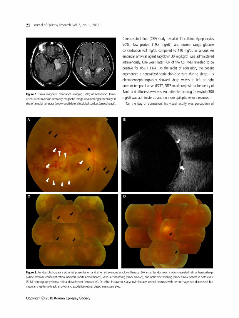
Figure 2. Fundus photographs at initial presentation and after intravenous acyclovir therapy. (A) Initial fundus examination revealed retinal hemorrhage
(white arrows), confluent retinal necrosis (white arrow heads), vascular sheathing (black arrows), and optic disc swelling (black arrow heads) in both eyes.
(B) Ultrasonography shows retinal detachment (arrows). (C, D). After intravenous acyclovir therapy, retinal necrosis with hemorrhage was decreased, but
vascular sheathing (black arrows) and exudative retinal detachment persisted.
Cerebrospinal fluid (CSF) study revealed 11 cells/mL (lymphocytes
90%), low protein (19.3 mg/dL), and normal range glucose
concentration (63 mg/dL compared to 110 mg/dL in serum). An
empirical antiviral agent (acyclovir 30 mg/kg/d) was administered
intravenously. One week later PCR of the CSF was revealed to be
positive for HSV-1 DNA. On the night of admission, the patient
experienced a generalized tonic-clonic seizure during sleep. His
electroencephalography showed sharp waves in left or right
anterior temporal areas (F7T7, F8T8 maximum) with a frequency of
1/min and diffuse slow waves. An antiepileptic drug (phenytoin 300
mg/d) was administered and no more epileptic seizure recurred.
On the day of admission, his visual acuity was perception of
C D
A B

Sang Jin Kim, et al. HSV Encephalitis Following ARN 23
www.kes.or.kr
light in both eyes. Fundus examination showed retinal hemorrhage,
confluent retinal necrosis affecting almost the whole retina, vascular
sheathing involving both arterioles and venules, optic disc
swelling, and exudative retinal detachment in both eyes (Fig. 2A).
Fluorescein angiography revealed multiple vascular occlusions and
vascular leakage. Ultrasonography shows retinal detachment in both
eyes (Fig. 2B). Infectious retinitis including acute retinal necrosis was
suspected, but in terms of the location of necrosis, his clinical
features were not typical of ARN Diagnostic vitrectomy confirmed the
presence of HSV-1. Finally, he was diagnosed with ARN with optic
neuropathy by HSV-1.
After administration of intravenous acyclovir for 14 days, his
systemic symptoms consistent with encephalitis slowly resolved, but
his vision was not restored. On follow-up examination, retinal
necrosis did not progress, but he still suffered from severe bilateral
ocular pain with partial improvement of the exudative retinal
detachment (Fig. 2C, D). Thus, intravitreal ganciclovir injection
(5,000/0.05 μg/mL) causes pain relief and decreased intraocular
inflammation. But his visual acuity could not perceptany light.
Bilateral rhegmatogenous retinal detachment with optic atrophy was
found six weeks later, thus vitrectomy with silicone oil tamponade
was done in the right eye.
Discussion
Cases with ARN following HSE have been uncommonly reported,5-9
and to our knowledge, this is the first report of the reverse situation,
HSE following ARN, in even an immune-competent patient. ARN
associated with HSE is known to be caused by axonal transmission of
a reactivated latent virus from the brain to the retina, and retinitis
occasionally occurs after the encephalitis. As shown by our patient,
opposite direct transmission from the retina to the brain seems to be
possible. This is consistent with the finding that herpes virus uses
bidirectional fast-axonal transport in neurons.11
ARN usually presents as retinal necrosis with discrete borders
located in the peripheral retina, rapid progression, circumferential
spread, occlusive vasculopathy with arterial involvement, and a
prominent inflammatory reaction in the vitreous and anterior
chamber.3 Our patient was initially diagnosed with retinal vasculitis
and treated with a high-dose systemic corticosteroid, which was not
effective. Corticosteroids are known to affect cellular immunity and
may promote viral replication. Intraocular or systemic corticosteroids
may induce retinal necrosis in patients with viral infection. Thus, it is
recommended that corticosteroids should be used in combination
with antiviral agents or after beginning antiviral therapy.12 In this
patient, the contralateral eye became infected, and HSE occurred
during initial corticosteroid therapy without antiviral medication. In
cases with an unusual clinical course, especially in retinal vasculitis
with arterial involvement, viral uveitis including HSV infection should
be suspected. Diagnostic vitrectomy to obtain a vitreous specimen is
appropriate in those cases.
In summary, ARN may be a risk factor for HSE, even in an
immune-competent patient. The virus may reach the brain from the
eye by a trans-axonal route. Systemic steroid therapy may have
exacerbated herpes viral infection in the retina, with subsequent
spread to the contralateral eye and brain.
References
1. Ganatra JB, Chandler D, Santos C, Kuppermann B, Margolis TP. Viral causes of the acute retinal necrosis syndrome. Am J Ophthalmol 2000;129:166-72.
2. Van Gelder RN, Willig JL, Holland GN, Kaplan HJ. Herpes simplex virus type 2 as a cause of acute retinal necrosis syndrome in young patients. Ophthalmology 2001;108:869-76.
3. Holland GN. Standard diagnostic criteria for the acute retinal necrosis syndrome. Executive committee of the American Uveitis Society. Am J Ophthalmol 1994;117:663-7.
4. Bodaghi B, Rozenberg F, Cassoux N, Fardeau C, LeHoang P. Nonnec-rotizing herpetic retinopathies masquerading as severe posterior uveitis. Ophthalmology 2003;110:1737-43.
5. Pavesio CE, Conrad DK, McCluskey PJ, Mitchell SM, Towler HM, Lightman S. Delayed acute retinal necrosis after herpetic encephalitis. Br J Ophthalmol 1997;81:415-6.
6. Vandercam T, Hintzen RQ, De Boer JH, Van der Lelij A. Herpetic ence-phalitis is a risk factor for acute retinal necrosis. Neurology 2008;71: 1268-74.
7. Kianersi F, Masjedi A, Ghanbari H. Acute Retinal Necrosis after Herpetic Encephalitis. Case Rep Ophthalmol 2010;1:85-9.
8. Perry JD, Gikin CA, Miller NR, Kerr DA. Herpes simplex encephalitis and bilateral acute retinal necrosis syndrome after craniotomy. Am J Ophthalmol 1998;126:456-60.
9. Klein A, Lefebvre P. Three consecutive episodes of acute retinal ne-crosis due to herpes simplex-1 over twelve years following herpetic encephalitis. Ocul Immunol Inflamm 2007;15:411-3.
10. Wittles KN, Goold LA, Gilhotra JS. Herpes simplex encephalitis pre-senting after steroid treatment of panuveitis. Med J Aust 2011;195: 87-8.
11. Smith GA, Gross SP, Enquist LW. Herpesviruses use bidirectional fast-axonal transport to spread in sensory neurons. Proc Natl Acad Sci

24 Journal of Epilepsy Research Vol. 2, No. 1, 2012
Copyright ⓒ 2012 Korean Epilepsy Society
USA 2001;98:3466-70.12. Tibbetts MD, Shah CP, Young LH, Duker JS, Maguire JI, Morley MG.
Treatment of acute retinal necrosis. Ophthalmology 2010;117:818-24.