journal of critical care -...
TRANSCRIPT
Journal of Critical Care 31 (2016) 101–109
Contents lists available at ScienceDirect
Journal of Critical Care
j ourna l homepage: www. jcc journa l .o rg
Thoracic ultrasound: Potential new tool for physiotherapists in
respiratory management. A narrative review☆,☆☆Aymeric Le Neindre a,⁎, Silvia Mongodi b,c, François Philippart a, Bélaïd Bouhemad d,e
a Intensive Care Unit and Department of Critical Care Medicine, Groupe Hospitalier Paris Saint-Joseph, Paris, Franceb Department of Clinical-Surgical, Diagnostic and Pediatric Sciences, University of Pavia, Pavia, Italyc Department of Anesthesia and Intensive Care, Fondazione IRCCS Policlinico S Matteo, Pavia, Italyd Department of Anesthesia and Surgical Intensive Care Unit, C.H.U. de Dijon, Dijon, Francee Faculty of Medicine, University of Burgundy, Dijon, France
a b s t r a c ta r t i c l e i n f o
☆ Support was provided solely by departmental sources☆☆ None of the authors have any financial interest in thequipment discussed or in any competing materials.⁎ Corresponding author. Tel.: +33 6 72 04 10 01.
E-mail address: [email protected] (A. Le N
http://dx.doi.org/10.1016/j.jcrc.2015.10.0140883-9441/© 2015 Elsevier Inc. All rights reserved.
Downloaded for Anonymous User (n/a) For personal use only
Keywords:
ChestOutcome assessmentPhysiotherapyUltrasoundRespiratory therapyThe use of diagnostic ultrasound by physiotherapists is not a new concept; it is frequently performed in muscu-loskeletal physiotherapy. Physiotherapists currently lack accurate, reliable, sensitive, and validmeasurements forthe assessment of the indications and effectiveness of chest physiotherapy. Thoracic ultrasound may be a prom-ising tool for the physiotherapist and could be routinely performed at patients' bedsides to provide real-time andaccurate information on the status of pleura, lungs, and diaphragm; this would allow for assessment of lung aer-ation from interstitial syndrome to lung consolidation with much better accuracy than chest x-rays or ausculta-tion. Diaphragm excursion and contractility may also be assessed by ultrasound. This narrative review refers tolung and diaphragmultrasound semiology and describes how physiotherapists could use this tool in their clinicaldecision-making processes in various cases of respiratory disorders. The use of thoracic ultrasound semiologyalongside typical examinations may allow for the guiding, monitoring, and evaluating of chest physiotherapytreatments. Thoracic ultrasound is a potential new tool for physiotherapists.
.e subject matter, materials, or
eindre).
at Blackpool Teaching Hospitals NHS Foundation Trust. No other uses without permission. Copyright ©2017. E
© 2015 Elsevier Inc. All rights reserved.
1. Introduction
The use of diagnostic ultrasound by physiotherapists is not a newconcept [1]; it is frequently performed in musculoskeletal physiothera-py. Diagnostic ultrasound is noninvasive, is ionization-free, and can beperformed rapidly at the patient's bedside for assessment and monitor-ing. The use of ultrasound for lung examination is increasingly gainingwide acceptance in emergency and critical care medicine [2,3]. A recentnarrative review by Leech et al [4] notes that physiotherapists typicallyuse pulmonary auscultation and chest x-rays to assess and monitortheir interventions. However, based on previous studies regarding aus-cultation, Leech et al [4] report a low interrater reliability and a low ac-curacy for identifying pleural effusion, alveolar consolidation, andalveolar interstitial syndrome (61%, 36%, and 55%, respectively). Thoseauthors also highlight the fact that chest x-ray sensitivity and specificityare low at detecting pleural effusion (42% and 89%, respectively), inter-stitial syndrome (53% and 90%, respectively), and lung consolidation(53% and 90%, respectively) [4]. Finally, chest x-rays are not necessarilyperformed during a physiotherapist's intervention, which could yield
inaccurate information about lung status. Thus, the limits of ausculta-tion and chest x-rays are well described: the real effectiveness of chestphysiotherapy is probably underestimated or overestimated, leadingto excessive or inadequate treatment [4]. Thus, physiotherapists cur-rently lack accurate, reliable, sensitive, and valid measurements for pa-tientmonitoring and assessments of the indications and effectiveness ofchest physiotherapy [5]. The use of lung ultrasound (LUS) examinationby physiotherapists to evaluate lung status in real time offers new in-sights [6]. The assessment of diaphragm function is another promisingapplication of ultrasound, particularly in intensive care units (ICUs) dur-ing weaning frommechanical ventilation [7]. Leech et al [4] discuss thebenefits for critical care physiotherapists of implementing LUS in theirclinical practices and briefly propose applications in certain clinical sit-uations. For example, LUS could be used to differentiate pleural effusionfrom lung collapse in the case of chest x-ray opacity. In this situation,LUS allowing a physiotherapist to know if a lung is recruitable or notcould have the potential to influence physiotherapy treatment.
The objective of this narrative review is to detail the development ofLUS for physiotherapists. This review refers to lung and diaphragm ul-trasound semiology, and how thoracic ultrasound should be used atthe patient's bedside for lung and diaphragm assessment. This reviewexplores the literature and the LUS experiences of the authors to de-scribe how physiotherapists may use this potential new tool in theirclinical decision-making process.
from ClinicalKey.com by Elsevier on July 26, 2017.lsevier Inc. All rights reserved.
102 A. Le Neindre et al. / Journal of Critical Care 31 (2016) 101–109
2. Basics of LUS
2.1. Examination protocol
A simple machinewith amicroconvex probe (with a frequency of 2-4MHz) is sufficient for performing a LUS examination. To explore the entirethorax, it is necessary to define examination areas. Two lines delimit theaxillary space: the anterior and posterior axillary lines [8]. These lines de-fine areas of investigation for each hemithorax—anterior, lateral, andposterior—each of which is divided into upper and lower parts. Eachhemithorax includes 6 examination areas [2,8]. The locations of the liverand spleen allow for the identification of the right and left diaphragm cu-polas, which separate the thoracic and abdominal compartments [2,9].The probe explores the intercostal spaces, which form acoustic windows.The baseline examination uses a longitudinal view but can also use atransverse view [2]. By following this procedure, the physiotherapist canexplore the entire pleura-pulmonary complex. Lung ultrasonographyhas 2 modes [8]: B mode (brightness, real-time), which allows for a 2-dimensional view, andMmode (motion), which allows for the visualiza-tion of structure motion as a function of time.
2.2. Normal lung ultrasonography
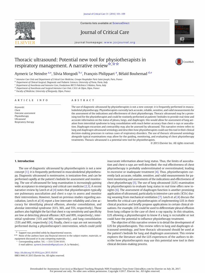
Ultrasound is not transmitted through well-aerated lungs [2]. Lungultrasound is partially based on the analysis of generated air artifacts,and the LUS of a normal lung (aerated) is characterized by the presenceof the bat sign, pleural sliding, andA lines [2,3]. Only thepleura is visible,and its movement (ie, pleural sliding) is typically evident. The A linesare reverberation artifacts from this pleural line [2,3] (Fig. 1A).
2.3. Alveolar-interstitial syndrome
B lines are vertical artifacts originating from the pleural line; they eraseA lines and reach the edge of the screen. B lines are generated by the pres-ence of airmixedwithwater, which causes amismatch of the acoustic im-pedance [10]. Edematous interlobular septa in contact with air-filledalveoli generate multiple and well-defined B lines (Fig. 1B): this isknownas interstitial syndrome (seeVideo S1 in the online supporting infor-mation). When alveoli are partially filled with fluid, the B lines becomeconfluent and alveolar-interstitial syndrome is present [10,11].
The presence of multiple and diffuse B lines is characteristic ofalveolar-interstitial syndrome [2,3]. Investigations are typically per-formed at the anterior and lateral thoracic areas. A positive region is de-fined by the presence of 3 or more B lines in a longitudinal planebetween 2 ribs, and a positive examination result suggests the presenceof 2 or more positive regions. The number of B lines is correlated withthe loss of aeration and is not specific. Unilateral and focal B lines are as-sociated with pneumonia, atelectasis, and pulmonary contusions. Bilat-eral and diffuse B lines are associated with pulmonary edema and acuterespiratory distress syndrome. A gain or a loss in lung aeration inducedbypositive-end expiratory pressure [12], dialysis [13], or antibiotics [14]can be assessed semiquantitatively using LUS by monitoring the num-ber of B lines present.
2.4. Lung consolidation
Lung consolidationmay have a variety of causes, including pneumo-nia, pulmonary embolism, lung cancer and metastasis, compressive orobstructive atelectasis, and pulmonary contusion [8]. Lung consolida-tion appears as a tissue-like structure with irregular deep marginscontacting the aerated lung or regular deep margins when the entirelobe is involved [8,15] (Fig. 1D, E). The presence of air bronchograms,fluid bronchograms (see Video S2 in the online supporting informa-tion), and vascular patterns can be observed within the consolidation[8,15]. Lung ultrasound may be helpful in determining the type ofconsolidation [8].
Downloaded for Anonymous User (n/a) at Blackpool Teaching Hospitals NHFor personal use only. No other uses without permission
Pneumonia is defined by the presence of subpleural consolidation[16], which can be hemilobar with irregular deep margins or lobarwith regular deep margins [15]. This tissue-like structure contains airbronchograms, which can be static and/or dynamic (see Video S3 inthe online supporting information) [16], but fluid bronchograms mayalso be present, which indicate fluid-filled airways [17–19]. Tree-likevascularities may also be observed within the consolidation. ColorDoppler ultrasound allows for the differentiation of tree-like vascular-ities and fluid bronchograms (Fig. 1E) [17,20].
Atelectasis ultrasoundfindings are characterized by a tissue-like pat-tern, abolished lung sliding, and static air bronchograms [21] (see VideoS4 in the online supporting information). As with chest x-rays, the ipsi-lateral attraction of mediastinum structures can also be observed. Thepresence of a dynamic air bronchogram rules out atelectasis [22]. Thisbronchogram is associated with nonretractile consolidation and indi-cates the absence of airway congestion [22]. At an early stage precedingtotal aeration loss, atelectasis appears as A lines or possibly B lines, andlung sliding is replaced by lungpulsations synchronizedwith cardiac ac-tivity, which is known as the lung pulse [23].
2.5. Pleural effusion
Pleural effusion appears as dependent and anechoic (ie, free of ech-oes) structures [8]. Pleural effusion occurs between the diaphragm andthe pleura, and conducts ultrasounds, allowing for deeper structures tobe visualized, such as the thoracic aorta [2]. In Mmode, pleural effusionis characterized by the sinusoid sign (Fig. 1C) [3,8].
The type of pleural effusion (ie, transudate or exudate) cannot be de-termined with certainty only using ultrasound examination, but someultrasound patterns are characteristic of their nature. Transudates arealways anechoic, whereas exudates are echoic and loculated [24].
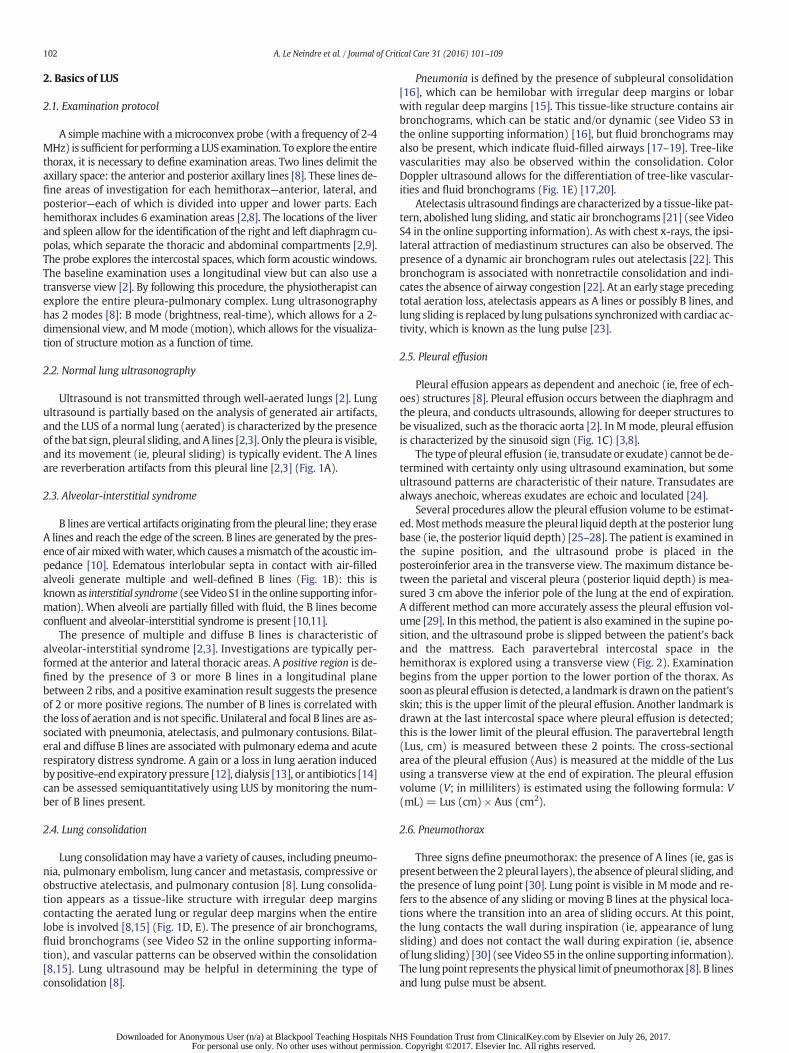
Several procedures allow the pleural effusion volume to be estimat-ed.Mostmethodsmeasure the pleural liquid depth at the posterior lungbase (ie, the posterior liquid depth) [25–28]. The patient is examined inthe supine position, and the ultrasound probe is placed in theposteroinferior area in the transverse view. The maximum distance be-tween the parietal and visceral pleura (posterior liquid depth) is mea-sured 3 cm above the inferior pole of the lung at the end of expiration.A different method can more accurately assess the pleural effusion vol-ume [29]. In this method, the patient is also examined in the supine po-sition, and the ultrasound probe is slipped between the patient's backand the mattress. Each paravertebral intercostal space in thehemithorax is explored using a transverse view (Fig. 2). Examinationbegins from the upper portion to the lower portion of the thorax. Assoon as pleural effusion is detected, a landmark is drawnon the patient'sskin; this is the upper limit of the pleural effusion. Another landmark isdrawn at the last intercostal space where pleural effusion is detected;this is the lower limit of the pleural effusion. The paravertebral length(Lus, cm) is measured between these 2 points. The cross-sectionalarea of the pleural effusion (Aus) is measured at the middle of the Lususing a transverse view at the end of expiration. The pleural effusionvolume (V; in milliliters) is estimated using the following formula: V(mL) = Lus (cm) × Aus (cm2).
2.6. Pneumothorax
Three signs define pneumothorax: the presence of A lines (ie, gas ispresent between the2 pleural layers), the absence of pleural sliding, andthe presence of lung point [30]. Lung point is visible in Mmode and re-fers to the absence of any sliding or moving B lines at the physical loca-tions where the transition into an area of sliding occurs. At this point,the lung contacts the wall during inspiration (ie, appearance of lungsliding) and does not contact the wall during expiration (ie, absenceof lung sliding) [30] (see Video S5 in the online supporting information).The lungpoint represents thephysical limit of pneumothorax [8]. B linesand lung pulse must be absent.
S Foundation Trust from ClinicalKey.com by Elsevier on July 26, 2017.. Copyright ©2017. Elsevier Inc. All rights reserved.
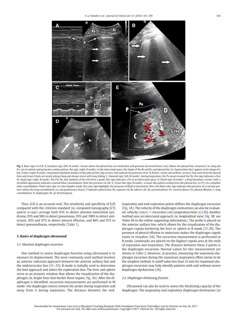
Fig. 1. Basic signs in LUS. A, Seashore sign (left, Mmode): tissues above the pleural line are motionless and generate horizontal lines (sea). Below the pleural line, structures (ie, lung andPv) are inmotion and generate a sandy pattern. Bat sign (right, Bmode): in the intercostal space, the shape of the Rs and Ri, and pleural line (ie, hyperechoic line) appear in the shape of abat. A lines (right, Bmode): horizontal repetition artifacts of the pleural line (big arrows) that indicate the presence of air. B, B lines: comet-tail artifacts (arrows) that arise from the pleurallines and erase A lines are nearly always long and always movewith lung sliding. C, Sinusoid sign (left, Mmode): during inspiration, the Pvmoves toward the Pp; this sign indicates a freePe. Quad sign (right, B mode): the Pvl, Pp, and shadows of the ribs form a quad; this sign indicates a Pe in an intercostal space. D, Shred sign (B mode): a deep boundary (arrow) with ashredded appearance indicates a partial lobar consolidation. Note the presence of a Br. E, Tissue-like sign (Bmode): a tissue-like pattern arising from the pleural line (or Pv) of a completelobar consolidation. Fluid-color sign: in color Doppler mode, the color sign highlights the presence of fluid inmovement. Here, the fluid-color sign indicates the presence of a vascular pat-tern within the lung consolidation (ie, intrapulmonary shunt). P indicates pleural line; Rs, superior rib; Ri, inferior rib; Pp, parietal pleura; Pv, visceral pleura; Pe, pleural effusion; C, lungconsolidation; D, diaphragm; Br, air bronchogram.
103A. Le Neindre et al. / Journal of Critical Care 31 (2016) 101–109
Thus, LUS is an accurate tool. The sensitivity and specificity of LUScompared with the criterion standard (ie, computed tomography [CT]and/or x-rays) average both 93% to detect alveolar-interstitial syn-drome, 93% and 98% to detect pneumonia, 93% and 100% to detect atel-ectasis, 93% and 97% to detect pleural effusion, and 86% and 97% todetect pneumothorax, respectively (Table 1).
3. Basics of diaphragm ultrasound
3.1. Maximal diaphragm excursion
One method to assess diaphragm function using ultrasound is tomeasure its displacement. The most commonly used method involvesan anterior subcostal approach between the anterior axillary line andthe midclavicular line [31–35]. B mode is initially used to determinethe best approach and select the exploration line. The liver and spleenserve as an acoustic window that allows the visualization of the dia-phragm (ie, bright lines that border these organs; Fig. 3A). After the di-aphragm is identified, excursion measurements are performed in Mmode: the diaphragm moves toward the probe during inspiration andaway from it during expiration. The distance between the end-
Downloaded for Anonymous User (n/a) at Blackpool Teaching Hospitals NHS For personal use only. No other uses without permission. C
inspiration and end-expiration points defines the diaphragm excursion(Fig. 3A). The velocity of the diaphragm contraction can also be evaluat-ed: velocity (cm/s)= excursion (cm)/inspiratory time (s) [36]. Anothermethod uses an intercostal approach (ie, longitudinal view; Fig. 3B; seeVideo S6 in the online supporting information). The probe is placed onthe anterior axillary line, which allows for the visualization of the dia-phragm cupola bordering the liver or spleen in B mode [37,38]. Thepresence of pleural effusion or atelectasis makes the diaphragm cupolaeasier to visualize [38]. The excursion measurement is performed inB mode. Landmarks are placed on the highest cupola area at the endsof expiration and inspiration. The distance between these 2 points isthe diaphragm excursion. Normal values for this measurement areshown in Table 2. However, in practice, measuring the maximum dia-phragm excursion during the maximum inspiratory effort seems to bethe simplest method. A cutoff value less than 25mm for maximum dia-phragm excursion may help identify patients with and without severediaphragm dysfunction [38].
3.2. Diaphragm thickening fraction
Ultrasound can also be used to assess the thickening capacity of thediaphragm. The inspiratory and expiratory diaphragm thicknesses (in
Foundation Trust from ClinicalKey.com by Elsevier on July 26, 2017.opyright ©2017. Elsevier Inc. All rights reserved.
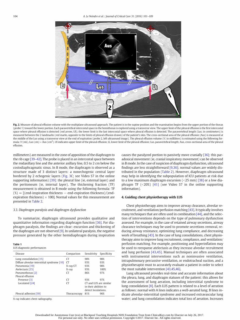
Fig. 2.Measure of pleural effusion volumewith themultiplane ultrasound approach. The patient is in the supine position and the examination begins from the upper portion of the thorax(probe 1) toward the lower portion. Each paravertebral intercostal space in the hemithorax is exploredusing a transverse view. The upper limit of the pleural effusion is the first intercostalspace where pleural effusion is detected (red arrow, Ul); the lower limit is the last intercostal space where pleural effusion is detected. The paravertebral length (Lus; in centimeters) ismeasured between the 2 landmarks (redmarks, opposite to the limits of pleural effusion drawn) of the patient's skin. The cross-sectional area of the pleural effusion (Aus) ismeasured atthe middle of the Lus using a transverse view at the end of expiration (probe 2, left ultrasound image). The pleural effusion volume (V; in milliliters) is estimated using the following for-mula: V (mL), Lus (cm) × Aus (cm2). Ul indicates upper limit of the pleural effusion; Ll, lower limit of the pleural effusion; Lus, paravertebral length; Aus, cross-sectional area of the pleuraleffusion.
104 A. Le Neindre et al. / Journal of Critical Care 31 (2016) 101–109
millimeters) aremeasured in the zone of apposition of the diaphragm tothe rib cage [39–42]. The probe is placed in an intercostal space betweenthe midaxillary line and the anterior axillary line, 0.5 to 2 cm below thecostodiaphragmatic sinus. In B mode, the diaphragm is observed as astructure made of 3 distinct layers: a nonechogenic central layerbordered by 2 echogenic layers (Fig. 3C; see Video S7 in the onlinesupporting information) [39]: the pleural line (ie, external layer) andthe peritoneum (ie, internal layer). The thickening fraction (TF)measurement is obtained in B mode using the following formula: TF(%) = [(end-inspiration thickness − end-expiration thickness)/(end-expiration thickness) × 100]. Normal values for this measurement arepresented in Table 2.
3.3. Diaphragm paralysis and diaphragm dysfunction
To summarize, diaphragm ultrasound provides qualitative andquantitative information regarding diaphragm function [36]. For dia-phragm paralysis, the findings are clear: excursion and thickening ofthe diaphragm are not observed [9]. In unilateral paralysis, the negativepressure generated by the other hemidiaphragm during inspiration
Table 1LUS diagnostic performances
Disease Comparison Sensitivity Specificity
Lung consolidation [15] CT 90% 98%Diffuse alveolar-interstitial syndrome [10] CT 93% 93%Pneumonia [19] X-ray/CT 93% 98%Atelectasis [23] X-ray 93% 100%Pneumothorax [2] CT 86% 97%Pleural effusionPresence [3] CT 93% 97%Loculated [24] CT CT and LUS are similar
in their abilities todetect loculations.
Pleural adhesion [59] Thoracoscopy 81% 96%
X-ray indicates chest radiography.
Downloaded for Anonymous User (n/a) at Blackpool Teaching Hospitals NHFor personal use only. No other uses without permission
causes the paralyzed portion to passively move cranially [36]; this par-adoxical movement (ie, cranial inspiratory movement) can be observedin Bmode. In the case of suspicion of diaphragmdysfunction, ultrasoundfindings are less straightforward [9,36]; normal values are widely dis-tributed in the population (Table 2). However, diaphragm ultrasoundmay help in identifying the subpopulation of ICU patients at risk dueto a low maximum diaphragm excursion (b25 mm) [38] or a low dia-phragm TF (b20%) [41] (see Video S7 in the online supportinginformation).
4. Guiding chest physiotherapy with LUS
Chest physiotherapy aims to improve airway clearance, alveolar re-cruitment, and ventilation/perfusionmatching [43]. It typically involvesmany techniques that are often used in combination [44], and the selec-tion of interventions depends on the type of pulmonary dysfunctionassessed. For example, in the case of retained airway secretions, airwayclearance techniques may be used to promote secretions removal, re-ducing airway resistance, optimizing lung compliance, and decreasingwork of breathing [43]. In the case of lung consolidations, chest physio-therapy aims to improve lung recruitment, compliance, and ventilation/perfusion matching. For example, positioning and hyperinflation maybe used to reexpanse atelectasis as they increase alveolar recruitmentand lung perfusion [43,45]. Manual techniques are often associatedwith instrumental interventions such as noninvasive ventilation,intrapulmonary percussive ventilation, or endotracheal suction, and aphysiotherapist must to accurately evaluate a patient in order to selectthe most suitable intervention [43,45,46].
Lung ultrasound provides real-time and accurate information aboutthe pleura, lung, and diaphragm statuses of the patient: this allows forthe assessment of lung aeration, including interstitial syndrome tolung consolidation [8]. Each LUS pattern is related to a level of aerationas follows: normal with A lines indicates a well-aerated lung; B lines in-dicate alveolar-interstitial syndrome and increased extravascular lungwater; and lung consolidation indicates total loss of aeration. Increases
S Foundation Trust from ClinicalKey.com by Elsevier on July 26, 2017.. Copyright ©2017. Elsevier Inc. All rights reserved.
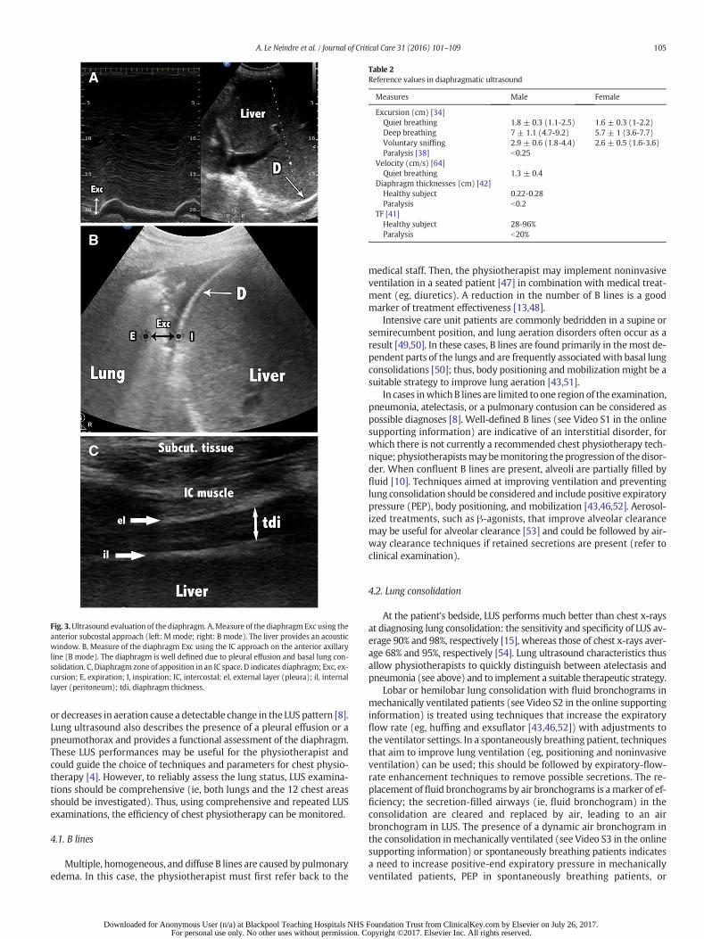
Fig. 3.Ultrasound evaluation of the diaphragm. A,Measure of the diaphragm Exc using theanterior subcostal approach (left: M mode; right: B mode). The liver provides an acousticwindow. B, Measure of the diaphragm Exc using the IC approach on the anterior axillaryline (B mode). The diaphragm is well defined due to pleural effusion and basal lung con-solidation. C, Diaphragm zone of apposition in an IC space. D indicates diaphragm; Exc, ex-cursion; E, expiration; I, inspiration; IC, intercostal; el, external layer (pleura); il, internallayer (peritoneum); tdi, diaphragm thickness.
Table 2Reference values in diaphragmatic ultrasound
Measures Male Female
Excursion (cm) [34]Quiet breathing 1.8 ± 0.3 (1.1-2.5) 1.6 ± 0.3 (1-2.2)Deep breathing 7 ± 1.1 (4.7-9.2) 5.7 ± 1 (3.6-7.7)Voluntary sniffing 2.9 ± 0.6 (1.8-4.4) 2.6 ± 0.5 (1.6-3.6)Paralysis [38] b0.25
Velocity (cm/s) [64]Quiet breathing 1.3 ± 0.4
Diaphragm thicknesses (cm) [42]Healthy subject 0.22-0.28Paralysis b0.2
TF [41]Healthy subject 28-96%Paralysis b20%
105A. Le Neindre et al. / Journal of Critical Care 31 (2016) 101–109
or decreases in aeration cause a detectable change in the LUS pattern [8].Lung ultrasound also describes the presence of a pleural effusion or apneumothorax and provides a functional assessment of the diaphragm.These LUS performances may be useful for the physiotherapist andcould guide the choice of techniques and parameters for chest physio-therapy [4]. However, to reliably assess the lung status, LUS examina-tions should be comprehensive (ie, both lungs and the 12 chest areasshould be investigated). Thus, using comprehensive and repeated LUSexaminations, the efficiency of chest physiotherapy can be monitored.
4.1. B lines
Multiple, homogeneous, and diffuse B lines are caused by pulmonaryedema. In this case, the physiotherapist must first refer back to the
Downloaded for Anonymous User (n/a) at Blackpool Teaching Hospitals NHS For personal use only. No other uses without permission. C
medical staff. Then, the physiotherapist may implement noninvasiveventilation in a seated patient [47] in combination with medical treat-ment (eg, diuretics). A reduction in the number of B lines is a goodmarker of treatment effectiveness [13,48].
Intensive care unit patients are commonly bedridden in a supine orsemirecumbent position, and lung aeration disorders often occur as aresult [49,50]. In these cases, B lines are found primarily in themost de-pendent parts of the lungs and are frequently associatedwith basal lungconsolidations [50]; thus, body positioning andmobilization might be asuitable strategy to improve lung aeration [43,51].
In cases inwhichB lines are limited to one region of the examination,pneumonia, atelectasis, or a pulmonary contusion can be considered aspossible diagnoses [8]. Well-defined B lines (see Video S1 in the onlinesupporting information) are indicative of an interstitial disorder, forwhich there is not currently a recommended chest physiotherapy tech-nique; physiotherapistsmay bemonitoring the progressionof thedisor-der. When confluent B lines are present, alveoli are partially filled byfluid [10]. Techniques aimed at improving ventilation and preventinglung consolidation should be considered and include positive expiratorypressure (PEP), body positioning, and mobilization [43,46,52]. Aerosol-ized treatments, such as β-agonists, that improve alveolar clearancemay be useful for alveolar clearance [53] and could be followed by air-way clearance techniques if retained secretions are present (refer toclinical examination).
4.2. Lung consolidation
At the patient's bedside, LUS performs much better than chest x-raysat diagnosing lung consolidation: the sensitivity and specificity of LUS av-erage 90% and 98%, respectively [15], whereas those of chest x-rays aver-age 68% and 95%, respectively [54]. Lung ultrasound characteristics thusallow physiotherapists to quickly distinguish between atelectasis andpneumonia (see above) and to implement a suitable therapeutic strategy.
Lobar or hemilobar lung consolidation with fluid bronchograms inmechanically ventilated patients (see Video S2 in the online supportinginformation) is treated using techniques that increase the expiratoryflow rate (eg, huffing and exsuflator [43,46,52]) with adjustments tothe ventilator settings. In a spontaneously breathing patient, techniquesthat aim to improve lung ventilation (eg, positioning and noninvasiveventilation) can be used; this should be followed by expiratory-flow-rate enhancement techniques to remove possible secretions. The re-placement of fluid bronchograms by air bronchograms is amarker of ef-ficiency; the secretion-filled airways (ie, fluid bronchogram) in theconsolidation are cleared and replaced by air, leading to an airbronchogram in LUS. The presence of a dynamic air bronchogram inthe consolidation in mechanically ventilated (see Video S3 in the onlinesupporting information) or spontaneously breathing patients indicatesa need to increase positive-end expiratory pressure in mechanicallyventilated patients, PEP in spontaneously breathing patients, or
Foundation Trust from ClinicalKey.com by Elsevier on July 26, 2017.opyright ©2017. Elsevier Inc. All rights reserved.
106 A. Le Neindre et al. / Journal of Critical Care 31 (2016) 101–109
implement measures such as mobilization and body positioning to im-prove alveolar ventilation [43,46]. With LUS, the physiotherapist moni-tors the consolidation and evaluates the effectiveness of treatments[14]. Whatever the chest physiotherapy treatment indicated and cho-sen, the decrease of the consolidation size and the number of B lines,and the appearance of A lines are markers of efficiency [55]. The unfa-vorable evolution of pneumonia is defined by increases in the signs de-scribed above, which can lead to pulmonary abscesses or empyema[17,20].
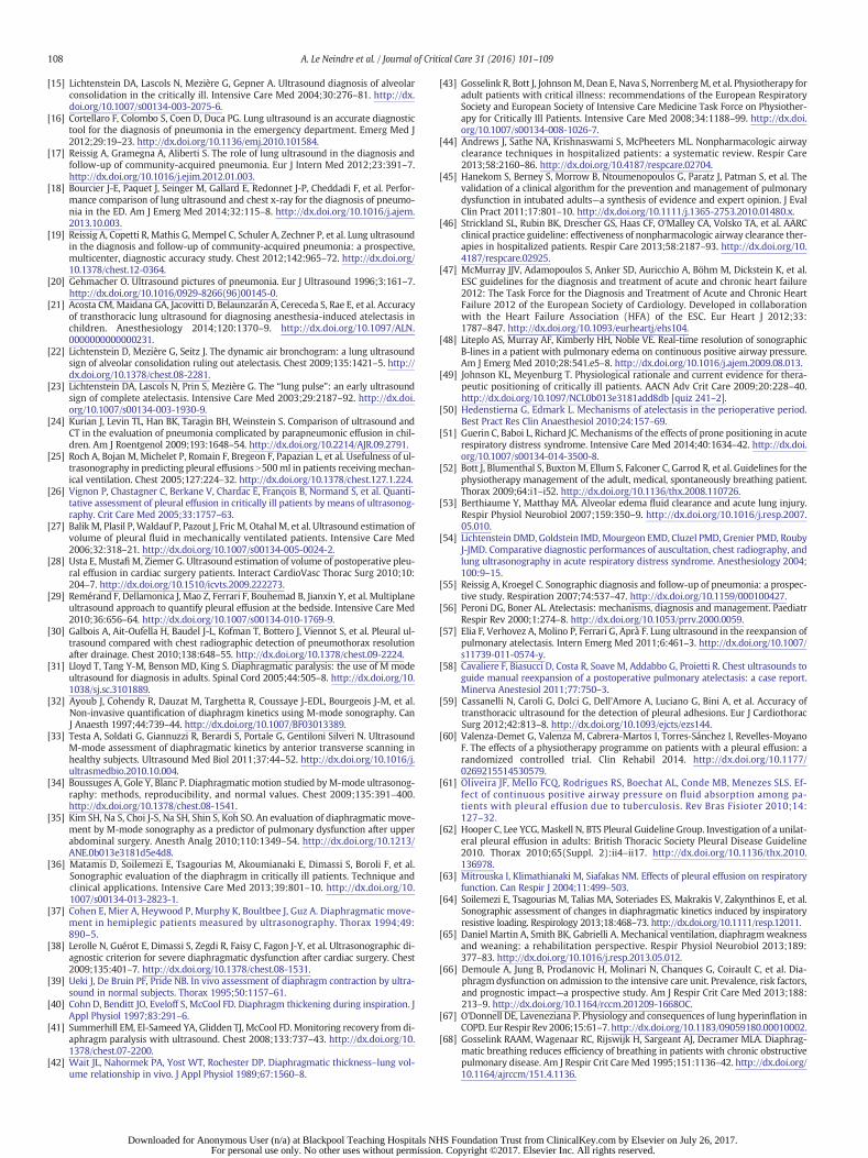
In cases of atelectasis, it is important to differentiate between ob-structive and passive mechanisms [50,56]. There are primarily 3 causesof atelectasis: absorptive (eg, airway obstruction), compression of lungtissue (eg, positioning and pleural effusion), and surfactant dysfunction.Management of atelectasis differs according the etiology [50,56]. Thepresence of a large pleural effusion in LUS is indicative of passive atelec-tasis related to lung compression. Thus, physiotherapist should firstrefer the patient to medical staff. If the patient's condition does not re-quire the placement of a chest drain, the physiotherapist may help de-crease the work of breathing and improve lung ventilation (eg,noninvasive ventilation) if required [43,46]. If there is an obstructive at-electasis caused by retained secretions (refer to clinical review), the im-plementation of lung recruitment techniques is indicated (eg,manual orventilator hyperinflation, PEP, body positioning, and expiratory-flow-rate enhancement techniques) to promote secretion removal [43]. Theresolution of obstructive atelectasis or an increase in aeration is charac-terized by the appearance of A lines or B lines, respectively (Fig. 4; seeVideo S4 in the online supporting information). These changes in theLUS pattern allow to assess the effectiveness of chest physiotherapytreatments [57,58].
4.3. Pleural effusion
Lung ultrasound is an excellent tool for pleural assessment and canhelp to locate effusion, estimate its volume, distinguish transudatefrom exudate, and detect loculations and the presence of pleural adhe-sions [59].
The quality and number of studies that assess the effectiveness ofchest physiotherapy in pleural effusion are limited [60,61]. None ofthese studies provide precision regarding the nature of pleural effusionand the patient's condition. If the physiotherapist identifies a pleural ef-fusion, it seems reasonable to address the patient and wait for medicalor surgical treatment to be completed [62]. Then, the physiotherapistmay help to implement supportive respiratory management if a chestdrain is not required (see above).
Moreover, in cases of diaphragm dyskinesia induced by pleural effu-sion, physiotherapist may measure improvement by ultrasoundassessing an increase in diaphragm function [63].
4.4. Diaphragm dysfunction
Diaphragm ultrasound could be a valuable tool for physiotherapistswhen evaluating diaphragm kinetics and strength in cases of difficult-to-wean ICU patients. It may also be helpful for the assessment ofchanges in diaphragm kinetics induced by inspiratory resistive loads[64] or the assessment of the effectiveness of inspiratory muscle train-ing [65].
Diaphragm dysfunction is observed as dyskinesia (ie, a decrease inthe maximum diaphragm excursion) or a decrease in the diaphragmTF. In ICU patients, diaphragm dysfunction frequently occurs (ie, inci-dence averages 60%), and the primary risk factors are sepsis and diseaseseverity [66]. Mechanical ventilation is often cited as a cause of dia-phragm dysfunction (ie, disuse atrophy). Mechanisms of diaphragm in-juries are complex and not completely known. Phrenic nerve injuriesafter major surgical procedures (ie, cardiac and thoracic) [36] are alsotypically observed (see Video S7 in the online supporting information).
Downloaded for Anonymous User (n/a) at Blackpool Teaching Hospitals NHFor personal use only. No other uses without permission
Regular monitoring of maximum diaphragm excursions or the dia-phragm TFs via ultrasound allows for the monitoring of diaphragm re-covery [41]. The regular monitoring of diaphragm function determinesthe evolving nature (ie, fast or slow) of diaphragm recovery and guidestreatment strategies (eg, waiting for a fast resolution or pursuinga tracheotomy).
Diaphragm ultrasound could be of interest for some controversial is-sues related to diaphragm physiotherapy. Patients with chronic ob-structive pulmonary disease have static and dynamic lunghyperinflation related to their disease severity (ie, degree of obstructionand severity of emphysema); this hyperinflation makes the diaphragmless efficient due to its unfavorable position [67]. The effectiveness of di-aphragmatic breathing exercises for these patients is controversial. In pa-tients with severe hyperinflation, a loss of mechanical efficiency in therespiratory muscles has been reported [68]. Conversely, in less severelyaffected patients, it appears that diaphragmatic breathing exercises im-proved the functional capacities of patients [69]. It is possible to measurethe effects of diaphragmatic breathing exercises on the maximum dia-phragm excursion and the diaphragm thickening ratio. Using ultrasoundin this way, the physiotherapist could rationally adapt their rehabilitationprograms for patients with chronic obstructive pulmonary disease (seeVideo S6 in the online supporting information) [70].
5. Limitations
First, lung disorders that do not reach the pleura cannot be assessedby ultrasound. Second, dressings, subcutaneous emphysema, and obesi-ty are obstacles to using LUS. The use of inadequate probes or settingsand noncompliance with examination conditions are also limitations[3]. Finally, LUS must be used alongside clinical evaluations and shouldnot be the only outcomemeasure used in clinical practice. As suggestedby Leech et al [4], LUSmay be used to answer a focused clinical question.The results of using tools currently available (eg, clinical review, auscul-tation, and chest x-rays) allow the physiotherapist to form an initial di-agnosis of patient's respiratory conditions. Then, the physiotherapistshould perform LUS if his clinical hypothesismust be confirmed. The pa-tient selection requiring LUS is helpful because nearly all mechanicallyventilated patients have pathologic findings in LUS but not alwayswith clinical significance [71]. In addition to clinical examinations, LUSmay guide the choice of chest physiotherapy strategies and help to de-termine if the patient requires medical intervention and no chest phys-iotherapy (eg, pneumothorax and severe pleural effusion).
Finally, an appropriate level of training is necessary to implementLUS in clinical practice [2,4]. As highlighted by Leech et al, physiothera-pists do not routinely use LUS for patient assessments. Standard trainingin LUS thus must be developed for physiotherapists.
6. Perspectives and research
For physiotherapists to perform LUS evaluations, they must possessextensive knowledge of anatomy, pulmonary physiopathology, and LUSsemiology. Rigorous training and experience are required for LUS use[4]. Lung ultrasound is currently a diagnostic tool for intensivists, emer-gency physicians, and pulmonologists [8]. It is quite conceivable that aphysiotherapist may use LUS to detect lung abnormalities and not toprovide amedical diagnosis in order to select and reassess his/her phys-iotherapy treatments. The application of LUS in physiotherapy maycover all medical specialties that require chest physiotherapy.
Xirouchaki et al [71] assessed the influence of LUS on medical prac-tices and observed a significant change in diagnoses and treatmentscompared to typical examinations. Because it is also a useful diagnostictool in chest physiotherapy, LUS's influence on physiotherapist's clinicaldecision-making process should be evaluated.
Lung ultrasound may be used in clinical research for chest physio-therapy because it is accurate, reliable, and reproducible; it is also a suit-able outcome for research on the impact of chest physiotherapy in lung
S Foundation Trust from ClinicalKey.com by Elsevier on July 26, 2017.. Copyright ©2017. Elsevier Inc. All rights reserved.
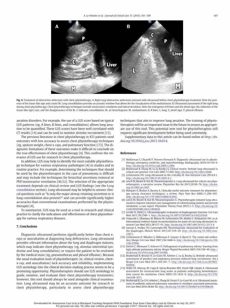
Fig. 4. Treatment of obstructive atelectasis with chest physiotherapy. A, Right lung obstructive atelectasis assessed with ultrasound before chest physiotherapy treatment. Note the pres-ence of the tissue-like sign and a static Br. Lung consolidation provides an acoustic window that allows for the visualization of themediastinum. B, Ultrasound assessment of the right lungduring chest physiotherapy. Chest physiotherapy techniques include noninvasive ventilation and lateral decubitus. Note the emergence of B lines and the shred sign, the reduction of thetissue-like sign's size, and the disappearance of the Br. C indicates consolidation; Br, air bronchogram; M, mediastinum; B, B lines; L, lung; S, shred sign; P, pleural effusion.
107A. Le Neindre et al. / Journal of Critical Care 31 (2016) 101–109
aeration disorders. For example, the use of a LUS score based on typicalLUS patterns (eg, A lines, B lines, and consolidation) allows lung aera-tion to be quantified. These LUS scores have been well correlated withCT results [14] and can be used to monitor alveolar recruitment [12].
The previous literature in chest physiotherapy in ICU patients usedoutcomes with low accuracy to assess chest physiotherapy techniques(eg, sputumweight, chest x-rays, and pulmonary function) [72]. The di-agnostic limitations of these outcomes make it difficult to conclude onthe true effectiveness of chest physiotherapy [4]. This confirms the rel-evance of LUS use for research in chest physiotherapy.
In addition, LUS may help to identify the most suitable physiothera-py technique for various respiratory pathologies [4] in studies and inroutine practice. For example, determining the techniques that shouldbe used by the physiotherapist in the case of pneumonia is difficultand may include the techniques for bronchial secretions removal orPEP/noninvasive ventilation [46,52]. The selection of the appropriatetreatment depends on clinical review and LUS findings (see the Lungconsolidation section). Lung ultrasoundmay be helpful to answer clini-cal questions such as “Is only themajor airway retaining secretions or islung consolidation also present?” and can provide significantly higheraccuracies than conventional examinations performed by the physio-therapist [4].
To summarize, LUS may be used as a tool in research and clinicalpractice to clarify the indications and effectiveness of chest physiother-apy for various respiratory diseases.
7. Conclusions
Diagnostic ultrasound performs significantly better than chest x-rays or auscultation at diagnosing lung deficiencies. Lung ultrasoundprovides relevant information about the lung and diaphragm statuses,which may indicate chest physiotherapy (eg, alveolar-interstitial syn-drome and lung consolidation) or require the patient to be reviewedby the medical team (eg, pneumothorax and pleural effusion). Becausethe usual evaluation tools of physiotherapist (ie, clinical review, chest-x-ray, and auscultation) lack accuracy and reliability, implementingLUS semiology into the clinical decision-making process seems to be apromising opportunity. Physiotherapists should use LUS semiology toguide, monitor, and evaluate their chest physiotherapy treatments;however, this tool should always be used alongside clinical examina-tion. Lung ultrasound may be an accurate outcome for research inchest physiotherapy, particularly to assess chest physiotherapy
Downloaded for Anonymous User (n/a) at Blackpool Teaching Hospitals NHS For personal use only. No other uses without permission. C
techniques that aim to improve lung aeration. The training of physio-therapistswill be an important issue in the future to ensure an appropri-ate use of this tool. This potential new tool for physiotherapists stillrequires significant development before being used commonly.
Supplementary data to this article can be found online at http://dx.doi.org/10.1016/j.jcrc.2015.10.014.
References
[1] McKiernan S, Chiarelli P, Warren-Forward H. Diagnostic ultrasound use in physio-therapy, emergency medicine, and anaesthesiology. Radiography 2010;16:154–9.http://dx.doi.org/10.1016/j.radi.2009.12.004.
[2] Bouhemad B, Zhang M, Lu Q, Rouby J-J. Clinical review: bedside lung ultrasound incritical care practice. Crit Care 2007;11:205. http://dx.doi.org/10.1186/cc5668.
[3] Lichtenstein DA. Lung ultrasound in the critically ill. Ann Intensive Care 2014;4:1.http://dx.doi.org/10.1186/2110-5820-4-1.
[4] LeechM, Bissett B, Kot M, Ntoumenopoulos G. Lung ultrasound for critical care phys-iotherapists: a narrative review. Physiother Res Int 2015;20:69–76. http://dx.doi.org/10.1002/pri.1607.
[5] Marques A, Bruton A, Barney A. Clinically useful outcome measures for physiother-apy airway clearance techniques: a review. Phys Ther Rev 2006;11:299–307.http://dx.doi.org/10.1179/108331906X163441.
[6] Leech M, Bissett B, Kot M, Ntoumenopoulos G. Physiotherapist-initiated lung ultra-sound to improve intensive caremanagement of a deteriorating patient and preventintubation: a case report. Physiother Theory Pract 2015;1–5. http://dx.doi.org/10.3109/09593985.2014.1003629.
[7] Lerolle N, Diehl J-L. Ultrasonographic evaluation of diaphragmatic function. Crit CareMed 2011;39:2760–1. http://dx.doi.org/10.1097/CCM.0b013e31822a55e9.
[8] Volpicelli G, Elbarbary M, Blaivas M, Lichtenstein DA, Mathis G, Kirkpatrick AW, et al.International evidence-based recommendations for point-of-care lung ultrasound. In-tensive Care Med 2012;38:577–91. http://dx.doi.org/10.1007/s00134-012-2513-4.
[9] Sarwal A, Walker FO, Cartwright MS. Neuromuscular ultrasound for evaluation ofthe diaphragm. Muscle Nerve 2013;47:319–29. http://dx.doi.org/10.1002/mus.23671.
[10] Lichtenstein D, Mézière G, Biderman P, Gepner A, Barré O. The comet-tail artifact.Am J Respir Crit Care Med 1997;156:1640–6. http://dx.doi.org/10.1164/ajrccm.156.5.96-07096.
[11] Sartori C, Allemann Y, Scherrer U. Pathogenesis of pulmonary edema: learning fromhigh-altitude pulmonary edema. Respir Physiol Neurobiol 2007;159:338–49. http://dx.doi.org/10.1016/j.resp.2007.04.006.
[12] Bouhemad B, Brisson H, Le-Guen M, Arbelot C, Lu Q, Rouby J-J. Bedside ultrasoundassessment of positive end-expiratory pressure-induced lung recruitment. Am JRespir Crit Care Med 2011;183:341–7. http://dx.doi.org/10.1164/rccm.201003-0369OC.
[13] Noble VE, Murray AF, Capp R, Sylvia-Reardon MH, Steele DJR, Liteplo A. Ultrasoundassessment for extravascular lung water in patients undergoing hemodialysis:time course for resolution. Chest 2009;135:1433–9. http://dx.doi.org/10.1378/chest.08-1811.
[14] Bouhemad B, Liu Z-H, Arbelot C, ZhangM, Ferarri F, Le-GuenM, et al. Ultrasound assess-ment of antibiotic-induced pulmonary reaeration in ventilator-associated pneumonia.Crit Care Med 2010;38:84–92. http://dx.doi.org/10.1097/CCM.0b013e3181b08cdb.
Foundation Trust from ClinicalKey.com by Elsevier on July 26, 2017.opyright ©2017. Elsevier Inc. All rights reserved.
108 A. Le Neindre et al. / Journal of Critical Care 31 (2016) 101–109
[15] Lichtenstein DA, Lascols N, Mezière G, Gepner A. Ultrasound diagnosis of alveolarconsolidation in the critically ill. Intensive Care Med 2004;30:276–81. http://dx.doi.org/10.1007/s00134-003-2075-6.
[16] Cortellaro F, Colombo S, Coen D, Duca PG. Lung ultrasound is an accurate diagnostictool for the diagnosis of pneumonia in the emergency department. Emerg Med J2012;29:19–23. http://dx.doi.org/10.1136/emj.2010.101584.
[17] Reissig A, Gramegna A, Aliberti S. The role of lung ultrasound in the diagnosis andfollow-up of community-acquired pneumonia. Eur J Intern Med 2012;23:391–7.http://dx.doi.org/10.1016/j.ejim.2012.01.003.
[18] Bourcier J-E, Paquet J, Seinger M, Gallard E, Redonnet J-P, Cheddadi F, et al. Perfor-mance comparison of lung ultrasound and chest x-ray for the diagnosis of pneumo-nia in the ED. Am J Emerg Med 2014;32:115–8. http://dx.doi.org/10.1016/j.ajem.2013.10.003.
[19] Reissig A, Copetti R, Mathis G, Mempel C, Schuler A, Zechner P, et al. Lung ultrasoundin the diagnosis and follow-up of community-acquired pneumonia: a prospective,multicenter, diagnostic accuracy study. Chest 2012;142:965–72. http://dx.doi.org/10.1378/chest.12-0364.
[20] Gehmacher O. Ultrasound pictures of pneumonia. Eur J Ultrasound 1996;3:161–7.http://dx.doi.org/10.1016/0929-8266(96)00145-0.
[21] Acosta CM, Maidana GA, Jacovitti D, Belaunzarán A, Cereceda S, Rae E, et al. Accuracyof transthoracic lung ultrasound for diagnosing anesthesia-induced atelectasis inchildren. Anesthesiology 2014;120:1370–9. http://dx.doi.org/10.1097/ALN.0000000000000231.
[22] Lichtenstein D, Mezière G, Seitz J. The dynamic air bronchogram: a lung ultrasoundsign of alveolar consolidation ruling out atelectasis. Chest 2009;135:1421–5. http://dx.doi.org/10.1378/chest.08-2281.
[23] Lichtenstein DA, Lascols N, Prin S, Mezière G. The “lung pulse”: an early ultrasoundsign of complete atelectasis. Intensive Care Med 2003;29:2187–92. http://dx.doi.org/10.1007/s00134-003-1930-9.
[24] Kurian J, Levin TL, Han BK, Taragin BH, Weinstein S. Comparison of ultrasound andCT in the evaluation of pneumonia complicated by parapneumonic effusion in chil-dren. Am J Roentgenol 2009;193:1648–54. http://dx.doi.org/10.2214/AJR.09.2791.
[25] Roch A, Bojan M, Michelet P, Romain F, Bregeon F, Papazian L, et al. Usefulness of ul-trasonography in predicting pleural effusions N500ml in patients receivingmechan-ical ventilation. Chest 2005;127:224–32. http://dx.doi.org/10.1378/chest.127.1.224.
[26] Vignon P, Chastagner C, Berkane V, Chardac E, François B, Normand S, et al. Quanti-tative assessment of pleural effusion in critically ill patients by means of ultrasonog-raphy. Crit Care Med 2005;33:1757–63.
[27] Balik M, Plasil P,Waldauf P, Pazout J, Fric M, OtahalM, et al. Ultrasound estimation ofvolume of pleural fluid in mechanically ventilated patients. Intensive Care Med2006;32:318–21. http://dx.doi.org/10.1007/s00134-005-0024-2.
[28] Usta E, MustafiM, Ziemer G. Ultrasound estimation of volume of postoperative pleu-ral effusion in cardiac surgery patients. Interact CardioVasc Thorac Surg 2010;10:204–7. http://dx.doi.org/10.1510/icvts.2009.222273.
[29] Remérand F, Dellamonica J, Mao Z, Ferrari F, Bouhemad B, Jianxin Y, et al. Multiplaneultrasound approach to quantify pleural effusion at the bedside. Intensive Care Med2010;36:656–64. http://dx.doi.org/10.1007/s00134-010-1769-9.
[30] Galbois A, Ait-Oufella H, Baudel J-L, Kofman T, Bottero J, Viennot S, et al. Pleural ul-trasound compared with chest radiographic detection of pneumothorax resolutionafter drainage. Chest 2010;138:648–55. http://dx.doi.org/10.1378/chest.09-2224.
[31] Lloyd T, Tang Y-M, Benson MD, King S. Diaphragmatic paralysis: the use of M modeultrasound for diagnosis in adults. Spinal Cord 2005;44:505–8. http://dx.doi.org/10.1038/sj.sc.3101889.
[32] Ayoub J, Cohendy R, Dauzat M, Targhetta R, Coussaye J-EDL, Bourgeois J-M, et al.Non-invasive quantification of diaphragm kinetics using M-mode sonography. CanJ Anaesth 1997;44:739–44. http://dx.doi.org/10.1007/BF03013389.
[33] Testa A, Soldati G, Giannuzzi R, Berardi S, Portale G, Gentiloni Silveri N. UltrasoundM-mode assessment of diaphragmatic kinetics by anterior transverse scanning inhealthy subjects. Ultrasound Med Biol 2011;37:44–52. http://dx.doi.org/10.1016/j.ultrasmedbio.2010.10.004.
[34] Boussuges A, Gole Y, Blanc P. Diaphragmatic motion studied byM-mode ultrasonog-raphy: methods, reproducibility, and normal values. Chest 2009;135:391–400.http://dx.doi.org/10.1378/chest.08-1541.
[35] Kim SH, Na S, Choi J-S, Na SH, Shin S, Koh SO. An evaluation of diaphragmatic move-ment by M-mode sonography as a predictor of pulmonary dysfunction after upperabdominal surgery. Anesth Analg 2010;110:1349–54. http://dx.doi.org/10.1213/ANE.0b013e3181d5e4d8.
[36] Matamis D, Soilemezi E, Tsagourias M, Akoumianaki E, Dimassi S, Boroli F, et al.Sonographic evaluation of the diaphragm in critically ill patients. Technique andclinical applications. Intensive Care Med 2013;39:801–10. http://dx.doi.org/10.1007/s00134-013-2823-1.
[37] Cohen E, Mier A, Heywood P, Murphy K, Boultbee J, Guz A. Diaphragmatic move-ment in hemiplegic patients measured by ultrasonography. Thorax 1994;49:890–5.
[38] Lerolle N, Guérot E, Dimassi S, Zegdi R, Faisy C, Fagon J-Y, et al. Ultrasonographic di-agnostic criterion for severe diaphragmatic dysfunction after cardiac surgery. Chest2009;135:401–7. http://dx.doi.org/10.1378/chest.08-1531.
[39] Ueki J, De Bruin PF, Pride NB. In vivo assessment of diaphragm contraction by ultra-sound in normal subjects. Thorax 1995;50:1157–61.
[40] Cohn D, Benditt JO, Eveloff S, McCool FD. Diaphragm thickening during inspiration. JAppl Physiol 1997;83:291–6.
[41] Summerhill EM, El-Sameed YA, Glidden TJ, McCool FD. Monitoring recovery from di-aphragm paralysis with ultrasound. Chest 2008;133:737–43. http://dx.doi.org/10.1378/chest.07-2200.
[42] Wait JL, Nahormek PA, Yost WT, Rochester DP. Diaphragmatic thickness–lung vol-ume relationship in vivo. J Appl Physiol 1989;67:1560–8.
Downloaded for Anonymous User (n/a) at Blackpool Teaching Hospitals NHFor personal use only. No other uses without permission
[43] Gosselink R, Bott J, JohnsonM, Dean E, Nava S, NorrenbergM, et al. Physiotherapy foradult patients with critical illness: recommendations of the European RespiratorySociety and European Society of Intensive Care Medicine Task Force on Physiother-apy for Critically Ill Patients. Intensive Care Med 2008;34:1188–99. http://dx.doi.org/10.1007/s00134-008-1026-7.
[44] Andrews J, Sathe NA, Krishnaswami S, McPheeters ML. Nonpharmacologic airwayclearance techniques in hospitalized patients: a systematic review. Respir Care2013;58:2160–86. http://dx.doi.org/10.4187/respcare.02704.
[45] Hanekom S, Berney S, Morrow B, Ntoumenopoulos G, Paratz J, Patman S, et al. Thevalidation of a clinical algorithm for the prevention and management of pulmonarydysfunction in intubated adults—a synthesis of evidence and expert opinion. J EvalClin Pract 2011;17:801–10. http://dx.doi.org/10.1111/j.1365-2753.2010.01480.x.
[46] Strickland SL, Rubin BK, Drescher GS, Haas CF, O'Malley CA, Volsko TA, et al. AARCclinical practice guideline: effectiveness of nonpharmacologic airway clearance ther-apies in hospitalized patients. Respir Care 2013;58:2187–93. http://dx.doi.org/10.4187/respcare.02925.
[47] McMurray JJV, Adamopoulos S, Anker SD, Auricchio A, Böhm M, Dickstein K, et al.ESC guidelines for the diagnosis and treatment of acute and chronic heart failure2012: The Task Force for the Diagnosis and Treatment of Acute and Chronic HeartFailure 2012 of the European Society of Cardiology. Developed in collaborationwith the Heart Failure Association (HFA) of the ESC. Eur Heart J 2012;33:1787–847. http://dx.doi.org/10.1093/eurheartj/ehs104.
[48] Liteplo AS, Murray AF, Kimberly HH, Noble VE. Real-time resolution of sonographicB-lines in a patient with pulmonary edema on continuous positive airway pressure.Am J Emerg Med 2010;28:541.e5–8. http://dx.doi.org/10.1016/j.ajem.2009.08.013.
[49] Johnson KL, Meyenburg T. Physiological rationale and current evidence for thera-peutic positioning of critically ill patients. AACN Adv Crit Care 2009;20:228–40.http://dx.doi.org/10.1097/NCI.0b013e3181add8db [quiz 241–2].
[50] Hedenstierna G, Edmark L. Mechanisms of atelectasis in the perioperative period.Best Pract Res Clin Anaesthesiol 2010;24:157–69.
[51] Guerin C, Baboi L, Richard JC. Mechanisms of the effects of prone positioning in acuterespiratory distress syndrome. Intensive Care Med 2014;40:1634–42. http://dx.doi.org/10.1007/s00134-014-3500-8.
[52] Bott J, Blumenthal S, BuxtonM, Ellum S, Falconer C, Garrod R, et al. Guidelines for thephysiotherapy management of the adult, medical, spontaneously breathing patient.Thorax 2009;64:i1–i52. http://dx.doi.org/10.1136/thx.2008.110726.
[53] Berthiaume Y, Matthay MA. Alveolar edema fluid clearance and acute lung injury.Respir Physiol Neurobiol 2007;159:350–9. http://dx.doi.org/10.1016/j.resp.2007.05.010.
[54] Lichtenstein DMD, Goldstein IMD,Mourgeon EMD, Cluzel PMD, Grenier PMD, RoubyJ-JMD. Comparative diagnostic performances of auscultation, chest radiography, andlung ultrasonography in acute respiratory distress syndrome. Anesthesiology 2004;100:9–15.
[55] Reissig A, Kroegel C. Sonographic diagnosis and follow-up of pneumonia: a prospec-tive study. Respiration 2007;74:537–47. http://dx.doi.org/10.1159/000100427.
[56] Peroni DG, Boner AL. Atelectasis: mechanisms, diagnosis and management. PaediatrRespir Rev 2000;1:274–8. http://dx.doi.org/10.1053/prrv.2000.0059.
[57] Elia F, Verhovez A, Molino P, Ferrari G, Aprà F. Lung ultrasound in the reexpansion ofpulmonary atelectasis. Intern Emerg Med 2011;6:461–3. http://dx.doi.org/10.1007/s11739-011-0574-y.
[58] Cavaliere F, Biasucci D, Costa R, Soave M, Addabbo G, Proietti R. Chest ultrasounds toguide manual reexpansion of a postoperative pulmonary atelectasis: a case report.Minerva Anestesiol 2011;77:750–3.
[59] Cassanelli N, Caroli G, Dolci G, Dell'Amore A, Luciano G, Bini A, et al. Accuracy oftransthoracic ultrasound for the detection of pleural adhesions. Eur J CardiothoracSurg 2012;42:813–8. http://dx.doi.org/10.1093/ejcts/ezs144.
[60] Valenza-Demet G, Valenza M, Cabrera-Martos I, Torres-Sánchez I, Revelles-MoyanoF. The effects of a physiotherapy programme on patients with a pleural effusion: arandomized controlled trial. Clin Rehabil 2014. http://dx.doi.org/10.1177/0269215514530579.
[61] Oliveira JF, Mello FCQ, Rodrigues RS, Boechat AL, Conde MB, Menezes SLS. Ef-fect of continuous positive airway pressure on fluid absorption among pa-tients with pleural effusion due to tuberculosis. Rev Bras Fisioter 2010;14:127–32.
[62] Hooper C, Lee YCG, Maskell N, BTS Pleural Guideline Group. Investigation of a unilat-eral pleural effusion in adults: British Thoracic Society Pleural Disease Guideline2010. Thorax 2010;65(Suppl. 2):ii4–ii17. http://dx.doi.org/10.1136/thx.2010.136978.
[63] Mitrouska I, Klimathianaki M, Siafakas NM. Effects of pleural effusion on respiratoryfunction. Can Respir J 2004;11:499–503.
[64] Soilemezi E, Tsagourias M, Talias MA, Soteriades ES, Makrakis V, Zakynthinos E, et al.Sonographic assessment of changes in diaphragmatic kinetics induced by inspiratoryresistive loading. Respirology 2013;18:468–73. http://dx.doi.org/10.1111/resp.12011.
[65] Daniel Martin A, Smith BK, Gabrielli A. Mechanical ventilation, diaphragmweaknessand weaning: a rehabilitation perspective. Respir Physiol Neurobiol 2013;189:377–83. http://dx.doi.org/10.1016/j.resp.2013.05.012.
[66] Demoule A, Jung B, Prodanovic H, Molinari N, Chanques G, Coirault C, et al. Dia-phragm dysfunction on admission to the intensive care unit. Prevalence, risk factors,and prognostic impact—a prospective study. Am J Respir Crit Care Med 2013;188:213–9. http://dx.doi.org/10.1164/rccm.201209-1668OC.
[67] O'Donnell DE, Laveneziana P. Physiology and consequences of lung hyperinflation inCOPD. Eur Respir Rev 2006;15:61–7. http://dx.doi.org/10.1183/09059180.00010002.
[68] Gosselink RAAM, Wagenaar RC, Rijswijk H, Sargeant AJ, Decramer MLA. Diaphrag-matic breathing reduces efficiency of breathing in patients with chronic obstructivepulmonary disease. Am J Respir Crit Care Med 1995;151:1136–42. http://dx.doi.org/10.1164/ajrccm/151.4.1136.
S Foundation Trust from ClinicalKey.com by Elsevier on July 26, 2017.. Copyright ©2017. Elsevier Inc. All rights reserved.

109A. Le Neindre et al. / Journal of Critical Care 31 (2016) 101–109
[69] YamagutiWP, Claudino RC, Neto AP, ChammasMC, Gomes AC, Salge JM, et al. Diaphrag-matic breathing training program improves abdominal motion during natural breathingin patients with chronic obstructive pulmonary disease: a randomized controlled trial.Arch Phys Med Rehabil 2012;93:571–7. http://dx.doi.org/10.1016/j.apmr.2011.11.026.
[70] Smargiassi A, Inchingolo R, Tagliaboschi L, Di Marco Berardino A, Valente S,Corbo GM. Ultrasonographic assessment of the diaphragm in chronic obstruc-tive pulmonary disease patients: relationships with pulmonary function and
Downloaded for Anonymous User (n/a) at Blackpool Teaching Hospitals NHS For personal use only. No other uses without permission. C
the influence of body composition - a pilot study. Respiration 2014;87:364–71.http://dx.doi.org/10.1159/000358564.
[71] Xirouchaki N, Kondili E, Prinianakis G, Malliotakis P, Georgopoulos D. Impact of lungultrasound on clinical decision making in critically ill patients. Intensive Care Med2014;40:57–65. http://dx.doi.org/10.1007/s00134-013-3133-3.
[72] Stiller K. Physiotherapy in intensive care: an updated systematic review. Chest 2013;144:825–47. http://dx.doi.org/10.1378/chest.12-2930.
Foundation Trust from ClinicalKey.com by Elsevier on July 26, 2017.opyright ©2017. Elsevier Inc. All rights reserved.