journal of ayurveda and integrative medicineheart diseases and skin disorders. intestinal...
TRANSCRIPT
lable at ScienceDirect
Journal of Ayurveda and Integrative Medicine 7 (2016) 209e217
Contents lists avai
Journal of Ayurveda and Integrative Medicine
journal homepage: http : / /e lsevier .com/locate/ ja im
Original Research Article (Experimental)
Preconditioning with Azadirachta indica ameliorates cardiorenaldysfunction through reduction in oxidative stress and extracellularsignal regulated protein kinase signalling
Temidayo Olutayo Om�ob�ow�al�e a, Ademola Adetokunbo Oyagbemi b, *,Olumuyiwa Abiola Adejumobi a, Eguonor Vivian Orherhe a, Adetayo Sadudeen Amid c,Adeolu Alex Adedapo b, Helen Olubukola Nottidge a, Momoh Audu Yakubu d
a Department of Veterinary Medicine, Faculty of Veterinary Medicine, University of Ibadan, Nigeriab Departments of Veterinary Physiology, Biochemistry and Pharmacology, Faculty of Veterinary Medicine, University of Ibadan, Nigeriac Department of Veterinary Surgery and Reproduction, Faculty of Veterinary Medicine, University of Ibadan, Nigeriad Department of Environmental and Interdisciplinary Sciences, College of Science, Technology and Engineering, Texas Southern University, 3100 CleburneAvenue, Houston, TX 77004, USA
a r t i c l e i n f o
Article history:Received 10 June 2016Received in revised form2 August 2016Accepted 5 August 2016Available online 25 November 2016
Keywords:Azadirachta indicaVitamin CIntestinal ischaemia-reperfusion injuryOxidative stressChemoprevention
* Corresponding author.E-mail address: [email protected] review under responsibility of Transdisciplin
http://dx.doi.org/10.1016/j.jaim.2016.08.0060975-9476/© 2016 Transdisciplinary University, BangCC BY-NC-ND license (http://creativecommons.org/li
a b s t r a c t
Background: Azadirachta indica is widely distributed in Africa, Asia and other tropical parts of the world.A. indica (AI) is traditionally used for the treatment of several conditions including cancer, hypertension,heart diseases and skin disorders. Intestinal ischaemia-reperfusion is a common pathway for manydiseases and may lead to multiple organ dysfunction syndrome and death.Objective: In this study, we investigated the ameliorative effects of AI on intestinal ischaemia-reperfusioninjury-induced cardiorenal dysfunction.Materials and methods: Sixty rats were divided into 6 groups; each containing 10. Corn oil was orallyadministered to group A (control) rats for 7 days without intestinal ischaemia-reperfusion injury. GroupB underwent intestinal ischaemia-reperfusion injury (IIRI) without any pre-treatment. Groups C, D, E andF were pre-treated orally for 7 days with 100 mg/kg AI (100 and (200 mg/kg) vitamin C (100 and 200 mg/kg) respectively and thereafter underwent IIRI on the 8th day.Results: The cardiac and renal hydrogen peroxide increased significantly whereas serum xanthine oxi-dase and myeloperoxidase levels were significantly elevated (p < 0.05) in IIRI only when compared to thecontrol. The cardiac and renal reduced glutathione, glutathione peroxidase, protein thiol, non-proteinthiol and serum nitric oxide (NO) decreased (p < 0.05) significantly following IIRI. Immunohistochem-ical evaluation of cardiac and renal tissues showed reduced expressions of the extracellular signalregulated kinase (ERK1/2) in rats with IIRI only. However, pre-treatment with A. indica and vitamin Csignificantly reduced markers of oxidative stress and inflammation together with improvement inantioxidant status. Also, reduced serum NO level was normalised in rats pre-treated with A. indica andvitamin C with concomitant higher expressions of cardiac and renal ERK1/2.Conclusions: Together, A. indica and vitamin C prevented IRI-induced cardiorenal dysfunction viareduction in oxidative stress, improvement in antioxidant defence system and increase in the ERK1/2expressions. Therefore, A. indica can be a useful chemopreventive agent in the prevention and treatmentof conditions associated with intestinal ischaemia-reperfusion injury.© 2016 Transdisciplinary University, Bangalore and World Ayurveda Foundation. Publishing Services byElsevier B.V. This is an open access article under the CC BY-NC-ND license (http://creativecommons.org/
licenses/by-nc-nd/4.0/).
(A.A. Oyagbemi).ary University, Bangalore.
alore and World Ayurveda Foundcenses/by-nc-nd/4.0/).
1. Introduction
Intestinal ischaemia results from any condition which leads toarterial occlusion by embolism or thrombi [1,2]. It may also be thesequelae of non-occlusive processes as is found in conditions causinglowmesenteric blood flow like cardiac insufficiency and sepsis [3,4].
ation. Publishing Services by Elsevier B.V. This is an open access article under the
T.O. Om�ob�ow�al�e et al. / Journal of Ayurveda and Integrative Medicine 7 (2016) 209e217210
However, important features of acute mesenteric ischaemia includebacterial translocation, systemic inflammatory response syndromeand reperfusion injury [5]. In order to prevent irreversible damage toan ischaemic organ, restoration of blood flow is essential, however;reperfusion may accentuate the injury produced by ischaemia alone[6e8]. Cellular damage caused by the reperfusion of a previouslyviable ischaemic tissue is defined as ischaemia-reperfusion injury[9]. This reperfusion injury exacerbates the ischaemic damage of theintestinalmicrocirculation togetherwith a negative outcome [10,11].Reperfusion of splanchnic arteries following occlusion may precipi-tate circulatory shock with the consequent activation and adhesionof polymorphonuclear neutrophils, release of proinflammatorysubstances and formation of both oxidative and nitrosative stress[12e14]. Intestinal ischaemia-reperfusion is a common pathway formanydiseases andmay lead tomultiple organdysfunctionanddeath[15]. In humans, thrombosis of the mesenteric venous vessels canresult in haemorrhagic infarction with acute mesenteric ischaemiaand irreversible severe tissue pathology [16e18]. Complex in-teractions between the endothelium and several cell types can beprovoked by ischaemia-reperfusion with resultant microvascularinjury, cellular necrosis and/or apoptosis [19e21]. In severe condi-tions, resulting inflammatory responses from ischaemia-reperfusioninjurymay lead to systemic inflammatory response syndrome (SIRS)and multiple organ dysfunction syndrome (MODS) [22,23]. There-fore, I-R injurymayextendbeyond the ischaemic area at risk to causeinjury of remote non-ischaemic organs [8].
Azadirachta indica, a plant belonging to the family Meliaceae andwidely distributed in Africa, Asia and other tropical parts of theworld has been extensively utilised in traditional medical practices.It has been reported that various parts of the plant have variousmedicinal and pharmacological properties [24e29] The differentcomponents of A. indica have been indicated to possess antioxidant,anti-inflammatory, anti-proliferative and modulation of varioussignalling pathways [28,29]. These properties make A. indica atherapeutic candidate that can be traditionally used for the treat-ment of several conditions characterized by free radical generation,inflammatory reactions, cellular proliferations and dysregulationcellular signalling pathways such as in cancer, hypertension, heartdiseases and skin disorders [31e34]. Intestinal ischaemia-reperfu-sion injury is a challenging and life-threatening clinical problemwith diverse causes and high mortality rate. With the plethoric ac-tions and possible beneficial effects of A. indica, we have evaluatedthe ameliorative effects and the possiblemechanism of action of themethanol extract of A. indica and Vitamin C on IIRI- induced car-diorenal dysfunction and oxidative stress in rats.
2. Materials and methods
2.1. Extraction of plant material
Fresh leaves of A. indica were collected from the Botanical Gar-den, University of Ibadan and deposited in the herbarium withvoucher number UIH-22527. The leaves were cleaned, air-dried andcrushed into coarse powder using an electric blender. Thepowdered leaves were soaked in n-hexane for 72 h and agitated atintervals, then filtered and afterward soaked in methanol for 24 hand agitated at intervals. The mixture was filtered thereafter andfiltrate was concentrated in-vacuo at 40 �C using a rotary evapo-rator to give a semi-solid methanol extract of A. indica that wasfinally used for this study.
2.2. Chemicals
Vitamin C, Sulphosalicyclic acid, 2-dichloro-4-nitrobenzene(CDNB), 5,50-dithio-bis-2-nitrobenzoic acid (DTNB), trichloroacetic
acid (TCA), thiobarbituric acid (TBA), reduced glutathione (GSH),hydrogen peroxide (H2O2), sodium hydroxide (NaOH) pellets,epinephrine, xylenol orange, Sorbitol, were purchased from SigmaAldrich (USA). Normal goat serum, Biotinylated antibody and HorseRadish Peroxidase (HRP) System was purchased from (KPL, Inc.,Gaithersburg, Maryland, USA). Extracellular signal regulated kinase(ERK) antibody was purchased from (Bioss Inc. Woburn, Massa-chusetts, USA) while 3, 30-Diaminobenzidine (DAB) tablets werepurchased from (AMRESCO LLC. OHio, USA). All other chemicalsusedwereof analytical grade andobtained fromBritishDrugHouses(Poole, Dorset, UK).
2.3. Experimental animals
Sixty male Wistar rats were obtained from the experimentalanimal house of the Faculty of Veterinary Medicine, University ofIbadan and housed in well-ventilated cages. The rats were fed withcommercial rat chow and water was provided ad libitum. The ratswere subjected to natural photoperiod of about 12 h light and 12 hdarkness daily. The animals were acclimatized for seven (7) daysprior to the commencement of the experiment. The protocols usedwere in conformity with the guidelines of the National Institutes ofHealth (NIH) guidelines for laboratory animal care and use [35].
2.4. Experimental protocol
The animals were randomly divided into six (6) experimentalgroups with ten (10) animals in each group, and the treatment wasas follows:
Group A: Administered with corn oil orally for seven dayswithout intestinal ischaemia-reperfusionGroup B: Administered with corn oil orally for seven days withintestinal ischaemia-reperfusion on the 8th dayGroup C: Administered with 100 mg/kg body weight of A. indicaorally for seven days with intestinal ischaemia-reperfusion onthe 8th day (AI1)Group D: Administered with 200 mg/kg body weight A. indicaorally for seven days with intestinal ischaemia-reperfusion onthe 8th day (AI2).Group E: Administered with 100 mg/kg body weight of vitaminC orally for seven days and intestinal ischaemia-reperfusion onthe 8th day (Vit C1).Group F: Administered with 200 mg/kg body weight of vitaminC orally for seven days and intestinal -reperfusion on the 8th day(Vit C2).
2.5. Surgical procedure for the induction of intestinal ischaemia-reperfusion injury
Rats were anaesthetized with Ketamine (90 mg/kg; i.m.) andXylazine (10 mg/kg; i.m.). A ventral midline laparotomy was per-formed after shaving and local cleaning with antiseptic solution. Toinduce intestinal ischaemia, the superior mesenteric artery (SMA)was dissected out and carefully clamped with an atraumatic micro-vascular clip. Thereafter, the intestines were returned into theabdomen and the incision was closed temporarily. The clip wasremoved following 30 min of occlusion of the SMA and reperfusionwas allowed for 45 min. The animals were thereafter sacrificed bycervical dislocation.
2.6. Serum collection
About 5 ml of blood was collected from the retro-orbital venousplexus into sterile plain tubes and left for about 30 min to clot. The
Table 1The effect of A. indica and vitamin C on the level of hydrogen peroxide (H2O2)generated in cardiac and renal tissues of experimental rats with ischaemia-reperfusion injury.
Groups Treatment (mg/Kg) H2O2 (heart)(mmole/mg protein)
H2O2 (kidney)(mmole/mg protein)
A Control 14.89 ± 0.38 15.29 ± 0.74B IRI only 16.83 ± 0.65a 20.56 ± 0.14a
C IRI þ AI1 15.89 ± 0.58a,b 18.68 ± 0.76a,b
D IRI þ AI2 16.14 ± 0.14a,b 18.10 ± 0.25a,b
E IRI þ Vit. C1 15.98 ± 0.25a,b 17.04 ± 1.05a,b
F IRI þ Vit C2 15.06 ± 0.58b 17.03 ± 0.69a,b
The results above are shown as Mean ± Standard deviation for each group of eight(8) rats per group.IRI ¼ Ischaemia-reperfusion injury; AI1 ¼ Azadirachta indica (100 mg/kg);AI2 ¼ Azadirachta indica (200 mg/kg); Vit C1 ¼ Vitamin C (100 mg/kg); VitC2 ¼ Vitamin C (200 mg/kg).
a p < 0.05 when compared with the corn oil control group.b p < 0.05 when compared with ischaemia-reperfusion injury group.
T.O. Om�ob�ow�al�e et al. / Journal of Ayurveda and Integrative Medicine 7 (2016) 209e217 211
clotted blood was thereafter centrifuged at 4000 rpm for 10 min.Serumwas decanted into eppendorf tubes and stored at �40 �C tillthe time of analysis.
2.7. Preparation of tissues for analysis
The blood samples were then centrifuged at 4, 000 rpm for10 min. The serum was collected and stored in the refrigeratorat �4
�C. The heart and kidney tissues were removed, minced with
scissors and homogenized in ice-cold 0.1 M phosphate buffer, pH7.4. The resultant homogenates were centrifuged at 10, 000 g at 4
�C
for 15 min. The post mitochondrial fractions were collected andprocessed for biochemical assays.
2.8. Biochemical assays
The post-mitochondrial fractions of the heart and kidneys wereassayed for the estimation of reduced GSH at 412 nm according tothe method by Beutler et al. [36] The Glutathione-S-transferase(GST) was measured by the method of Habig et al. [37] and gluta-thione peroxidase (GPx) activity was determined as described byRotruck et al. [38] The sulfhydryl (total thiol) and non-protein thiol(NPT) content was determined as described by Ellman [39]. Theactivity of xanthine oxidase (XO) was determined according tomethod of Akaike et al. [40] The serum myeloperoxidase (MPO)activity was determined according to the method of Xia and Zweier[41]. The malondialdehyde (MDA) level was calculated as describedby Varshney and Kale [42]. Lipid peroxidation in mmol MDAformed/mg protein as a marker of oxidative stress was computedwith a molar extinction coefficient of 1.56 � 105 M�1cm�1.Hydrogen peroxide generation was estimated as described Wolff[43]. Nitric oxide (NO) in the serum was measured as earlierdescribed [44,45]. The concentration of nitrite in the sample wasdetermined from a sodium nitrite (NaNO2) standard curve and wasexpressed as mmol/L. Protein concentration was determined byBiuret method as described by Gornal et al. [46].
2.9. Immunohistochemistry of cardiac and renal extracellular signalregulated kinase (ERK)
Immunohistochemistry of paraffin embedded tissue of theheart and kidneys was performed after the tissues were obtainedfrom buffered formalin perfused rats. Paraffin sections weremelted at 60
�C in the oven. Dewaxing of the samples in xylene
was followed by passage through ethanol of decreasing concen-tration (100e80%). Peroxidase quenching in 3% H2O2/methanolwas carried out with subsequent antigen retrieval performed bymicrowave heating in 0.01 M citrate buffer (pH 6.0) to boil. All thesections were blocked in normal goat serum (10%, HistoMark®,KPL, Gaithersburg MD, USA) and probed with ERK antibody (Bioss,San Diego, California, USA), 1:300 for 16 h in a refrigerator.Detection of bound antibody was carried out using biotinylated(goat anti-rabbit, 2.0 mg/ml) secondary antibody and subsequently,streptavidin peroxidase (Horse Radish Peroxidase-streptavidin)according to manufacturer's protocol (HistoMark®, KPL, Gaithers-burg, MD, USA). Reaction product was enhanced with dia-minobenzidine (DAB, Amresco®, USA) for 6e10 min andcounterstained with high definition haematoxylin (Enzo®,NYeUSA), with subsequent dehydration in ethanol. The slideswere covered with coverslips and sealed with resinous solution.The immunoreactive positive expression of ERK intensive regionswere viewed starting from low magnification on each slice thenwith 100 � magnifications using a photo microscope (Olympus)and a digital camera (Toupcam®, Touptek Photonics, Zhejiang,China).
2.10. Statistical analysis
All values are expressed as mean ± standard deviation (SD). Thetest of significance between two groups was estimated by Student'st-test. One way Analysis of Variance (ANOVA) with Tukey's post-hoc test using GraphPad Prism 5.0 was also performed with p-values < 0.05 considered statistically significant.
3. Results
3.1. Effect of A. indica and vitamin C on markers of oxidative stressmarkers and antioxidant defence system
In Table 1, there was a significant (p < 0.05) increase in thecardiac and renal H2O2 level of rats that underwent ischaemia-reperfusion injury only when compared to the control. However,pre-treatment with AI1 (100 mg/kg) and AI2 (200 mg/kg) caused asignificant decrease (p < 0.05) in the H2O2 levels when comparedwith the rats which underwent IIRI only. Furthermore, a significant(p < 0.05) reduction in the H2O2 levels of the rats pre-treated withvit C1 (100 mg/kg) and vit C2 (200 mg/kg) was obtained whencompared with the rats that underwent IIRI only (Table 1). Ourstudy also shows that the rats which underwent ischaemia-reperfusion injury only, had significant (p < 0.05) reduction in thecontent of cardiac and renal reduced glutathione (GSH) whencompared to the control (Table 2). Further, pre-treatment with AI1(100 mg/kg) and AI2 (200 mg/kg) caused a significant increase(p < 0.05) in the GSH content when compared with the rats whichunderwent IIRI only. Similarly, there was a significant increase(p < 0.05) in the GSH levels of the groups pre-treated with vit C1and vit C2 when compared with the rats that underwent IIRI only(Table 2).
In the heart tissues, there was a significant decrease (p < 0.05) inprotein thiol level of rats that underwent IIRI only when comparedwith control, while there was a significant increase (p < 0.05) inprotein thiol levels of rats pre-treated with AI1 and Vit C1 whencompared with IIRI only (Table 3). In the renal tissues, there was asignificant decrease (p < 0.05) in the level of protein thiol of ratsthat underwent IIRI only when compared with the control, how-ever, there was a significant increase (p < 0.05) in the rats pre-treated with AI1, AI2, Vit C1and Vit C2 when compared with therats that underwent IIRI only (Table 3). In another experiment,there was a significant decrease (p < 0.05) in non-protein thiol levelin the heart and kidney tissues of rats that underwent IIRI onlywhen compared to the control while there was a significant in-crease (p < 0.05) in non-protein thiol level in the heart and kidney
Table 2The effect of A. indica and vitamin C on the level of reduced glutathione (GSH) in thecardiac and renal tissues of experimental rats with ischaemia-reperfusion injury.
Groups Treatment (mg/Kg) GSH (heart)(mmole/mg protein)
GSH (kidney)(mmole/mg protein)
A Control 9.08 ± 1.04 56.33 ± 1.16B IRI only 7.94 ± 0.13a 53.58 ± 1.26a
C IRI þ AI1 9.19 ± 0.55b 56.75 ± 1.06b
D IRI þ AI2 8.65 ± 0.14b 58.75 ± 1.26a,b
E IRI þ Vit C1 8.75 ± 0.20b 55.96 ± 0.51b
F IRI þ Vit C2 9.00 ± 0.46b 56.40 ± 0.55b
The results above are shown as Mean ± Standard deviation for each group of eight(8) rats per group.IRI ¼ Ischaemia-reperfusion injury; AI1 ¼ Azadirachta indica (100 mg/kg);AI2 ¼ Azadirachta indica (200 mg/kg); Vit C1 ¼ Vitamin C (100 mg/kg); VitC2 ¼ Vitamin C (200 mg/kg).
a p < 0.05 when compared with the corn oil control group.b p < 0.05 when compared with ischaemia-reperfusion injury group.
Table 3The effect of A. indica and vitamin C on the level of protein thiol (PT) in the cardiacand renal tissues of experimental rats with ischaemia-reperfusion injury.
Groups Treatment (mg/Kg) PT (heart)(nmole/mg protein)
PT (kidney)(nmole/mg protein)
A Control 45.81 ± 3.71 62.19 ± 5.04B IRI only 34.83 ± 6.58a 56.33 ± 4.40a
C IRI þ AI 1 65.20 ± 7.65a,b 107.92 ± 3.09a,b
D IRI þ AI2 38.54 ± 3.47a 76.00 ± 6.97a,b
E IRI þ Vit C1 42.00 ± 5.22b 67.67 ± 6.78b
F IRI þ Vit C2 38.96 ± 2.09a 67.35 ± 6.80b
The results above are shown as Mean ± Standard deviation for each group of eight(8) rats per group.IRI ¼ Ischaemia-reperfusion injury; AI1 ¼ Azadirachta indica (100 mg/kg);AI2 ¼ Azadirachta indica (200 mg/kg); Vit C1 ¼ Vitamin C (100 mg/kg); VitC2 ¼ Vitamin C (200 mg/kg).
a p < 0.05 when compared with the corn oil control group.b p < 0.05 when compared with ischaemia-reperfusion injury group.
Table 5The effect of A. indica and vitamin C on the level of glutathione peroxidase (GPx) inthe cardiac and renal tissues of experimental rats with ischaemia-reperfusion injury.
Groups Treatment(mg/kg)
GPx (heart) (mmoleof GSH/min/mg protein)
GPx (kidney) (mmole ofGSH/min/mg protein)
A Control 141.36 ± 4.77 98.47 ± 6.19B IRI only 122.47 ± 4.00a 75.82 ± 6.35a
C IRI þ AI1 133.20 ± 6.81a,b 88.83 ± 4.97a,b
D IRI þ AI2 132.33 ± 4.80a,b 100.91 ± 4.03b
E IRI þ Vit C1 136.17 ± 5.01a,b 93.45 ± 2.06a,b
F IRI þ Vit C2 139.55 ± 7.97b 94.06 ± 4.21b
The results above are shown as Mean ± Standard deviation for each group of eight(8) rats per group.IRI ¼ Ischaemia-reperfusion injury; AI1 ¼ Azadirachta indica (100 mg/kg);AI2 ¼ Azadirachta indica (200 mg/kg); Vit C1 ¼ Vitamin C (100 mg/kg); VitC2 ¼ Vitamin C (200 mg/kg).
a p < 0.05 when compared with the corn oil control group.b p < 0.05 when compared with ischaemia-reperfusion injury group.
T.O. Om�ob�ow�al�e et al. / Journal of Ayurveda and Integrative Medicine 7 (2016) 209e217212
tissues of rats that were pre-treated with AI1, AI2, Vit C1and Vit C2when compared with the rats that underwent IIRI only (Table 4).
The activity of renal and cardiac GPx decreased (p < 0.05)significantly in rats that underwent ischaemia-reperfusion injuryonly when compared to the control (Table 5). In contrary, pre-treated of rats with AI1 and AI2 and vit C1 and vit C significantlyincreased (p < 0.05) the antioxidant activity of GPx when comparedwith the rats which underwent IIRI only (Table 5).
In the heart tissues, there was a significant increase (p < 0.05) inthe GST level of rats which underwent IIRI only when comparedwith control, however, there was a significant decrease (p < 0.05) in
Table 4The effect of A. indica and vitamin C on the level of non-protein thiol (NPT) in thecardiac and renal tissues of experimental rats with ischaemia-reperfusion injury.
Groups Treatment(mg/Kg)
NPT (heart)(nmole/mg protein)
NPT (kidney)(nmole/mg protein)
A Control 94.35 ± 2.50 116.95 ± 3.60B IRI only 85.15 ± 2.97a 104.92 ± 0.72a
C IRI þ AI1 94.35 ± 7.04b 118.56 ± 2.18b
D IRI þ AI2 89.14 ± 2.81a,b 115.63 ± 5.13b
E IRI þ Vit C1 104.58 ± 9.61a,b 114.50 ± 3.67b
F IRI þ Vit C2 110.63 ± 5.79a,b 127.40 ± 2.04a,b
The results above are shown as Mean ± Standard deviation for each group of eight(8) rats per group.IRI ¼ Ischaemia-reperfusion injury; AI1 ¼ Azadirachta indica (100 mg/kg);AI2 ¼ Azadirachta indica (200 mg/kg); Vit C1 ¼ Vitamin C (100 mg/kg); VitC2 ¼ Vitamin C (200 mg/kg).
a p < 0.05 when compared with the corn oil control group.b p < 0.05 when compared with ischaemia-reperfusion injury group.
the GST level of rats thatwere pre-treatedwith AI1, AI2, Vit C1andVitC2 when compared with the rats that underwent IIRI only (Table 6).In the kidney tissues, therewas no significant difference (p>0.05) inthe GST level of rats that underwent IIRI only and control (Table 6).However, there was a significant (p < 0.05) increase in the rats pre-treated with AI1, AI2 and Vit C1 when compared with the rats thatunderwent IRI only whereas there was no significant difference(p > 0.05) in the rats that were pre-treated with Vit C2 whencompared with the rats that underwent IIRI only (Table 6).
3.2. Effect of A. indica and vitamin C on markers of inflammationand renal damage
The result from Table 7 shows that the rats which underwentischaemia-reperfusion injury only had significantly (p < 0.05)increased serum level of xanthine oxidase (XO) when compared tothe control. However, a significant decrease (p < 0.05) in serumlevel of xanthine oxidase (XO) was obtained in the rats pre-treatedwith AI1 and AI2 and vit C1 and vit C2 when compared with the ratswhich underwent IIRI only (Table 7). The serum level of myelo-peroxidase (MPO) increased (p < 0.05) significantly in rats thatunderwent ischaemia-reperfusion injury only when compared tothe control (Table 8) indicating inflammation, oxidative stress andcardiac damage. On the other hand, however, there was a signifi-cant (p < 0.05) decrease in the levels of myeloperoxidase (MPO) inthe rats pre-treated with AI1 and AI2 and vit C1 and vit C2 whencompared with the rats which underwent IIRI only (Table 8).
Table 6The effect of A. indica and vitamin C on the level of glutathione-S-transferase (GST)in the cardiac and renal tissues of experimental rats with ischaemia-reperfusioninjury.
Groups Treatment(mg/Kg)
GST (heart) (mmolCDNB-GSH complexformed/min/mg protein)
GST (kidney) (mmolCDNB-GSH complexformed/min/mg protein)
A Control 0.739 ± 0.060 1.001 ± 0.373B IRI only 2.347 ± 0.343a 0.979 ± 0.374C IRI þ AI1 0.643 ± 0.032a,b 2.388 ± 0.078a,b
D IRI þ AI2 0.696 ± 0.034b 1.695 ± 0.648a,b
E IRI þ Vit C1 0.605 ± 0.083a,b 2.086 ± 0.111a,b
F IRI þ Vit C2 2.010 ± 0.275a,b 1.052 ± 0.438
The results above are shown as Mean ± Standard deviation for each group of eight(8) rats per group.IRI ¼ Ischaemia-reperfusion injury; AI1 ¼ Azadirachta indica (100 mg/kg);AI2 ¼ Azadirachta indica (200 mg/kg); Vit C1 ¼ Vitamin C (100 mg/kg) VitC2 ¼ Vitamin C (200 mg/kg).
a p < 0.05 when compared with the corn oil control group.b p < 0.05 when compared with ischaemia-reperfusion injury group.
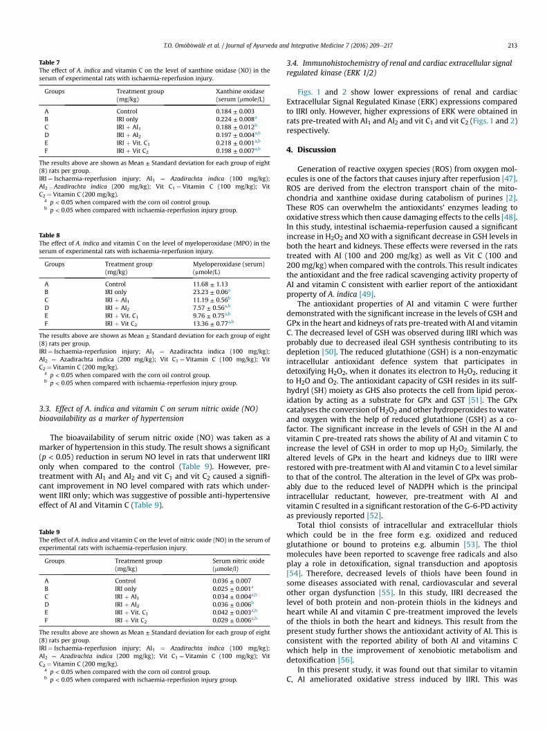
Table 7The effect of A. indica and vitamin C on the level of xanthine oxidase (XO) in theserum of experimental rats with ischaemia-reperfusion injury.
Groups Treatment group(mg/kg)
Xanthine oxidase(serum (mmole/L)
A Control 0.184 ± 0.003B IRI only 0.224 ± 0.008a
C IRI þ AI1 0.188 ± 0.012b
D IRI þ AI2 0.197 ± 0.004a,b
E IRI þ Vit. C1 0.218 ± 0.001a,b
F IRI þ Vit C2 0.198 ± 0.007a,b
The results above are shown as Mean ± Standard deviation for each group of eight(8) rats per group.IRI ¼ Ischaemia-reperfusion injury; AI1 ¼ Azadirachta indica (100 mg/kg);AI2 ¼ Azadirachta indica (200 mg/kg); Vit C1 ¼ Vitamin C (100 mg/kg); VitC2 ¼ Vitamin C (200 mg/kg).
a p < 0.05 when compared with the corn oil control group.b p < 0.05 when compared with ischaemia-reperfusion injury group.
Table 8The effect of A. indica and vitamin C on the level of myeloperoxidase (MPO) in theserum of experimental rats with ischaemia-reperfusion injury.
Groups Treatment group(mg/kg)
Myeloperoxidase (serum)(mmole/L)
A Control 11.68 ± 1.13B IRI only 23.23 ± 0.06a
C IRI þ AI1 11.19 ± 0.56b
D IRI þ AI2 7.57 ± 0.56a,b
E IRI þ Vit. C1 9.76 ± 0.75a,b
F IRI þ Vit C2 13.36 ± 0.77a,b
The results above are shown as Mean ± Standard deviation for each group of eight(8) rats per group.IRI ¼ Ischaemia-reperfusion injury; AI1 ¼ Azadirachta indica (100 mg/kg);AI2 ¼ Azadirachta indica (200 mg/kg); Vit C1 ¼ Vitamin C (100 mg/kg); VitC2 ¼ Vitamin C (200 mg/kg).
a p < 0.05 when compared with the corn oil control group.b p < 0.05 when compared with ischaemia-reperfusion injury group.
T.O. Om�ob�ow�al�e et al. / Journal of Ayurveda and Integrative Medicine 7 (2016) 209e217 213
3.3. Effect of A. indica and vitamin C on serum nitric oxide (NO)bioavailability as a marker of hypertension
The bioavailability of serum nitric oxide (NO) was taken as amarker of hypertension in this study. The result shows a significant(p < 0.05) reduction in serum NO level in rats that underwent IIRIonly when compared to the control (Table 9). However, pre-treatment with AI1 and AI2 and vit C1 and vit C2 caused a signifi-cant improvement in NO level compared with rats which under-went IIRI only; which was suggestive of possible anti-hypertensiveeffect of AI and Vitamin C (Table 9).
Table 9The effect of A. indica and vitamin C on the level of nitric oxide (NO) in the serum ofexperimental rats with ischaemia-reperfusion injury.
Groups Treatment group(mg/kg)
Serum nitric oxide(mmole/l)
A Control 0.036 ± 0.007B IRI only 0.025 ± 0.001a
C IRI þ AI1 0.034 ± 0.004a,b
D IRI þ AI2 0.036 ± 0.006b
E IRI þ Vit. C1 0.042 ± 0.003a,b
F IRI þ Vit C2 0.029 ± 0.006a,b
The results above are shown as Mean ± Standard deviation for each group of eight(8) rats per group.IRI ¼ Ischaemia-reperfusion injury; AI1 ¼ Azadirachta indica (100 mg/kg);AI2 ¼ Azadirachta indica (200 mg/kg); Vit C1 ¼ Vitamin C (100 mg/kg); VitC2 ¼ Vitamin C (200 mg/kg).
a p < 0.05 when compared with the corn oil control group.b p < 0.05 when compared with ischaemia-reperfusion injury group.
3.4. Immunohistochemistry of renal and cardiac extracellular signalregulated kinase (ERK 1/2)
Figs. 1 and 2 show lower expressions of renal and cardiacExtracellular Signal Regulated Kinase (ERK) expressions comparedto IIRI only. However, higher expressions of ERK were obtained inrats pre-treated with AI1 and AI2 and vit C1 and vit C2 (Figs. 1 and 2)respectively.
4. Discussion
Generation of reactive oxygen species (ROS) from oxygen mol-ecules is one of the factors that causes injury after reperfusion [47].ROS are derived from the electron transport chain of the mito-chondria and xanthine oxidase during catabolism of purines [2].These ROS can overwhelm the antioxidants’ enzymes leading tooxidative stress which then cause damaging effects to the cells [48].In this study, intestinal ischaemia-reperfusion caused a significantincrease in H2O2 and XOwith a significant decrease in GSH levels inboth the heart and kidneys. These effects were reversed in the ratstreated with AI (100 and 200 mg/kg) as well as Vit C (100 and200 mg/kg) when compared with the controls. This result indicatesthe antioxidant and the free radical scavenging activity property ofAI and vitamin C consistent with earlier report of the antioxidantproperty of A. indica [49].
The antioxidant properties of AI and vitamin C were furtherdemonstrated with the significant increase in the levels of GSH andGPx in the heart and kidneys of rats pre-treatedwith AI and vitaminC. The decreased level of GSH was observed during IIRI which wasprobably due to decreased ileal GSH synthesis contributing to itsdepletion [50]. The reduced glutathione (GSH) is a non-enzymaticintracellular antioxidant defence system that participates indetoxifying H2O2, when it donates its electron to H2O2, reducing itto H2O and O2. The antioxidant capacity of GSH resides in its sulf-hydryl (SH) moiety as GHS also protects the cell from lipid perox-idation by acting as a substrate for GPx and GST [51]. The GPxcatalyses the conversion of H2O2 and other hydroperoxides towaterand oxygen with the help of reduced glutathione (GSH) as a co-factor. The significant increase in the levels of GSH in the AI andvitamin C pre-treated rats shows the ability of AI and vitamin C toincrease the level of GSH in order to mop up H2O2. Similarly, thealtered levels of GPx in the heart and kidneys due to IIRI wererestored with pre-treatment with AI and vitamin C to a level similarto that of the control. The alteration in the level of GPx was prob-ably due to the reduced level of NADPH which is the principalintracellular reductant, however, pre-treatment with AI andvitamin C resulted in a significant restoration of the G-6-PD activityas previously reported [52].
Total thiol consists of intracellular and extracellular thiolswhich could be in the free form e.g. oxidized and reducedglutathione or bound to proteins e.g. albumin [53]. The thiolmolecules have been reported to scavenge free radicals and alsoplay a role in detoxification, signal transduction and apoptosis[54]. Therefore, decreased levels of thiols have been found insome diseases associated with renal, cardiovascular and severalother organ dysfunction [55]. In this study, IIRI decreased thelevel of both protein and non-protein thiols in the kidneys andheart while AI and vitamin C pre-treatment improved the levelsof the thiols in both the heart and kidneys. This result from thepresent study further shows the antioxidant activity of AI. This isconsistent with the reported ability of both AI and vitamins Cwhich help in the improvement of xenobiotic metabolism anddetoxification [56].
In this present study, it was found out that similar to vitaminC, AI ameliorated oxidative stress induced by IIRI. This was
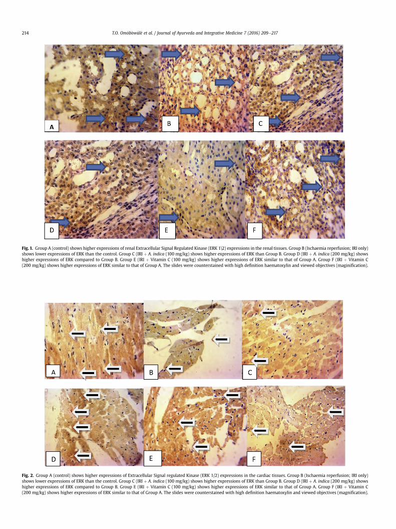
Fig. 1. Group A (control) shows higher expressions of renal Extracellular Signal Regulated Kinase (ERK 1\2) expressions in the renal tissues. Group B (Ischaemia reperfusion; IRI only)shows lower expressions of ERK than the control. Group C (IRI þ A. indica (100 mg/kg) shows higher expressions of ERK than Group B. Group D (IRI þ A. indica (200 mg/kg) showshigher expressions of ERK compared to Group B. Group E (IRI þ Vitamin C (100 mg/kg) shows higher expressions of ERK similar to that of Group A. Group F (IRI þ Vitamin C(200 mg/kg) shows higher expressions of ERK similar to that of Group A. The slides were counterstained with high definition haematoxylin and viewed objectives (maginification).
Fig. 2. Group A (control) shows higher expressions of Extracellular Signal regulated Kinase (ERK 1/2) expressions in the cardiac tissues. Group B (Ischaemia reperfusion; IRI only)shows lower expressions of ERK than the control. Group C (IRI þ A. indica (100 mg/kg) shows higher expressions of ERK than Group B. Group D (IRI þ A. indica (200 mg/kg) showshigher expressions of ERK compared to Group B. Group E (IRI þ Vitamin C (100 mg/kg) shows higher expressions of ERK similar to that of Group A. Group F (IRI þ Vitamin C(200 mg/kg) shows higher expressions of ERK similar to that of Group A. The slides were counterstained with high definition haematoxylin and viewed objectives (magnification).
T.O. Om�ob�ow�al�e et al. / Journal of Ayurveda and Integrative Medicine 7 (2016) 209e217214
T.O. Om�ob�ow�al�e et al. / Journal of Ayurveda and Integrative Medicine 7 (2016) 209e217 215
consistent with the reduction in the level of serum xanthineoxidase levels in IRI which can contribute to the severe organdamage observed after reperfusion in ischaemic tissues [57]. Inaddition; xanthine oxidase degrades xanthine to uric acid. In thepurine degradation pathway, it oxidizes nicotinamide adeninedinucleotide (NADH) to generate superoxide anion radical (O2
e)and H2O2 during reperfusion. The increase in serum xanthineoxidase activity was directly proportional to the serum uric acidwhich has been implicated as a biomarker of oxidative stress incardiorenal diseases and also a mediator of hypertension [58,59].Hence, the level of xanthine oxidase in the serum was associatedwith IIRI. However, pre-treatment of rats with AI and vitamin Ccaused a significant reduction in the serum xanthine oxidaseactivity normalizing hyperuricaemia (high levels of uric acid inthe blood) which is a diagnostic marker for renal damage andhypertension.
Similarly, AI and vitamin C also ameliorated the oxidative stress,inflammation and cardiac damage signified by the decreased levelof myeloperoxidase in the serum of the rats pre-treated with AI andvitamin C. The MPO catalyses the cycle that produces oxidizingagents such as HOCl, oxidation of NO and reduction in NObioavailability [60]. In patients with cardiovascular diseases, MPO isusually increased [61]. AI contains important bioactive compoundswhich include phytosterols (sitosterols, sigmasterol and campas-terol) and flavonoids (rutin and quercetin), commonly known fortheir antioxidant, anti-inflammatory and antimicrobial activities[62]. Also, consistent with observation is an earlier study in whichAI was reported to decrease the activity of colonic MPO [56]. Since,AI could normalize the aforementioned elevated MPO, suggestingAI as an antioxidant, anti-inflammatory and cardioprotectivephytonutrient.
In this study, the level of serum NO was reduced followingischaemia-reperfusion injury. NO has been shown to be amediator and/or protector of the vascular systems in several vitalorgans including the heart, liver, lungs and kidneys. These pro-tective actions of nitric oxide in ischaemia-reperfusion injury aredue to its potential as an antioxidant, anti-adhesion, and anti-inflammatory agent [63]. Also, normalization of nitric oxidebioavailability is an important factor in the amelioration of hy-pertension by preventing platelet aggregation, improving smoothmuscle relaxation keeping blood vessels patent thereby loweringblood pressure. However, low levels of NO lead can to imbalancebetween dilation and vasoconstriction in favour of constrictionincreasing blood pressure with decreased flow leading to hy-pertension [64]. More importantly, NO may combine with su-peroxide anion radical to form peroxynitrite (ONOO-) which is acytototic agent. The formation of peroxynitrite may alsocontribute significantly to the reduced bioavailability of NO andhence to the development of cardiovascular and renal dysfunc-tion. In this study, AI and Vitamin C ameliorated ischaemia-reperfusion injury-induced reduction in NO levels in the serumespecially in the rats pre-treated with AI (200 mg/k) and Vit C(100 mg/kg). However, one of the limitations of the present studyis that we could not take the blood pressure measurement as aclinical parameter for proper correlation of observable reducedbioavailability of NO as an indication of hypertensive state.Furthermore, we endeavour to take this into consideration thesignificance of this clinical parameter in our future study afterthe surgical procedure.
The results in the present study showed that IIRI crashed thelevels of GSH and GPx activity in both the heart and kidneys.However, IIRI increased the level of GST in the heart. The increasemight be attributable to adaptive responses of cells to oxidativestress, whereas the pre-treated rats had significantly decreasedlevel of GST. At 200 mg/kg vitamin C, there was a significant
increase in the level of GST which was probably due to the ability ofvitamin C to function as pro-oxidants at high concentration [65].However, in the kidneys; IIRI did not reduce the levels of GST ac-tivity as the level was similar to that of the control without IIRI.Meanwhile, AI and vitamin C increased the levels of GST but at200 mg/kg vitamin C, the level of GST was similar to that IRI onlywhich might be suggestive of vitamin C as a pro-oxidant at highconcentration. Hence, caution must be exercised in the use ofsynthetic antioxidant such as vitamin C and the interpretation ofthe results thereof.
The extracellular signal-regulated protein kinases 1 and 2 (ERK1/2) signalling pathway is a cascade consisting of at least threefamilies of protein kinases, including Raf (MAPKKK or MEKK),MAPKKs (MEK1 and MEK2), MAPK (ERK1 and ERK2 or p42/p44MAPKs). The ERK pathway not only regulates a wide range ofcellular behaviours, such as growth, proliferation, migration, dif-ferentiation, apoptosis and autophagy, but also mediates inflam-matory responses [66,67]. The ERK pathway can be activated by avariety of extracellular stimuli such as growth factors, cytokines,mitogens, hormones and oxidative or heat stress [68]. It has beendemonstrated that activation of ERK 1/2 mediated neuroprotectionof dexmedetomine, a potent and highly selective a2-adrenoceptoragonist in transient cerebral ischaemia-reperfusion [69]. The acti-vation of the Reperfusion Injury Salvage Kinase (RISK) pathway,which incorporates phosphatidylinositol-3-OH kinase (PI3K), AKT/Protein Kinase B (PKB) and p44/42 Mitogen Activated Protein Ki-nase (MAPK) underlies protection against IIRI [70]. In the presentstudy, IIRI reduced the activation of the RISK pathway therebyreducing the expression of ERK as shown by reduction in theimmuneepositive reaction IRI only group. However, AI and vitaminC ameliorated tissue damage following IIRI by increasing the ex-pressions of ERK (a survival protein) as mediated in the RISKpathway.
The results in the present study have shown that intestinalischaemia reperfusion injury does not only have deleterious ef-fect on the intestines, but also on the heart and kidneys whichwas shown by the inhibition of both the enzymic and non-enzymic antioxidants and increased generation of ROS. Howev-er, AI was able to ameliorate these deleterious effects byincreasing the in vivo antioxidant status, reduction in markers ofoxidative stress, inflammation, cardiac and renal damagetogether with improvement in NO bioavailability. Tissue survivalwas also mediated via increase in the expressions of ERK.
5. Conclusion
Together, A. indica and vitamin C prevented IRI-induced car-diorenal dysfunction via reduction in oxidative stress, improve-ment in antioxidant defence system and increase in the ERK1/2expressions. Therefore, A. indica can be a useful chemotherapeuticagent in the prevention and treatment of conditions induced byintestinal ischaemia-reperfusion injury.
Conflict of interest
There is no conflict of interest (political, religious, academic orfinancial) whatsoever attached with this manuscript.
Acknowledgment
Dr. Ebunoluwa R. Asenuga for her contribution during the Lab-oratory work of this research. We wish to acknowledge the Car-negie African Diaspora Fellowship Program support to Momoh A.Yakubu, PhD of Texas Southern University, Houston, TX to facilitatethe collaborations between the authors.
T.O. Om�ob�ow�al�e et al. / Journal of Ayurveda and Integrative Medicine 7 (2016) 209e217216
References
[1] Sharkey LM, Russell NK, Rutter CS, Middleton SJ, Bradley JA, Jamieson NV, et al.Urgent multivisceral transplantation for widespread splanchnic ischemia.J Am Coll Surg 2016;222(5):760e5.
[2] Cerqueira NF, Hussni CA, Yoshida WB. Pathophysiology of mesentericischemia/reperfusion: a review. Acta Cir Bras 2005;20(4):336e43.
[3] Moulin L, Rumbo C, Romero P, Pedraza N, Garcia Herv�a D, Orce G, et al. Casereport: multivisceral transplantation for an extensive cystic Lymphangioma ofthe mesenteric root. Transpl Proc 2016;48(2):543e5.
[4] Efthymiou CA, Weir WI. Salmonella sepsis simulating gastrointestinalischaemia following cardiopulmonary bypass. Interact Cardiovasc Thorac Surg2011;12(2):334e6.
[5] Zanoni FL, Benabou S, Greco KV, Moreno AC, Cruz JW, Filgueira FP, et al.Mesenteric microcirculatory dysfunctions and translocation of indigenousbacteria in a rat model of strangulated small bowel obstruction. Clin (SaoPaulo) 2009;64(9):911e9.
[6] Ozturk H, Terzi EH, Ozgen U, Duran A, Ozturk H. Lithospermic acid andischemia/reperfusion injury of the rat small intestine prevention. Adv Clin ExpMed 2012;21(4):433e9.
[7] Nardo B, Beltempo P, Bertelli R, Montalti R, Vivarelli M, Cescon M, et al.Combined heart and liver transplantation in four adults with familialamyloidosis: experience of a single center. Transpl Proc 2004;36(3):645e7.
[8] Eltzschig HK, Collard CD. Vascular ischaemia and reperfusion injury. Br MedBull. 2004;70(1):71e86.
[9] Carden DL, Granger DN. Pathophysiology of ischaemia-reperfusion injury.J Pathol 2000;190:255e66.
[10] Radonak J, Lakyov�a L, Toporcer T, Bober J. Mesenteric ischemiaelate diagnosisor managed disease? Rozhl Chir 2010;89(4):242e6.
[11] Abboud B, Daher R, Boujaoude J. Acute mesenteric ischemia after cardio-pulmonary bypass surgery. World J Gastroenterol 2008;4(35):5361e70.
[12] Jian J, Xuan F, Qin F, Huang R. The antioxidant, anti-inflammatory and anti-apoptotic activities of the bauhinia championii flavone are connected withprotection against myocardial ischemia/reperfusion injury. Cell Physiol Bio-chem 2016;38(4):1365e75.
[13] Cuzzocrea S, Chatterjee P, Mazzon E, Dugo L, De Sarro A, Van de Loo FAJ, et al.Role of induced nitric oxide in the initiation of the inflammatory responseafter postischemic injury. Shock 2002;18:169e76.
[14] Macarengo RSS, Takahagi RU, Bardella LC, Sequeira JL, Yoshida WB. Estudo daaç~ao do extrato de Gingko biloba e amido hidroxietílico hipertonico naatenuaç~ao de alteraç~oes decorrentes de isquemia e reperfus~ao de �org~aosesplancnicos em ratos. Acta Cir Bras 2001;16:139e45.
[15] Pierro A, Eaton S. Intestinal ischemia reperfusion injury and multisystem or-gan failure. Semin Pediatr Surg 2004;13(1):11e7.
[16] Foley TR, Rogers RK. Endovascular therapy for chronic mesenteric ischemia.Curr Treat Options Cardiovasc Med 2016;18(6):39.
[17] Carver TW, Vora RS, Taneja A. Mesenteric ischemia. Crit Care Clin 2016;32(2):155e71.
[18] McKinsey JF, Gewertz BL. Isquemia mesent�erica aguda. In: Schwartz LB,Gewertz BL, editors. Isquemia mesent�erica. Rio de Janeiro: Interlivros; 1997.p. 313e24.
[19] Kojima M, Iwakiri R, Wu B, Fujise T, Watanabe K, Lin T, et al. Effects of anti-oxidative agents on apoptosis induced by ischaemia-reperfusion in rat in-testinal mucosa. Aliment Pharmacol Ther 2003;1(18 Suppl):139e45.
[20] Li Q, Zhang Q, Wang C, Liu X, Qu L, Gu L, et al. Altered distribution of tightjunction proteins after intestinal ischaemia/reperfusion injury in rats. J CellMol Med 2009;13(9B):4061e76.
[21] Massberg S, Messmer K. The nature of ischemia/reperfusion injury. TransplProc 1998;30:4217e23.
[22] Cibri�an D, Ajamieh H, Berlanga J, Le�on OS, Alba JS, Kim MJ, et al. Use ofgrowth-hormone-releasing peptide-6 (GHRP-6) for the prevention of multipleorgan failure. Clin Sci (Lond) 2006;110(5):563e73.
[23] Savas C, Ozogul C, Kara€oz E, Delibas N, Ozgüner F. Splenectomy reducesremote organ damage after intestinal ischaemia-reperfusion injury. Acta ChirBelg 2003;103(3):315e20.
[24] Satyanarayana K, Sravanthi K, Shaker IA, Ponnulakshmi R. Molecular approachto identify antidiabetic potential of Azadirachta indica. J Ayurveda Integr Med2015;6(3):165e74.
[25] Biswas K, Chattopadhyay I, Banerjee RK, Bandyopadhyay U. Biological activ-ities and medicinal properties of neem (Azadirachta indica). Curr Sci2002;82(11):1336e45.
[26] Omobowale TO, Oyagbemi AA, Oyewunmi OA, Adejumobi OA. Chemo-preventive effect of methanol extract of Azadirachta indica on experimentalTrypanosoma brucei induced oxidative stress in dogs. Pharmacogn Res2015;7(3):249e58.
[27] Lakshmi T, Krishnan V, Rajendran R, Madhusudhanan N. Azadirachta indica: aherbal panacea in dentistry e an update. Pharmacogn Rev 2015;9(17):41e4.
[28] Dallaqua B, Saito FH, Rodrigues T, Calderon IM, Rudge MV, Volpato GT, et al.Azadirachta indica treatment on the congenital malformations of fetuses fromrats. J Ethnopharmacol 2013;150(3):1109e13.
[29] Vijayan V, Tiwari PK, Meshram GP. Inhibitory effects of neem seed oil and itsextract on various direct acting and activation-dependant mutagens-inducedbacterial mutagenesis. Pharm Biol 2013;51(12):1525e30.
[31] Babu TA, Ananthakrishnan S. Idiopathic intracranial hypertension secondaryto ingestion of Morinda coreia and Azadirachta indica leaves extract in infant.J Pharmacol Pharmacother 2013;4(4):298e9.
[32] Koul A, Goyal R, Bharati S. Protective effect of Azadirachta indica a. Juss againstdoxorubicin-induced cardiac toxicity in tumour bearing mice. Indian J ExpBiol 2014;52(4):323e31.
[33] Arora N, Bansal MP, Koul A. Modulatory effects of Azadirachta indica leafextract on cutaneous and hepatic biochemical status during promotion phaseof DMBA/TPA-induced skin tumorigenesis in mice. Indian J Biochem Biophys2013;50(2):105e13.
[34] Peer PA, Trivedi PC, Nigade PB, Ghaisas MM, Deshpande AD. Cardioprotectiveeffect of Azadirachta indica A. Juss. on isoprenaline induced myocardialinfarction in rats. Int J Cardiol 2008;126(1):123e6.
[35] Garber JC, Barbee RW, Bielitzki JT, Clayton LA, Donovan JC, Hendriksen CFM.Guide for the care and use of laboratory animals, vol. 8. Washington DC: TheNational Academic Press; 2011. p. 220.
[36] Buetler E, Duron O, Kelly BM. Improved method for the determination ofblood glutathione. J Lab Clin Med 1963;61:882e8.
[37] Habig WH, Pabst MJ, Jacoby WB. Glutathione-S-transferase activity: theenzymic step in mercapturic acid formation. J Biol Chem 1974;249:130e9.
[38] Rotruck JT, Pope AL, Ganther HE, Swanson AB, Hafeman DG, Hoekstra WG.Seleniu biochemical role as a component of glutathione peroxidase. Sci1973;179:588e90.
[39] Ellman GL. Tissue sulfhydryl groups. Arch Biochem Biophys 1959;82:70e7.[40] Akaike T, Ando M, Oda T, Doi T, Ijiri S, Araki S, et al. Dependence on O2-
generation by xanthine oxidase of pathogenesis of influenza virus infection inmice. J Clin Invest 1990;85(3):739e45.
[41] Xia Y, Zweier JL. Measurement of myeloperoxidase in leukocyte-containintissues. Anal Biochem 1997;245:93e6.
[42] Varshney R, Kale RK. Effect of calmodulin antagonists on radiation inducedlipid peroxidation in microsomes. Intern J Biol 1990;158:733e41.
[43] Wolff SF. Ferrous ion oxidation in the presence of ferric ion indicator xylenolorange for measurement of hydrogen peroxides. Methods Enzymol 1994;233:182e9.
[44] Green LC, Ruiz de Luzuriaga K, Wagner DA. Nitrate biosynthesis in man. ProcNatl Acad Sci U. S. A 1981;78:7764e8.
[45] Crespo E, Macías M, Pozo D, Escames G, Martín M, Vives F, et al. Melatonininhibits expression of the inducible NO synthase II in liver and lung andprevents endotoxemia in lipopolysaccharide-induced multiple organdysfunction syndrome in rats. FASEB J 1999;13(12):1537e56.
[46] Gornal AG, Bardawill JC, David MM. Determination of serum proteins bymeans of Biuret reaction. J Biol Chem 1949;177:751e66.
[47] Galagudza MM, Sonin DL, Vlasov TD, Kurapeev DI, Shlyakhto EV. Remote vs.local ischaemic preconditioning in the rat heart: infarct limitation, suppres-sion of ischaemic arrhythmia and the role of reactive oxygen species. Int J ExpPathol 2016 Feb;97(1):66e74.
[48] Ray R, Shah AM. NADPH oxidase and endothelial cell function. Clin Sci2005;109(3):217e26.
[49] Charan AA, Gupta P. Comparative analysis of antibacterial, antioxidant andphotosynthetic activity of Azardirachta indica, Rosa indica and Moringa olieferacultivars. Int J Curr Res 2013;15:556e61.
[50] Kimura Y, Pierro A, Eaton S. Glutathione synthesis in intestinal ischaemia-reperfusion injury: effects of moderate hypothermia. J Pediatr Surg2009;44(2):353e7.
[51] Masella R, Di Benedetto R, Varì R, Filesi C, Giovannini C. Novel mechanisms ofnatural antioxidant compounds in biological systems: involvement of gluta-thione and glutathione-related enzymes. J Nutri Biochem 2005;16(10):577e86.
[52] Shailey S, Basir SF. Strengthening of antioxidant defense by Azadirachta indicain alloxan-diabetic rat tissues. J Ayurveda Integ Med 2012;3(3):130.
[53] Carter DC, Ho JX. Structure of serum albumin. Adv Protein Chem 2004;45:153e203.
[54] Jones DP, Carlson JL, Mody VC. Redox state of glutathione in human plasma.Free Radic Biol Med 2000;28:625e35.
[55] Colombo G, Reggiani F, Podest�a MA, Garavaglia ML, Portinaro NM, Milzani A,et al. Plasma protein thiolation index (PTI) as a biomarker of thiol-specificoxidative stress in haemodialyzed patients. Free Radic Biol Med 2015;89:443e51.
[56] Gautam MK, Goel S, Ghatule RR, Singh A, Joshi VK, Goel RK. Azadirachta indicaattenuates colonic mucosal damage in experimental colitis induced by trini-trobenzene sulfonic acid. Indian J Pharm Sci 2013;75(5):602e6.
[57] Ohtsubo T, Rovira II , Starost MF, Liu C, Finkel T. Xanthine oxidoreductase is anendogenous regulator of cyclooxygenase-2. Circu Res 2004;95(11):1118e24.
[58] Cantu-Medellin N, Kelley EE. Xanthine oxidoreductase-catalyzed reactivespecies generation: a process in critical need of reevaluation. Redox biology2013;1(1):353e8.
[59] Riegersperger M, Covic A, Goldsmith D. Allopurinol, uric acid, and oxidativestress in cardiorenal disease. Intern Urolo Nephrol 2011;43(2):441e9.
[60] Abu-Soud HM, Hazen SL. Nitric oxide is a physiological substrate formammalian peroxidases. J Biol Chem 2000;275(48):37524e32.
[61] Kutter D, Devaquet P, Vanderstocken G, Paulus JM, Marchal V, Gothot A.Consequences of total and subtotal myeloperoxidase deficiency: risk orbenefit? Acta Haematol 2000;104:10e5.
T.O. Om�ob�ow�al�e et al. / Journal of Ayurveda and Integrative Medicine 7 (2016) 209e217 217
[62] Ghatule RR, Goel S, Gautam MK, Singh A, Joshi VK, Goel RK. Effect of Azadir-achta indica leaves extract on AA-Induced colitis in rats: role of antioxidants,free radicals and myeloperoxidase. Asian Pac J Trop Dis 2012:S651e7.
[63] Phillips L, Toledo AH, Lopez-Neblina F, Anaya-Prador Toled Pereyra LH. Nitricoxide mechanism of protection in ischaemia and reperfusion injury. J InvestSurg 2009;22(1):46e55.
[64] Raij L. Nitric oxide in the pathogenesis of cardiac disease. J Clin Hypertens(Greenwich) 2006;8(12 Suppl 4):30e9.
[65] Miura Y. Oxidative stress, radiation- adaptive responses and ageing. J RadiatRes 2004;45:357e72.
[66] Cagnol S, Chambard JC. ERK and cell death: mechanisms of ERK-induced celldeatheapoptosis, autophagy and senescence. FEBS 2010;277(1):2e21.
[67] Gorelik G, Richardson B. Key role of ERK pathway signaling in lupus. Auto-immunity 2010;43(1):17e22.
[68] Mebratu Y, Tesfaigzi. How ERK1/2 activation controls cell proliferation andcell death: is subcellular localization the answer? Cell cycle 2007;8:1168e75.
[69] Zhu YM, Wang CC, Chen L, Qian LB, Ma LL. Both PI3K/Akt and ERK1/2 path-ways participate in the protection by dexmedetomidine against transientfocal cerebral ischaemia/reperfusion injury in rats. Brain Res 2013;1494:1e8.
[70] Smith CC, Mocanu MM, Brown J, Wynne AM, Simpkin JC, Dixon RA, et al.Temporal changes in myocardial salvage kinases during reperfusion followingischaemia: studies involving the cardiprotective adipocytokine apelin. Car-divasc Drugs Ther 2007;21(6):409e14.