jemds.d… · web viewmost cyst but not all, are lined by epithelium.(1) the term cyst is derived...
TRANSCRIPT

Rushton Bodies: A Rare entity in Radicular Cyst
Dr. Shubhangi Khandekar, Dr. Alka Dive, Dr. Divya Sao, Dr. Anand Rajderkar
Abstract
Radicular cyst is the most common inflammatory odontogenic cyst. It is a consequence
to pulp necrosis of carious teeth. In this case report, we present a rare case of radicular cyst
with Rushton bodies in a 42 years male patient.
Keywords – Inflammatory cyst, Radicular cyst, Rushton bodies.
Introduction
Cyst is defined as “a pathological cavity having fluid, semifluid or gaseous contents
and which are not created by the accumulation of pus”. Most cyst but not all, are lined by
epithelium.(1) The term cyst is derived from the Greek word Kystis meaning sac or bladder.(2)
Radicular cysts are the most common inflammatory cysts and arise from the epithelial
residues in the periodontal ligament as a result of periapical periodontitis following death
and necrosis of the pulp (1) They may also be found on the lateral aspects of the roots in
relation to lateral accessory root canals. Quite often a radicular cyst remains behind in the
jaws after removal of the offending tooth and this is referred to as a residual cyst. (3) It is the
most common cystic lesions in the jaws. The high frequency of occurrence of cyst is
maxillary anterior region. It occurs at the age between third and fifth decades of life.(1)
CASE REPORT
A 42 years old male patient reported to the department with a chief complaint of pain in
upper front region of jaw since 15 days. Pain was dull and intermittent. Patient had a
history of an accident 20 years back that lead to discoloration of maxillary right lateral
incisor over a period.

Intra-oral examination revealed a single diffuse swelling on the palate of approximately
2x4 cm in size extending from 15 to 22 region. Swelling was oval in shape, soft in
consistency and of normal mucosal color. On palpation, swelling was non tender. The
vitality test was negative with maxillary right lateral incisor.
Radiographic examination revealed a radiolucent area which was well defined, roughly
oval in shape of approximately 2x4 cm in size and was extending from maxillary right
second premolar to maxillary left lateral incisor. Divergence was seen between the roots of
maxillary right lateral incisors and maxillary canine.
Figure - 1
On correlating the clinical & radiographical findings, the case was provisionally
diagnosed as “radicular cyst”.
After routine hematological investigation, the patient underwent root canal treatment of
maxillary right lateral incisor and enucleation of the cystic lesion was done under local
anesthesia. Specimen was sent for histopathological examination.
Gross examination revealed a soft tissue specimen which was grayish black in colour,
oval in shape, of approximately 2x4 cm in size and soft to firm in consistency with rough
surface texture. Cut section revealed a cystic sac.
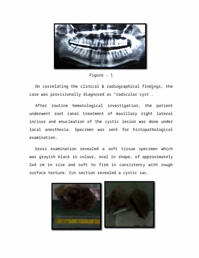
Figure - 2
Microscopic examination of H & E stained section showed cystic lumen with thin
lining of non-keratinized stratified squamous epithelium of variable thickness. The
connective tissue capsule consisted of fibrocellular stroma with dense chronic
inflammatory cell infiltrate chiefly of lymphocytes and plasma cells. Endothelial lined
blood capillaries were present few of which were engorged with RBC’s. Numerous
extravasated RBC’s were also seen at periphery. Rushton bodies were seen within the
epithelial lining.
Figure – 3, H & E Stained Sections

These Rushton bodies were positive with Papanicolaou (PAP) & Masson's Trichrome
stains. PAP stain showed pink colour, while with Masson’s Trichrome they appeared red in
color.
Based on clinical, radiological and histopathological findings, diagnosis of “radicular
cyst with Rushton bodies” was made.
Figure – 4. Pap Stain Figure – 5. Masson’s Trichrome Stain
Discussion
Usually, radicular cysts are symptomless and are discovered by chance when periapical
radiographs are taken of teeth with non-vital pulps. The radiological differential diagnosis
of radicular cyst includes periapical granuloma, surgical defect or periapical scar,
periapical cemento-osseous dysplasia, Traumatic Bone Cyst. It occasionally includes
odontogenic tumors, giant cell lesions, metastatic disease and primary osseous tumors.(1, 4)
Histopathologically, almost all radicular cysts are lined wholly or in part by non-
keratinized stratified squamous epithelium. These linings may be discontinuous in part and
range in thickness from one to 50 cell layers. The epithelial linings may be proliferating
and show arcading with an intense associated inflammatory process or be quiescent and
fairly regular with a certain degree of differentiation. The inflammatory cell infiltrate in the
proliferating epithelial linings consists predominantly of polymorphonuclear leucocytes
whereas the adjacent fibrous capsule is infiltrated mainly by chronic inflammatory cells. In
some cases of radicular cysts cholesterol clefts, Rushton bodies and pulse/seed granulomas
are occasionally seen.

Rushton bodies within the epithelium in odontogenic cyst were first described in detail
by MA Rushton and hence are often referred to as Rushton bodies.(6) These hyaline bodies
are found in approximately 10% of periapical cysts. However, available data from
published studies suggests a total incidence of 8%.(6) The bodies occur -only in the
epithelium or on its surface and are irregularly distributed.(1, 5) They are exclusively seen in
odontogenic cysts and not seen in non-odontogenic cysts such as nasopalatine duct cyst and
fissural cysts. A single case of Rushton Bodies in plexiform ameloblastoma has been
reported in the lesions other than jaw cysts.(6)
Microscopically, on histopathological examination Rushton bodies appear as structures
tucked within the epithelium that are acellular, eosinophilic, linear, straight or curved or
hairpin shaped, circular or polycyclic forms often with a granular core and at times
laminated concentrically. They were refractile but not birefringent.(6)
Rushton bodies gives negative result with Periodic acid Schiff and Von Kossa’s stains
for mucopolysaccharides and calcium respectively. Although Rushton bodies shows strong
positivity with Papanicolaou stain, Masson’s Trichrome, Orcein stains and Prussian blue.
Rushton bodies may also show positivity with Gomori stain. (5, 6)
According to Rushton, Rushton bodies originated from odontogenic epithelium as a
keratin product. Some authors postulated that they were of hematogenous origin or thought
that they were formed due to elastotic degeneration. Alternatively, they were also thought
to be formed as a cellular reaction to extravasated serum.(6)
In our case, Rushton bodies within the epithelial lining appeared as acellular,
eosinophilic and curvilinear shaped structures. These Rushton bodies showed positivity
with Papanicolaou (Pap) & Masson's Trichrome stains.
Conclusion
Radicular cyst usually goes unobserved and is discovered accidently in the radiographs.
This paper represents a case of radicular cyst showing Rushton bodies which is very rare
and the clinical significance of Rushton bodies is still not clear in the present scenario.

References
1. Shear M & Speight PM. Radicular & Rasidual Cyst. Cysts of the Oral Regions, 3rd edtion, Boston, Wright, 1992 .136-70.
2. Nair PNR. New perspectives on radicular cysts: do they heal? International Endodontic Journal (1998) 31.155–160.
3. Suhail Latoo, Ajaz. A. Shah, Suhail. M. Jan, S Qadir, I Ahmed, A.R . Purra, A .H Malik. Radicular Cyst. www.Jkscience.Org.Vol. 11(4), Oct-december 2009.
4. Rajendran R. Cysts & tumors of odontogenic origin. Shafer’s text book of oral pathology. 5th edition. Elsevier, 2010.268-269
5. Rushton MA. Hyaline bodies in the epithelium of dental cysts. Proc R Soc Med 1955;48: 407-9.
6. Sunitha Jacob.Rushton bodies or hyaline bodies in radicular cyst –a morphologic curiosity.. IJPM. Vol. 53. Oct.- Dec. 2010.