jalal jalal shokouhi-md diffusion imaging of cholesteatomas
TRANSCRIPT
Jalal jalal shokouhi-MD
DIFFUSION IMAGING OF CHOLESTEATOMAS

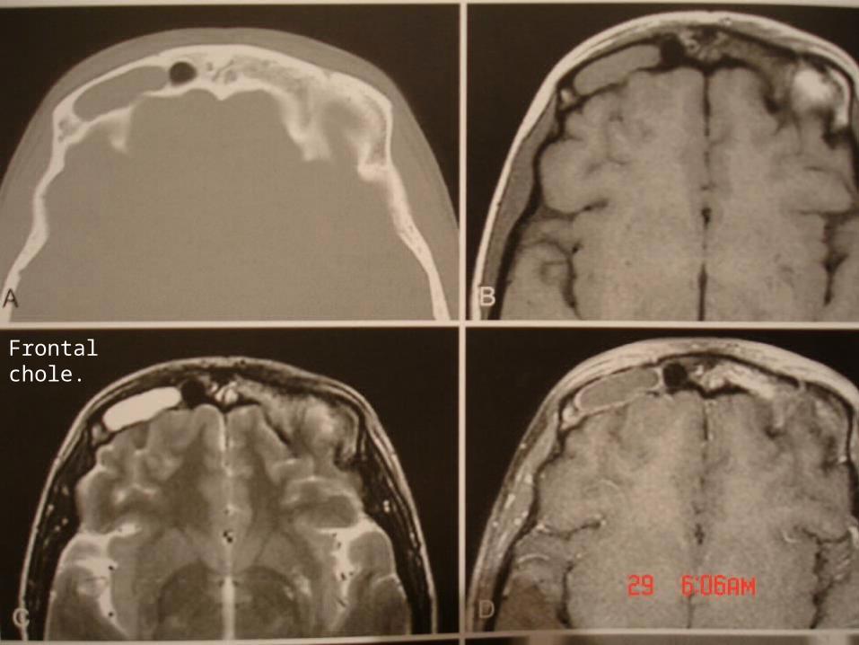
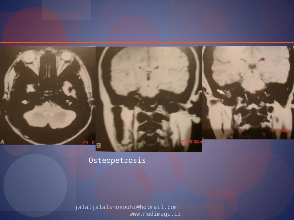
cholesteatomas
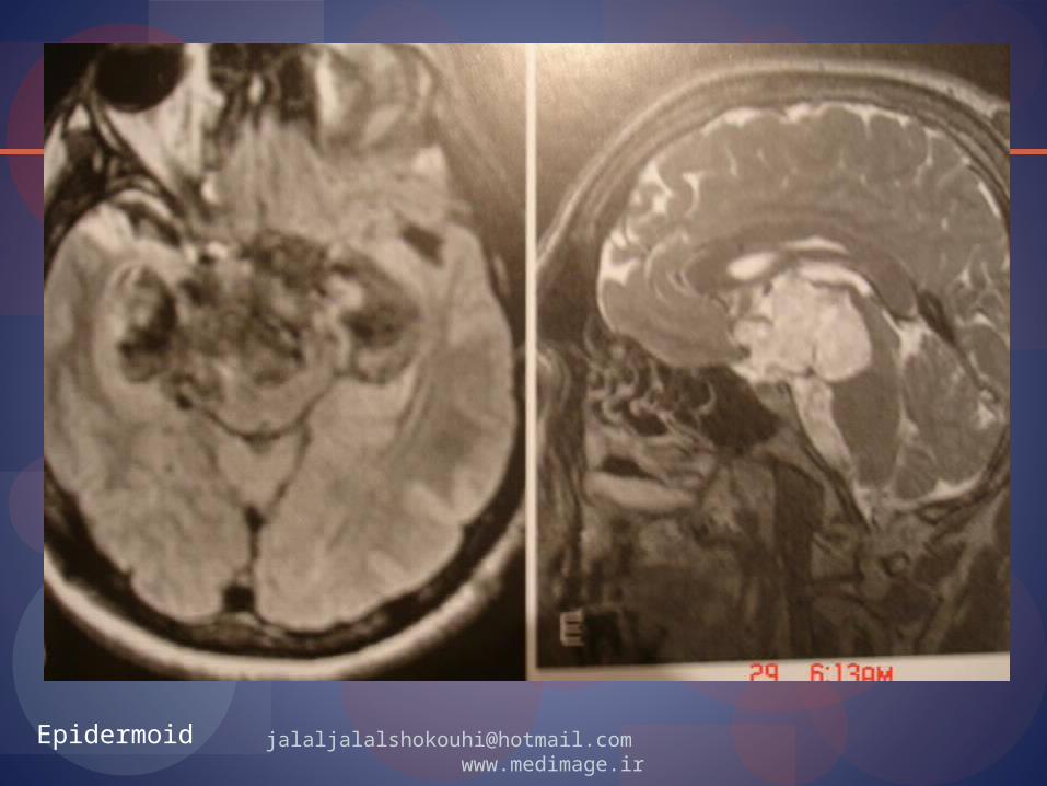
Epidermoid inclusion cystCystic creatinin-filled massLined by stratified squamous epitheliumCommon in frontal bone and earCause: secondary to trauma and implanted
inner table, outer table
[email protected] www.medimage.ir
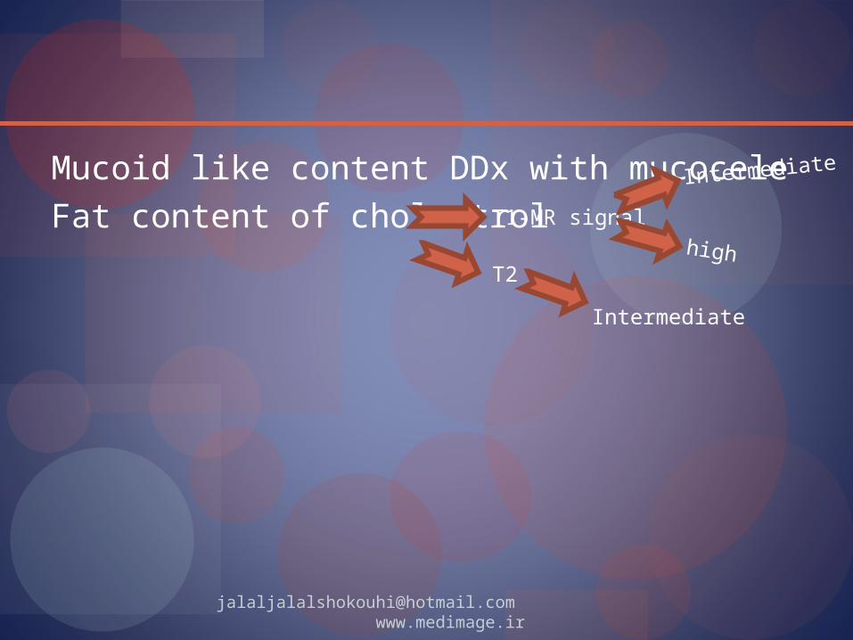
Mucoid like content DDx with mucoceleFat content of cholestrol T1-MR signal
T2
Intermediate
Intermediate
high
[email protected] www.medimage.ir
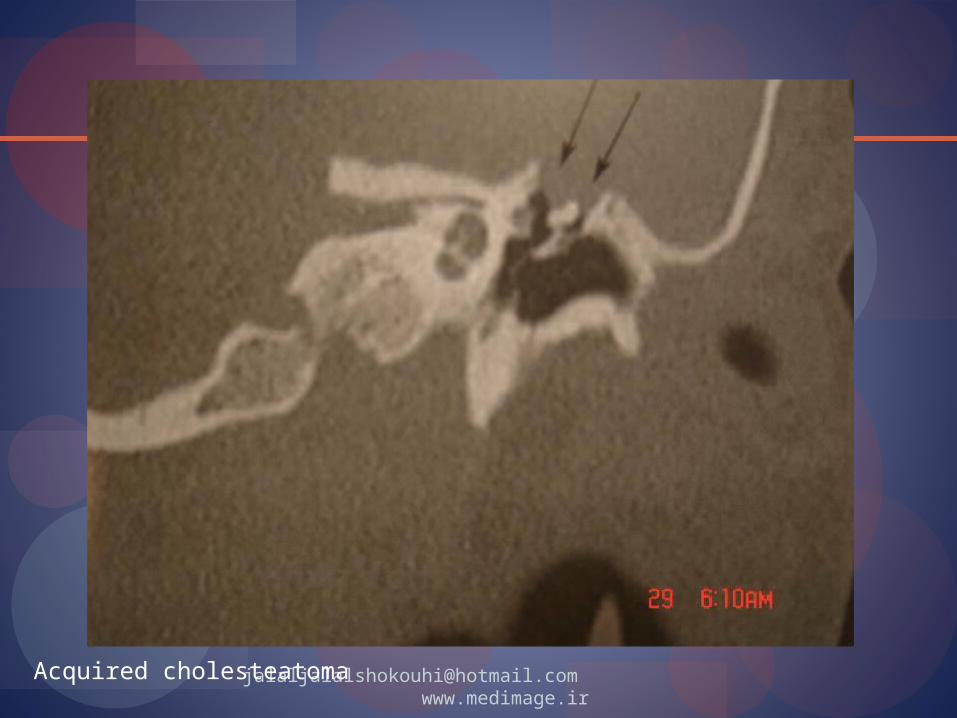
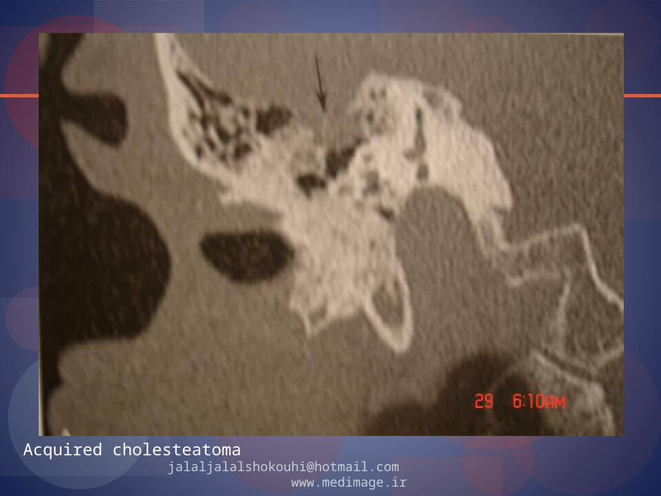
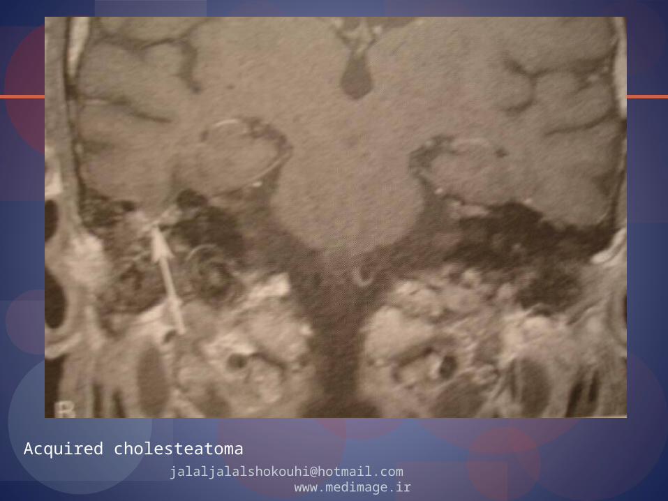
Acquired cholesteatoma Expansile concentrically enlarging
collection of exfoliated keratin lined by keratinizing stratified squamous epithelium
Not neoplasm May or may not contain cholesterol crystals Congenital (epidermoid) 2% Acquired in middle ear 98%
[email protected] www.medimage.ir


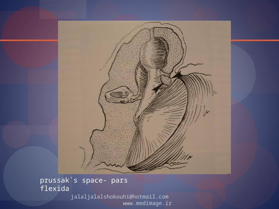
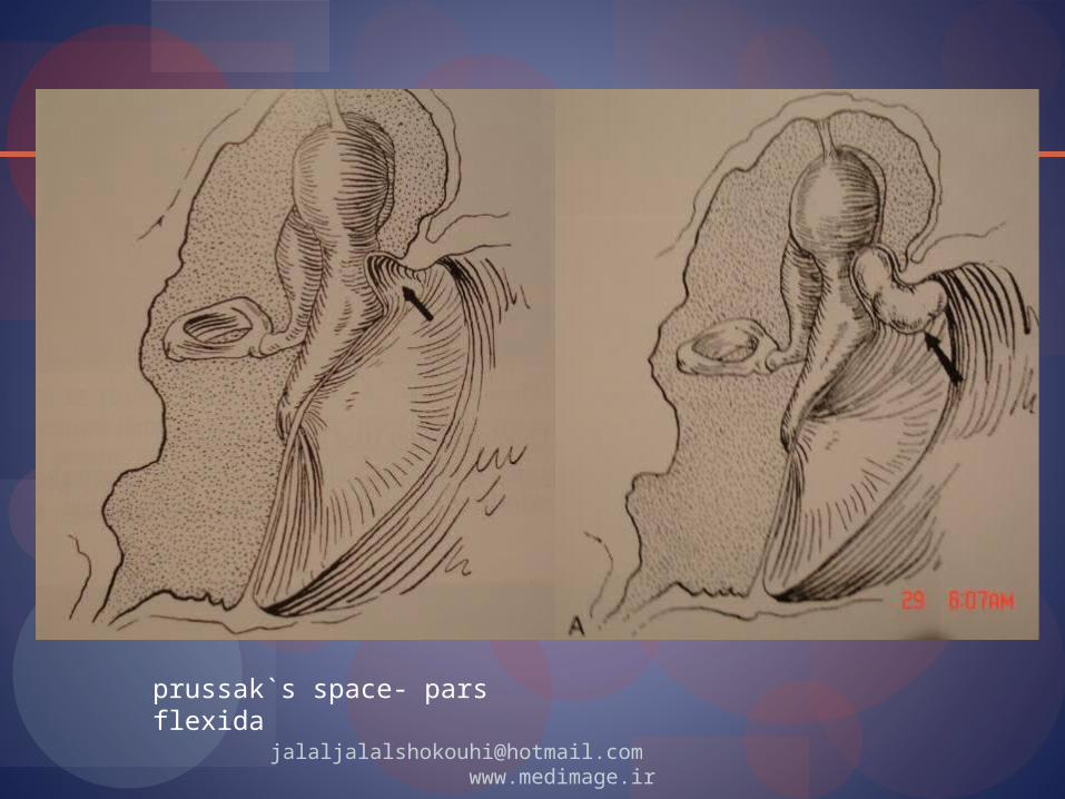
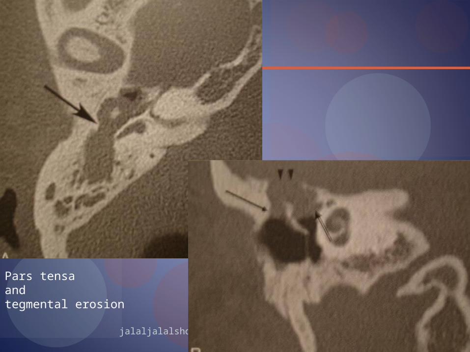
Acquired: Pars flexia (attic) -> prussak`s space -> mastoid Pars tensa
Acquired: Primary acquired (no infection) Secondary acquired (infection)
Etiology: retraction*, papillary proliferation, immigration, metplasia
[email protected] www.medimage.ir

[email protected] www.medimage.ir
[email protected] www.medimage.ir

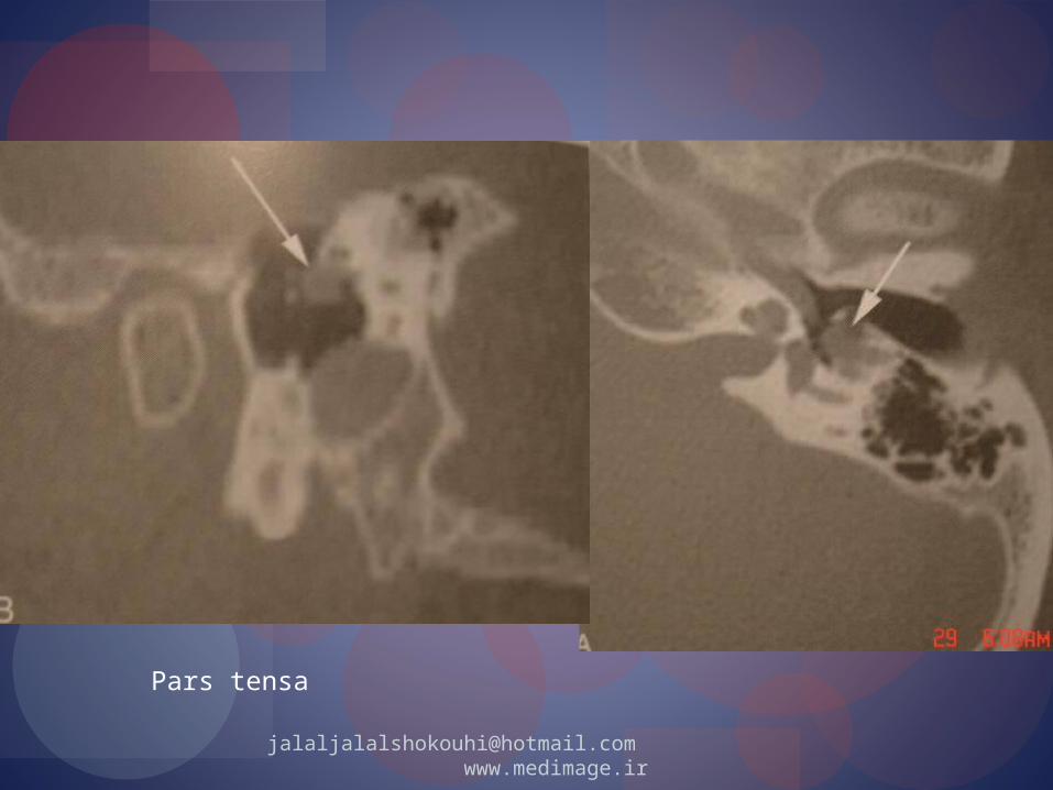
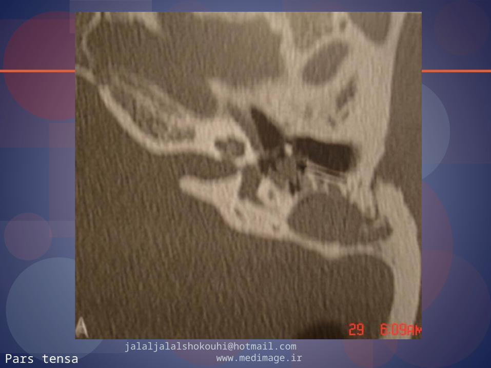
[email protected] www.medimage.irPars tensa

[email protected] www.medimage.irPars tensa
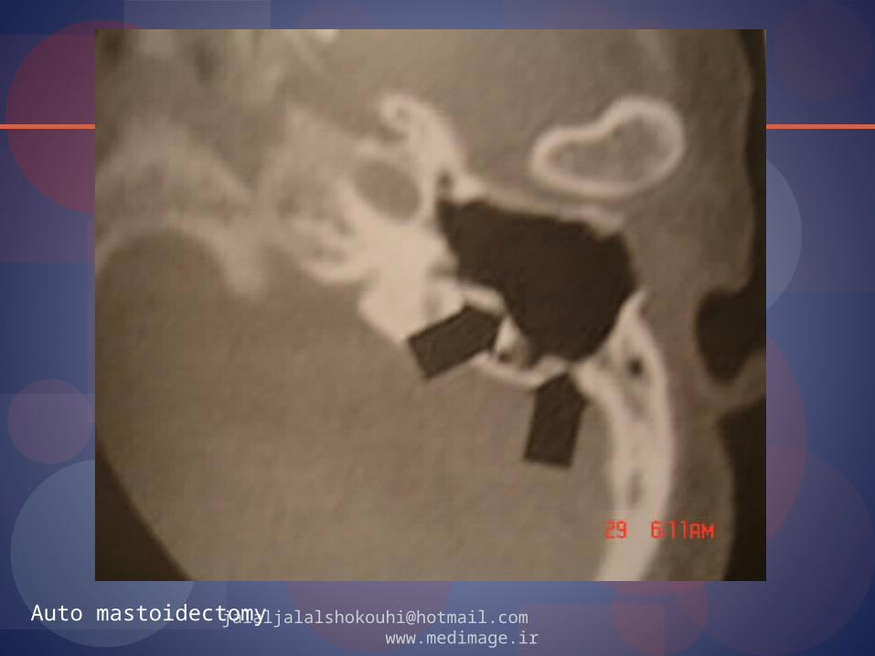
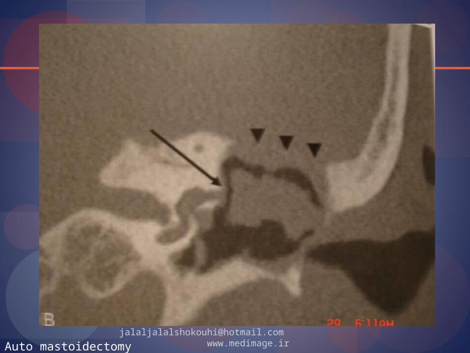
[email protected] www.medimage.irAuto mastoidectomy

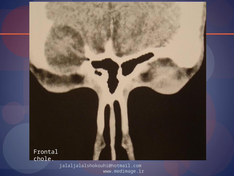
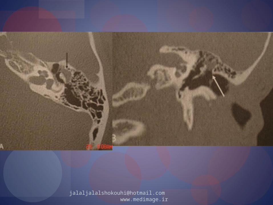
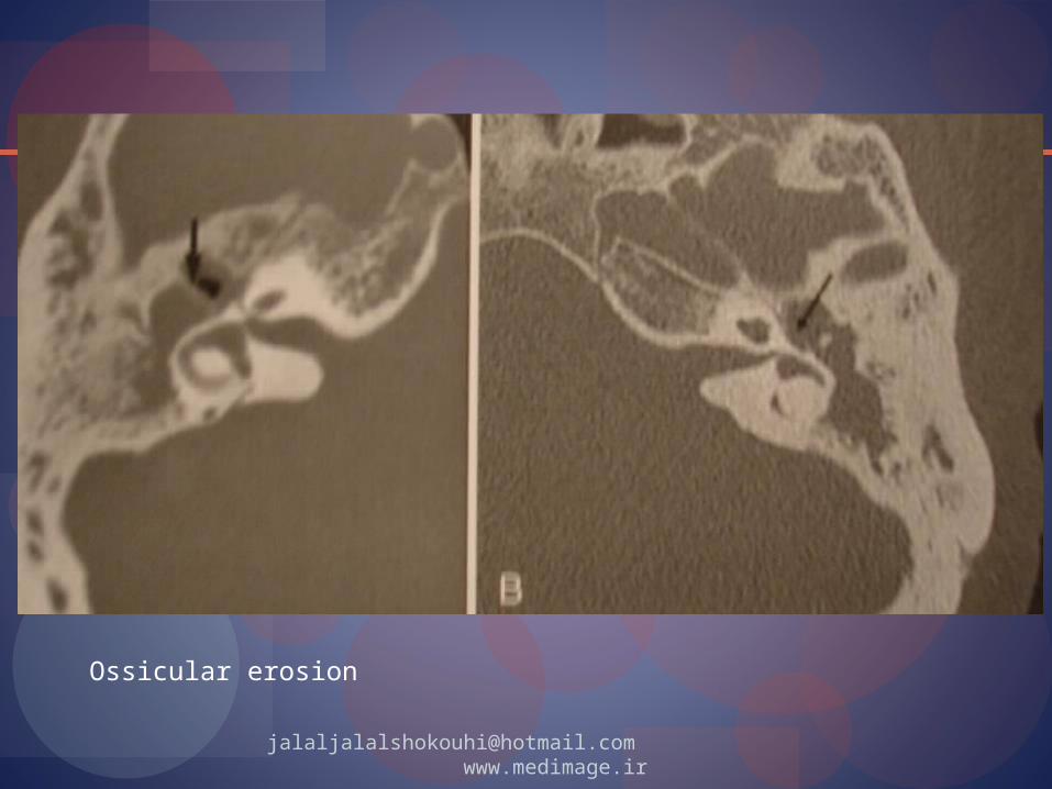
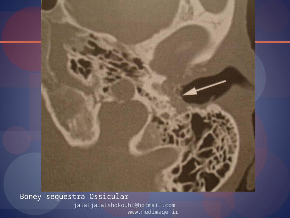
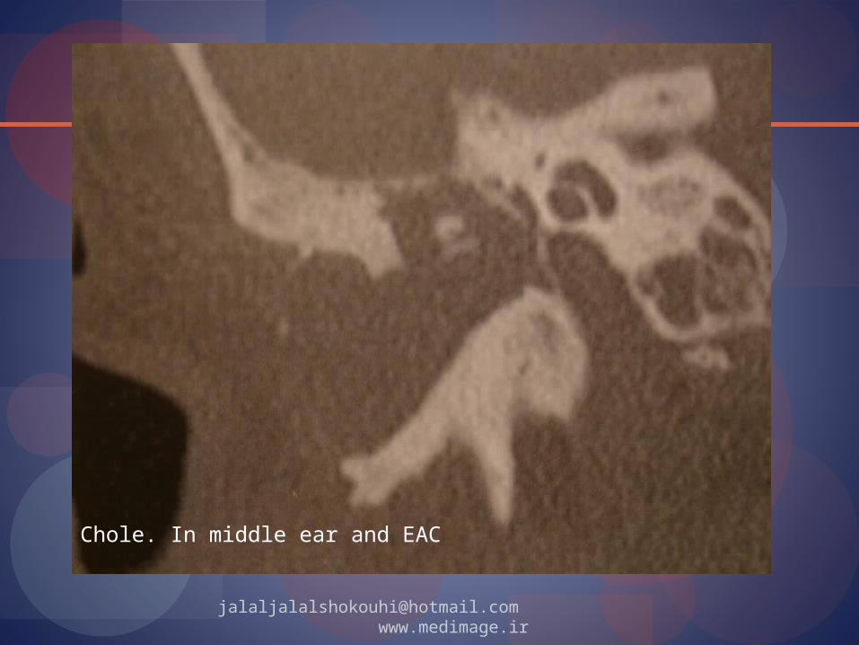
Imaging:X-ray CT bone destruction, soft tissue
demo. And complications, uncommon granulation tissue
MR spine-echo non-specific signal moderately Hyperintense (better for tegmen tympani and sinus) exclude
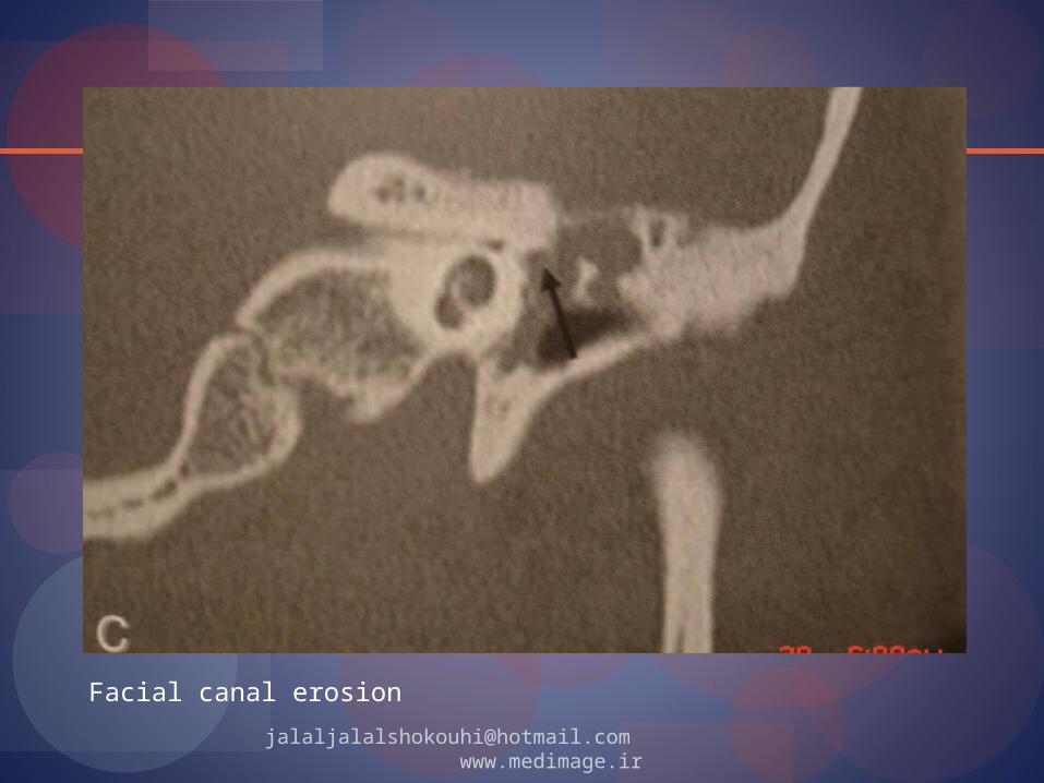
facial nerve involvement GD for granulation tissue versus non-enhancing
cholesteatoma
[email protected] www.medimage.ir


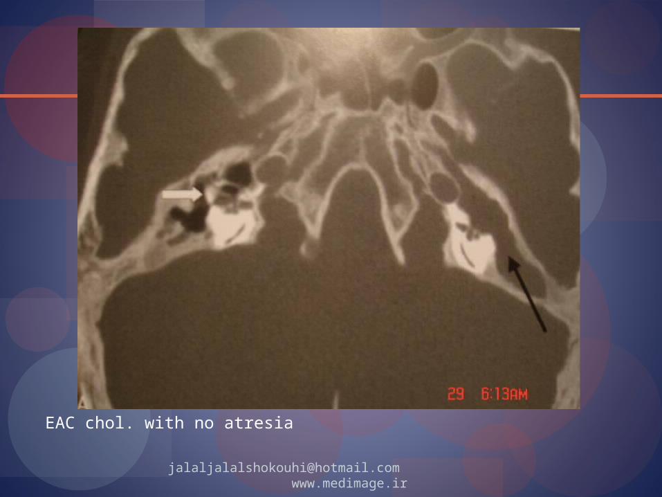


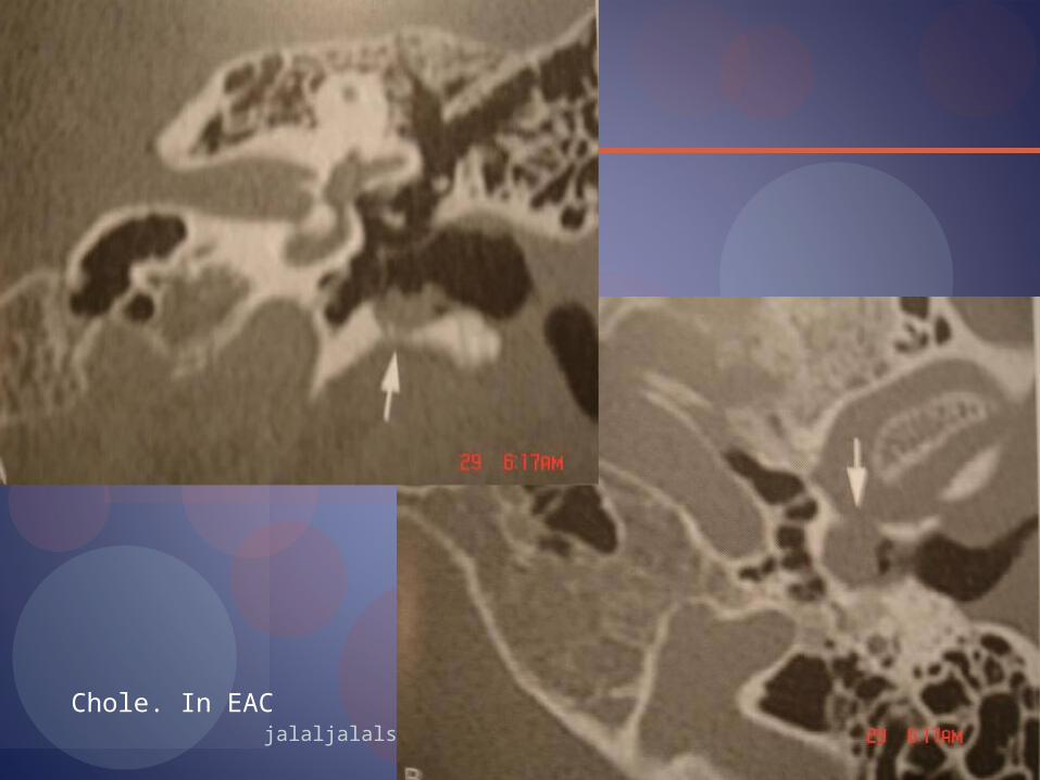
Cholesteatoma of EAC with atresia
CH. In vestibula

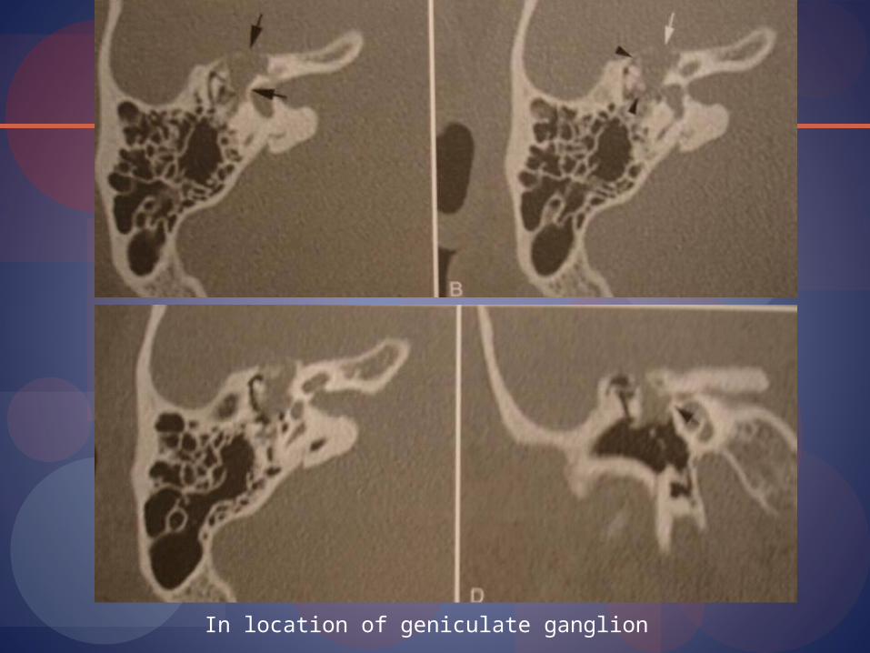
In location of geniculate ganglion

Diffusion-weighted imagingCholesteatoma is bright means restricted diffusion and
(T2 shine through) Please be aware eddy current artifacts, susceptibility
artifacts, ghosting artifacts, chemical shift and motion artifacts all are prevalent
Higher magnetic fields increase potential of these artifacts in echo-planar DWI
Turbo spin-echo DWI in known to limit, these distortions
Multi shot fast spin-echo periodically rotated overlapping parallel lines with enhanced reconstructions (PROPELLER) DWI= enhanced detection of the lesion (and limiting artifact in high fields)
[email protected] www.medimage.ir
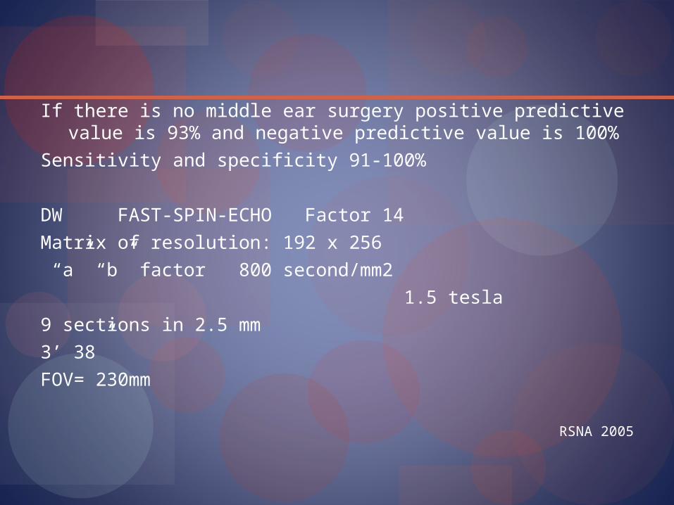
If there is no middle ear surgery positive predictive value is 93% and negative predictive value is 100%
Sensitivity and specificity 91-100%
DW FAST-SPIN-ECHO Factor 14
Matrix of resolution: 192 x 256
“a” “b” factor 800 second/mm2
1.5 tesla
9 sections in 2.5 mm
3’ 38”
FOV= 230mm
RSNA 2005
DW non-echoplanarDW of middle ear cholesteatoma differ from
abscess and infected cholesteatoma AJNR
DW for post-operative recurrent JU-radiology
[email protected] www.medimage.ir
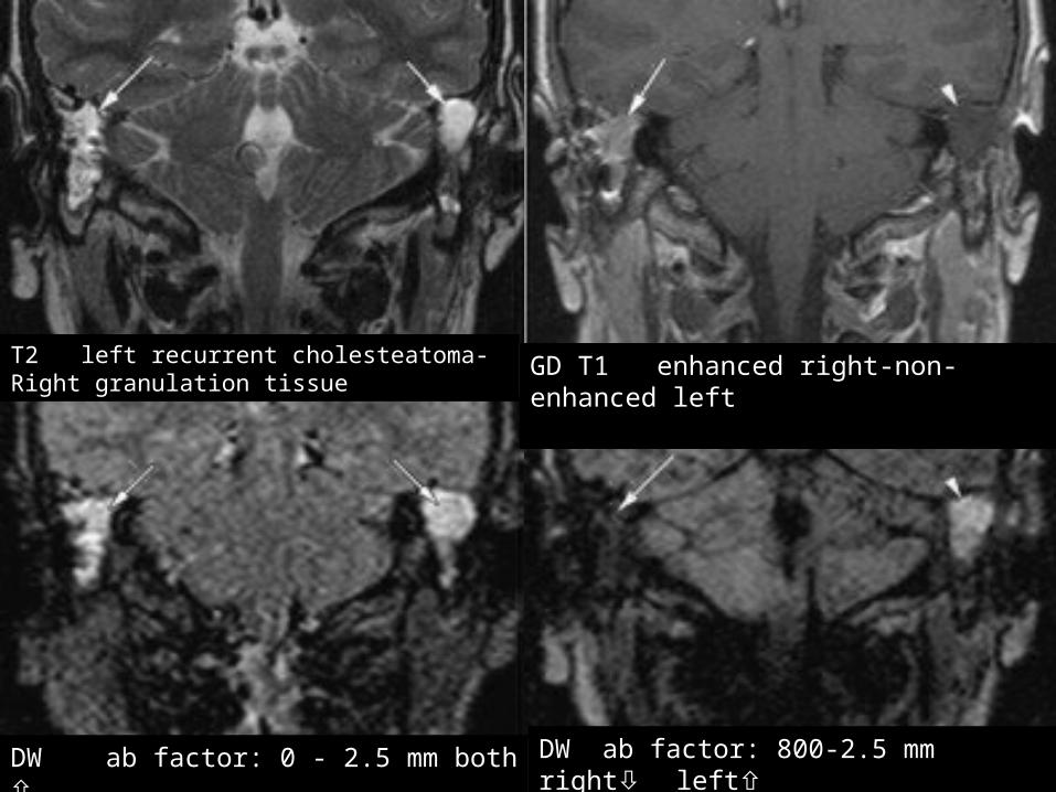
T2 left recurrent cholesteatoma-Right granulation tissue
GD T1 enhanced right-non-enhanced left
DW ab factor: 0 - 2.5 mm both DW ab factor: 800-2.5 mm right left

All complications related to bone destruction (mechanical)
C.O.M., vertigo, labyrinthin fistula(more morbidity) in lat. Semicircular canal (18-49)
Facial nerve palsy or paresis (1%)
[email protected] www.medimage.ir
[email protected] www.medimage.ir