iv brazilian guidelines for autoantibodies on hep-2 cells
TRANSCRIPT
www.reumatologia.com.br
REVISTA BRASILEIRA DE REUMATOLOGIA
R E V B R A S R E U M A T O L . 2 0 1 3 ; 5 4 ( 1 ) : 4 4 – 5 0
Original article
IV Brazilian Guidelines for autoantibodies on HEp-2 cells
Paulo Luiz Carvalho Francescantonioa, Wilson de Melo Cruvinela, Alessandra Dellavance b, Luis Eduardo Coelho Andrade b,c, Ben HurTalibertid, Carlos Alberto von Mühlene, Carlos David Araújo Bicharaf, Cleonice Buenog, Cristóvão Luis Pitangueira Mangueirah,i, Darlene Gonçalves Carvalho j, Eloísa S.D. de O. Bonfák, Fabiano de Almeida Brito j,l, Flávia Ikeda e Araújom, Jozelia Rêgon, Kaline Medeiros Costa Pereirab, Lisiane Maria Enriconi dos Anjoso,p, Maria de Fatima Bissoliq, Mittermayer Barreto Santiagor, Natalya Zaidan Malufs, Rossana Rassi Alvarengaa, Suzane Pretti Figueiredo Nevesl, Valeria Valimq, Wilton Silva dos Santost,u
a Pontifícia Universidade Católica de Goiás (PUC-Goiás), Goiânia, GO, Brazilb Fleury Medicina e Saúde, São Paulo, SP, Brazilc Discipline of Rheumatology, Universidade Federal de São Paulo (UNIFESP), São Paulo, SP, Brazil d Service of Rheumatology, Hospital das Clínicas, Universidade Federal de Uberlândia (UFU), Uberlândia, MG, Brazile Centro de Diagnósticos Médicos e Rheuma Clínica de Doenças Reumáticas de Porto Alegre, Porto Alegre, RS, Brazil f Amaral Costa Medicina Diagnóstica, Belém, PA, Brazilg Laboratórios de Investigação Médica, Hospital das Clínicas, Faculdade de Medicina, Universidade de São Paulo (FM-USP), São Paulo, SP, Brazilh Laboratório Central, Hospital das Clínicas, Faculdade de Medicina, Universidade de São Paulo (FM-USP), São Paulo, SP, Brazili Department of Clinical Pathology, Hospital Israelita Albert Einstein, São Paulo, SP, Brazilj Instituto Hermes Pardini, Belo Horizonte, MG, Brazilk Faculdade de Medicina, Universidade de São Paulo (USP), São Paulo, SP, Brazill Faculdade de Medicina, Universidade Federal de Minas Gerais (UFMG), Belo Horizonte, MG, Brazilm Universidade Católica de Brasília (UCB), Brasília, DF, Braziln Faculdade de Medicina, Universidade Federal de Goiás (UFG), Goiânia, GO, Brazilo Universidade do Sul de Santa Catarina (UNISUL), Florianópolis, SC, Brazilp Universidade do Vale do Itajaí (UNIVALE), Florianópolis, SC, Brazilq Universidade Federal do Espírito Santo (UFES), Vitória, ES, Brazilr Escola Bahiana de Medicina e Saúde Pública (EBMSP), Salvador, BA, Brazils Grupo DASA, São Paulo, SP, Brazilt Escola Superior de Ciências da Saúde do Distrito Federal, Brasília, DF, Brazilu Laboratório Sabin, Brasília, DF, Brazil
* Corresponding author.
E-mail: [email protected] (W.M. Cruvinel).
0482-5004/$ - see front matter. © 2014 Elsevier Editora Ltda. All rights reserved.
http://dx.doi.org/10.1016/j.rbre.2014.02.0062255-5021/$

45R E V B R A S R E U M A T O L . 2 0 1 3 ; 5 4 ( 1 ) : 4 4 – 5 0
IV Consenso Brasileiro para pesquisa de autoanticorpos em células HEp-2
Palavras-chave:
Autoanticorpos
Células HEp-2
Anticorpos antinúcleo
Imunofluorescência indireta
Consenso de FAN-HEp-2
r e s u m o
Objetivo: O IV Consenso Brasileiro para Pesquisa de Autoanticorpos em Células HEp-2 (FAN)
realizado em Vitória (ES), no dia 18 de setembro de 2012, objetivou discutir estratégias e
recomendações relacionadas ao procedimento técnico, à padronização e à interpretação
dos resultados da pesquisa de autoanticorpos em células HEp-2.
Métodos: Participaram do evento 23 pesquisadores e especialistas de Universidades e labo-
ratórios brasileiros. Foram abordados diferentes tópicos, discutidos amplamente a fim de
se estabelecer recomendações específicas.
Resultados e conclusão: O IV Consenso integrou à árvore de decisão o padrão citoplas-
mático em Anéis e Bastões, o padrão nuclear pontilhado Quasi-homogêneo (QH) e o
padrão misto CENP-F. Discutiu-se ainda a necessidade de atenção para a classificação
do padrão misto relacionado à presença de anticorpos anti-DNA topoisomerase I (Scl-
70), compreendendo os componentes nuclear pontilhado fino, nucleolar homogêneo,
NOR na placa metafásica e citoplasmático pontilhado fino. Foram sugeridas diretrizes
para o controle de qualidade do teste, diluição de triagem e diluição de esgotamento,
e foi emitido alerta quanto à necessidade de atenção em relação à heterogeneidade de
substratos disponíveis no mercado e a utilização de metodologias automatizadas para
detecção de autoanticorpos.
© 2014 Elsevier Editora Ltda. Todos os direitos reservados.
a r t i c l e i n f o
Article history:
Received 4 September 2013
Accepted 13 October 2013
Keywords:
Autoantibodies
HEp-2 cells
Antinuclear antibodies
Indirect immunofluorescence
ANA consensus
a b s t r a c t
Objective: The Fourth Brazilian Consensus for Autoantibodies Screening in HEp-2 Cells
(ANA) was held in Vitória, Espírito Santo, and aimed to discuss strategies and recommen-
dations about the technique, standardization, interpretation and quality control of the in-
direct immunofluorescence reaction on HEp-2 cells.
Methods: Twenty three ANA experts from university centers and private laboratories in dif-
ferent areas from Brazil discussed and agreed upon recommendations for the fourth edi-
tion of the Brazilian Consensus for Autoantibodies Screening in HEp-2 Cells.
Results and conclusion: The 4th ANA Consensus included three novel patterns into the ex-
isting algorithm (cytoplasmic Rods and Rings, nuclear Quasi-homogeneous, and CENP-F).
Emphasis was given to the need of attention in describing the peculiar mixed pattern elic-
ited by anti-DNA topoisomerase I (Scl-70) autoantibodies, comprising nuclear fine specked,
nucleolar homogeneous pattern, NOR staining in metaphase plates, and cytoplasmic fine
speckled patterns. The group also emphasized the need for continuous quality control in
indirect immunofluorescence assays, the establishment of screening dilutions, as well as
conjugate titration. An alert was made regarding the heterogeneity of commercial kits in
defining patterns and the use of solid phase methodologies to determine the presence of
autoantibodies.
© 2014 Elsevier Editora Ltda. All rights reserved.
Introduction
Over the last decade, the evaluation of autoantibodies against cellular antigens by indirect immunofluorescence (IIF) on HEp-2 cells underwent elaborate process of national standardization. These actions began in 2000, bringing sev-eral repercussions in Brazilian territory for the completion and interpretation of the test,1-4 stimulating even similar in-ternational initiatives.5-8
The first Consensuses were aimed at establishing of ac-tions in order to standardizing criteria for reading and in-terpreting the different patterns of autoantibodies on HEp-2
cells. Classification algorithms, based on morphological cri-teria and establishing five main groups of patterns (nuclear, nucleolar, cytoplasmic, mitotic apparatus and mixed) were prepared. Each algorithm was presented with orientation manuals, and the main clinical relevance of the different findings have also been addressed.1,2
The III Consensus, conducted in 2007, aimed to upgrade the clinical relevance of the test, to suggest effective mea-sures for quality control, and to evaluate the difficulties in implementing the standardization norms.3,4 The definition of still controversial aspects of the definition of nucleolus positivity was also discussed in III Consensus, and it was suggested the incorporation, to the algorithms, of two new
46 R E V B R A S R E U M A T O L . 2 0 1 3 ; 5 4 ( 1 ) : 4 4 – 5 0
fluorescence patterns that remained reserved, so that fur-ther studies were performed to obtain scientific evidence for its recognition – Cytoplasmic Rods/Rings Pattern and Speckled Quasi-homogeneous Pattern.3,4 In view of continu-ing education and of the need to keep up with scientific de-velopments , the IV Brazilian Consensus for Autoantibodies in HEp - 2 cells was held. The event was held in Vitória - ES, and gathered 23 experts on the subject from different regions of Brazil, and again the difficulties and advances in standardization of the test were discussed. The IV Consen-sus integrated, to the decision tree, the rings/rods pattern, the nuclear quasi-homogeneous speckled pattern (QH), and the mixed pattern observed with the presence of anti-CENP-F antibodies. Also was discussed In the Consensus the need for attention to the classification of mixed nuclear pattern associated with the presence of anti-DNA topoisomerase antibodies.9 Guidelines for quality control for the test and for the screening and exhaustion dilutions were suggested. Finally, warnings were issued about the heterogeneity of substrates available on the market and about the use of au-tomated methodologies for detection of autoantibodies.
The results of the IV Consensus allowed further progress in improving the criteria that enable the satisfactory control and utilization of the potentiality of this auxiliary method of diagnosis.
Work methodology
During the XXIX Brazilian Congress of Rheumatology (BCR), 23 researchers and experts from universities and private laboratories in different regions of Brazil took part in the IV Consensus in Vitória (ES), on September 18, 2012. In this event, recommendations related to technical procedures, standardization in the implementation and interpretation of test results were discussed and approved. Commercial rep-resentatives of different manufacturing companies attended the meeting as listeners, without commenting, voting, or presentation rights. Different topics related to the descrip-tion of new patterns, the technical procedure of the test, the dilution and titration of sera, the reproducibility of differ-ent brands of substrates, the use of automated methods for identifying autoantibodies, and presentation of reports were discussed. Each of the topics was presented to members of the assembly by relators and discussed widely, in order to establish recommendations. Each relator was based on data from the literature and from the presentation of personal studies.
General recommendations
I- Cytoplasmic rods/rings pattern
The rods/rings pattern was presented during the III Consen-sus, although at that time without defined immunological identity and only with preliminary scientific evidence. There-fore, this pattern was not incorporated into the decision tree at that occasion, with the recommendation of additional studies. After these studies, inosine monophosphate dehy-
drogenase 2 (IMPDH2) and cytidine triphosphate synthase 1 (CTPS1) were recognized as antigenic targets.10 These are es-sential enzymes in the biosynthesis of cytidine triphosphate and guanosine triphosphate, respectively. CTP is involved in the biosynthesis of nucleic acids (DNA, RNA) and phospholip-ids, with an important role in cell proliferation.11 IMPDH2 cat-alyzes the NAD-dependent oxidation of inosine monophos-phate to xanthosine monophosphate, an essential process in the biosynthesis of guanosine monophosphate – therefore, an activity also closely related to the cell proliferation mecha-nism.12 From the pharmacological inhibition of CTPS1 (6-di-azo-5-oxo-L-norleucine [Acivicin]) and IMPDH2 (Ribavirin), a dose-dependent induction of cytoplasmic rods/rings struc-tures on substrates of neoplastic cells, including HEp-2 cells, was evidenced.10
The Keppeke et al. (2012) study confirmed the close as-sociation between cytoplasmic rods/rings pattern and HCV infection. In a sample of 597 subjects with several clinical conditions, antibodies associated with the rods/rings pat-tern occurred exclusively in patients with HCV. Among 342 patients with HCV, the autoantibody occurred in 38% of those treated with ribavirin and interferon alpha, but in none of the other patients, including those receiving one of these drugs as monotherapy. Demographic parameters, time since diagno-sis, response to treatment, virus genotype or viral load were not correlated with the pattern.13 In the study by Covini et al. (2012), the production of autoantibodies against cytoplasmic structures after treatment with Ribavirin/IFN was observed in 15 HCV-positive participants.14
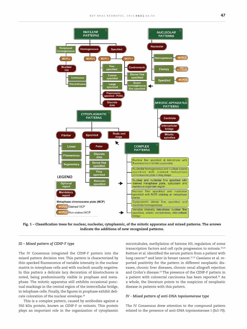
The IV Consensus integrated the rods/rings pattern to the decision tree (Fig. 1); this pattern was classified as an inde-pendent Cytoplasmic Pattern, not entailed to other cytoplas-mic patterns. A relevant information on the characterization of this pattern was related to the fact that the rods/rings structures are not expressed in all commercial substrates.15 Therefore, the IV Consensus suggested that, in the report, be informed that the recognition of this pattern is dependent on substrate.
II - Quasi-homogeneous speckled pattern (QH)
The IV Consensus integrated the nuclear quasi-homoge-neous speckled (QH) pattern into the nuclear pattern deci-sion-making tree. This pattern fits into the interpretation guide as optional (Fig. 1) and is characterized by extremely thin specked nuclear fluorescence, approaching the homo-geneous texture, with the metaphase plate similarly stained. This is a pattern distinct of the nuclear homogeneous and of the nuclear dense fine speckled patterns, where a single an-tigenic specificity is not noted, but miscellaneous antigenic targets are recognized. França et al. showed that the quasi-homogeneous fine speckled pattern exhibits a autoantibody profile intermediate between that of the dense fine speck-led pattern and the homogeneous pattern. Also, the clinical profile associated with quasi-homogeneous fine speckled pattern lies in a gray area between the dense fine speckled pattern and the homogeneous pattern.16 Therefore, the iden-tification of this pattern suggests further investigation of the clinical diagnosis, because it can be related to systemic auto-immune rheumatic diseases.17
47R E V B R A S R E U M A T O L . 2 0 1 3 ; 5 4 ( 1 ) : 4 4 – 5 0
III – Mixed pattern of CENP-F type
The IV Consensus integrated the CENP-F pattern into the mixed pattern decision tree. This pattern is characterized by thin specked fl uorescence of variable intensity in the nuclear matrix in interphase cells and with nucleoli usually negative. In this pattern a delicate lacy decoration of kinetochores is noted, being predominantly visible in prophase and meta-phase. The mitotic apparatus still exhibits occasional punc-tual markings in the central region of the intercellular bridge, in telophase cells. Finally, the fi gures in prophase exhibit deli-cate coloration of the nuclear envelope.18
This is a complex pattern, caused by antibodies against a 350 kDa protein, known as CENP-F or mitosin. This protein plays an important role in the organization of cytoplasmic
microtubules, methylation of histone H3, regulation of some transcription factors and cell cycle progression to mitosis.19,20 Rattner et al. identifi ed the serum pattern from a patient with lung cancer18 and later in breast cancer.21.22 Cassiano et al. re-ported positivity for the pattern in different neoplastic dis-eases, chronic liver diseases, chronic renal allograft rejection and Crohn's disease.23 The presence of the CENP-F pattern in a patient with colorectal carcinoma has been reported.24 As a whole, the literature points to the suspicion of neoplastic disease in patients with this pattern.
IV - Mixed pattern of anti-DNA topoisomerase type
The IV Consensus drew attention to the compound pattern related to the presence of anti-DNA topoisomerase I (Scl-70).
Fig. 1 – Classifi cation trees for nuclear, nucleolar, cytoplasmic, of the mitotic apparatus and mixed patterns. The arrows indicate the additions of new recognized patterns.
48 R E V B R A S R E U M A T O L . 2 0 1 3 ; 5 4 ( 1 ) : 4 4 – 5 0
In the literature, the classic description of the pattern associ-ated with anti-DNA topoisomerase I antibodies is restricted to the nucleus and nucleolus, with no specificity in this finding. It was recently shown that anti-DNA topoisomerase I anti-bodies cause an extremely specific pattern, characterized by decoration of five cellular domains, namely, nucleus, nucleo-lus, cytoplasm, nucleolus organizer region and metaphase plate chromosomes.9
V - Titration of the conjugate and quality control of the assay
The IV Consensus stressed again the need for a rigorous qual-ity control of the assay in order to restrict false-positive reac-tions in non- autoimmune individuals, but with request for the test and with the purpose of minimizing the differences of results between different laboratories. The continued need for titration of the conjugate for equalization of Brazilian laboratories’ systems and the use of adjacent negative and positive controls was recommended. This orientation, previ-ously established and detailed in III Consensus,4 underscores the need for the Brazilian laboratories ensure the quality of the test. In this sense, it must be emphasized the need for specific training and qualification of technical staff, in addi-tion to considering the heterogeneity of commercial kits and of optical equipment between different services. The need of performing the titration of the conjugate for each new batch of commercial kit was reinforced, based on the use of com-mercial reference sera, or of sera coming from other services.4
Considering that the investigation of autoantibodies on HEp-2 cells depends technically on multiple factors (micro-scope power lamp ranging from 20, 50 or 100 W; concentration and protein/fluorescein rate of the conjugate; minimal reac-tivity of control sera in dilution 1/80; qualification and inher-ent subjectivity of the observer), it appears that the titration of the conjugate is a fundamental parameter and subject to adjustment, in order to ensure the recognition of the nominal title of control sera. This measure is considered vital, in order to achieve objectivity and accuracy for the method.4
VI – Screening dilution and titration of sera
The IV Consensus urged the Brazilian laboratories the use of a screening dilution of 1 /80. This recommendation is based on the fact that some autoimmune patients may have titles of 1/80, although most of them present moderate (1/160 and 1/320) to high (≥ 1/640) autoantibody titles on HEp - 2 cells, while healthy subjects tend to have low titers (1/40 and 1/80).17,25 Another aspect which reinforced the need for this recommendation was the fact that the test continues to be requested by a variety of specialists that attend patients from various types in services, where the autoimmune rheumatic diseases are less prevalent. The IV Consensus emphasizes that the test should be requested in the presence of a com-pelling clinical suspicion of autoimmune disease, preventing that the request of the test in an inappropriate clinical con-text (low pretest probability) result in confusion in the clinical reasoning.4
Another orientation was related to depletion of serum up to 1/640: the positive samples can be released as ≥ 640, for it has been shown that up to this title there is substantial gain
in terms of positive predictive value for the diagnosis of auto-immune rheumatic diseases.17,26 In some circumstances, the continuity of dilution for discriminating one (or more than one) concurrent pattern may be desirable.
VII-Reproducibility of different patterns
The IV Consensus warned about the reproducibility of differ-ent patterns among different commercial brands. There is a degree of variation among different commercial substrates available in the Brazilian market, and this variability can af-fect differently the definition of different patterns. The vari-ations may be related to the batches, being inherent to the manufacturing process of the kits.
In a recent study, Dellavance et al. (2013 ) analyzed 17 pat-terns of recognized diagnostic relevance in eight substrates. The processing of reactions and the reading were done blindly and independently by three diagnostic centers. Gen-erally a good reproducibility of the 17 tested patterns was evidenced.27 However, some patterns showed significant vari-ability of recognition in some commercial substrates, such as the CENP-F pattern, the cytoplasmic fine speckled (associated with anti-Jo-1) pattern, and the nuclear speckled pattern type PCNA pleomorphic. Such patterns have been recognized in only two of the eight tested substrates.27 This study showed that the most part of the patterns was adequately recognized in most antigenic substrates analyzed. Possibly one of the as-pects that subsidized this high rate of reproducibility was the fact that the samples used in the study were immunologically and morphologically well characterized. These results cannot be extrapolated to the situation of samples with fluorescence patterns less well characterized.
Considering that autoimmune patients do not always present monospecific sera, IV Consensus warned to the need to use a panel of control samples for validation of batches and commercial brands of HEp-2 cells used in laboratories, because this measure will ensure greater reliability and safety in the application of the results by clinicians. Furthermore, the use of more than one commercial brand of substrate for specific cases is recommended, and that, for each new batch or slide brand, the reference sera, representing the different cellular regions and patterns, be tested.
VIII - Automated methods for autoantibodies screening
The IV Consensus does not recommend the use of automated assays (EIA and chemiluminescence) in the screening of au-toantibodies. There is considerable supply of commercial kits for screening of autoantibodies based in solid phase immuno-assays and with antigenically distinct formulations. Despite the considerable progress of the industry in improving these products, its diagnostic performance do not superpose on the traditional indirect immunofluorescence assay with HEp-2 cells. False-negatives in ANA ELISA, for example, can create serious diagnostic problems, with unsuspected consequenc-es.28,29 In addition, that test allows the preliminary analysis of the likely autoantibodies present in a certain serum, with careful interpretation of the immunofluorescence pattern, while the solid phase immunoassays only provide a numeri-cal result.
49R E V B R A S R E U M A T O L . 2 0 1 3 ; 5 4 ( 1 ) : 4 4 – 5 0
IX - Detection of specifi c antibodies
The IV Consensus alert to the choice of methods for identi-fi cation of specifi c autoantibodies such as anti-native DNA antibodies and antibodies against extractable nuclear anti-gens. The Consensus emphasizes the need to take care for the excessive sensitivity of the immunoenzymatic methods, given that the standardization of the detection of specifi c au-toantibodies and their clinical correlations were originally de-scribed based on the method of double immunodiffusion and its counterpart, counterimmunoelectrophoresis.
Automated methods are more sensitive, and its positive predictive value is generally lower, being therefore suitable for carrying screening services in general, but not in special-ized laboratories. For the other hand, the double immunodif-fusion is a method that ensures excellent clinical correla-tion, being relevant as a confi rmatory test when automated methods are used in a fi rst phase, or as method of choice for supportive laboratories for rheumatology. The use of more sensitive methods tends to produce positive results in clini-cal contexts different than those in which autoantibodies are expected, and this may impair the diagnostic process. Ulti-mately, it is possible that the widespread use of ultrasensitive methods compromise the reputation of these autoantibodies as specifi c biomarkers.
With the use of solid phase immunoassays, it was recom-mended that the results be confi rmed by specifi c methods (double immunodiffusion, counterimmunoelectrophoresis, Crithidia luciliae immunofl uorescence, immunoblotting, etc.), to ensure high specifi city to the fi nal result. This recom-mendation is essential in defi ning the diagnosis, being less relevant in monitoring the patient. Therefore, the choice of method of identifi cation must be deployed with care, based on the profi le of patients attending the service.
X- The report
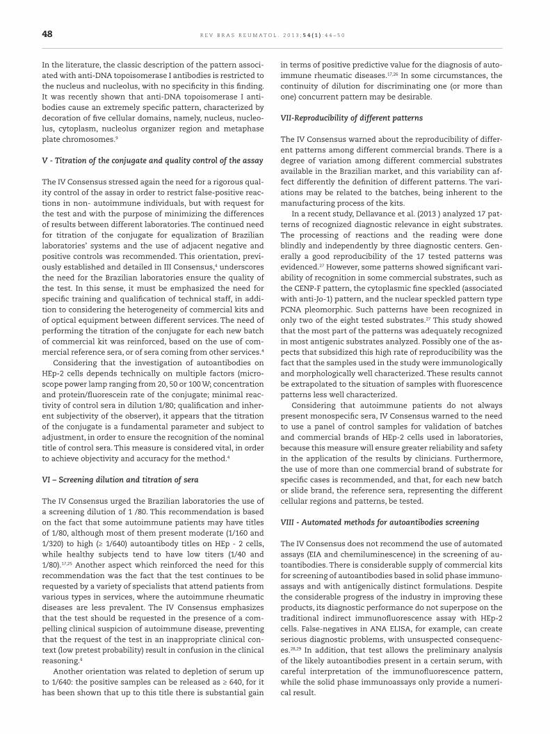
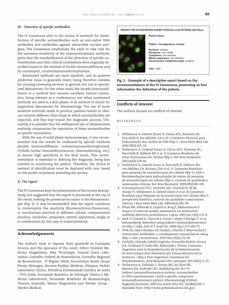
The IV Consensus kept the presentation of the results descrip-tively, but suggested that the report is presented at the top of the result, making the presentation easier to the Rheumatolo-gist (Fig. 2). It was recommended that the report continues to contemplate the reactivity (fl uorescent/non-fl uorescent, or reactive/non-reactive) in different cellular compartments (nucleus, nucleolus, cytoplasm, mitotic apparatus), singly or in combination (in the case of mixed patterns).
Acknowledgements
The Authors wish to express their gratitude to Fundação Aroeira and the sponsors of the event, Albert Einstein Me-dicina Diagnóstica, Alka Tecnologia, Amaral Costa Labo-ratório, Conselho Federal de Biomedicina, Conselho Regional de Biomedicina - 3ª Região, DASA, Euroimmun Brasil, Grupo Fleury, Hemagen, Hermes Pardini, Medivax, Olimpus, Padrão Laboratório Clínico, Pontifícia Universidade Católica de Goiás - PUC-Goiás, Sociedade Brasileira de Patologia Clínica e Me-dicina Laboratorial, Sociedade Brasileira de Reumatologia, Thermo Scientifi c, Wama Diagnóstica and Werfen Group - Werfen Medical.
Confl icts of interest
The authors declare no confl icts of interest.
R E F E R E N C E S
1. Dellavance A, Gabriel Júnior A, Cintra AFU, Ximenes AC, Nuccitelli B, von Mühlen CA et al. I Consenso Nacional para Padronização dos Laudos de FAN HEp-2. J Bras Patot Med Lab. 2002;38(3):201–16.
2. Dellavance A, Grabriel Junior A, Cintra AFU, Ximenes AC, Nuccitelli B, Taliberti BH et al. II Consenso Brasileiro de Fator Antinuclear em Células HEp-2. Rev Bras Reumatol. 2003;43(3):129–40.
3. Dellavance A, Gabriel Júnior A, Nuccitelli B, Taliberti BH, von Mühlen CA, Bichara CDA et al. 3o Consenso Brasileiro para pesquisa de autoanticorpos em células HEp-2 ( FAN ). Recomendações para padronização do ensaio de pesquisa de autoanticorpos em células HEp-2, controle de qualidade e associações clínicas. Rev Bras Reumatol. 2009;49(2):89–109.
4. Francescantonio PLC, Andrade LEC, Cruvinel W de M, Araújo FI, Dellavance A, Gabriel Júnior A et al. III Consenso Brasileiro para Pesquisa de Autoanticorpos em Células HEp-2: perspectiva histórica, controle de qualidade e associações clínicas. J Bras Patot Med Lab. 2009;45(3):185–99.
5. Pham BN, Albarede S, Guyard A, Burg E, Maisonneuve P. Impact of external quality assessment on antinuclear antibody detection performance. Lupus. 2005 Jan;14(2):113–9.
6. Sack U, Conrad K, Csernok E, Frank I, Hiepe F, Krieger T, et al. Autoantibody detection using indirect immunofl uorescence on HEp-2 cells. Ann N Y Acad Sci. 2009 Sep;1173:166–73.
7. Wiik AS, Høier-Madsen M, Forslid J, Charles P, Meyrowitsch J. Antinuclear antibodies: a contemporary nomenclature using HEp-2 cells. J Autoimmun. 2010 Nov;35(3):276–90.
8. Carballo, Orlando Gabriel Ingénito, Fernanda Beatriz Ginaca AA, Carabajal P, Costa MA, Balbaryski J. Primer Consenso Argentino para la Estandarización de la Determinación de Anticuerpos Anti-Nucleares por Inmunofl uorescencia Indirecta – HEp-2 First Argentine Consensus for Standardization. Acta Bioquím Clín Latinoam. 2012;46(1):3–13.
9. Dellavance A, Gallindo C, Soares MG, da Silva NP, Mortara RA, Andrade LEC. Redefi ning the Scl-70 indirect immunofl uorescence pattern: autoantibodies to DNA topoisomerase I yield a specifi c compound immunofl uorescence pattern. Rheumatology (Oxford, England) [Internet]. 2009 Jun [cited 2012 Oct 15];48(6):632–7. Available from: http://www.pubmedcentral.nih.gov/
Fig. 2 – Example of a descriptive report based on the recommendations of the IV Consensus, presenting as fi rst information the defi nition of the pattern.
PSEARCH FOR AUTOANTIBODIES AGAINST INTRACELLULAR ANTIGENS (ANA HEp-2)
Patient Name
Pattern: homogeneous nuclear
Nucleus: reactiveNucleolus: non visibleCytoplasm: non reactiveMitotic apparatus: non reactiveChromosome metaphase plate: positive
Title: 1280
50 R E V B R A S R E U M A T O L . 2 0 1 3 ; 5 4 ( 1 ) : 4 4 – 5 0
articlerender.fcgi?artid=2681287&tool=pmcentrez&rendertype=abstract
10. Carcamo WC, Satoh M, Kasahara H, Terada N, Hamazaki T, Chan JYF, et al. Induction of cytoplasmic rods and rings structures by inhibition of the CTP and GTP synthetic pathway in mammalian cells. PloS one. 2011 Jan;6(12):e29690.
11. Kursula P, Flodin S, Ehn M, Hammarström M, Schüler H, Nordlund P, et al. Structure of the synthetase domain of human CTP synthetase, a target for anticancer therapy. Acta Crystallogr Sect F Struct Biol Cryst Commun. 2006 Jul 1;62(Pt 7):613–7.
12. Carr SF, Papp E, Wu JC, Natsumeda Y. Characterization of human type I and type II IMP dehydrogenases. J Biol Chem. 1993 Dec 25;268(36):27286–90.
13. Keppeke GD, Nunes E, Ferraz MLG, Silva EAB, Granato C, Chan EKL, et al. Longitudinal study of a human drug-induced model of autoantibody to cytoplasmic rods/rings following HCV therapy with ribavirin and interferon-�. PloS one. 2012 Jan;7(9):e45392.
14. Covini G, Carcamo WC, Bredi E, von Mühlen CA, Colombo M, Chan EKL. Cytoplasmic rods and rings autoantibodies developed during pegylated interferon and ribavirin therapy in patients with chronic hepatitis C. Antivir Ther. 2012 Jan;17(5):805–11.
15. Stinton LM, Myers RP, Coffin CS, Fritzler MJ. Clinical associations and potential novel antigenic targets of autoantibodies directed against rods and rings in chronic hepatitis C infection. BMC gastroenterology. BMC Gastroenterology; 2013 Mar 19;13(1):50.
16. Franca NR, Dellavance, Alessandra Rodrigues SH, Perazzio SF, Silva NP, Andrade LEC. Quasi-homogeneous ANA-HEp-2 pattern reflects an autoantibody profile intermediate to the homogeneous and dense fine speckled nuclear patterns. Arthritis Rheum. 2011;63 Suppl(10):2307.
17. Mariz HA, Sato EI, Barbosa SH, Rodrigues SH, Dellavance A, Andrade LEC. Pattern on the antinuclear antibody-HEp-2 test is a critical parameter for discriminating antinuclear antibody-positive healthy individuals and patients with autoimmune rheumatic diseases. Arthritis Rheum. 2011 Jan;63(1):191–200.
18. Rattner JB, Rao A, Fritzler MJ, Valencia DW, Yen TJ. CENP-F is a .ca 400 kDa kinetochore protein that exhibits a cell-cycle dependent localization. Cell Motil Cytoskeleton. 1993 Jan;26(3):214–26.
19. Du J, Li Y, Zhu X. Involvement of CENP-F in histone methylation. Acta Biochim Biophys Sin (Shanghai). 2010;42(3):173–6.
20. Moynihan KL, Pooley R, Miller PM, Kaverina I, Bader DM. Murine CENP-F regulates centrosomal microtubule nucleation and interacts with Hook2 at the centrosome. Mol Biol Cell. 2009;20:4790–803.
21. Rattner JB, Rees J, Whitehead CM, Casiano CA, Tan EM, Humbel RL, et al. High frequency of neoplasia in patients with autoantibodies to centromere protein CENP-F. Clin Invest Med. 1997 Oct;20(5):308–19.
22. O’Brien SL, Fagan A, Fox EJP, Millikan RC, Culhane AC, Brennan DJ, et al. CENP-F expression is associated with poor prognosis and chromosomal instability in patients with primary breast cancer. Int J Cancer. 2007 Apr 1;120(7):1434–43.
23. Casiano CA, Humbel RL, Peebles C, Covini G, Tan EM. Autoimmunity to the cell cycle-dependent centromere protein p330d/CENP-F in disorders associated with cell proliferation. J Autoimmun. 1995 Aug;8(4):575–86.
24. Bonaci-Nikolic B, Andrejevic S, Bukilica M, Urosevic I, Nikolic M. Autoantibodies to mitotic apparatus: association with other autoantibodies and their clinical significance. J Clin Immunol. 2006 Sep;26(5):438–46.
25. Tan EM, Feltkamp TE, Smolen JS, Butcher B, Dawkins R, Fritzler MJ, et al. Range of antinuclear antibodies in “healthy” individuals. Arthritis Rheum. 1997 Sep;40(9):1601–11.
26. Satoh M, Chan EKL, Ho LA, Rose KM, Parks CG, Cohn RD, et al. Prevalence and sociodemographic correlates of antinuclear antibodies in the United States. Arthritis Rheum. 2013;64(7):2319–27.
27. Dellavance A, Cruvinel W de M, Francescantonio PLC, Mangueira CLP, Drugowick IC, Rodrigues SH, et al. Variability in the recognition of distinctive immunofluorescence patterns in different brands of HEp-2 cell slides. J Bras Patot Med Lab. 2013;49(3):182–90.
28. Kroshinsky D, Stone JH, Bloch DB, Sepehr A. Case records of the Massachusetts General Hospital. Case 5-2009. A 47-year-old woman with a rash and numbness and pain in the legs. N Engl J Med. 2009 Mar 12;360(7):711–20.
29. Nordal EB, Songstad NT, Berntson L, Moen T, Straume B, Rygg M. Biomarkers of chronic uveitis in juvenile idiopathic arthritis: predictive value of antihistone antibodies and antinuclear antibodies. J Rheumatol. 2009 Aug;36(8):1737–43.