isolated abdominal vasculitis as an atypical presentation of wegener’s granulomatosis
TRANSCRIPT
BRIEF CASE REPORTS
Isolated Abdominal Vasculitisas an Atypical Presentationof Wegener’s GranulomatosisYa Ju Chang, M.D., and Leslie Dubin Kerr, M.D.Division of Rheumatology, Department of Medicine, MountSinai Medical Center, New York, New York
ABSTRACTAlthough current classifications characterize vasculitic syn-dromes based upon the size of the vessels involved, thehistopathology, and the presence or absence of antineutro-phil cytoplasmatic antibodies ANCA (1–3), those occa-sional patients with vasculitis whose features are not typicalmay evade diagnosis and effective treatment. We report onesuch patient who presented with bilateral refractory uveitisand abdominal angina who had a positive C-ANCA. Be-cause of his atypical presentation, this patient’s diseaseprogressed over 8 yr despite an extensive gastrointestinalevaluation, before a diagnosis of vasculitis was establishedangiographically, and immunosuppressive therapy wasbegun. (Am J Gastroenterol 2000;95:297–298. © 2000 byAm. Coll. of Gastroenterology)
CASE REPORT
This 47-yr-old man presented with an 8-yr history of bilat-eral refractory uveitis. He was treated with topical steroidswith some suppression of symptoms; however, he had aflare of uveitis every time he tried to decrease the number ofsteroid eye drops. He developed the insidious onset ofpostprandial back pain followed by frontal crampy abdom-inal pain lasting approximately 20 min to 1 h. He thenstarted to lose weight. The pain gradually intensified, be-coming severe, diffuse and incapacitating, making it impos-sible to eat. He lost.100 pounds over 8 yr. Over the years,he underwent an evaluation consisting of abdominal CTscan that was negative, a colonoscopy that showed diver-ticulosis without inflammatory change, and a magnetic res-onance imaging plus MRA, both of which were normal. Hewas treated with 2 wk of ciprofloxacin without improve-ment. Two years before presentation, bilateral Achilles ten-dinitis and left shoulder pain developed. Both resolved withthe use of NSAIDs. He continued to experience recurrentepisodes of this incapacitating abdominal pain, which wastriggered by eating. In the 6 months before presentation, hedeveloped the new onset of hypertension. He denied ahistory of conjunctivitis, oral or genital ulcers, skin rash, anyneurological symptoms, muscle weakness, sinusitis, hemop-tysis, or renal disease. His medications consisted of topicalsteroid eye drops, losartan, loperamide, and multivitamins.
His exam upon presentation to us was pertinent for a2-pound weight loss, normal blood pressure of 140/80 mmHg plus a completely unremarkable HEENT, cardiac, pul-monary, abdominal, and neurological examination. No vas-culitic skin lesions were seen. Chest x-ray showed no evi-dence of infiltrates, nodules or cavities. Laboratory valueswere: white blood cells 10,7003 1,000/ml, hemoglobin13.8 g/100 ml, hematocrit 40.3%, platelet 331,000/mm3,ANA, RPR, and Lyme titer were negative. The C-ANCAwas positive at 1:160, and the P-ANCA was negative. TheESR was 60 mm/h.
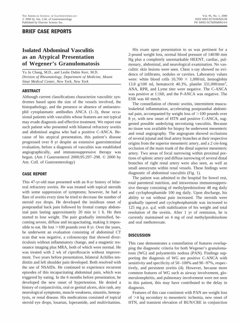
The constellation of chronic uveitis, intermittent muscu-loskeletal inflammation, accelerating postprandial abdomi-nal pain, accompanied by weight loss of.100 pounds over8 yr, with new onset of HTN and positive C-ANCA, sug-gested possible underlying necrotizing vasculitis. Becauseno tissue was available for biopsy he underwent mesentericand renal angiography. The angiogram showed occlusionsof several jejunal and ileal artery branches at their respectiveorigins from the superior mesenteric artery, and a 2 cm-longocclusion of the main trunk of the distal superior mesentericartery. Two areas of focal narrowing within proximal por-tions of splenic artery and diffuse narrowing of several distalbranches of right renal artery were also seen, as well assmall aneurysms within renal vessels. These findings werediagnostic of abdominal vasculitis (Fig. 1).
The patient was admitted to the hospital for bowel rest,total parenteral nutrition, and intravenous immunosuppres-sive therapy consisting of methylprednisolone 48 mg dailyand cyclophosphamide 100 mg daily. Upon discharge, hisability to eat without pain increased. The steroids weregradually tapered and cyclophosphamide was increased to125 mgp.o. q.d. with stabilization of his weight and totalresolution of the uveitis. After 1 yr of remission, he iscurrently maintained on 4 mg of oral methylprednisoloneand oral methotrexate.
DISCUSSION
This case demonstrates a constellation of features overlap-ping the diagnostic criteria for both Wegener’s granuloma-tosis (WG) and polyarteritis nodosa (PAN). Findings sup-porting the diagnosis of WG are positive C-ANCA withsensitivity and specificity of 50–100% and 90–97%, respec-tively, and persistent uveitis (4). However, because morecommon features of WG such as airway involvement, glo-merulonephritis, and pulmonary involvement were not seenin this patient, this may have contributed to the delay indiagnosis.
Features of this case consistent with PAN are weight lossof .4 kg secondary to mesenteric ischemia, new onset ofHTN, and transient elevation of BUN/CRE in conjunction
THE AMERICAN JOURNAL OF GASTROENTEROLOGY Vol. 95, No. 1, 2000© 2000 by Am. Coll. of Gastroenterology ISSN 0002-9270/00/$20.00Published by Elsevier Science Inc. PII S0002-9270(99)00614-0
with positive celiac and renal angiograms, per the AmericanCollege of Rheumatology (ACR) criteria for the diagnosisof PAN, with a sensitivity and specificity of 82% and 86%respectively (5). However the presence of uveitis and thepositive C-ANCA make this case atypical of PAN and mostconsistent with WG (4, 5).
Based upon the above clinical features, the positive se-rology for C-ANCA plus angiographic findings, we believethat this patient has an atypical presentation of WG. Themost important feature of this case is the morbidity experi-enced by this patient as a consequence of his unusual,indolent overlapping symptoms, which delayed his diagno-sis of vasculitis and the appropriate treatment, which ulti-mately was lifesaving.
This case serves as a reminder to remain vigilant to thepossibility of systemic vasculitis despite the atypical con-stellation of the clinical and serological features.
Reprint requests and correspondence:Ya Ju Chang, M.D.,Division of Rheumatology, Box 1244, Mount Sinai Medical Cen-ter, 1 Gustave L. Levy Place, New York, NY 10029-6574.
Received Aug. 4, 1998; accepted Jan. 11, 1999.
REFERENCES
1. Jennette JC, Falk RJ, Andrassy K, et al. Nomenclature ofsystemic vasculitides: Proposal of an international consensusconference. Arth Rheum 1994;37:187–92.
2. Gross LR, Antineutrophil cytoplasmatic autoantibody testingin vasculitides. Rheum Dis Clin North Am 1995;21:987–1011.
3. Lie JT. Illustrated histopathologic classification criteria forselected vasculitis syndromes. Arth Rheum 1990;33:1088–93.
4. Duna GF, Galperin C, Hoffman G. Wegener’s granulomatosis.Rheuma Dis Clin North Am 1995;21:949–85.
5. Lhote F, Guillevin L. Polyarteritis nodosa microscopic poly-angiitis, and Churg-Strauss syndrome. Rheum Dis Clin NorthAm 1995;21:911–47.
Precipitation of Iron Overload andHereditary Hemochromatosis AfterSuccessful Treatment of Celiac DiseaseMichael A. Heneghan, M.D., Kenneth M. Feeley, M.D.,Fiona M. Stevens, M.D., Malcolm P. G. Little, M.D., andCiaran F. McCarthy, M.D.Departments of Medicine and Pathology, Clinical ScienceInstitute, University College Hospital, Galway, Ireland
INTRODUCTION
Hereditary hemochromatosis (HH) is inherited as an auto-somal recessive trait. It is characterized by increased ab-sorption of dietary iron, resulting in excess iron depositionin the parenchymal cells of the liver, heart, and certainendocrine glands. Premature death can occur from compli-cations of chronic liver disease, hepatocellular carcinoma,and heart failure, whereas considerable morbidity is as-sociated with nonspecific constitutional symptoms andmalaise. It is estimated that up to 5 persons per 1000 ofEuropean descent are homozygous for the condition (1,2). It is speculated that intact mucosal epithelium of theproximal small bowel may be critical for the manifesta-tion of the condition.
CASE REPORT
In February 1986, a 61-yr-old woman presented with weightloss of 8 kg and malaise. She had a history of anemia for 30yr and was treated with intermittent oral iron. A nonsmokerand nondrinker, her family history was noncontributory.Examination revealed a woman of 45 kg with clubbing butan otherwise normal examination. Hemoglobin was 11.0g/dl (normal range [NR] 12–16 g/dl), mean corpuscularvolume 94.9 fL (NR 77–91 fL), platelets 5173 109/L (NR160–4403 109/L) with Howell Jolly bodies on blood film.Vitamin B12 and folate levels were normal. Serum iron was11 (NR 11–26), iron binding capacity 39mmol/L (NR45–72mmol/L), and serum ferritin 462 ng/ml (NR 10–200ng/ml). Celiac disease was confirmed on duodenal biopsy bythe presence of a flat mucosa, increased intraepithelial lym-phocytes, and chronic inflammatory cells in the laminapropria. Serumalkaline phosphatase was 167 u/L (NR30 –115 u/L), with other liver function tests normal. Thepatient was discharged from the hospital after 3 wk on agluten-free diet (GFD).
By November 1986, she was pigmented. Tests for Addi-
Figure 1. (1) Occlusion of multiple ileal and jejunal branches atorigin of superior mesenteric artery. (2) Occlusion of ileocolicartery with reconstitution via collateral. (3) Right colic arteryocclusion.
298 Brief Case Reports AJG – Vol. 95, No. 1, 2000