isis shoulder dystocia final - glowm shoulder dystocia.pdf · shoulder dystocia page 3 of 44 1....
TRANSCRIPT
Shoulder Dystocia Page 1 of 44
ISIS CURRICULUM
Shoulder Dystocia
Created: September 05, 2007 Modified: May 24, 2010
Shoulder Dystocia Page 2 of 44
Curriculum Outline
Author Contact Information
Description of Curriculum
Target Trainees
Prerequisite Knowledge and Skills
Goals and Objectives
Instructor Notes
Common Errors & Prevention Strategies
Cognitive Training
Skill Training
Equipment Setup
Assessment Methods
Appendices
11
10
9
8
5
4
3
12
2
1
6
7
Shoulder Dystocia Page 3 of 44
1. Author Contact Information
Thomas J Benedetti, MD, MHA
Professor Obstetrics and Gynecology
Box 356460 University of Washington 1959 NE Pacific Street Seattle, WA 98195
Phone: (206)543‐3729 Email: [email protected]
Michael Fialkow, MD MPH
Assistant Professor Obstetrics and Gynecology
Box 356460 University of Washington 1959 NE Pacific Street Seattle WA 98195
Phone: (206)543‐3045 Fax: (206)616‐9479
Email: [email protected]
Sara Kim, PhD Chair, Interprofessional Education and Practice Committee
Institute for Simulation and Interprofessional Studies Box 357240
University of Washington 1959 NE Pacific Street Seattle, WA 98195
Phone: (206) 616‐0597 Fax: (206) 598‐0809
Acknowledgements:
Jane Hitti, MD
Megan Sherman
Sarah Waller, MD
Shoulder Dystocia Page 4 of 44
2. Description of Curriculum
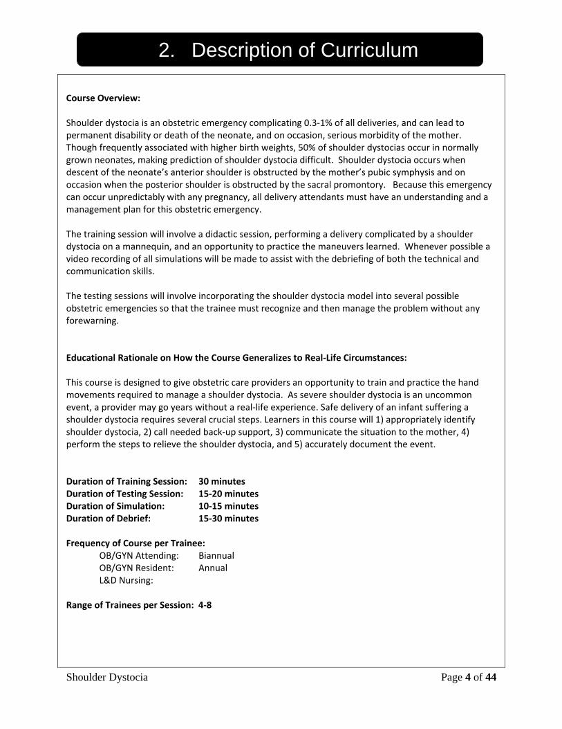
Course Overview: Shoulder dystocia is an obstetric emergency complicating 0.3‐1% of all deliveries, and can lead to permanent disability or death of the neonate, and on occasion, serious morbidity of the mother. Though frequently associated with higher birth weights, 50% of shoulder dystocias occur in normally grown neonates, making prediction of shoulder dystocia difficult. Shoulder dystocia occurs when descent of the neonate’s anterior shoulder is obstructed by the mother’s pubic symphysis and on occasion when the posterior shoulder is obstructed by the sacral promontory. Because this emergency can occur unpredictably with any pregnancy, all delivery attendants must have an understanding and a management plan for this obstetric emergency. The training session will involve a didactic session, performing a delivery complicated by a shoulder dystocia on a mannequin, and an opportunity to practice the maneuvers learned. Whenever possible a video recording of all simulations will be made to assist with the debriefing of both the technical and communication skills. The testing sessions will involve incorporating the shoulder dystocia model into several possible obstetric emergencies so that the trainee must recognize and then manage the problem without any forewarning. Educational Rationale on How the Course Generalizes to Real‐Life Circumstances: This course is designed to give obstetric care providers an opportunity to train and practice the hand movements required to manage a shoulder dystocia. As severe shoulder dystocia is an uncommon event, a provider may go years without a real‐life experience. Safe delivery of an infant suffering a shoulder dystocia requires several crucial steps. Learners in this course will 1) appropriately identify shoulder dystocia, 2) call needed back‐up support, 3) communicate the situation to the mother, 4) perform the steps to relieve the shoulder dystocia, and 5) accurately document the event. Duration of Training Session: 30 minutes Duration of Testing Session: 15‐20 minutes Duration of Simulation: 10‐15 minutes Duration of Debrief: 15‐30 minutes Frequency of Course per Trainee: OB/GYN Attending: Biannual OB/GYN Resident: Annual L&D Nursing: Range of Trainees per Session: 4‐8
Shoulder Dystocia Page 5 of 44
3. Target Trainees
Primary: OB/GYN residents
Secondary: OB/GYN Attending, Labor and Delivery nursing staff, CNM, Licensed midwives & Anesthesia Faculty

Shoulder Dystocia Page 6 of 44
4. Prerequisite Knowledge and Skills While trainees participating in the course will have varied backgrounds and experiences, all will be obstetric care providers and therefore at risk to encounter a shoulder dystocia. Prior to receiving training in this course, trainees should have the following background knowledge: Medical Knowledge: Normal labor; spontaneous vaginal delivery; Labor dystocia; Clinical Skills: Vaginal delivery
Shoulder Dystocia Page 7 of 44
5. Goals and Objectives Goal 1: Experience Shoulder Dystocia The learner will experience and manage a severe shoulder dystocia in a simulated setting. (ACGME Competencies: Medical Knowledge, Patient Care, Interpersonal and Communication Skills, Professionalism, System‐based Practice, Practice‐based Learning)
a. Objective: The learner will be able to identify a shoulder dystocia, and recognize a need for further assistance. (1,2)
b. Objective: The learner will be able to modify and apply new knowledge to deliveries
complicated by shoulder dystocia. (1,5,6)
c. Objective: The learner will formulate a mental checklist of maneuvers and appropriate help, and apply this to management of shoulder dystocias. (6)
Goal 2: Teamwork and Interprofessional Communication Physicians and Midwives will have the opportunity to interact with nursing staff in a patient scenario, away from the patient care setting, and to interact with simulated patients in times of stress (ACGME: Interpersonal and Communication Skills and Patient Care) (ACGME Competencies: Medical Knowledge, Patient Care, Interpersonal and Communication Skills, Professionalism, System‐based Practice, Practice‐based Learning)
a. Objective: The team will be able to demonstrate components of communication such as TeamSTEPPS principles, which have demonstrated improvement in patient outcomes in crisis situation. (1,2,3)
b. Objective: The learner will identify components of communication with a patient, which
facilitate delivery, and allow the patient to maintain confidence in the team. (2,3,4) Goal 3: Self‐assessment of Technical and Communication Skills Review the video‐recorded shoulder dystocia simulation session, and improve technical and communication skills. (ACGME Competencies: Practice‐Based Learning and Improvement)
a. Objective: After reviewing the video, the learner will identify aspects of the simulation that were both successful, and those that need improvement.
b. Objective: The learner will evaluate his/her performance during the simulation event by
completing a self‐assessment form. Goal 4: Documentation of Events Document the key components of a shoulder dystocia for the medical record. (ACGME Competencies: Medical Knowledge, Patient Care, Systems Based Practice)
a. Objective: The learner will be able to accurately and thoroughly document the shoulder dystocia for the medical record, recording the key maneuvers, personnel present, and outcome of the neonate and patient.
Shoulder Dystocia Page 8 of 44
6. Instructor Notes * TeamSTEPPS terms in Bold All caps – see appendix I for definitions • Prepare the simulation room as described and shown in section 10
Image with description • At the start of each training/testing session: • Orient trainees to the simulation • Explain cameras will be used for the debriefing • Read the scenario
• During a training session the “nurse”(instructor) provides the scenario in SBAR form as the trainee enters the room.
• During a testing session the physician is called to the room from the “board” immediately after completion of the “BRIEF” if one is indicated. The brief should include the history of the patient supporting potential issues in her care.
• Execution of the simulation by the trainer
• Instruct the trainee to verbalize each step of the exercise. When the trainee wants to do a procedure, they should communicate which procedure they want to do, and verbalize each step of the process (“thinking out loud”).
• The simulation lasts until successful delivery of the baby, or 5 minutes, whichever comes first. • Following the simulation, instruct the participants to write a delivery/procedure note • Following the simulation, conduct a debriefing session to discuss the experiences of each
participant, and to address concerns regarding implementing the skills learned into practice on the labor floor. During the debriefing, the video of the shoulder dystocia simulations will be available to review.
• Instructions to Trainees:
“Remember to:” 1. Treat the scenario as real as possible 2. Use mask/gloves/gown as needed 3. You may ask for instruments or medications as needed 4. Request assistance if needed 5. Ignore the camera if one is present 6. Ask questions if you have them
• Answers to Common questions
General “Call pediatrics?” ‐ If they ask for pediatrics or other help, tell them that they are on their way –
if pediatrics involved in the simulation they can then arrive. “What else do we know about the patient?” ‐ You do not have any more history than what the
resident received before they walked into the room. “Can you give her X?” ‐ You do not know any of the medication doses, but can give anything
they ask for. “Can you get me X instrument?” – No additional instruments are available “What are the patient’s vitals?” – Restate the original scenario vitals “Let's move to the OR?” – This is OK if the management of the shoulder dystocia did not relieve
the SD however if the trainee has not tried all efforts, this effectively ends the scenario.
Shoulder Dystocia Page 9 of 44
7. Common Errors and Prevention Strategies • Poor communication with patient
o Example: “Patient” continues to ask questions “What is going on? Why won’t the baby come out? It hurts!”
o Management: The instructor and trainee will review the scenario video to identify when further communication with the patient might have been helpful.
• Difficulty understanding role of assistant(s), and using the assistant(s) to help with maneuvers o Management: To alleviate confusion, the “nurse” will be identified prior to beginning of
the scenario o Management: To solidify the role, the nurse may ask the trainee questions about what
he/she could do to aid the trainee during the exercise.
• Attempt to perform posterior arm extraction with suprapubic pressure still being given o Management: The instructor or nurse may directly ask the trainee if he/she would like
to maintain suprapubic pressure. o Management: The instructor and trainee will review each maneuver, after the
completion of the scenario, to explain how continuous suprapubic pressure could impede delivery.
• Attempt to enter vagina without appropriate hand configuration
o Management: Learners will be instructed before the simulation on correct hand configuration (palming the thumb) to allow easier entry into the vagina for performing posterior arm extraction as well as rotational maneuvers.
• Use of excessive force to deliver the infant o Management: The instructor and trainee will review the force monitor data, allowing
the trainee to watch the monitor as the instructor puts increasing amounts of force on the fetal head during delivery.
• Difficulties using the mannequin – some people have scratched their hands! o Management: The instructor will alert all exercise participants to treat the scenario as a
real situation even though the mannequin is not as pliable as skin. The instructor will have gloves readily available to avoid scratching.
Shoulder Dystocia Page 10 of 44
8. Cognitive Training Included • Syllabus material • E‐Training modules
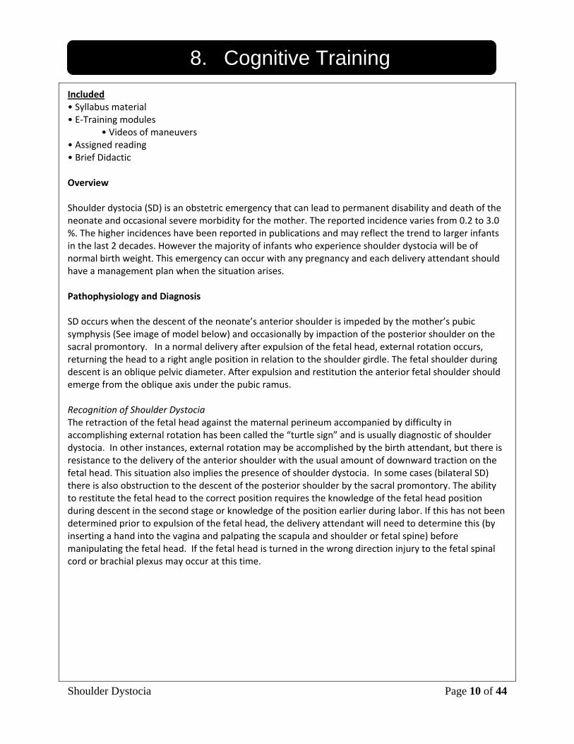
• Videos of maneuvers • Assigned reading • Brief Didactic Overview Shoulder dystocia (SD) is an obstetric emergency that can lead to permanent disability and death of the neonate and occasional severe morbidity for the mother. The reported incidence varies from 0.2 to 3.0 %. The higher incidences have been reported in publications and may reflect the trend to larger infants in the last 2 decades. However the majority of infants who experience shoulder dystocia will be of normal birth weight. This emergency can occur with any pregnancy and each delivery attendant should have a management plan when the situation arises. Pathophysiology and Diagnosis SD occurs when the descent of the neonate’s anterior shoulder is impeded by the mother’s pubic symphysis (See image of model below) and occasionally by impaction of the posterior shoulder on the sacral promontory. In a normal delivery after expulsion of the fetal head, external rotation occurs, returning the head to a right angle position in relation to the shoulder girdle. The fetal shoulder during descent is an oblique pelvic diameter. After expulsion and restitution the anterior fetal shoulder should emerge from the oblique axis under the pubic ramus. Recognition of Shoulder Dystocia The retraction of the fetal head against the maternal perineum accompanied by difficulty in accomplishing external rotation has been called the “turtle sign” and is usually diagnostic of shoulder dystocia. In other instances, external rotation may be accomplished by the birth attendant, but there is resistance to the delivery of the anterior shoulder with the usual amount of downward traction on the fetal head. This situation also implies the presence of shoulder dystocia. In some cases (bilateral SD) there is also obstruction to the descent of the posterior shoulder by the sacral promontory. The ability to restitute the fetal head to the correct position requires the knowledge of the fetal head position during descent in the second stage or knowledge of the position earlier during labor. If this has not been determined prior to expulsion of the fetal head, the delivery attendant will need to determine this (by inserting a hand into the vagina and palpating the scapula and shoulder or fetal spine) before manipulating the fetal head. If the fetal head is turned in the wrong direction injury to the fetal spinal cord or brachial plexus may occur at this time.
Shoulder Dystocia Page 11 of 44
8. Cognitive Training
SD occurs because of a relative disproportion between the fetal size and maternal pelvic capacity. Fetal weight shows the strongest correlation with the risk of SD, however the trunk and chest circumferences relative to head circumference along with the body configuration have also been shown to be influential. Because of the strong correlation with fetal weight, the risk factors for SD are those known to accelerate fetal growth or fetal disproportion, such as diabetes. There is, however, no single associated condition or combination of antenatal risk factors that provide clinically useful positive predictive values for SD. The majority of cases, however, occur with no risk factors and shoulder dystocia is, therefore usually an unpredictable event. Risks for shoulder dystocia based on known (but not estimated) fetal weight are listed in the table below. Factors associated with shoulder dystocia
Pre‐labor Intrapartum
Previous shoulder dystocia Prolonged first stage of labor
Macrosomia Active phase arrest of labor
Diabetes Prolonged second stage of labor
Raised maternal body mass index Vacuum assisted vaginal delivery
Excessive weight gain in pregnancy Precipitous second state of labor
Management Because of the potential dire consequences to the infant and mother when a SD occurs there is typically an urgency or panic that occurs in response to recognition of the problem. There are no randomized clinical trials to guide physicians in the selection of maneuvers to perform and in which order. The lone published random trial found that prophylactic shoulder dystocia maneuvers did not reduce the incidence of shoulder dystocia. Recent studies, however, have shown that a previously uncompromised
Image 1: Anterior Shoulder against the mother’s pubic symphysis
Shoulder Dystocia Page 12 of 44
8. Cognitive Training fetus should be able to withstand 5‐7 minutes of SD without suffering very low five minute apgar score or metabolic acidosis on umbilical artery pH. Unfortunately the best evidence available shows fetal injury to be associated with all described maneuvers to relieve shoulder dystocia, however, some maneuvers are clearly associated with unacceptable risk of fetal injury and should be avoided in the panic:
• Strong lateral downward traction on the fetal head • Uterine fundal pressure.
The following sequence of maneuvers to manage a SD delivery, without undue increased risk of injury, represents the best available evidence (a. – action; b. – principle; c ‐ evidence). Proceed through and repeat the steps until successful delivery. Traction on the fetal head during the maneuvers should always be gentle and downward during a maternal push
1. Instruct the patient to push only when asked to do so by you.
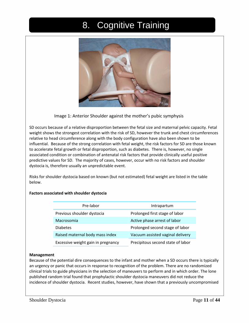
2. McRobert’s position.
a. Mother or birth attendants grasp her posterior thigh and flexing the legs against her abdomen
b. McRobert’s position causes cephalic rotation of the pubic symphysis and flattening of the sacrum, both of which will aid in relieving both anterior and posterior shoulder impaction.
c. A retrospective case review found that the McRobert's maneuver was sufficient to achieve delivery in 77/94 (79%) cases of shoulder dystocia (Nocon et al.,1993). Baskett & Allen (1995) found this maneuver was associated with less neonatal trauma. Laboratory tests with maternal pelvic and fetal models demonstrated a reduction in the amount of traction used (Allen et al., 1994). Gonik et al., (1993) compared lateral pelvic x‐rays of the lithotomy position and the McRobert's maneuver to show the angle of pelvic inclination was reduced from 26 to 10 degrees. The symphysis rotated superiorly by a distance of 8 cms. The dimensions of the true pelvis are not altered. It is the rotation of the symphysis superiorly which aids release of the impacted anterior shoulder.
Shoulder Dystocia Page 13 of 44
8. Cognitive Training
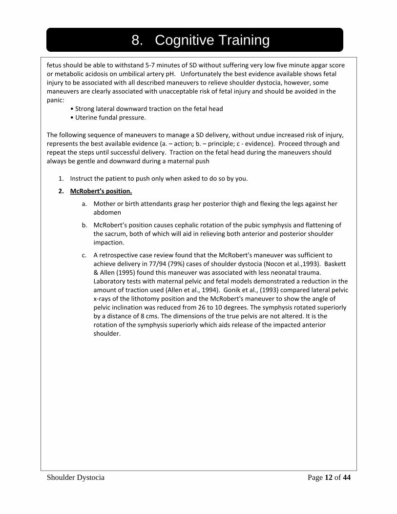
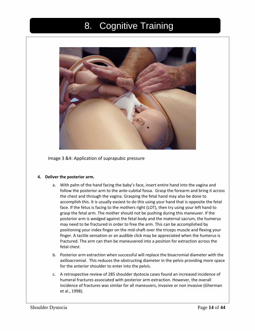
3. Suprapubic pressure.
a. In an oblique diameter, beginning with pressure applied toward the side opposite the fetal face (ie. if the fetus is facing the mother’s right side, pressure should be applied from the mother’s left side with force intended to “shove” or collapse the shoulder.
b. Suprapubic pressure reduces the bisachromial diameter, rotating the anterior shoulder into the oblique
Image 2 : McRobert’s maneuver
Shoulder Dystocia Page 14 of 44
8. Cognitive Training
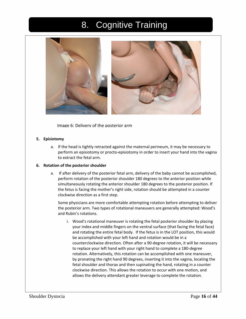
4. Deliver the posterior arm.
a. With palm of the hand facing the baby’s face, insert entire hand into the vagina and follow the posterior arm to the ante‐cubital fossa. Grasp the forearm and bring it across the chest and through the vagina. Grasping the fetal hand may also be done to accomplish this. It is usually easiest to do this using your hand that is opposite the fetal face. If the fetus is facing to the mothers right (LOT), then try using your left hand to grasp the fetal arm. The mother should not be pushing during this maneuver. If the posterior arm is wedged against the fetal body and the maternal sacrum, the humerus may need to be fractured in order to free the arm. This can be accomplished by positioning your index finger on the mid‐shaft over the triceps muscle and flexing your finger. A tactile sensation or an audible click may be appreciated when the humerus is fractured. The arm can then be maneuvered into a position for extraction across the fetal chest.
b. Posterior arm extraction when successful will replace the bisacromial diameter with the axilloacromial. This reduces the obstructing diameter in the pelvis providing more space for the anterior shoulder to enter into the pelvis.
c. A retrospective review of 285 shoulder dystocia cases found an increased incidence of humeral fractures associated with posterior arm extraction. However, the overall incidence of fractures was similar for all maneuvers, invasive or non invasive (Gherman et al., 1998).
Image 3 &4: Application of suprapubic pressure

Shoulder Dystocia Page 15 of 44
8. Cognitive Training
Image 5: Delivery of the posterior arm
Shoulder Dystocia Page 16 of 44
8. Cognitive Training
5. Episiotomy
a. If the head is tightly retracted against the maternal perineum, it may be necessary to perform an episiotomy or procto‐episiotomy in order to insert your hand into the vagina to extract the fetal arm.
6. Rotation of the posterior shoulder
a. If after delivery of the posterior fetal arm, delivery of the baby cannot be accomplished, perform rotation of the posterior shoulder 180 degrees to the anterior position while simultaneously rotating the anterior shoulder 180 degrees to the posterior position. If the fetus is facing the mother’s right side, rotation should be attempted in a counter clockwise direction as a first step.
Some physicians are more comfortable attempting rotation before attempting to deliver the posterior arm. Two types of rotational maneuvers are generally attempted: Wood’s and Rubin’s rotations.
i. Wood’s rotational maneuver is rotating the fetal posterior shoulder by placing your index and middle fingers on the ventral surface (that facing the fetal face) and rotating the entire fetal body. If the fetus is in the LOT position, this would be accomplished with your left hand and rotation would be in a counterclockwise direction. Often after a 90‐degree rotation, it will be necessary to replace your left hand with your right hand to complete a 180‐degree rotation. Alternatively, this rotation can be accomplished with one maneuver, by pronating the right hand 90 degrees, inserting it into the vagina, locating the fetal shoulder and thorax and then supinating the hand, rotating in a counter clockwise direction. This allows the rotation to occur with one motion, and allows the delivery attendant greater leverage to complete the rotation.
Image 6: Delivery of the posterior arm
Shoulder Dystocia Page 17 of 44
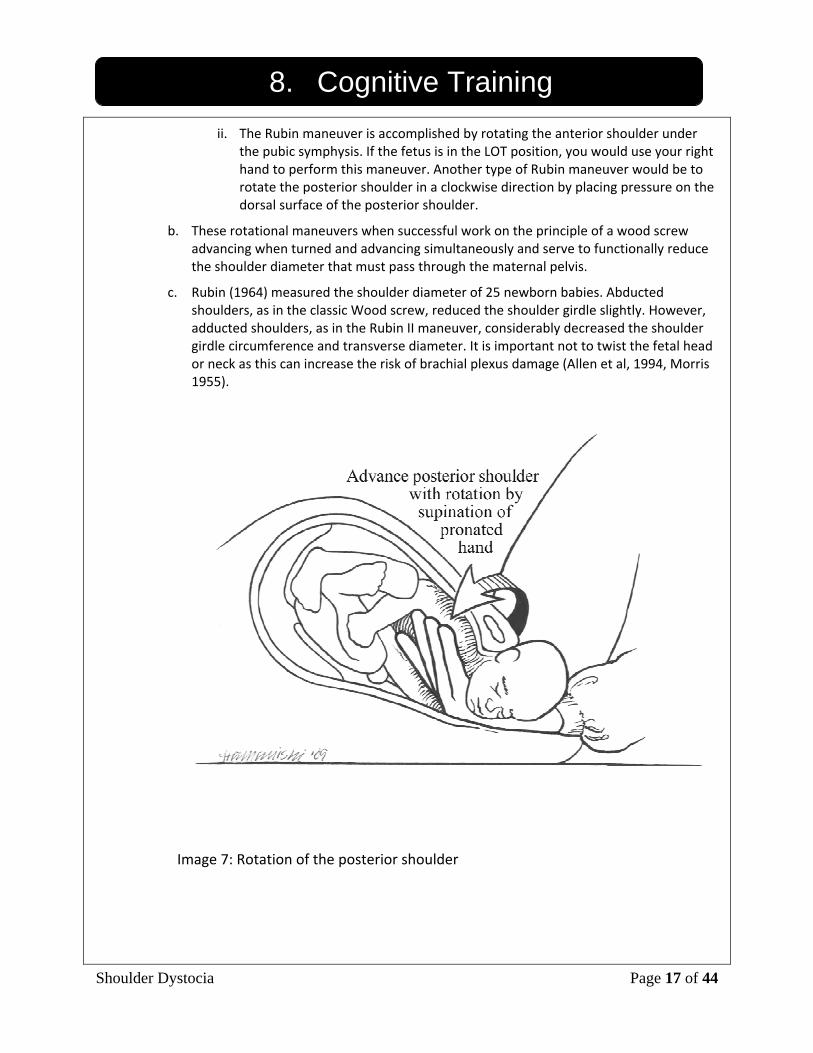
8. Cognitive Training ii. The Rubin maneuver is accomplished by rotating the anterior shoulder under
the pubic symphysis. If the fetus is in the LOT position, you would use your right hand to perform this maneuver. Another type of Rubin maneuver would be to rotate the posterior shoulder in a clockwise direction by placing pressure on the dorsal surface of the posterior shoulder.
b. These rotational maneuvers when successful work on the principle of a wood screw advancing when turned and advancing simultaneously and serve to functionally reduce the shoulder diameter that must pass through the maternal pelvis.
c. Rubin (1964) measured the shoulder diameter of 25 newborn babies. Abducted shoulders, as in the classic Wood screw, reduced the shoulder girdle slightly. However, adducted shoulders, as in the Rubin II maneuver, considerably decreased the shoulder girdle circumference and transverse diameter. It is important not to twist the fetal head or neck as this can increase the risk of brachial plexus damage (Allen et al, 1994, Morris 1955).
Image 7: Rotation of the posterior shoulder
Shoulder Dystocia Page 18 of 44
8. Cognitive Training
7. If neither rotational maneuvers nor extraction of the posterior arm is possible, bilateral shoulder dystocia may be present. In this case the anterior arm is lodged behind the symphysis pubis and the posterior shoulder is lodged high in the pelvis at or near the sacral promontory. It may be impossible to deliver the fetus with the previously described maneuvers.
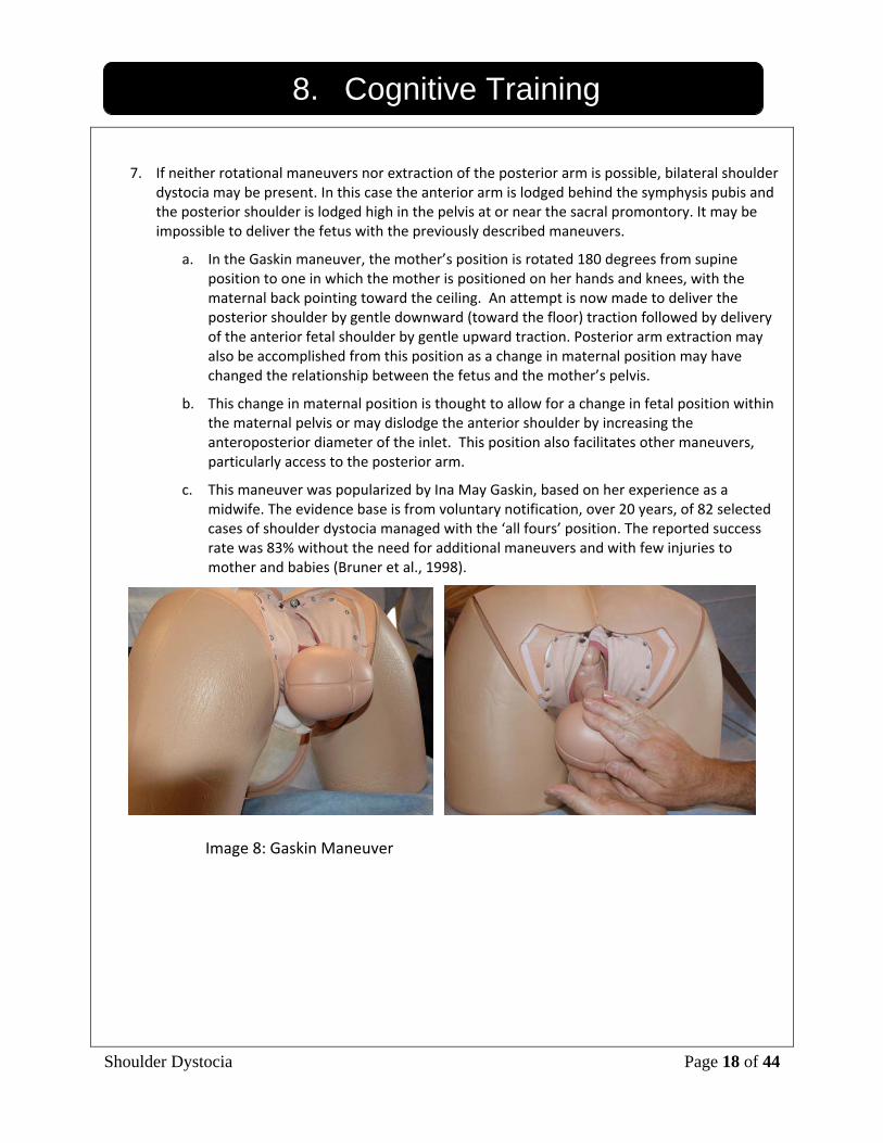
a. In the Gaskin maneuver, the mother’s position is rotated 180 degrees from supine position to one in which the mother is positioned on her hands and knees, with the maternal back pointing toward the ceiling. An attempt is now made to deliver the posterior shoulder by gentle downward (toward the floor) traction followed by delivery of the anterior fetal shoulder by gentle upward traction. Posterior arm extraction may also be accomplished from this position as a change in maternal position may have changed the relationship between the fetus and the mother’s pelvis.
b. This change in maternal position is thought to allow for a change in fetal position within the maternal pelvis or may dislodge the anterior shoulder by increasing the anteroposterior diameter of the inlet. This position also facilitates other maneuvers, particularly access to the posterior arm.
c. This maneuver was popularized by Ina May Gaskin, based on her experience as a midwife. The evidence base is from voluntary notification, over 20 years, of 82 selected cases of shoulder dystocia managed with the ‘all fours’ position. The reported success rate was 83% without the need for additional maneuvers and with few injuries to mother and babies (Bruner et al., 1998).
Image 8: Gaskin Maneuver
Shoulder Dystocia Page 19 of 44
8. Cognitive Training 8. Cephalic Replacement If the mother cannot assume the all fours position or delivery cannot be accomplished in this position, cephalic replacement may be necessary. To accomplish delivery using cephalic replacement the fetal head must be returned to the occiput anterior position. The fetal head is then flexed by simultaneously putting downward pressure on the occiput with one hand and inward pressure on the fetal maxilla and face with the other hand. Once the fetal head is reintroduced into the vagina, emergency cesarean section should be performed. A fetal heart beat should be documented if at all possible before performing cesarean delivery Extraordinary Maneuvers:
Symphysiotomy: should be performed only by individuals who have knowledge and experience in this procedure. It should be uncommonly used in the United States. Fracture of the clavicle: Deliberate fracture of the clavicle has been advocated to release the impacted shoulder. This has been achieved by pressing the anterior clavicle upwards against the ramus of the pubis. Pressure should be applied at the midpoint of the clavicle to avoid injury to the subclavian vessel. However, deliberate fracture of the clavicle can be difficult to do. Attempts to fracture the fetal clavicle are usually unsuccessful and should generally be attempted only by those with prior experience and training. Bilateral Shoulder Dystocia In cases where bilateral shoulder dystocia is present and Gaskin Maneuver is unsuccessful the mother can be returned to the dorsal lithotomy position. An initial attempt to deliver the anterior or posterior shoulder should be attempted as the change in maternal position necessary for the Gaskin maneuver may have changed the anatomic relationship between the mother and the fetus and allow successful delivery. If these maneuvers still are not successful, some physicians have had success with deep insertion of the hand and forearm into the pelvis. The delivery attendants hand may in this case come to rest above the pelvic inlet. In some cases the posterior fetal arm will be found to be behind the fetal back, not in the usual position in front of the fetal abdomen. If the fetal humerus can be located it may be possible to move the arm across the midline , the hand then grasped and the posterior arm extracted. In some occasions, the fetal humerus may fracture with this maneuver however this may allow manipulation of the fetal arm across the midline and successful delivery.
Shoulder Dystocia Page 20 of 44
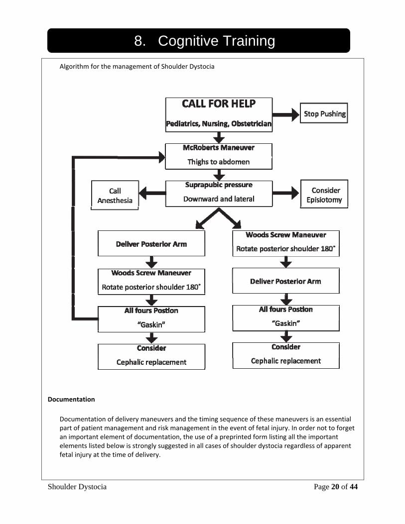
8. Cognitive Training Algorithm for the management of Shoulder Dystocia
Documentation
Documentation of delivery maneuvers and the timing sequence of these maneuvers is an essential part of patient management and risk management in the event of fetal injury. In order not to forget an important element of documentation, the use of a preprinted form listing all the important elements listed below is strongly suggested in all cases of shoulder dystocia regardless of apparent fetal injury at the time of delivery.
Shoulder Dystocia Page 21 of 44
8. Cognitive Training
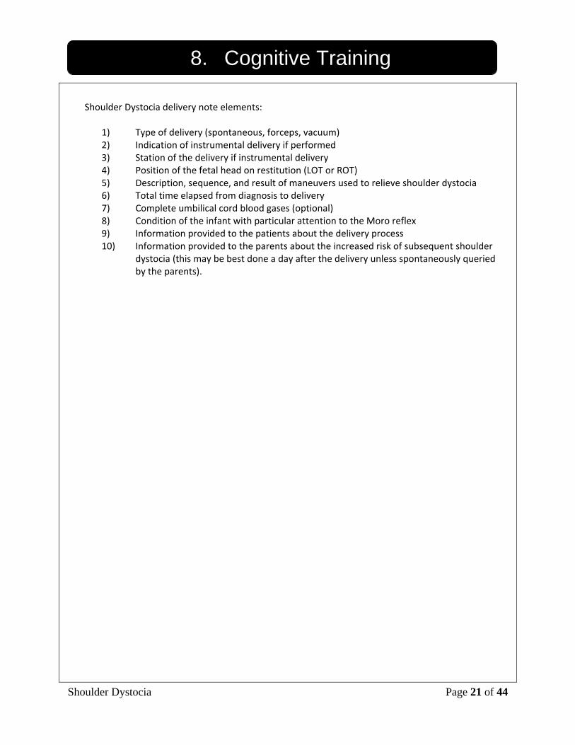
Shoulder Dystocia delivery note elements:
1) Type of delivery (spontaneous, forceps, vacuum) 2) Indication of instrumental delivery if performed 3) Station of the delivery if instrumental delivery 4) Position of the fetal head on restitution (LOT or ROT) 5) Description, sequence, and result of maneuvers used to relieve shoulder dystocia 6) Total time elapsed from diagnosis to delivery 7) Complete umbilical cord blood gases (optional) 8) Condition of the infant with particular attention to the Moro reflex 9) Information provided to the patients about the delivery process 10) Information provided to the parents about the increased risk of subsequent shoulder
dystocia (this may be best done a day after the delivery unless spontaneously queried by the parents).
Shoulder Dystocia Page 22 of 44
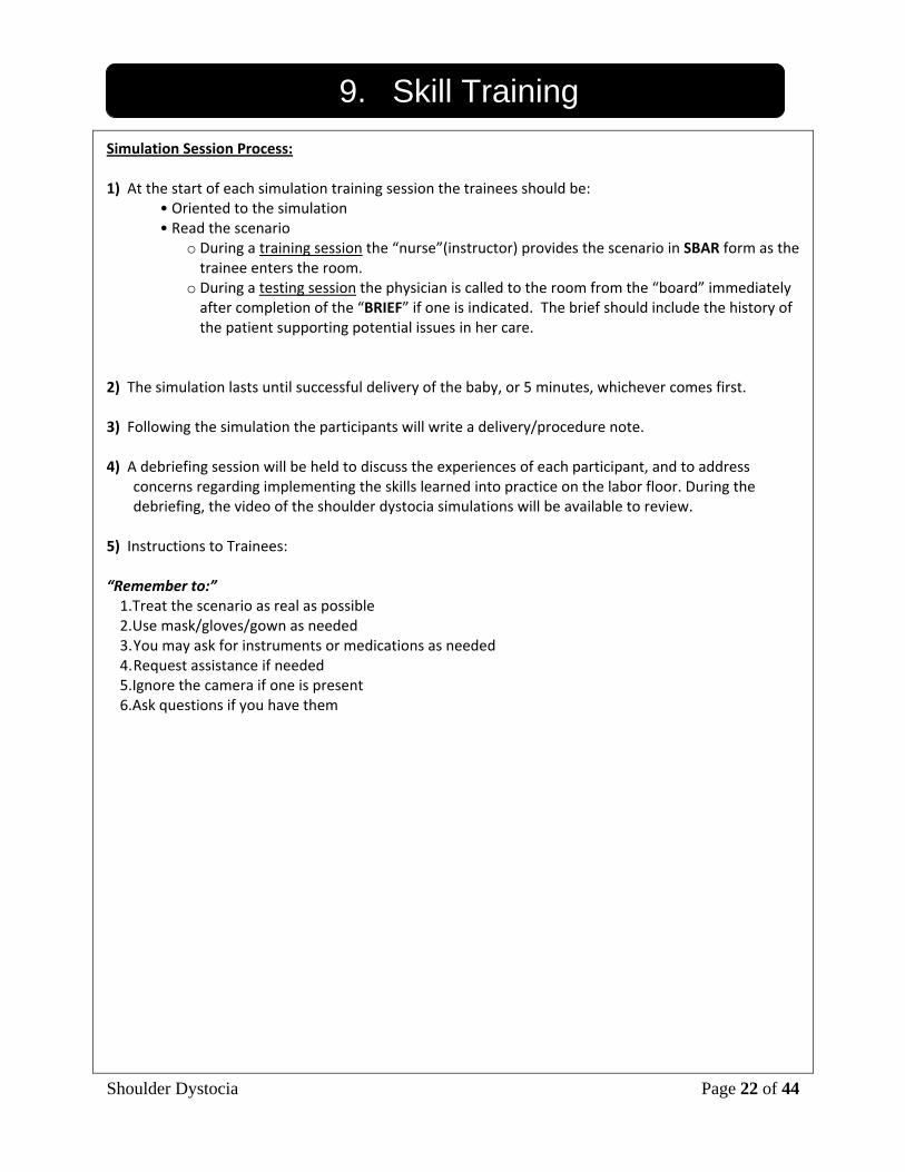
9. Skill Training Simulation Session Process: 1) At the start of each simulation training session the trainees should be: • Oriented to the simulation • Read the scenario
o During a training session the “nurse”(instructor) provides the scenario in SBAR form as the trainee enters the room.
o During a testing session the physician is called to the room from the “board” immediately after completion of the “BRIEF” if one is indicated. The brief should include the history of the patient supporting potential issues in her care.
2) The simulation lasts until successful delivery of the baby, or 5 minutes, whichever comes first. 3) Following the simulation the participants will write a delivery/procedure note. 4) A debriefing session will be held to discuss the experiences of each participant, and to address
concerns regarding implementing the skills learned into practice on the labor floor. During the debriefing, the video of the shoulder dystocia simulations will be available to review.
5) Instructions to Trainees: “Remember to:” 1.Treat the scenario as real as possible 2.Use mask/gloves/gown as needed 3. You may ask for instruments or medications as needed 4. Request assistance if needed 5.Ignore the camera if one is present 6.Ask questions if you have them
Shoulder Dystocia Page 23 of 44
9. Skill Training
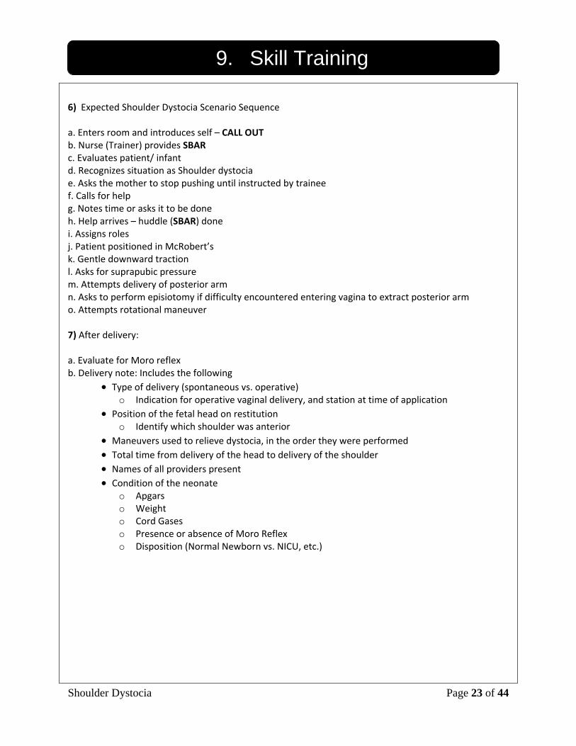
6) Expected Shoulder Dystocia Scenario Sequence a. Enters room and introduces self – CALL OUT b. Nurse (Trainer) provides SBAR c. Evaluates patient/ infant d. Recognizes situation as Shoulder dystocia e. Asks the mother to stop pushing until instructed by trainee f. Calls for help g. Notes time or asks it to be done h. Help arrives – huddle (SBAR) done i. Assigns roles j. Patient positioned in McRobert’s k. Gentle downward traction l. Asks for suprapubic pressure m. Attempts delivery of posterior arm n. Asks to perform episiotomy if difficulty encountered entering vagina to extract posterior arm o. Attempts rotational maneuver 7) After delivery: a. Evaluate for Moro reflex b. Delivery note: Includes the following
• Type of delivery (spontaneous vs. operative) o Indication for operative vaginal delivery, and station at time of application
• Position of the fetal head on restitution o Identify which shoulder was anterior
• Maneuvers used to relieve dystocia, in the order they were performed • Total time from delivery of the head to delivery of the shoulder • Names of all providers present • Condition of the neonate
o Apgars o Weight o Cord Gases o Presence or absence of Moro Reflex o Disposition (Normal Newborn vs. NICU, etc.)
Shoulder Dystocia Page 24 of 44
10. Equipment Set-up
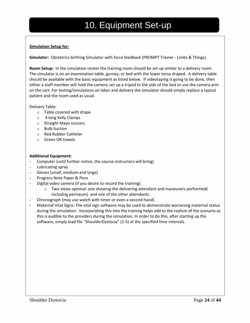
Simulation Setup for: Simulator: Obstetrics birthing Simulator with force feedback (PROMPT Trainer ‐ Limbs & Things). Room Setup: In the simulation center the training room should be set up similar to a delivery room. The simulator is on an examination table, gurney, or bed with the lower torso draped. A delivery table should be available with the basic equipment as listed below. If videotaping is going to be done, then either a staff member will hold the camera, set up a tripod to the side of the bed or use the camera arm on the cart. For testing/simulations on labor and delivery the simulator should simply replace a typical patient and the room used as usual. Delivery Table:
o Table covered with drape o 4 long Kelly Clamps o Straight Mayo scissors o Bulb Suction o Red Rubber Catheter o Green OR towels
Additional Equipment: - Computer (until further notice, the course instructors will bring) - Lubricating spray - Gloves (small, medium and large) - Progress Note Paper & Pens - Digital video camera (if you desire to record the training)
o Two views optimal: one showing the delivering attendant and maneuvers performed( including perineum) and one of the other attendants.
- Chronograph (may use watch with timer or even a second hand) - Maternal Vital Signs: The vital sign software may be used to demonstrate worsening maternal status
during the simulation. Incorporating this into the training helps add to the realism of the scenario as this is audible to the providers during the simulation. In order to do this, after starting up the software, simply load file “ShoulderDystocia” (1‐5) at the specified time intervals.
Shoulder Dystocia Page 25 of 44
11. Assessment Methods Trainer:
• Performance checklist o Assessment o Maneuvers o Delivery/Procedure note
Trainee:
• Pre‐test/ Post‐test
• Simulation evaluation
Shoulder Dystocia Page 26 of 44
12. Appendices
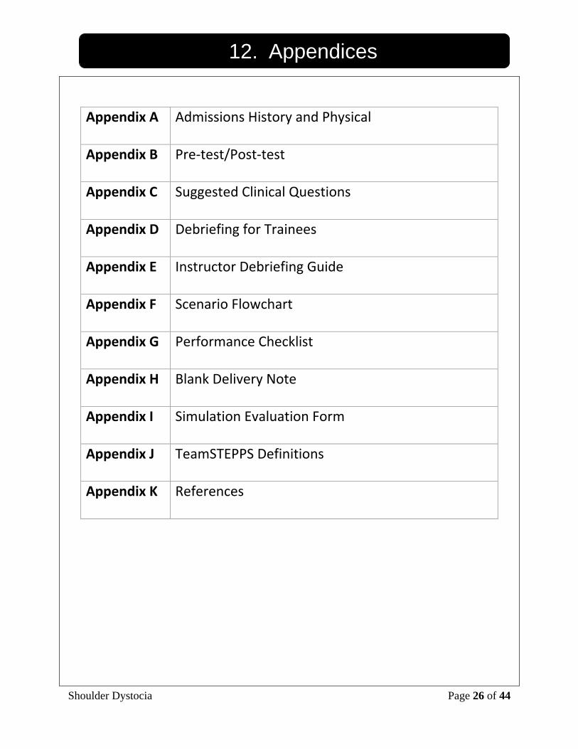
Appendix A Admissions History and Physical
Appendix B Pre‐test/Post‐test
Appendix C Suggested Clinical Questions
Appendix D Debriefing for Trainees
Appendix E Instructor Debriefing Guide
Appendix F Scenario Flowchart
Appendix G Performance Checklist
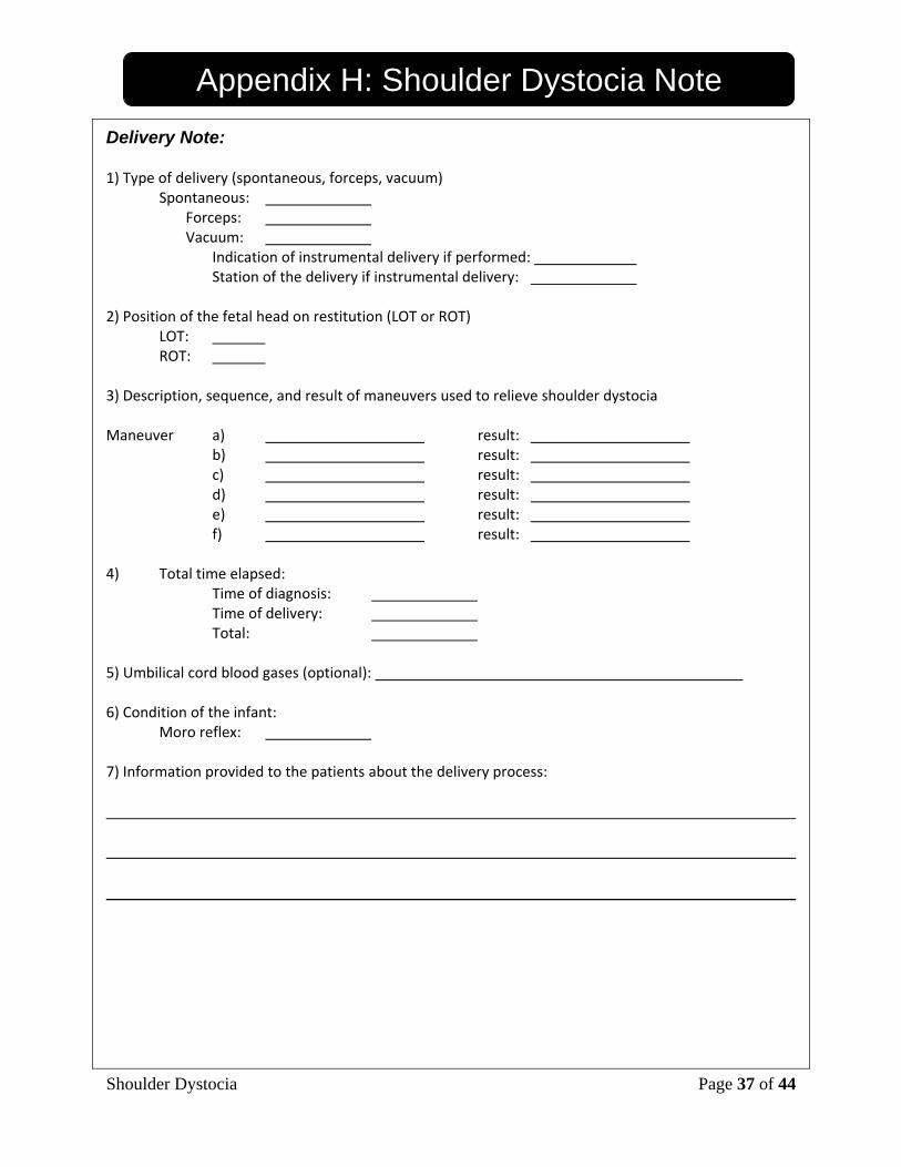
Appendix H Blank Delivery Note
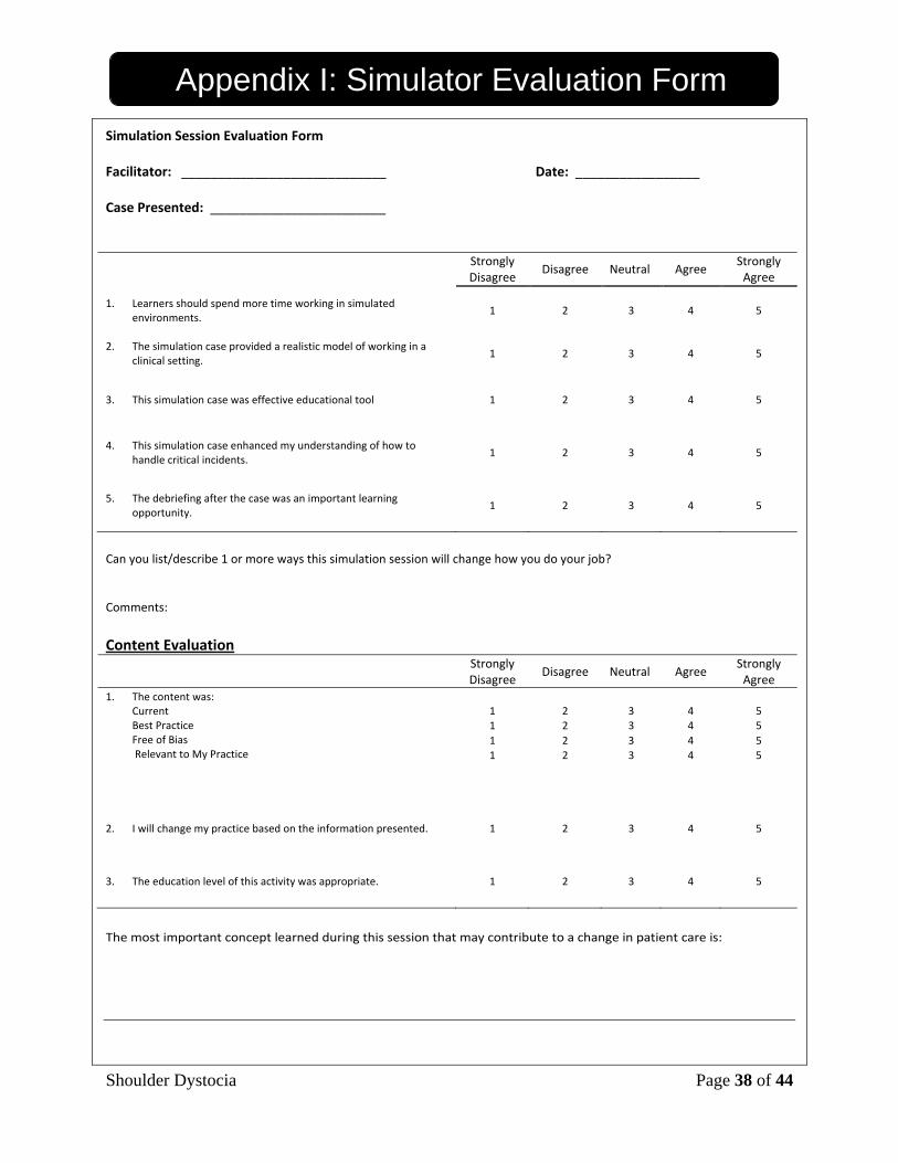
Appendix I Simulation Evaluation Form
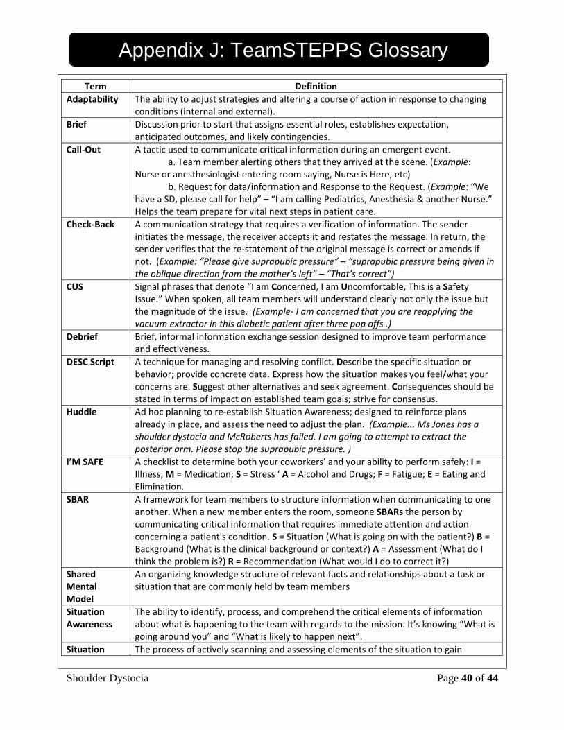
Appendix J TeamSTEPPS Definitions
Appendix K
References

Shoulder Dystocia Page 27 of 44
Appendix A: Admission Histories General Instructions for trainees: You may ask questions if you have them, and please remember to:
• Treat the scenario as real as possible • Use mask/gloves/gown as needed • Request assistance if needed • Ignore the camera • Please do not cut the simulator, but indicate if you would like to
Case Scenario Options Scenario 1: The trainee is asked to attend a delivery on the request of the midwife who has just delivered a baby’s head. The patient is a 28 yo G1. She has had a normal fist stage, and delivered the head after 90 minutes of pushing. She urgently requests the trainee’s presence because the head “seems stuck”. Scenario 2: The trainee is called emergently to a delivery room to assist a midwife in a shoulder dystocia. The trainee will know from board sign‐out that the patient is a 28 year old G2P1 with a history of an 8# baby, an elevated diabetic screen, but normal GTT. She has pushed for one hour. The trainee has been watching the tracing, and it had been overall reassuring, but has just developed deep variables with pushing. Scenario 3: The trainee is attending a delivery with an intern during the first week of July. After the head is delivered the intern seems flustered with what to do next. The trainee notices that the head has not restituted and shows the “Turtle Sign”. Scenario 4: One of your patients has labored normally at the birthing center. She is pushing well and has delivered the head. However, the shoulder did not deliver with your normal delivery maneuvers.
Shoulder Dystocia Page 28 of 44
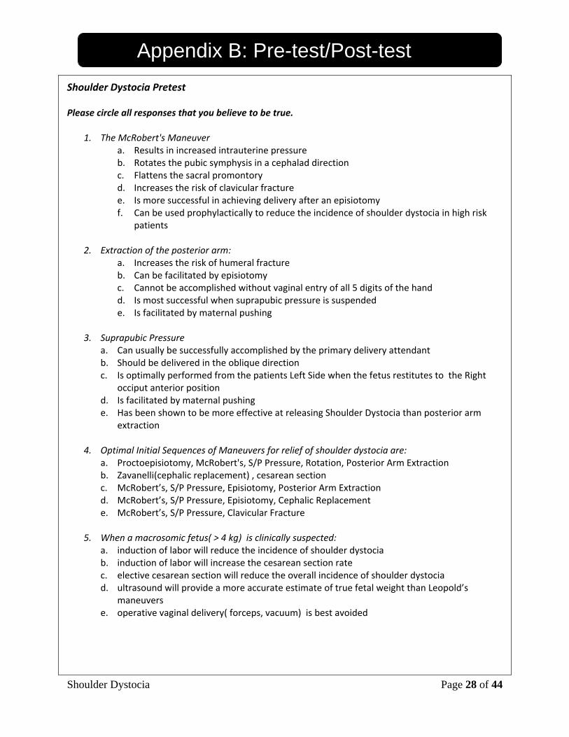
Appendix B: Pre-test/Post-test Shoulder Dystocia Pretest Please circle all responses that you believe to be true.
1. The McRobert's Maneuver a. Results in increased intrauterine pressure b. Rotates the pubic symphysis in a cephalad direction c. Flattens the sacral promontory d. Increases the risk of clavicular fracture e. Is more successful in achieving delivery after an episiotomy f. Can be used prophylactically to reduce the incidence of shoulder dystocia in high risk
patients
2. Extraction of the posterior arm: a. Increases the risk of humeral fracture b. Can be facilitated by episiotomy c. Cannot be accomplished without vaginal entry of all 5 digits of the hand d. Is most successful when suprapubic pressure is suspended e. Is facilitated by maternal pushing
3. Suprapubic Pressure
a. Can usually be successfully accomplished by the primary delivery attendant b. Should be delivered in the oblique direction c. Is optimally performed from the patients Left Side when the fetus restitutes to the Right
occiput anterior position d. Is facilitated by maternal pushing e. Has been shown to be more effective at releasing Shoulder Dystocia than posterior arm
extraction
4. Optimal Initial Sequences of Maneuvers for relief of shoulder dystocia are: a. Proctoepisiotomy, McRobert's, S/P Pressure, Rotation, Posterior Arm Extraction b. Zavanelli(cephalic replacement) , cesarean section c. McRobert’s, S/P Pressure, Episiotomy, Posterior Arm Extraction d. McRobert’s, S/P Pressure, Episiotomy, Cephalic Replacement e. McRobert’s, S/P Pressure, Clavicular Fracture
5. When a macrosomic fetus( > 4 kg) is clinically suspected:
a. induction of labor will reduce the incidence of shoulder dystocia b. induction of labor will increase the cesarean section rate c. elective cesarean section will reduce the overall incidence of shoulder dystocia d. ultrasound will provide a more accurate estimate of true fetal weight than Leopold’s
maneuvers e. operative vaginal delivery( forceps, vacuum) is best avoided
Shoulder Dystocia Page 29 of 44
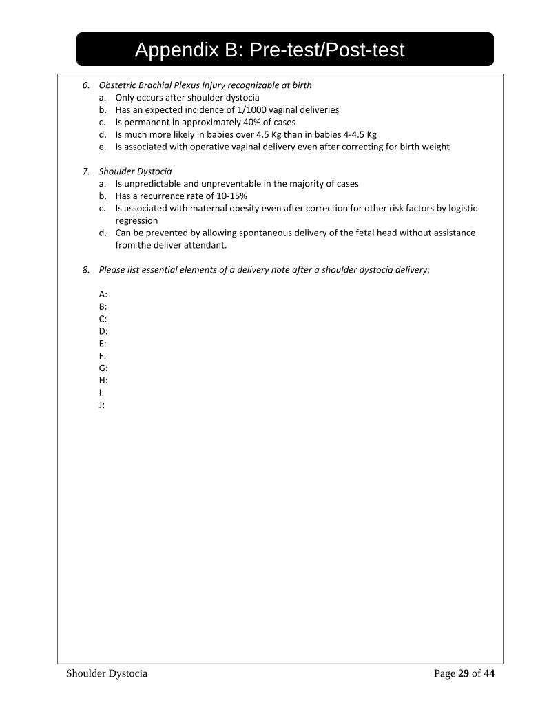
Appendix B: Pre-test/Post-test 6. Obstetric Brachial Plexus Injury recognizable at birth
a. Only occurs after shoulder dystocia b. Has an expected incidence of 1/1000 vaginal deliveries c. Is permanent in approximately 40% of cases d. Is much more likely in babies over 4.5 Kg than in babies 4‐4.5 Kg e. Is associated with operative vaginal delivery even after correcting for birth weight
7. Shoulder Dystocia
a. Is unpredictable and unpreventable in the majority of cases b. Has a recurrence rate of 10‐15% c. Is associated with maternal obesity even after correction for other risk factors by logistic
regression d. Can be prevented by allowing spontaneous delivery of the fetal head without assistance
from the deliver attendant.
8. Please list essential elements of a delivery note after a shoulder dystocia delivery:
A: B: C: D: E: F: G: H: I: J:
Shoulder Dystocia Page 30 of 44
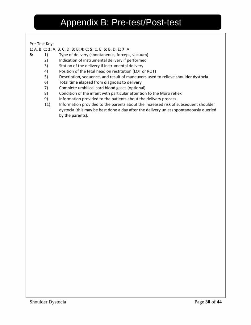
Appendix B: Pre-test/Post-test Pre‐Test Key: 1: A, B, C; 2: A, B, C, D; 3: B; 4: C; 5: C, E; 6: B, D, E; 7: A 8: 1) Type of delivery (spontaneous, forceps, vacuum)
2) Indication of instrumental delivery if performed 3) Station of the delivery if instrumental delivery 4) Position of the fetal head on restitution (LOT or ROT) 5) Description, sequence, and result of maneuvers used to relieve shoulder dystocia 6) Total time elapsed from diagnosis to delivery 7) Complete umbilical cord blood gases (optional) 8) Condition of the infant with particular attention to the Moro reflex 9) Information provided to the patients about the delivery process 11) Information provided to the parents about the increased risk of subsequent shoulder
dystocia (this may be best done a day after the delivery unless spontaneously queried by the parents).

Shoulder Dystocia Page 31 of 44
Appendix C: Suggested Clinical Questions Question 1 A 30 year old primigravid patient presents to your office at 39 weeks for routine prenatal visit. You notice that she has gained 6 pounds since her last visit one week ago and that her fundal height is now 42 cm, 4 cm greater than the last visit. Her glucola screen at 28 weeks was 128 mg %. You order an ultrasound and find she has normal amniotic fluid but that the fetal measurements put the baby above the 90% for growth at 4100 grams. The BPD‐AC difference is –1.2 cm. Your best management option is:
1‐ Induce the labor before the baby gets any larger 2‐ Recommend a cesarean section as the patient has at least a 5% incidence of shoulder dystocia. 3‐ Begin antepartum testing as she is probably an undetected gestational diabetic. 4‐ Wait at least another week for the spontaneous onset of labor.
Correct answer‐ 4 Question 2 Within one week of her visit at 39 weeks, during the labor of the same patient she has an active phase arrest of labor at 7 cm. You insert an IUPC and find inadequate uterine activity. With oxytocin stimulation she progresses to complete dilatation. After two and a half hours in the second state the baby’s head descends to +3/5 with 2+/4+ molding of the fetal scalp. However it does not descend further in the next hour despite good maternal pushing efforts. The fetal heart rate is reactive and the variability is good. She is having intermittent mild‐ moderate variable decelerations. Uterine contractions are coming every three minutes at 70mm HG. You should:
1‐ Allow the labor to progress another hour as the fetus is tolerating the labor without difficulty
2‐ Perform a forceps or vacuum delivery for the indication or arrest of descent 3‐ Increase the oxytocin to increase the UC frequency to every two minutes 4‐ Recommend cesarean section as the shoulder dystocia rate in this patient at this time
approaches 25%. Correct answer‐ 4
Shoulder Dystocia Page 32 of 44
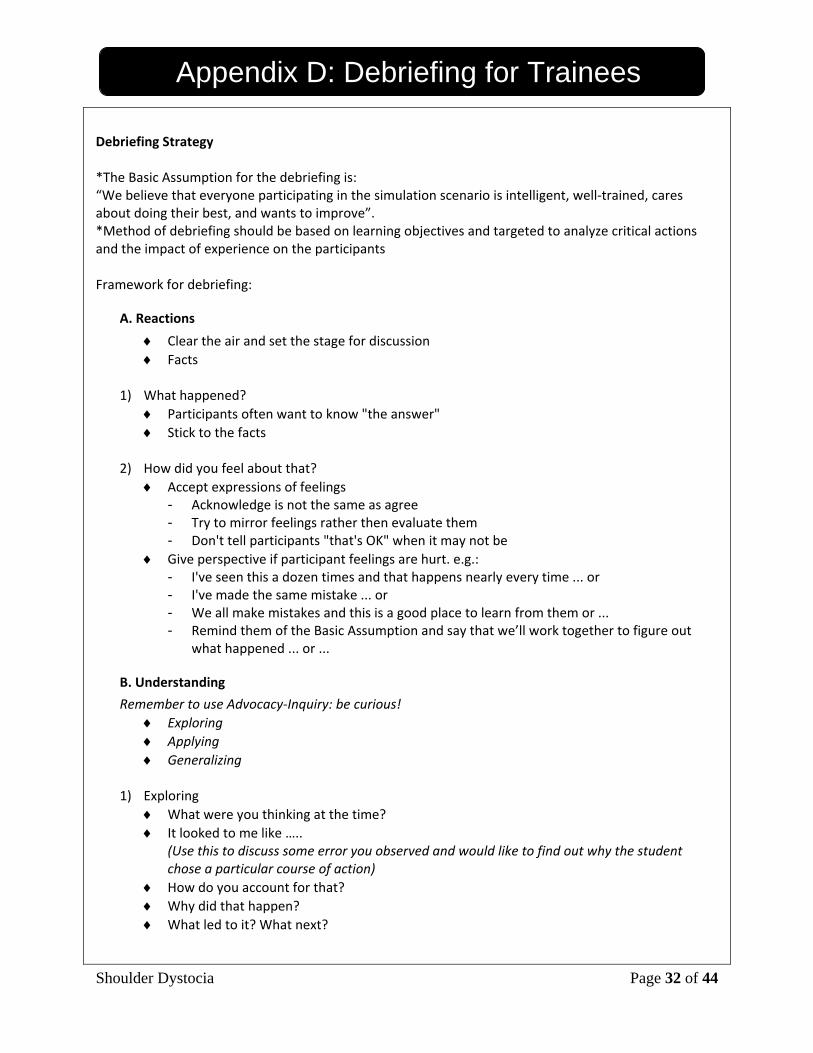
Appendix D: Debriefing for Trainees Debriefing Strategy
*The Basic Assumption for the debriefing is: “We believe that everyone participating in the simulation scenario is intelligent, well‐trained, cares about doing their best, and wants to improve”. *Method of debriefing should be based on learning objectives and targeted to analyze critical actions and the impact of experience on the participants Framework for debriefing:
A. Reactions
♦ Clear the air and set the stage for discussion ♦ Facts
1) What happened?
♦ Participants often want to know "the answer" ♦ Stick to the facts
2) How did you feel about that?
♦ Accept expressions of feelings - Acknowledge is not the same as agree - Try to mirror feelings rather then evaluate them - Don't tell participants "that's OK" when it may not be
♦ Give perspective if participant feelings are hurt. e.g.: - I've seen this a dozen times and that happens nearly every time ... or - I've made the same mistake ... or - We all make mistakes and this is a good place to learn from them or ... - Remind them of the Basic Assumption and say that we’ll work together to figure out
what happened ... or ...
B. Understanding
Remember to use Advocacy‐Inquiry: be curious! ♦ Exploring ♦ Applying ♦ Generalizing
1) Exploring
♦ What were you thinking at the time? ♦ It looked to me like …..
(Use this to discuss some error you observed and would like to find out why the student chose a particular course of action)
♦ How do you account for that? ♦ Why did that happen? ♦ What led to it? What next?
Shoulder Dystocia Page 33 of 44
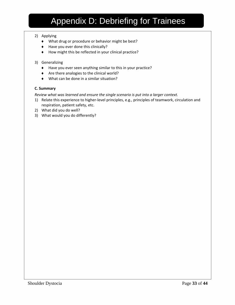
Appendix D: Debriefing for Trainees 2) Applying
♦ What drug or procedure or behavior might be best? ♦ Have you ever done this clinically? ♦ How might this be reflected in your clinical practice?
3) Generalizing
♦ Have you ever seen anything similar to this in your practice? ♦ Are there analogies to the clinical world? ♦ What can be done in a similar situation?
C. Summary
Review what was learned and ensure the single scenario is put into a larger context. 1) Relate this experience to higher‐level principles, e.g., principles of teamwork, circulation and
respiration, patient safety, etc. 2) What did you do well? 3) What would you do differently?
Shoulder Dystocia Page 34 of 44
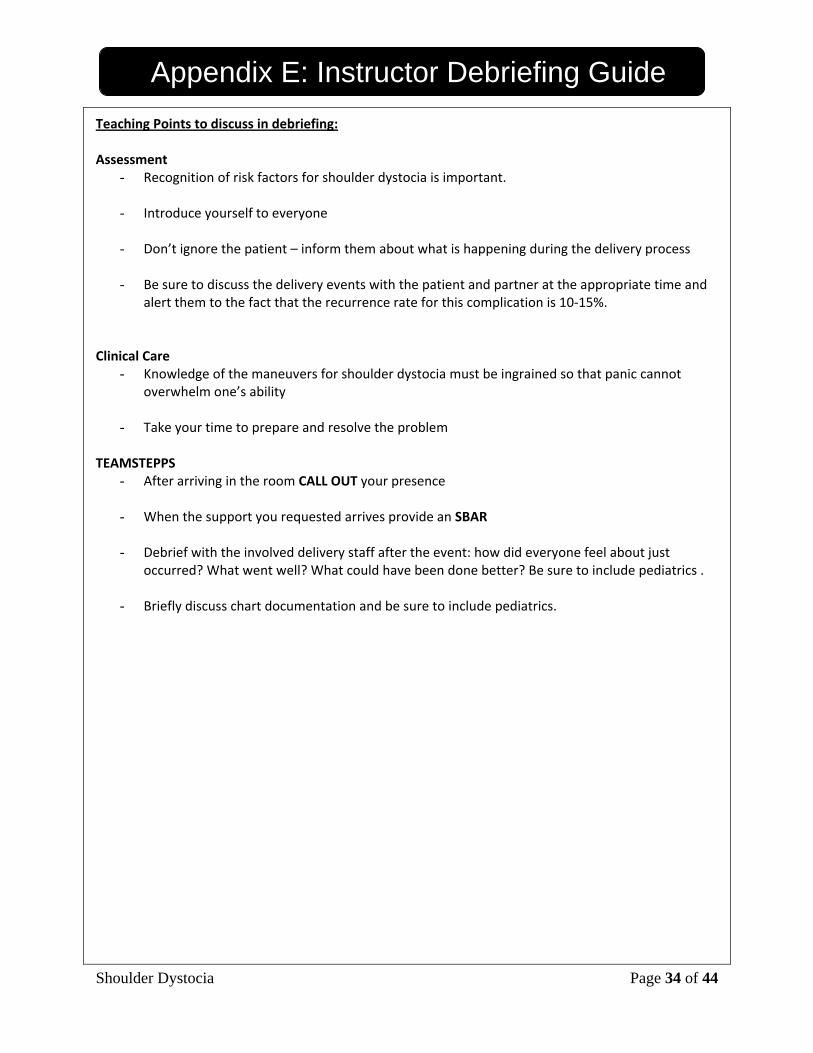
Appendix E: Instructor Debriefing Guide Teaching Points to discuss in debriefing: Assessment
- Recognition of risk factors for shoulder dystocia is important.
- Introduce yourself to everyone
- Don’t ignore the patient – inform them about what is happening during the delivery process
- Be sure to discuss the delivery events with the patient and partner at the appropriate time and alert them to the fact that the recurrence rate for this complication is 10‐15%.
Clinical Care
- Knowledge of the maneuvers for shoulder dystocia must be ingrained so that panic cannot overwhelm one’s ability
- Take your time to prepare and resolve the problem TEAMSTEPPS
- After arriving in the room CALL OUT your presence
- When the support you requested arrives provide an SBAR
- Debrief with the involved delivery staff after the event: how did everyone feel about just occurred? What went well? What could have been done better? Be sure to include pediatrics .
- Briefly discuss chart documentation and be sure to include pediatrics.
Shoulder Dystocia Page 35 of 44
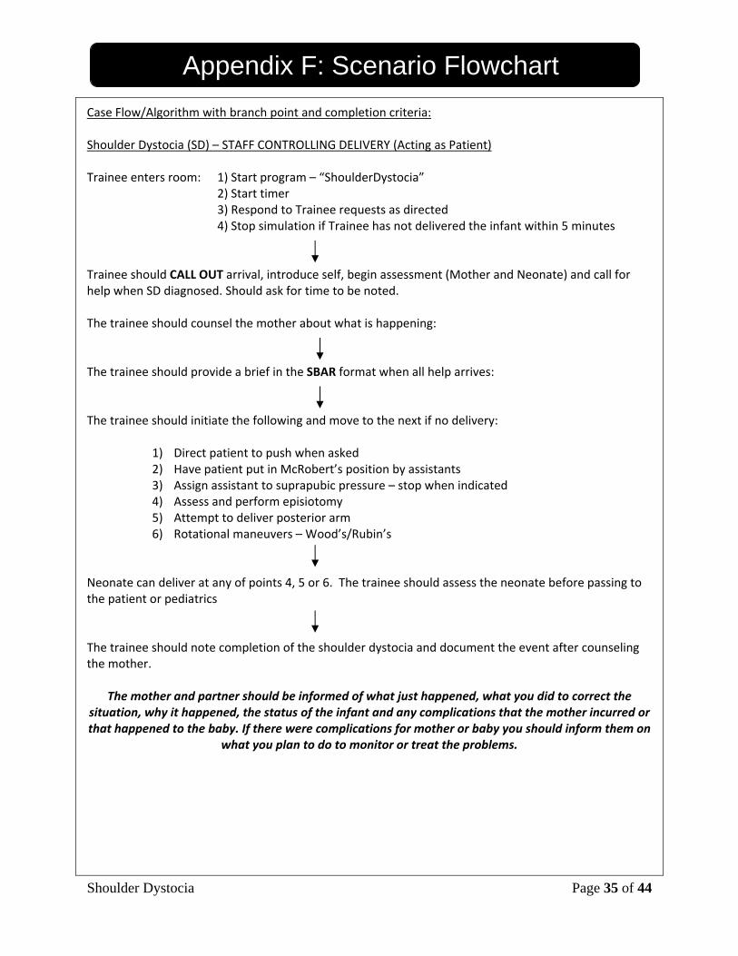
Appendix F: Scenario Flowchart Case Flow/Algorithm with branch point and completion criteria: Shoulder Dystocia (SD) – STAFF CONTROLLING DELIVERY (Acting as Patient) Trainee enters room: 1) Start program – “ShoulderDystocia”
2) Start timer 3) Respond to Trainee requests as directed 4) Stop simulation if Trainee has not delivered the infant within 5 minutes
Trainee should CALL OUT arrival, introduce self, begin assessment (Mother and Neonate) and call for help when SD diagnosed. Should ask for time to be noted. The trainee should counsel the mother about what is happening: The trainee should provide a brief in the SBAR format when all help arrives: The trainee should initiate the following and move to the next if no delivery:
1) Direct patient to push when asked 2) Have patient put in McRobert’s position by assistants 3) Assign assistant to suprapubic pressure – stop when indicated 4) Assess and perform episiotomy 5) Attempt to deliver posterior arm 6) Rotational maneuvers – Wood’s/Rubin’s
Neonate can deliver at any of points 4, 5 or 6. The trainee should assess the neonate before passing to the patient or pediatrics The trainee should note completion of the shoulder dystocia and document the event after counseling the mother.
The mother and partner should be informed of what just happened, what you did to correct the situation, why it happened, the status of the infant and any complications that the mother incurred or that happened to the baby. If there were complications for mother or baby you should inform them on
what you plan to do to monitor or treat the problems.
Shoulder Dystocia Page 36 of 44
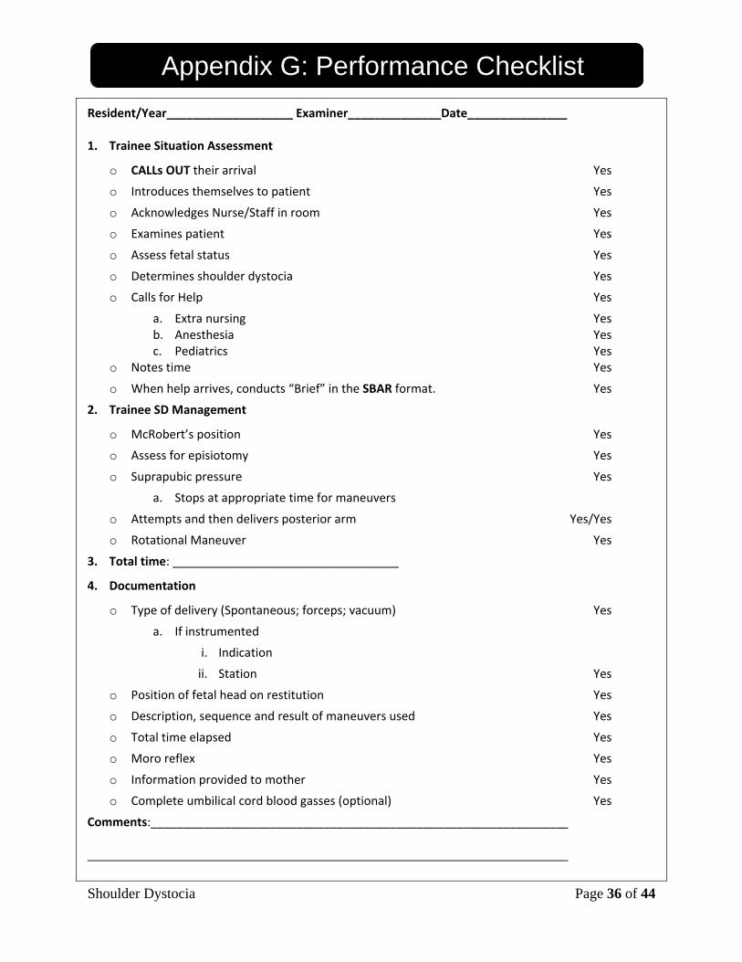
Appendix G: Performance Checklist Resident/Year___________________ Examiner______________Date_______________ 1. Trainee Situation Assessment
o CALLs OUT their arrival Yes
o Introduces themselves to patient Yes
o Acknowledges Nurse/Staff in room Yes
o Examines patient Yes
o Assess fetal status Yes
o Determines shoulder dystocia Yes
o Calls for Help Yes
a. Extra nursing Yes b. Anesthesia Yes c. Pediatrics Yes
o Notes time Yes
o When help arrives, conducts “Brief” in the SBAR format. Yes
2. Trainee SD Management
o McRobert’s position Yes
o Assess for episiotomy Yes
o Suprapubic pressure Yes
a. Stops at appropriate time for maneuvers
o Attempts and then delivers posterior arm Yes/Yes
o Rotational Maneuver Yes
3. Total time: __________________________________
4. Documentation
o Type of delivery (Spontaneous; forceps; vacuum) Yes
a. If instrumented
i. Indication
ii. Station Yes
o Position of fetal head on restitution Yes
o Description, sequence and result of maneuvers used Yes
o Total time elapsed Yes
o Moro reflex Yes
o Information provided to mother Yes
o Complete umbilical cord blood gasses (optional) Yes
Comments:_____________________________________________________________
Shoulder Dystocia Page 37 of 44
Appendix H: Shoulder Dystocia Note Delivery Note: 1) Type of delivery (spontaneous, forceps, vacuum) Spontaneous: Forceps: Vacuum:
Indication of instrumental delivery if performed: Station of the delivery if instrumental delivery:
2) Position of the fetal head on restitution (LOT or ROT) LOT: ROT: 3) Description, sequence, and result of maneuvers used to relieve shoulder dystocia Maneuver a) result:
b) result: c) result: d) result: e) result: f) result:
4) Total time elapsed:
Time of diagnosis: Time of delivery: Total:
5) Umbilical cord blood gases (optional): 6) Condition of the infant:
Moro reflex: 7) Information provided to the patients about the delivery process:
Shoulder Dystocia Page 38 of 44
Appendix I: Simulator Evaluation Form Simulation Session Evaluation Form Facilitator: ____________________________ Date: _________________ Case Presented: ________________________
Strongly Disagree
Disagree Neutral Agree Strongly Agree
1. Learners should spend more time working in simulated environments.
1 2 3 4 5
2. The simulation case provided a realistic model of working in a clinical setting.
1 2 3 4 5
3. This simulation case was effective educational tool 1 2 3 4 5
4. This simulation case enhanced my understanding of how to handle critical incidents.
1 2 3 4 5
5. The debriefing after the case was an important learning opportunity.
1 2 3 4 5
Can you list/describe 1 or more ways this simulation session will change how you do your job? Comments: Content Evaluation
Strongly Disagree
Disagree Neutral Agree Strongly Agree
1. The content was: Current Best Practice Free of Bias Relevant to My Practice
1 2 3 4 51 2 3 4 5 1 2 3 4 5 1 2 3 4 5
2. I will change my practice based on the information presented. 1 2 3 4 5
3. The education level of this activity was appropriate. 1 2 3 4 5
The most important concept learned during this session that may contribute to a change in patient care is:

Shoulder Dystocia Page 39 of 44
Appendix I: Simulator Evaluation Form Instructor
Strongly Disagree
Disagree Neutral Agree Strongly Agree
1. The instructor had a good command of the content. 1 2 3 4 5
2. The instructor’s presentation was clear and concise. 1 2 3 4 5
3. The instructor clearly demonstrated the required skills 1 2 3 4 5
4. The instructor created a safe environment for the debriefing. 1 2 3 4 5
5. The instructor was an effective facilitator 1 2 3 4 5
6. The instructor’s feedback was helpful 1 2 3 4 5
7. Overall, the instructor contributed to my learning 1 2 3 4 5
Comments:
Shoulder Dystocia Page 40 of 44
Appendix J: TeamSTEPPS Glossary Term Definition
Adaptability The ability to adjust strategies and altering a course of action in response to changing conditions (internal and external).
Brief Discussion prior to start that assigns essential roles, establishes expectation, anticipated outcomes, and likely contingencies.
Call‐Out A tactic used to communicate critical information during an emergent event. a. Team member alerting others that they arrived at the scene. (Example: Nurse or anesthesiologist entering room saying, Nurse is Here, etc) b. Request for data/information and Response to the Request. (Example: “We have a SD, please call for help” – “I am calling Pediatrics, Anesthesia & another Nurse.” Helps the team prepare for vital next steps in patient care.
Check‐Back A communication strategy that requires a verification of information. The sender initiates the message, the receiver accepts it and restates the message. In return, the sender verifies that the re‐statement of the original message is correct or amends if not. (Example: “Please give suprapubic pressure” – “suprapubic pressure being given in the oblique direction from the mother’s left” – “That’s correct”)
CUS Signal phrases that denote “I am Concerned, I am Uncomfortable, This is a Safety Issue.” When spoken, all team members will understand clearly not only the issue but the magnitude of the issue. (Example‐ I am concerned that you are reapplying the vacuum extractor in this diabetic patient after three pop offs .)
Debrief Brief, informal information exchange session designed to improve team performance and effectiveness.
DESC Script A technique for managing and resolving conflict. Describe the specific situation or behavior; provide concrete data. Express how the situation makes you feel/what your concerns are. Suggest other alternatives and seek agreement. Consequences should be stated in terms of impact on established team goals; strive for consensus.
Huddle Ad hoc planning to re‐establish Situation Awareness; designed to reinforce plans already in place, and assess the need to adjust the plan. (Example... Ms Jones has a shoulder dystocia and McRoberts has failed. I am going to attempt to extract the posterior arm. Please stop the suprapubic pressure. )
I’M SAFE A checklist to determine both your coworkers’ and your ability to perform safely: I = Illness; M = Medication; S = Stress ‘ A = Alcohol and Drugs; F = Fatigue; E = Eating and Elimination.
SBAR A framework for team members to structure information when communicating to one another. When a new member enters the room, someone SBARs the person by communicating critical information that requires immediate attention and action concerning a patient's condition. S = Situation (What is going on with the patient?) B = Background (What is the clinical background or context?) A = Assessment (What do I think the problem is?) R = Recommendation (What would I do to correct it?)
Shared Mental Model
An organizing knowledge structure of relevant facts and relationships about a task or situation that are commonly held by team members
Situation Awareness
The ability to identify, process, and comprehend the critical elements of information about what is happening to the team with regards to the mission. It’s knowing “What is going around you” and “What is likely to happen next”.
Situation The process of actively scanning and assessing elements of the situation to gain
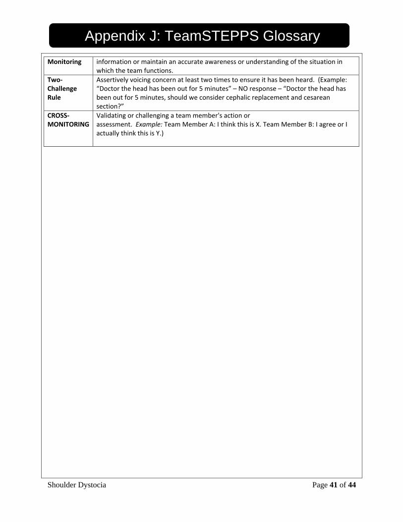
Shoulder Dystocia Page 41 of 44
Appendix J: TeamSTEPPS Glossary Monitoring information or maintain an accurate awareness or understanding of the situation in
which the team functions. Two‐Challenge Rule
Assertively voicing concern at least two times to ensure it has been heard. (Example: “Doctor the head has been out for 5 minutes” – NO response – “Doctor the head has been out for 5 minutes, should we consider cephalic replacement and cesarean section?”
CROSS‐MONITORING
Validating or challenging a team member's action or assessment. Example: Team Member A: I think this is X. Team Member B: I agree or I actually think this is Y.)
Shoulder Dystocia Page 42 of 44
Appendix K: References Key Reading in Shoulder Dystocia Management:
Benedetti T,J, & Gabba S.G. (1978) Shoulder dystocia; a complication of fetal macrosomia and prolonged second stage of labour with mid‐pelvic delivery. Obstetrics & Gynecology. 52: 526‐529.
Bruner J.P, Drummons S.B, Meeham A.L, & Garkin I.M. (1998) All‐fours maneuver for reducing shoulder dystocia during delivery. Journal of Reproductive Medicine. 43 (5): 439‐443.
Gherman RB, Goodwin TM, Souther I et al. The McRoberts maneuver for alleviation of shoulder dystocia: How successful is it? Am Jo Obstet Gynecol 1997; 176, 656‐661
Gonik B, Stringer C.A, & Held B. (1983). An alternative maneuver for management of shoulder dystocia. Am J. Obstet Gynecol. 145 (7): 882‐884
Poggi S, Sprong C, Allen R. Prioritizing Posterior Arm Delivery During Shoulder Dystocia. Obstet Gynecol 2003; 101, 1068‐1072
Rouse D.J & Owen J. (1999) Prophylactic cesarean delivery for fetal macrosomia diagnosed by means of ultrasonography – A Faustian bargain? Am J Obstet Gynecol. 181: 332‐338.
Rubin A. (1964) Management of shoulder dystocia. Journal of the American Medical Association. 189 (11): 835‐837.
Woods C.E. (1942) A principle of physics as applied to shoulder delivery. Am J Obstet Gynecol 45: 796‐850
Additional References:
Acker D.B, Sachs B.P, & Friedman E.A. (1986) Risk Factors for Shoulder Dystocia in the Average‐Weight Infant. Obstet & Gynecol 67: 614 ‐ 617.
Acker, D, Sachs BP, Friedman EA, Risk Factors for Shoulder Dystocia. Obstet Gynecol 1986; 67, 614‐618
ACOG practice bulletin: Shoulder dystocia. Number 40, November 2002
Al Hadi M, Geary M, Byrne P, & McKenna P. (2001) Shoulder dystocia: risk factors and maternal and perinatal outcome. Journal of Obstetrics & Gynaecology. 21 (4): 352‐354.
Allen R.H, Bankoski B.R, Butzin C.A, & Nagey D.A. (1994) Comparing clinician‐applied loads for routine, difficult, and shoulder dystocia deliveries. Am J Obstet Gynecol 171: 1621‐1627.
Bahar A.M. (1996) Risk factors and foetal outcome in cases of shoulder dystocia compared with normal deliveries of a similar birth weight. Br J Obstet & Gynecol 103: 868‐872
Baskett T.F, & Allen A,C. (1995) Perinatal implications of shoulder dystocia. Obstet & Gynecol. 86 (1): 14‐17.
Crofts JF, Bartlett C, Ellis D, Hunt LP, Fox R, Draycott TJ. (2006) Training for shoulder dystocia: a trial of simulation using low‐fidelity and high‐fidelity mannequins. Obstet Gynecol. 108(6):1477‐85.
Crofts JF, Bartlett C, Ellis D, Hunt LP, Fox R, Draycott TJ. (2007) Management of shoulder dystocia: skill
Shoulder Dystocia Page 43 of 44
Appendix K: References retention 6 and 12 months after training. Obstet Gynecol. 110(5):1069‐74.
Crofts JF, Ellis D, Draycott TJ, Winter C, Hunt LP, Akande VA. (2007) Change in knowledge of midwives and obstetricians following obstetric emergency training: a randomised controlled trial of local hospital, simulation centre and teamwork training. BJOG. 114(12):1534‐41.
Crofts JF, Bartlett C, Ellis D, Fox R, Draycott TJ. (2008) Documentation of simulated shoulder dystocia: accurate and complete? BJOG. 115(10):1303‐8.
Draycott TJ, Crofts JF, Ash JP, Wilson LV, Yard E, Sibanda T, Whitelaw A. (2008) Improving neonatal outcome through practical shoulder dystocia training. Obstet Gynecol. 112(1):14‐20.
Evans‐Jones G, Kay S.P.J, Weindling A.M, Cranny G, Ward A, Broadshaw A, & Hernon C. (2003) Arch Dis Child/Fetal Neonatal Ed. 88: 185‐189.
Gherman R.B, Ouzounain J.G, & Goodwon T.M. (1998) Obstetric maneuvres for shoulder dystocia and associated fetal morbidity. Am J Obstet Gynecol. 178 (6): 1126‐1130.
Gherman RB, Ousounian JG, Goodwin TM. Obstetric Maneuvers for shoulder dystocia and associated fetal morbidity. Am Jo Obstet Gynecol 1998; 1126‐1130
Gonik B, Zhung N, & Grimm MJ. (2003) Defining forces that are associated with shoulder dystocia: the use of a mathematical dynamic computer model. Am J Obstet Gynecol. 188 (4): 1068‐1072
Gross T.L, Sokol R.J, Williams T, & Thompson K. (1987) Shoulder dystocia: A fetal‐physician risk. Am J Obstet & Gynecol 156 (6):1408‐1418.
Hernandez C. & Wendel G.D. (1990) Shoulder dystocia. Clin. Obstet Gynecol. 33: 526‐533.
Hope P, Breslin S, Lamont L, Lucas A, Martin D, Moore I et al., (1998) Fatal shoulder dystocia: a review of 56 cases reported to the Confidential Enquiry into Stillbirths and Deaths in Infancy. Br J Obstet Gynaecol. 105: 1256‐1262.
McFarland MB, Langer O, Piper JM et al. Perinatal outcome and type and number of maneuvers in shoulder dystocia. Int J Gynecol Obstet 1996; 55, 219‐224
Modanlou HD, Komatsu G Dorchester W. Large for gestational age neonates: anthropometric reasons for shoulder dystocia. Obstet Gynecol 1982; 60, 417‐423
Morris W.I.C. (1955) “Reports of Societies,” J Obstet Gynaecol Br Empire, 62: 302‐306.
Naef R.W, Martin J.N. (1995) Emergent management of shoulder dystocia. Obstet & Gynecol Clin of North Am. 22: 247‐259.
Nesbitt S.T, Gilbert W.M, & Herrehen B. (1998) Shoulder dystocia and associated risk factors with macrosomic infants born in California. Am J Obstet Gynecol. 179: (2) 476‐480.
Nesbitt TS, Gilbert WM, Herrchen B. Shoulder Dystocia and associated risk factors with macrosomic infants born in California. Am Jo Obstet Gynecol 1998; 179, 476‐480
Nocon J.J, McKenzie D.K, Thomas L.J, & Hansell R.S. (1993) Shoulder dystocia: An analysis of risks and
Shoulder Dystocia Page 44 of 44
Appendix K: References obstetric manoeuvres. Am J Obstet & Gynecol 168:1732‐1739.
Randomized Controlled Trial of Prophylactic Maneuvers to Reduce Head to Body Time in Patients at Risk for Shoulder Dystocia. Obstet Gynecol 2003;102 ,31‐35
Richter H.E, Brumfield C.G, Cliver S.P, Burgio K.L, Needly C.L, & Varner R.E. (2002) Risk factors associated with anal sphincter tear: A comparison of primiparous patients, vaginal births after cesarean deliveries, and patients with previous vaginal deliveries. Am J. Obstet Gynecol. 187 (5): 1194‐1198.
Siassakos D, Crofts JF, Winter C, Weiner CP, Draycott TJ. (2009) The active components of effective training in obstetric emergencies. BJOG. 116(8):1028‐32.