ir-spectroscopy - ifm · ir-spectroscopy theory applications to biomolecules and proteins. infrared...
TRANSCRIPT
IR-spectroscopy
TheoryApplications to
biomolecules and proteins
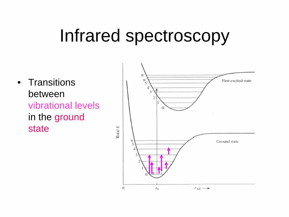
Infrared spectroscopy
• Transitions between vibrational levels in the ground state
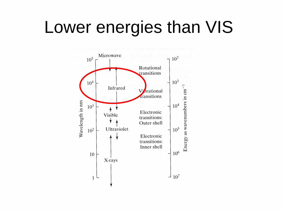
Lower energies than VIS
Infrared spectroscopy• Transitions between vibrational levels in the
ground state• The intensity is related to the transition dipole
moment• Dichroism can be observed in oriented states• Polymers give new spectral features compared
to monomers due to coupling between adjacent transition dipole moments
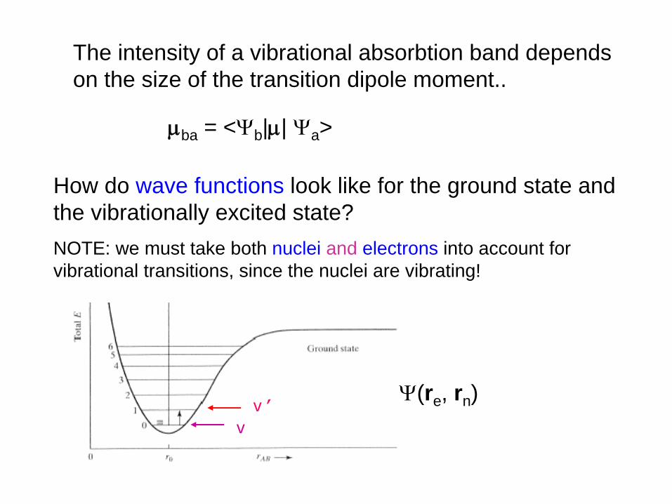
The intensity of a vibrational absorbtion band depends on the size of the transition dipole moment..
ba = <b || a >
How do wave functions look like for the ground state and the vibrationally excited state? NOTE: we must take both nuclei and electrons into account for vibrational transitions, since the nuclei are vibrating!
vv ’ (re , rn )
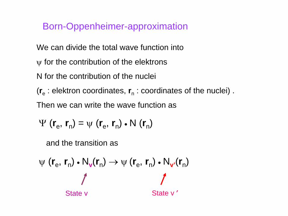
Born-Oppenheimer-approximation
We can divide the total wave function into
for the contribution of the elektrons
N for the contribution of the nuclei
(re : elektron coordinates, rn : coordinates of the nuclei) .
Then we can write the wave function as
State v State v ’
(re , rn ) =
(re , rn )
N (rn )
and the transition as
(re , rn )
Nv (rn ) (re , rn )
Nv’ (rn )
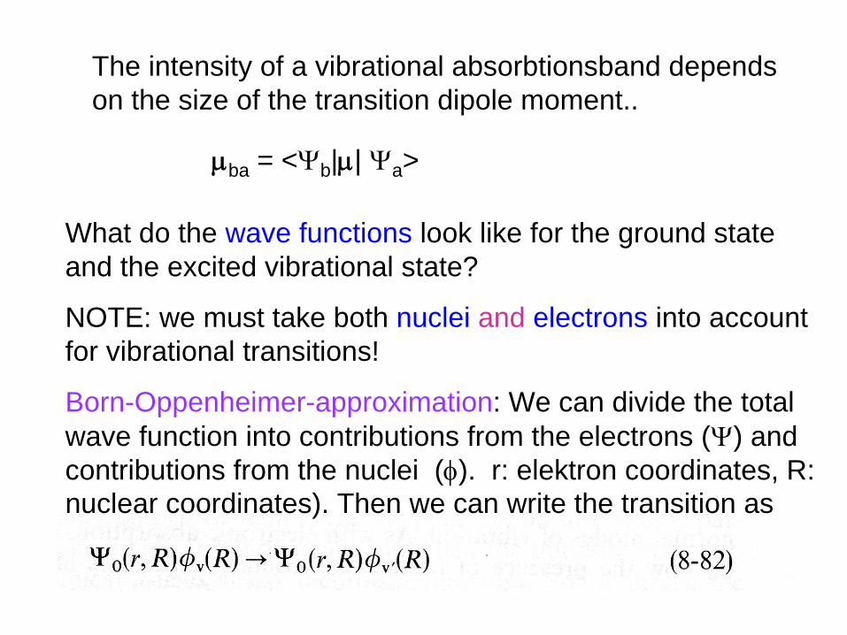
What do the wave functions look like for the ground state and the excited vibrational state?
NOTE: we must take both nuclei and electrons into account for vibrational transitions!
Born-Oppenheimer-approximation: We can divide the total wave function into contributions from the electrons () and contributions from the nuclei (). r: elektron coordinates, R: nuclear coordinates). Then we can write the transition as
The intensity of a vibrational absorbtionsband depends on the size of the transition dipole moment..
ba = <b || a >
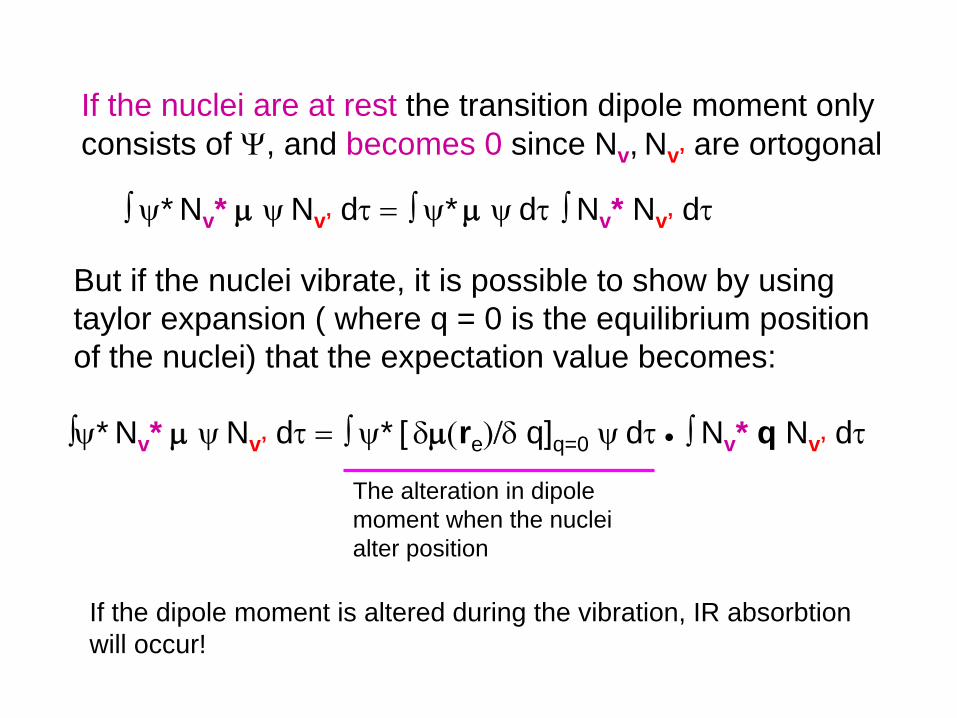
But if the nuclei vibrate, it is possible to show by using taylor expansion ( where q = 0 is the equilibrium position of the nuclei) that the expectation value becomes:
If the nuclei are at rest the transition dipole moment only consists of , and becomes 0 since Nv, Nv’ are ortogonal
The alteration in dipole moment when the nuclei alter position
* Nv *
Nv’ d
*
d
Nv * Nv’ d
* Nv *
Nv’ d
* [ re q]q=0 d
Nv * q Nv’ d
If the dipole moment is altered during the vibration, IR absorbtion will occur!
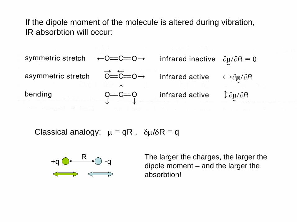
Classical analogy:
= qR , /R = q
+q -qR The larger the charges, the larger the dipole moment – and the larger the absorbtion!
If the dipole moment of the molecule is altered during vibration, IR absorbtion will occur:
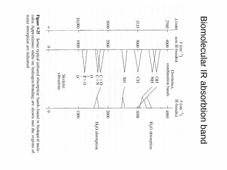
Biom
olecularIR absorbtion
band
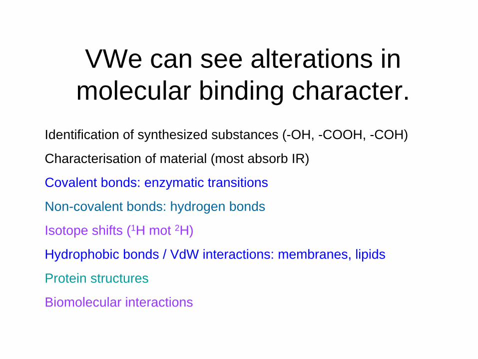
VWe can see alterations in molecular binding character.
Identification of synthesized substances (-OH, -COOH, -COH)
Characterisation of material (most absorb IR)
Covalent bonds: enzymatic transitions
Non-covalent bonds: hydrogen bonds
Isotope shifts (1H mot 2H)
Hydrophobic bonds / VdW interactions: membranes, lipids
Protein structures
Biomolecular interactions
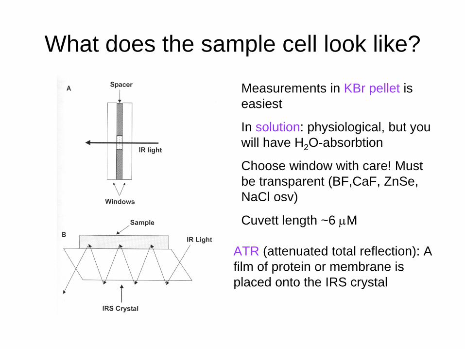
What does the sample cell look like?
ATR (attenuated total reflection): A film of protein or membrane is placed onto the IRS crystal
Measurements in KBr pellet is easiest
In solution: physiological, but you will have H2 O-absorbtion
Choose window with care! Must be transparent (BF,CaF, ZnSe, NaCl osv)
Cuvett length ~6 M

Effects of water
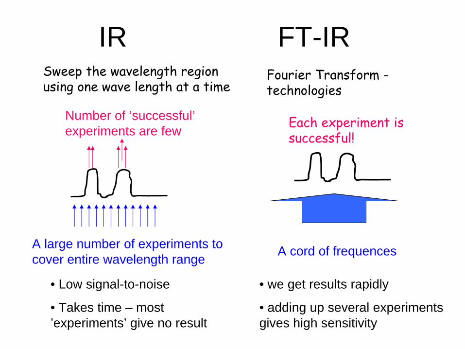
IR FT-IRSweep
the wavelength
region using
one
wave
length
at a timeFourier
Transform -
technologies
A large number of experiments to cover entire wavelength range
Number of ’successful’ experiments are few
A cord of frequences
Each
experiment is successful!
• Low signal-to-noise
• Takes time – most ’experiments’ give no result
• we get results rapidly
• adding up several experiments gives high sensitivity
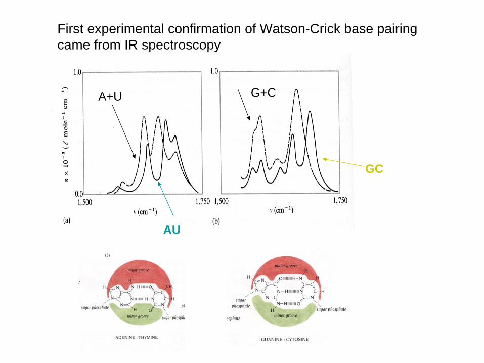
First experimental confirmation of Watson-Crick base pairing came from IR spectroscopy
A+U
AU
G+C
GC
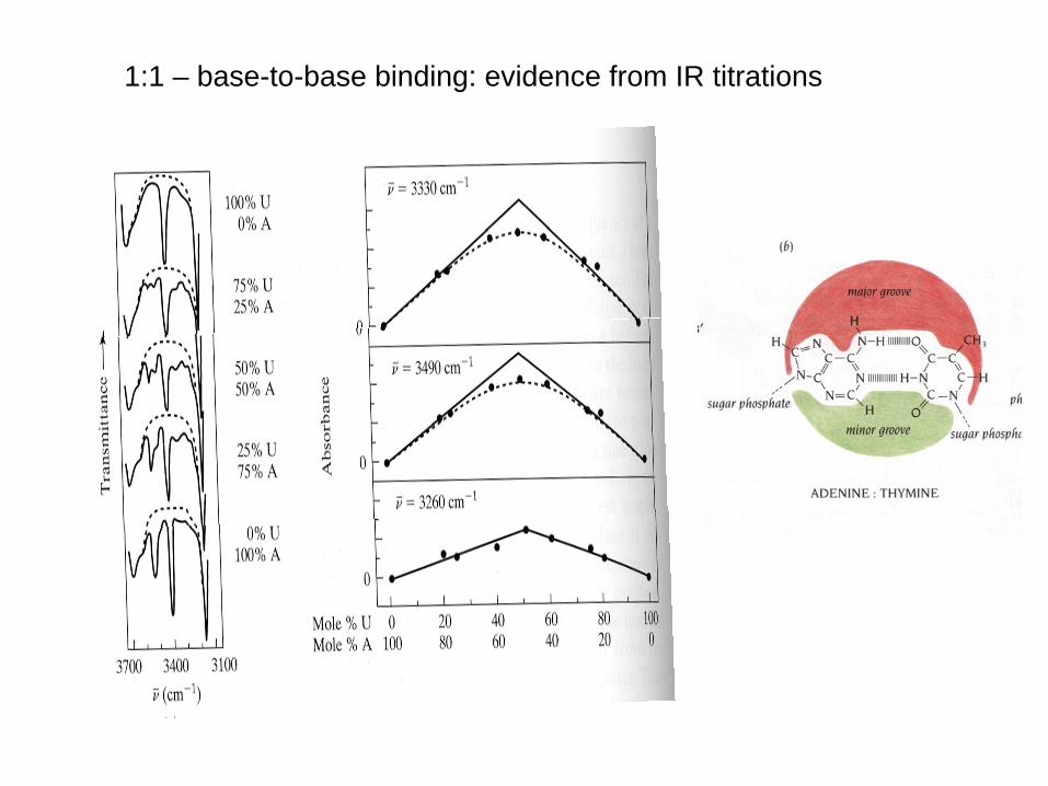
1:1 – base-to-base binding: evidence from IR titrations

Linear polarized light is widely applied in IR, since we can orient our samples onto the sample cell:
AII and A
can be related to molecular orientation.
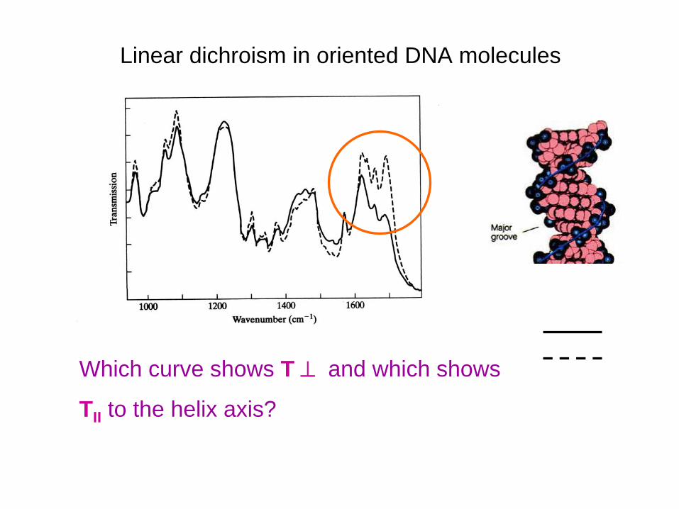
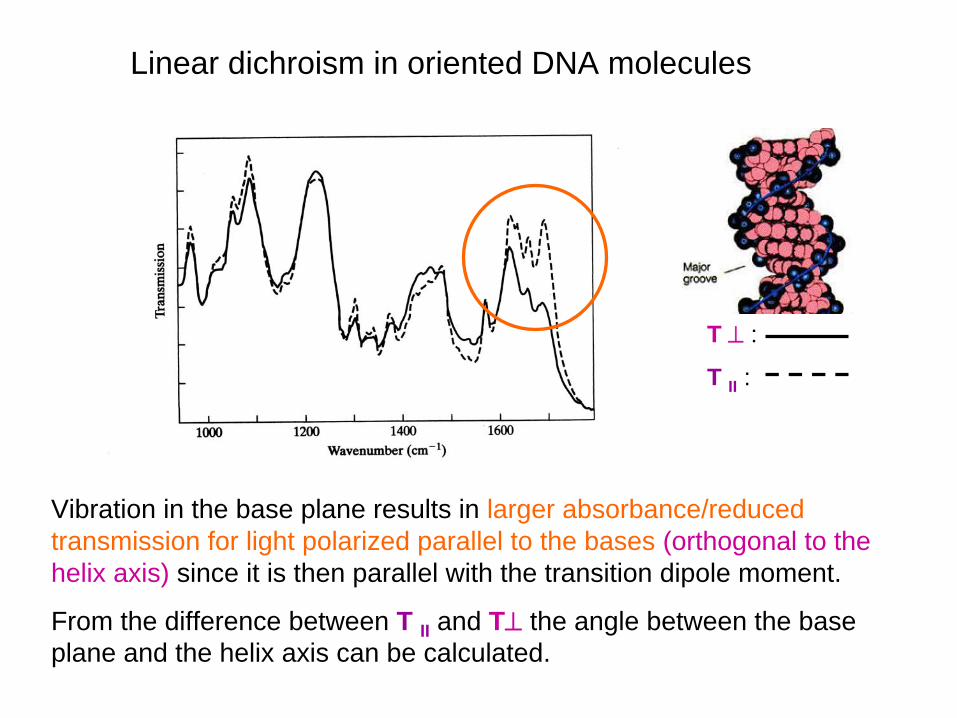
Linear dichroism in oriented DNA molecules
Which curve shows T
and which shows
TII to the helix axis?
Linear dichroism in oriented DNA molecules
Vibration in the base plane results in larger absorbance/reduced transmission for light polarized parallel to the bases (orthogonal to the helix axis) since it is then parallel with the transition dipole moment.
From the difference between T II and T
the angle between the base plane and the helix axis can be calculated.
T
:
T II :
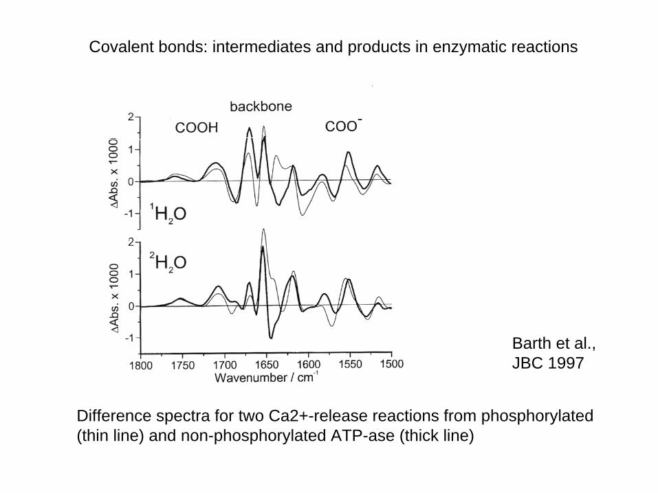
Difference spectra for two Ca2+-release reactions from phosphorylated (thin line) and non-phosphorylated ATP-ase (thick line)
Barth et al., JBC 1997
Covalent bonds: intermediates and products in enzymatic reactions
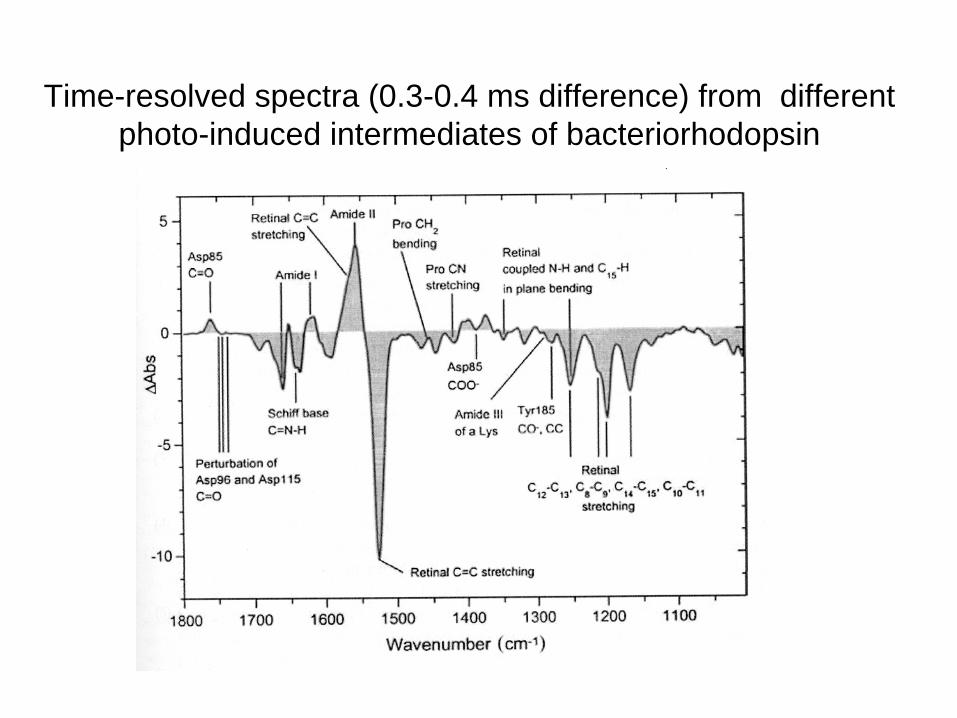
Time-resolved spectra (0.3-0.4 ms difference) from different photo-induced intermediates of bacteriorhodopsin
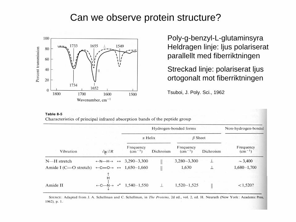
Can we observe protein structure?
Tsuboi, J. Poly. Sci., 1962
Poly-g-benzyl-L-glutaminsyra Heldragen linje: ljus polariserat parallellt med fiberriktningen
Streckad linje: polariserat ljus ortogonalt mot fiberriktningen
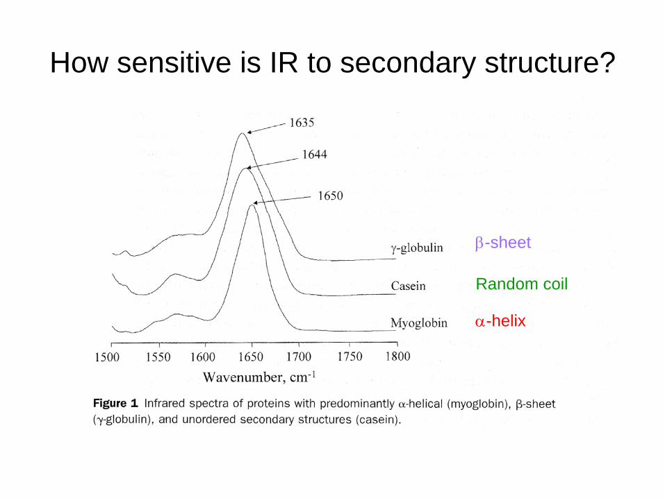
How sensitive is IR to secondary structure?
-helix
Random coil
-sheet
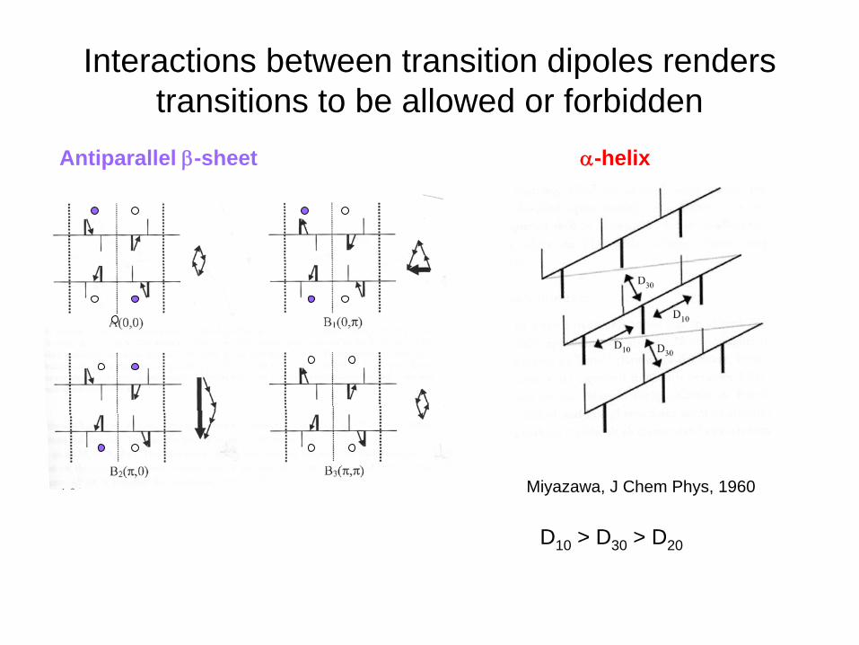
Interactions between transition dipoles renders transitions to be allowed or forbidden
Antiparallel -sheet -helix
Miyazawa, J Chem Phys, 1960
D10 > D30 > D20
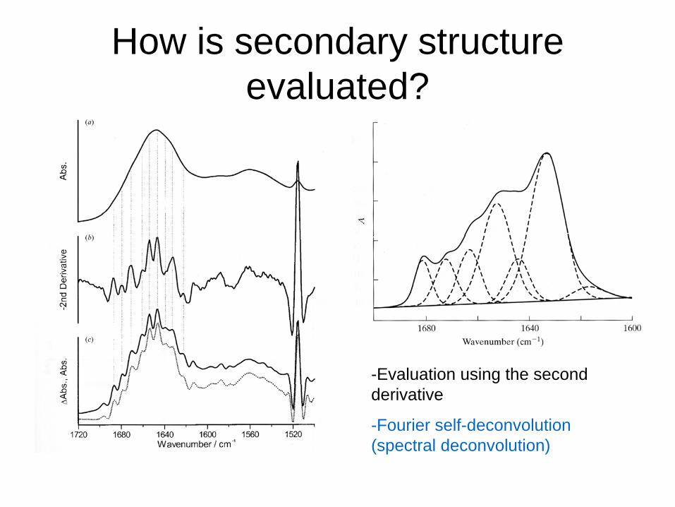
How is secondary structure evaluated?
-Evaluation using the second derivative
-Fourier self-deconvolution (spectral deconvolution)
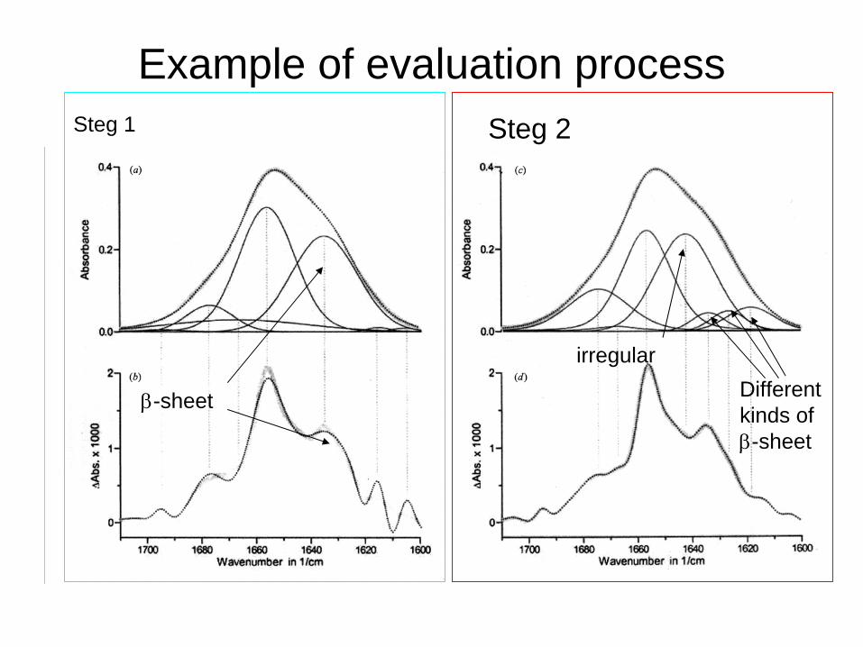
Example of evaluation process
-sheet
irregularDifferent kinds of -sheet
Steg 1 Steg 2
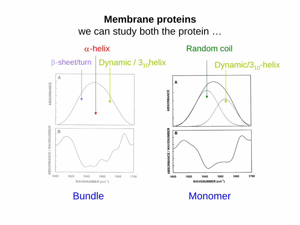
Membrane proteins we can study both the protein …
Bundle Monomer
Dynamic / 310 helix Dynamic/310 -helix
-helix-sheet/turn
Random coil
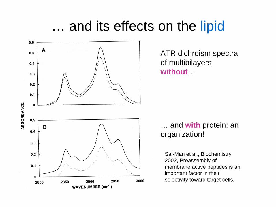
… and its effects on the lipid
ATR dichroism spectra of multibilayers without…
… and with protein: an organization!
Sal-Man et al., Biochemistry 2002, Preassembly of membrane active peptides is an important factor in their selectivity toward target cells.
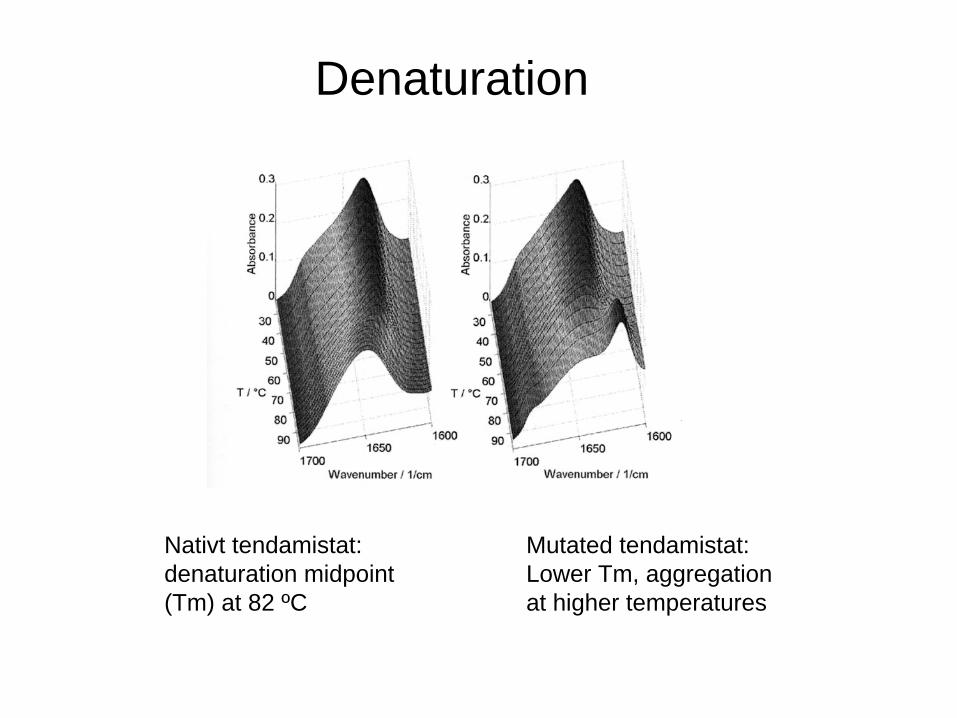
Denaturation
Nativt tendamistat: denaturation midpoint (Tm) at 82 ºC
Mutated tendamistat: Lower Tm, aggregation at higher temperatures
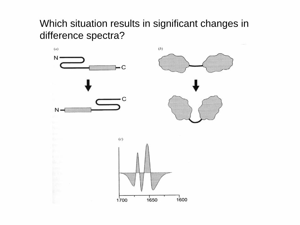
Which situation results in significant changes in difference spectra?