involvementofreceptoractivatorofnuclearfactor- b …and then subjected to time lapse imaging....
TRANSCRIPT

Involvement of Receptor Activator of Nuclear Factor-�BLigand (RANKL)-induced Incomplete Cytokinesis in thePolyploidization of Osteoclasts*□S
Received for publication, July 6, 2015, and in revised form, November 23, 2015 Published, JBC Papers in Press, December 15, 2015, DOI 10.1074/jbc.M115.677427
Noriko Takegahara‡§1, Hyunsoo Kim§, Hiroki Mizuno¶�, Asako Sakaue-Sawano**, Atsushi Miyawaki**,Michio Tomura‡‡§§, Osami Kanagawa¶¶, Masaru Ishii¶�, and Yongwon Choi§2
From the ‡Next Generation Optical Immune-imaging, WPI-Immunology Frontier Research Center, Osaka University, Suita, Osaka565-0871, Japan, the §Department of Pathology and Laboratory Medicine, University of Pennsylvania Perelman School ofMedicine, Philadelphia, Pennsylvania 19104, the ¶Department of Immunology and Cell Biology, Graduate School of Medicine andFrontier Biosciences, WPI-Immunology Frontier Research Center, Osaka University, 2-2 Yamada-oka, Suita, Osaka 565-0871,Japan, the � CREST, Japan Science and Technology Agency, 5 Sanban-cho, Chiyoda-ku, Tokyo 102-0075, Japan, the **Laboratoryfor Cell Function and Dynamics, Advanced Technology Development Group, Brain Science Institute, RIKEN, Wako-city, Saitama351-0198, Japan, the ‡‡Laboratory for Autoimmune Regulation, Research Center for Allergy and Immunology, RIKEN, YokohamaCity, Kanagawa 230-0045, Japan, the §§Laboratory of Immunology, Faculty of Pharmacy, Osaka-Ohtani University, 3-11-1Nishikiorikita, Tondabayashi-city, Osaka 584-8540, Japan, and the ¶¶Department of Molecular Preventive Medicine, GraduateSchool of Medicine, University of Tokyo, 7-3-1 Hongo, Bunkyo-ku, Tokyo 113-033, Japan
Osteoclasts are specialized polyploid cells that resorb bone.Upon stimulation with receptor activator of nuclear factor-�Bligand (RANKL), myeloid precursors commit to becomingpolyploid, largely via cell fusion. Polyploidization of osteoclastsis necessary for their bone-resorbing activity, but the mecha-nisms by which polyploidization is controlled remain to be deter-mined. Here, we demonstrated that in addition to cell fusion,incomplete cytokinesis also plays a role in osteoclast polyploidiza-tion. In in vitro cultured osteoclasts derived from mice expressingthe fluorescent ubiquitin-based cell cycle indicator (Fucci),RANKL induced polyploidy by incomplete cytokinesis as well ascell fusion. Polyploid cells generated by incomplete cytokinesis hadthe potential to subsequently undergo cell fusion. Nuclear polyp-loidy was also observed in osteoclasts in vivo, suggesting theinvolvement of incomplete cytokinesis in physiological polyp-loidization. Furthermore, RANKL-induced incomplete cytokine-sis was reduced by inhibition of Akt, resulting in impaired multi-nucleated osteoclast formation. Taken together, these resultsreveal that RANKL-induced incomplete cytokinesis contributes topolyploidization of osteoclasts via Akt activation.
Polyploidy, in which a cell has more than the diploid comple-ment of chromosomes, is a widespread physiological phenom-
enon observed especially in plants, fungi, and insects (1).Although it is less common in mammals, polyploidizationoccurs in selected tissues, including the placenta, liver, heart,skeletal muscle, and bone marrow during normal developmentand aging (2). During developmental programs, cells obtainadditional sets of chromosomes by various mechanisms,including endocycles, endomitosis, incomplete cytokinesis, andcell fusion. In endocycles and endomitosis, the cell undergoessuccessive rounds of DNA replication without interveningmitosis or karyokinesis (abort mitosis during metaphase).These cycles do not produce two nuclei in the cell. The best-studied examples of endocycles and endomitosis are tropho-blast giant cells and megakaryocytes, respectively (3– 6). Inincomplete cytokinesis, the cell undergoes karyokinesis butskips cytokinesis, resulting in a cell with two nuclei; this processhas been implicated in the normal development of hepatocytesand cardiomyocytes (7, 8). Cell fusion, which involves mergingof the plasma membrane and cytoplasmic mixing, is observedduring development of skeletal muscle cells and osteoclasts (9,10). Endocycles, endomitosis, and incomplete cytokinesis aredirectly associated with the proliferative state of the cell. Bycontrast, cell fusion is entirely independent of cell proliferation(10).
Polyploidy is a hallmark of mature osteoclasts, which are spe-cialized multinucleated giant cells that resorb bone (11). Thesecells are hematopoietic in origin and are derived from myeloidprecursors that also give rise to macrophages and dendriticcells. When myeloid precursors receive signals mediated by theosteoclast differentiation factor RANKL,3 which is mainly pro-duced by osteoblasts, they commit to becoming pre-osteoclastsand ultimately differentiate into multinucleated osteoclasts(12). The importance of polyploidization in osteoclast forma-
* This work was supported by the Uehara Memorial Foundation, theNakatomi Foundation, Tomizawa Jun-ichi and Keiko Fund of the MolecularBiology Society of Japan for Young Scientists, the Pharma-link betweenAcademia and Shionogi, the Takeda Science Foundation (to N.T.), and inpart by National Institutes of Health Grants AR055903 and AR067726 (toY. C.). The authors declare that they have no conflicts of interest withthe contents of this article. The content is solely the responsibility of theauthors and does not necessarily represent the official views of theNational Institutes of Health.Author’s Choice—Final version free via Creative Commons CC-BY license.
□S This article contains supplemental Movies S1–S5.1 To whom correspondence may be addressed: Osaka University, Suita, Osaka
565-0871, Japan. Tel.: 81-06-6879-3881; Fax: 81-06-6879-3889; E-mail:[email protected].
2 To whom correspondence may be addressed. Tel.: 215-746-6404; Fax: 215-573-0888; E-mail: [email protected].
3 The abbreviations used are: RANKL, receptor activator of nuclear factor-�Bligand; MGC, multinucleated giant cell; BMM, bone marrow-derivedmacrophage; dTg, double-transgenic; 7-AAD, 7-aminoactinomycin; BP,band path filter; mAG, monomeric Azami Green.
THE JOURNAL OF BIOLOGICAL CHEMISTRY VOL. 291, NO. 7, pp. 3439 –3454, February 12, 2016Author’s Choice © 2016 by The American Society for Biochemistry and Molecular Biology, Inc. Published in the U.S.A.
crossmark
FEBRUARY 12, 2016 • VOLUME 291 • NUMBER 7 JOURNAL OF BIOLOGICAL CHEMISTRY 3439
by guest on February 3, 2020http://w
ww
.jbc.org/D
ownloaded from

tion is demonstrated by the impaired bone-resorbing activity ofosteoclasts that cannot achieve polyploidy (13).
Although generation of polyploid osteoclasts is thought tooccur due to cell fusion, independently of cell proliferation (14),some researchers have pointed out a relationship between cellproliferation and osteoclast differentiation. For example, inosteoclast progenitors, progression and subsequent withdrawalfrom the cell cycle are required for differentiation into oste-oclasts (15–17). In addition, stimulation with RANKL also trig-gers a signaling pathway that is essential for cell cycle progres-sion (18). These reports prompted us to investigate whether cellcycle progression has an impact on polyploidization duringosteoclastogenesis and, if so, how and to what extent the cellcycle regulates the polyploidization of osteoclasts. The fluores-cent ubiquitination-based cell cycle indicator (Fucci) is a pow-erful tool for studying coordination of the cell cycle with otherdevelopmental processes (19 –22). The Fucci probe was gener-ated by fusing red fluorescent protein and green fluorescentprotein to human Cdt1 and human geminin, respectively.These two chimeric proteins accumulate reciprocally in thenuclei during the cell cycle, labeling the nuclei of cells in G0/G1phase red and those in S/G2/M phase green. Thus, these pro-teins function as effective G0/G1 and S/G2/M markers.
Here, using monocytes derived from Fucci transgenic mice,we show that RANKL-induced polyploidization occurs notonly by cell fusion but also by incomplete cytokinesis. In thesecells, RANKL stimulation transiently increased basal prolifera-tion and induced incomplete cytokinesis as well as cell fusion.Also, cells that underwent incomplete cytokinesis had thepotential to undergo cell fusion. In addition, fluorescence insitu hybridization revealed that some of osteoclasts exhibitednuclear polyploidy (i.e. they contained nuclei with more thanthe diploid complement of chromosomes (�2N)) in vivo, sug-gesting that cells that undergo incomplete cytokinesis areinvolved in physiological polyploidization of osteoclasts. Fur-thermore, RANKL stimulation induced phosphorylation ofAkt, which is required for efficient polyploidization by eitherincomplete cytokinesis or cell fusion. Collectively, our findingsreveal an unexpected pattern of cell division and fusion duringthe generation of polyploid osteoclasts.
Experimental Procedures
Mice—Fucci transgenic mouse lines FucciS/G2/M-#474 andFucciG1-#639 were obtained from the RIKEN BioResourceCenter. These lines were cross-bred to obtain Fucci double-transgenic mice. The generation of TRAP promoter-tdTomatotransgenic mice and V-type H�-ATPase a3 subunit-GFP fusionprotein expressing mice (a3-GFP) were described previously(23). All animal work was performed under veterinary supervi-sion in an accredited facility using protocols approved by theAnimal Care and Use Committee of the University of Pennsyl-vania and Osaka University.
In Vitro Osteoclast and Multinucleated Giant Cell Dif-ferentiation—Bone marrow-derived macrophages (BMMs)from wild type (WT) or Fucci double-transgenic (dTg) micewere obtained from cultures of bone marrow collected from 6-to 8-week-old male tibias and femurs, as described previously.In brief, bone marrow progenitor cells were cultured with
M-CSF (60 ng/ml) in �-minimal essential medium containing10% FCS. After 3– 4 days, cells were gathered as BMMs. Forosteoclast differentiation, BMMs were cultured for 3 days in�-minimal essential medium containing M-CSF (60 ng/ml) andRANKL (150 ng/ml). After culture for 3 days, the cells werefixed with 3.7% formaldehyde in PBS for 10 min and thenstained for TRAP using the acid phosphatase, leukocyte(TRAP) kit (Sigma). TRAP-positive multinucleated cells con-taining three or more nuclei were counted. For differentiationof multinucleated giant cells (MGCs), BMMs derived from wildtype mice were cultured in the presence of IL-3 (100 ng/ml)(R&D Systems) and IL-4 (100 ng/ml) (PeproTech) for 2 days.MGCs were stained with May-Grunewald Giemsa stain (LifeTechnologies, Inc.). For inhibitor assays, BMMs were culturedwith M-CSF (60 ng/ml) and RANKL (150 ng/ml) or with IL-4(100 ng/ml) plus IL-3 (100 ng/ml) in the presence of Y27632 (10�M) (Wako), Akt inhibitor VIII (5 �M) (Sigma), SN50 (50�g/ml) (BioVision), LY294003 (5 �M) (Wako), aphidicolin (500nM) (Sigma), or DMSO for 72 h.
BrdU Incorporation Assay—Wild Type-BMMs were cul-tured with M-CSF (60 ng/ml) in the presence or absence ofRANKL (150 ng/ml) or cultured with IL-4 (10 ng/ml) plus IL-3(100 ng/ml) for the indicated amount of time (in hours). BrdU (10�M) were added to the culture for the last 6 h. After culture, cellswere washed with PBS and detached using enzyme-free cell disso-ciation buffer (Millipore) at 37 °C for 5 min. After generation ofsingle-cell suspensions, cells were counted and suspended at 1 �106/ml in PBS in polystyrene tubes. The cells were stained withLIVE/DEAD Aqua (Life Technologies, Inc.) for 30 min on ice toexclude dead cells. Cells were washed with PBS with 2% FCS andfixed with BrdU fixation buffer (eBioscience) for 1 h on ice fol-lowed by DNase treatment. Cells were stained with FITC-labeledanti-BrdU antibody (clone Bu20a, eBioscience) for 20 min, andafter washing, cells were further stained with 7-AAD. Finally, thecells were analyzed using a FACSCanto II (BD Biosciences). Num-bers indicate the percentages of S phase cells. Results are represen-tative of three independent experiments.
Ploidy Analysis—dTg-BMMs stimulated with RANKL forthe indicated times in the presence of M-CSF were washed withPBS and detached using enzyme-free cell dissociation buffer(Millipore) at 37 °C for 5 min. After generation of single-cellsuspensions, cells were counted and suspended at 1 � 106/ml inphenol red-free �-minimal essential medium containing 2%FCS and 2 mM EDTA in polystyrene tubes. Then the cells werestained with 5 �M Vybrant DyeCycle Violet (Life Technologies,Inc.) at 37 °C for 60 min. Finally, the cells were stained withTO-PRO-3 (Life Technologies, Inc.) to exclude dead cells andanalyzed using an LSR (BD Biosciences). Excitation laser linesand emission filters were as follows: Vybrant DyeCycle Violet:excitation, 405-nm laser line, and emission, 450/50 BP; mAG:excitation, 488-nm laser line, and emission, 515/20 BP;mKO2: excitation, 532-nm laser line, and emission, 585/42 BP;TO-PRO-3: excitation, 640-nm laser line, and emission, 660/20BP. Data were analyzed using the FlowJo software (Tree Star).
Time-lapse Microscopy and Analysis—dTg-BMMs culturedin a glass-bottom dish were stimulated with RANKL (150ng/ml) in the presence of M-CSF (60 ng/ml) or stimulated withIL-4 (100 ng/ml) and IL-3 (100 ng/ml) for the indicated times,
Osteoclast Polyploidization via Incomplete Cytokinesis
3440 JOURNAL OF BIOLOGICAL CHEMISTRY VOLUME 291 • NUMBER 7 • FEBRUARY 12, 2016
by guest on February 3, 2020http://w
ww
.jbc.org/D
ownloaded from

and then subjected to time lapse imaging. Cell-tracking ex-periments were performed using a Deltavision microscope(Applied Precision) from an Olympus IX70 microscopeequipped with a �20 or �40 objective, 300-watt xenon lamp,and a Photometrics CoolSNAP HQ 12-bit monochrome-cooled CCD camera. A series of images was collected every 5min at 37 °C. To acquire large square fields of view, multipointtime-lapse imaging was performed. Image acquisition and pro-cessing were performed using the Deltavision SoftWorx soft-ware (Applied Precision), and image analysis was performedusing the Fiji software (National Institutes of Health). Cellswere manually tracked. Mononucleated cell fusion was countedas “cell fusion.” Mononucleated or binucleated cells that wentthrough cell fusion following incomplete cytokinesis, or daugh-ter cells of a binucleated cell that underwent cell fusion, werecounted as “cell fusion involving incomplete cytokinesis.” Dou-bling times of cytokinesis/incomplete cytokinesis were assessedusing a Nikon BioStation IMQ imaging system, and image anal-ysis was performed using the Imaris software (Bitplane). Thetime from the end of cytokinesis/incomplete cytokinesis to thenext end of cytokinesis/incomplete cytokinesis was measuredas the doubling time. Final graphs were generated usingGraphPad Prism (GraphPad Software).
Flow Cytometry—dTg-BMMs cultured with M-CSF (60ng/ml) in the presence or absence of RANKL (150 ng/ml) for48 h were washed with PBS and detached using enzyme-freecell dissociation buffer (Millipore) at 37 °C for 5 min. After gen-eration of single-cell suspensions, cells were counted and sus-pended at 1 � 106/ml in PBS in polystyrene tubes. Cells werestained with 1 �M LIVE/DEAD NearIR (Life Technologies, Inc.)on ice for 30 min to exclude dead cells, washed with PBS with2% FCS, and stained with allophycocyanin-labeled antibodiesagainst integrin �3 or F4/80 (integrin �3, clone 2C9.G3; F4/80,clone MB8 (eBioscience)) on ice for 30 min. After washing, cellswere further stained with 5 �M Vybrant DyeCycle Violet (LifeTechnologies, Inc.) at 37 °C for 60 min. A Fucci transgenicmouse line FucciG1-#610 obtained from the RIKEN BioRe-source Center and a3-GFP mice line were cross-bred to obtainFucci-mKO2 and a3-GFP expressing mice (Fucci-mKO2/a3-GFP). BMMs were prepared from Fucci-mKO2/a3-GFP miceand cultured with M-CSF (60 ng/ml) in the presence or absenceof RANKL (150 ng/ml) for the indicated times. After generationof single-cell suspensions, cells were stained with 5 �M VybrantDyeCycle Violet (Life Technologies, Inc.) at 37 °C for 60 min.Finally, the cells were stained with TO-PRO-3 (Life Technolo-gies, Inc.) to exclude dead cells. Peripheral blood cells or bonemarrow cells of Fucci-mAG transgenic mice were stained withallophycocyanin-labeled antibodies against Ly6C (clone HK1.4(eBioscience)) on ice for 30 min. After washing, cells were fur-ther stained with 5 �M Vybrant DyeCycle Violet (Life Technol-ogies, Inc.) at 37 °C for 60 min. Finally, the cells were stainedwith 7-AAD (eBioscience) to exclude dead cells. Cells were ana-lyzed using LSR (BD Biosciences), and data were analyzed usingthe FlowJo software (Tree Star).
Gelatin Degradation Assay—Fluorescein isothiocyanate(FITC) gelatin-coated glass bottom dishes were prepared asdescribed previously (24). dTg-BMMs were cultured on theFITC gelatin-coated dishes for 24 h with M-CSF (60 ng/ml) and
RANKL (150 ng/ml) and then subjected to time-lapse imagingusing a confocal A1 microscope system (Nikon). A series ofimages was collected every 30 min at 37 °C. Image analysis wasperformed using NIS-elements software (Nikon).
Fluorescence in Situ Hybridization—TRAP promoter-tdTomato transgenic mice were perfused with 4% paraformal-dehyde plus sucrose for fixation, and bone tissues were furtherfixed with 4% paraformaldehyde plus sucrose for 3 h at 4 °C.Next, the bones were incubated with 10% EDTA for 2 weeks fordecalcification and embedded in O.C.T. compound (Tissue-Tek). Sections 5 �m thick were prepared using Kawamoto’sfilm method and treated with pepsin, and then FISH probemixture was added. The sections were denatured at 90 °C for 10min and then kept overnight at 37 °C for hybridization. Afterhybridization, the sections were washed with 5% formamide,2� SSC for 20 min at 37 °C, and then 1� SSC for 10 min atroom temperature. After washing, the sections were stainedwith DAPI, and images were acquired using the Leica CW-4000system (Leica) with a �100 objective.
Western Blotting—After treatment of cells with the indicatedcytokines or inhibitors for the indicated times, cells werewashed twice with ice-cold PBS and scraped from the plasticplate with a cell lifter (Costar), and then whole-cell lysates wereisolated in RIPA buffer (1% Nonidet P-40, 0.5% sodium deoxy-cholate, 0.1% SDS, 25 mM Tris-HCl, pH 7.6, 150 mM NaCl)supplemented with protease inhibitor mixture and PhosSTOP(Roche Applied Science). Equivalent amounts of protein(20 – 40 �g) were subjected to 10% SDS-PAGE, and immuno-blotting was performed using antibodies specific for phospho-Akt (Ser-473), Akt, phospho-p38 MAPK (Thr-180/Tyr-182),p38, phospho-p44/42 MAPK (ERK1/2) (Thr-202/Tyr-204),ERK1/2, JNK (Cell Signaling), and phospho-JNK (Thr-183/Tyr-185) (BD Biosciences). For RhoA activation assay, celllysates were incubated with Rhotekin-RBD protein beads andblotted with anti-RhoA mAb (Cytoskeleton).
Statistical Analysis—Data were analyzed using one-way anal-ysis of variance or unpaired t test with Welch’s correction andare presented as means � S.D. A p value � 0.05 was consideredsignificant.
Results
RANKL Stimulation Increases Basal Proliferation of BMMs—To determine the impact of RANKL stimulation on the cellcycle during osteoclast development, we first examined theproportions of cells in the G1 and S/G2/M phases duringRANKL-induced osteoclast differentiation. Fucci double-transgenic mouse-derived bone marrow monocytes (dTg-BMMs) were stimulated with or without RANKL in the pres-ence of M-CSF, and the proportions of the cells positive forgreen fluorescence (S/G2/M phase) and red fluorescence (G1phase) were measured by flow cytometry. The proportion ofgreen cells increased 24 h after RANKL stimulation, but thisincrease disappeared 48 h after stimulation (Fig. 1, A and B).M-CSF alone did not significantly influence the proportion ofgreen or red cells during this period (Fig. 1, A and B). Theseresults suggested that RANKL stimulation transiently pro-motes cell cycle progression. The increase in cell cycle progres-sion was confirmed by BrdU incorporation assay (Fig. 1C).
Osteoclast Polyploidization via Incomplete Cytokinesis
FEBRUARY 12, 2016 • VOLUME 291 • NUMBER 7 JOURNAL OF BIOLOGICAL CHEMISTRY 3441
by guest on February 3, 2020http://w
ww
.jbc.org/D
ownloaded from

Using time-lapse imaging, we measured the doubling time ofdTg-BMMs cultured with M-CSF in the presence or absence ofRANKL for 48 h. RANKL significantly reduced doubling timein a dose-dependent manner (Fig. 1D), along with increasingformation of multinucleated osteoclasts (Fig. 1E). These results
support the idea that RANKL stimulation increases the basalproliferation of dTg-BMMs.
RANKL Stimulation Induces Polyploid Cells Not Only by CellFusion but Also by Incomplete Cytokinesis—We next performedploidy analysis during osteoclast formation. dTg-BMMs were
FIGURE 1. RANKL stimulation increases basal cell proliferation. A, flow cytometry analysis of dTg-BMMs during osteoclast differentiation. dTg-BMMs werecultured with M-CSF (60 ng/ml) in the presence or absence of RANKL (150 ng/ml) for the indicated times. Cells were harvested at the indicated times, and cellspositive for red (mKO2) or green (mAG) fluorescence were detected by flow cytometry. Results are representative of three to five independent experiments. B,ratios of red (mKO2) fluorescence-positive cells to green (mAG) fluorescence-positive cells. Each circle represents the result of an independent flow cytometryexperiment. Bars indicate means � S.D. C, BrdU incorporation assay. WT-BMMs were cultured with M-CSF (60 ng/ml) in the presence or absence of RANKL (150ng/ml) for the indicated amount of time (in hours). BrdU (10 �M) was added for the last 6 h. Incorporated BrdU was stained with FITC-labeled anti-BrdUantibody. DNA was stained with 7-AAD and analyzed by flow cytometry. Numbers indicate the percentages of S phase cells. Results are representative of threeindependent experiments. D, doubling time of dTg-BMMs during osteoclast differentiation. dTg-BMMs were cultured with M-CSF (60 ng/ml) in the presencethe indicated dose of RANKL (ng/ml) for 48 h. Each circle represents the result of a cell. Bars indicate means � S.D. *, p � 0.05; ***, p � 0.001. E, in vitro osteoclastdifferentiation. dTg-BMMs were cultured with M-CSF (60 ng/ml) in the presence of the indicated dose of RANKL (ng/ml) for 96 h. Percentages of multinucleatedcells containing more than five nuclei are shown. Scale bars, 100 �m.
Osteoclast Polyploidization via Incomplete Cytokinesis
3442 JOURNAL OF BIOLOGICAL CHEMISTRY VOLUME 291 • NUMBER 7 • FEBRUARY 12, 2016
by guest on February 3, 2020http://w
ww
.jbc.org/D
ownloaded from

stimulated with RANKL for the indicated times in the presenceof M-CSF, and ploidy was analyzed by flow cytometry (Fig. 2).As expected, stimulation with RANKL induced generation ofpolyploid cells (red fluorescence-positive 4C, 6C, 8C, and�10C) (Fig. 2). Among these polyploid cells, 4C and 8C cellswere detected first after RANKL stimulation for 24 h (Fig. 2). Bycontrast, 6C cells were not detected until 48 h after the onset ofRANKL stimulation, and 6C cells were less common than 8Ccells (Fig. 2 and Table 1).
To examine how these polyploid cells were generated, weperformed time-lapse imaging. In these experiments, dTg-BMMs were stimulated by RANKL for various times (1–16 h) inthe presence of M-CSF, and then cell cycle progression andpolyploidization were observed by time-lapse imaging. Regard-less of the period of RANKL stimulation, almost all cells weremononucleated at the beginning of imaging. No cell fusion wasobserved within 24 h, but fusion was observed at and after 36 hof RANKL stimulation (average time of the beginning of fusionwas 50.5 � 5.63 h; Fig. 3A). The majority of cells that wentthrough cell fusion were red fluorescence-positive mononu-cleated cells (Fig. 3B and Table 2), and the resultant fused cellsrarely went through mitosis (Table 2). Instead, they continued
to undergo cell fusion, and finally became large multinucleatedosteoclast-like cells with red nuclei. These observations sug-gested that 4C and 8C cells detected after 24 h of RANKL stim-ulation were not fusion products. Unexpectedly, incompletecytokinesis was observed during this period (Fig. 4A and sup-plemental Movie 1). The incomplete cytokinesis resulted in for-mation of 4C binucleated cells (Fig. 4A). Some of these cellsre-entered mitosis but failed to complete cytokinesis, resultingin formation of binucleated 8C cells (Fig. 4A). Consistent with
FIGURE 2. RANKL induces polyploid cells. Ploidy analysis of dTg-BMMs during osteoclast differentiation. dTg-BMMs were cultured with M-CSF (60 ng/ml) andRANKL (150 ng/ml) for the indicated times. Cells were harvested at the indicated times, stained with DNA staining dye (Vybrant DyeCycle Violet), and examinedby flow cytometry. Top row shows flow cytometry results of dTg-BMMs cultured with RANKL for the indicated times. Middle row shows flow cytometry resultsof red fluorescence (mKO2) versus Vybrant DyeCycle Violet. Bottom row provides quantitation of the flow cytometry results shown above. 2C, 4C, 6C, 8C, and�10C cells are gated. Numbers indicate the percentages of red (mKO2) positive cells in each bin. Results are representative of three independent experiments.
TABLE 1Percentage of diploid and polyploid cells among mKO2� cellsEach row represents the result of an independent flow cytometry experiment.
2C 4C 6C 8C >10C
Monocytes 98.32 1.00Monocytes 97.93 1.20 0.03Monocytes 94.25 3.89Monocytes 92.42 5.36 0.04 0.08Monocytes 93.39 4.11 0.09 0.09OC (day2) 84.77 12.94 0.25 0.49 0.02OC (day2) 83.17 13.04 0.17 0.73OC (day2) 77.27 16.02 0.66 1.27OC (day3) 77.92 13.10 1.32 1.47 1.61OC (day3) 81.81 13.80 0.59 1.13
Osteoclast Polyploidization via Incomplete Cytokinesis
FEBRUARY 12, 2016 • VOLUME 291 • NUMBER 7 JOURNAL OF BIOLOGICAL CHEMISTRY 3443
by guest on February 3, 2020http://w
ww
.jbc.org/D
ownloaded from

this, after RANKL stimulation for 24 h, flow cytometric analysisdetected S/G2/M phase cells between the 4C and 8C peaks (Fig.2), consistent with the observation that some of the tetraploidcells re-entered the cell cycle. Together, these observations sug-gested that 4C and 8C cells detected at 24 h after RANKL stim-ulation resulted from incomplete cytokinesis.
Cells That Undergo Incomplete Cytokinesis Have the Poten-tial for Cell Fusion—Because generation of polyploidy byincomplete cytokinesis has been observed in certain patholog-ical contexts (25, 26), we speculated that cells that undergoincomplete cytokinesis might merely be abnormal, rather thancritical intermediates in the formation of mature osteoclasts.To address this issue, we continued time-lapse imaging to tracethe fates of cells that underwent incomplete cytokinesis. Cell-tracking analysis revealed that binucleated cells generated byincomplete cytokinesis could undergo cell fusion (Fig. 5A andsupplemental Movie 2). Indeed, 11% of fused cells had previ-ously undergone incomplete cytokinesis (Table 2). Theseresults indicated that such cells had the potential to undergocell fusion during RANKL-induced osteoclast formation.
Some of the binucleated cells generated by incomplete cyto-kinesis ultimately succeeded in undergoing cytokinesis andformed mononucleated polyploid cells (Fig. 4B and supplemen-tal Movie 3). These mononucleated polyploid cells also had thepotential to undergo cell fusion (Fig. 5B and supplementalMovie 4). Notably, �50% of 4C cells sorted from dTg-BMMscultured with RANKL for 66 h were mononucleated.4 Theseobservations strongly suggested that mononucleated cellsinvolved in cell fusion consisted of both 2C and polyploid cells.During time-lapse imaging, target cells often migrated awayfrom the field of view. In this case, the fate of the target cellscould not be traced, and consequently those cells wereuncounted. Thus, the observed percentage of cell fusion involv-ing cells that undergo incomplete cytokinesis might representan underestimate. Collectively, these results revealed thatpolyploid cells detected after RANKL stimulation for morethan 48 h consisted not only of fusion products but also of cellsthat underwent incomplete cytokinesis.
Characterization of Cells That Undergo Incomplete Cyto-kinesis—To understand the phenotype of cells that had under-gone incomplete cytokinesis, we first examined the expressionprofiles of integrin �3, F4/80, and V-type H�-ATPase a3 sub-unit in RANKL-stimulated BMMs. Integrin �3 is an importantcell adhesion molecule that plays an important role in oste-oclast biology (27), and F4/80 is a macrophage marker that isbarely expressed on osteoclasts (28). Expression of both integ-rin �3 and F4/80 was detected on dTg-BMMs and changedafter 48 h of stimulation with RANKL (Fig. 6A). We separateddTg-BMMs into four groups depending on their cell cyclephase and DNA content as follows: (i) mononucleated cells (G1 �2C); (ii) cells in S phase (S); (iii) cells that underwent incom-plete cytokinesis or were generated by cell fusion (G1 �4C, G1 � 8C), and (iv) cells that underwent incomplete cytoki-nesis and re-entered S phase (S (re-enter)) (Fig. 6B). The expres-sion of integrin �3 and F4/80 was examined in each of thesegroups. Flow cytometry analysis revealed that the expressionlevels of these molecules were approximately the same in allfour groups (Fig. 6B), suggesting that the expression of integrin�3 and F4/80 was not affected by incomplete cytokinesis.
V-type H�-ATPase a3 subunit is a protein complex thatmediates H� transport into the resorption lacunae. Lack of a3subunit results in severe osteopetrosis due to impaired oste-oclast bone-resorbing function (29). BMMs derived fromFucci-mKO2 and a3-GFP-expressing mice (Fucci-mKO2/a3-GFP-BMMs) were cultured with M-CSF and RANKL for theindicated times, and the expression of a3-GFP was analyzed byflow cytometry. The expression of a3-GFP gradually increasedwith time (Fig. 6C) and was detected in diploids (G1 � 2C),
4 N. Takegahara and Y. Choi, unpublished observations.
FIGURE 3. Cell fusion occurs at and after 36 h of RANKL stimulation. A, timeof initiation of cell fusion after RANKL stimulation. Images were taken every 5min, and the time required for cell fusion was calculated. A total of 145 cellsthat underwent cell fusion are plotted. Red bar indicates means � S.D. B,representative snapshots of mononucleated cell-cell fusion. Fluorescenceand phase-contrast images were taken every 5 min. Blue arrowheads andboxes indicate cell fusion. White numbers in each image indicates time afterRANKL stimulation. Bottom schematically depicts the results observed in B.Scale bars, 20 �m.
TABLE 2Fusion summary of osteoclasts
EventFusion combination
mKO2 (DN) � mKO2 (DN) mKO2 (DN) � mAG mAG � mAG Total no. (%)
Cell fusion 132 12 1 145Cell fusion involving incomplete cytokinesis 16 0 0 16 (11.0)Cytokinesis after fusion 0 0 1 1 (0.69)
Osteoclast Polyploidization via Incomplete Cytokinesis
3444 JOURNAL OF BIOLOGICAL CHEMISTRY VOLUME 291 • NUMBER 7 • FEBRUARY 12, 2016
by guest on February 3, 2020http://w
ww
.jbc.org/D
ownloaded from

FIGURE 4. RANKL stimulation induces formation of polyploid cells by incomplete cytokinesis. A, representative snapshots of dTg-BMMs that underwentincomplete cytokinesis after RANKL stimulation. Fluorescence and phase-contrast images were taken every 5 min. B, representative snap shots of dTg-BMMsthat underwent cytokinesis after incomplete cytokinesis. Magenta boxes indicate incomplete cytokinesis, and the yellow box indicates cytokinesis. Whitenumbers in each image indicates time after RANKL stimulation. Scale bars, 20 �m. Results are representative of five independent experiments. Bottom showsthe scheme depicting the results observed in A and B.
Osteoclast Polyploidization via Incomplete Cytokinesis
FEBRUARY 12, 2016 • VOLUME 291 • NUMBER 7 JOURNAL OF BIOLOGICAL CHEMISTRY 3445
by guest on February 3, 2020http://w
ww
.jbc.org/D
ownloaded from

FIGURE 5. Cells that undergo incomplete cytokinesis have the potential to undergo cell fusion. A, representative snapshots of a binucleated cell that underwentincomplete cytokinesis and subsequently underwent cell fusion. Fluorescence and phase-contrast images were taken every 5 min. B, representative snapshots of amononucleated cell that underwent incomplete cytokinesis and subsequently underwent cell fusion. Blue arrowheads and blue boxes indicate cell fusion. The magentabox indicates incomplete cytokinesis, and the yellow box indicates cytokinesis. White numbers in each image indicate the time after RANKL stimulation. Scale bars, 20�m. Results are representative of five independent experiments. Bottom shows the scheme depicting the results observed in A and B.
Osteoclast Polyploidization via Incomplete Cytokinesis
3446 JOURNAL OF BIOLOGICAL CHEMISTRY VOLUME 291 • NUMBER 7 • FEBRUARY 12, 2016
by guest on February 3, 2020http://w
ww
.jbc.org/D
ownloaded from

polyploids (G1 � 4C, G1 � 6C, and G1 � 8C), and cells thatunderwent incomplete cytokinesis and re-entered S phase (S(re-enter)) (Fig. 6D). These results suggested that the expressionof a3-GFP was not affected by incomplete cytokinesis.
We next examined resorption activities of cells that hadundergone incomplete cytokinesis. dTg-BMMs were culturedon FITC-labeled gelatin-coated dishes in the presence ofM-CSF and RANKL for 24 h, and then time-lapse imaging was
FIGURE 6. Characterization of cells that undergo incomplete cytokinesis. A, dTg-BMMs were cultured with M-CSF (60 ng/ml) in the presence or absence ofRANKL (150 ng/ml). After 48 h of culture, cells were harvested and stained with anti-integrin �3 or anti-F4/80. The levels of these molecules were determinedby flow cytometry. B, dTg-BMMs described in C were divided into four groups depending on their cell cycle state and DNA contents as follows: mononucleatedcells (G1 � 2C); cells in S phase (S); cells that underwent incomplete cytokinesis or were generated by cell fusion (G1 � 4C, G1 � 8C); and cells that underwentincomplete cytokinesis and re-entered S phase (S (re-enter)). C, expression profiles of a3-GFP in Fucci-mKO2/a3-GFP-BMMs cultured with M-CSF (60 ng/ml) inthe presence or absence of RANKL (150 ng/ml) for the indicated times. D, Fucci-mKO2/a3-GFP-BMMs cultured with M-CSF (60 ng/ml) and RANKL (150 ng/ml)for 60 h were divided into diploids, polyploids, and cells in S phase (S (re-enter)) depending on their cell cycle state and DNA contents. E, left, representativesnapshots of dTg-BMMs cultured on FITC gelatin-coated culture dishes. dTg-BMMs cultured on FITC gelatin-coated dishes for 24 h were observed. Fluores-cence and bright field images were taken every 30 min. Yellow arrowheads indicate a cell in G1 phase that underwent cell fusion, and magenta arrowheadsindicate a cell that underwent incomplete cytokinesis and re-entered S phase (S (re-enter)). Black numbers in each image indicate time after RANKL stimulation(hours). Right, percentage of cells that degraded FITC gelatin during the imaging.
Osteoclast Polyploidization via Incomplete Cytokinesis
FEBRUARY 12, 2016 • VOLUME 291 • NUMBER 7 JOURNAL OF BIOLOGICAL CHEMISTRY 3447
by guest on February 3, 2020http://w
ww
.jbc.org/D
ownloaded from
performed. Resorption activity can be observed as degradationof the gelatin. Majority of cells that degraded FITC gelatin werein red (81.0%; Fig. 6E). In contrast, green� binucleated cells (i.e.the cells that underwent incomplete cytokinesis and re-enteredS phase) barely degraded FITC gelatin (Fig. 6E and supplemen-tal movie 5). These results suggested that cells that had under-gone incomplete cytokinesis and re-entered S phase had lowerresorption capacity than cells in G1 phase.
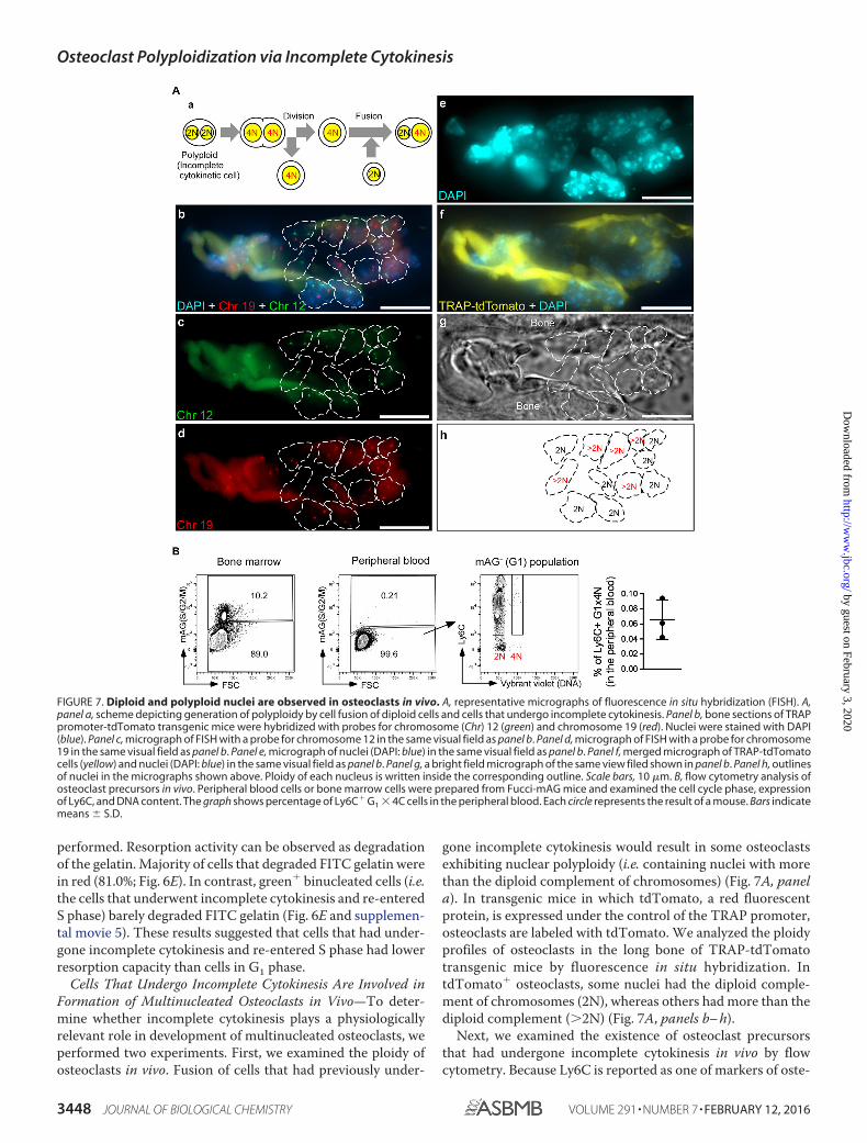
Cells That Undergo Incomplete Cytokinesis Are Involved inFormation of Multinucleated Osteoclasts in Vivo—To deter-mine whether incomplete cytokinesis plays a physiologicallyrelevant role in development of multinucleated osteoclasts, weperformed two experiments. First, we examined the ploidy ofosteoclasts in vivo. Fusion of cells that had previously under-
gone incomplete cytokinesis would result in some osteoclastsexhibiting nuclear polyploidy (i.e. containing nuclei with morethan the diploid complement of chromosomes) (Fig. 7A, panela). In transgenic mice in which tdTomato, a red fluorescentprotein, is expressed under the control of the TRAP promoter,osteoclasts are labeled with tdTomato. We analyzed the ploidyprofiles of osteoclasts in the long bone of TRAP-tdTomatotransgenic mice by fluorescence in situ hybridization. IntdTomato� osteoclasts, some nuclei had the diploid comple-ment of chromosomes (2N), whereas others had more than thediploid complement (�2N) (Fig. 7A, panels b– h).
Next, we examined the existence of osteoclast precursorsthat had undergone incomplete cytokinesis in vivo by flowcytometry. Because Ly6C is reported as one of markers of oste-
FIGURE 7. Diploid and polyploid nuclei are observed in osteoclasts in vivo. A, representative micrographs of fluorescence in situ hybridization (FISH). A,panel a, scheme depicting generation of polyploidy by cell fusion of diploid cells and cells that undergo incomplete cytokinesis. Panel b, bone sections of TRAPpromoter-tdTomato transgenic mice were hybridized with probes for chromosome (Chr) 12 (green) and chromosome 19 (red). Nuclei were stained with DAPI(blue). Panel c, micrograph of FISH with a probe for chromosome 12 in the same visual field as panel b. Panel d, micrograph of FISH with a probe for chromosome19 in the same visual field as panel b. Panel e, micrograph of nuclei (DAPI: blue) in the same visual field as panel b. Panel f, merged micrograph of TRAP-tdTomatocells (yellow) and nuclei (DAPI: blue) in the same visual field as panel b. Panel g, a bright field micrograph of the same view filed shown in panel b. Panel h, outlinesof nuclei in the micrographs shown above. Ploidy of each nucleus is written inside the corresponding outline. Scale bars, 10 �m. B, flow cytometry analysis ofosteoclast precursors in vivo. Peripheral blood cells or bone marrow cells were prepared from Fucci-mAG mice and examined the cell cycle phase, expressionof Ly6C, and DNA content. The graph shows percentage of Ly6C� G1 � 4C cells in the peripheral blood. Each circle represents the result of a mouse. Bars indicatemeans � S.D.
Osteoclast Polyploidization via Incomplete Cytokinesis
3448 JOURNAL OF BIOLOGICAL CHEMISTRY VOLUME 291 • NUMBER 7 • FEBRUARY 12, 2016
by guest on February 3, 2020http://w
ww
.jbc.org/D
ownloaded from

oclast precursors (30), and osteoclast precursors are circulatingin the periphery (31), we tried to detect Ly6C� G1 � 4C cells inthe peripheral blood. In the peripheral blood of Fucci-mAGtransgenic mice, green� cells were barely detected. In green�
(G1 phase) cells, Ly6C� 4C cells were detected (average per-centage was 0.07 � 0.03%; Fig. 7B). These results indicated thatalmost all circulating cells were in G1 phase under physiologicalconditions, and G1 � 4C osteoclast precursors could bedetected in the periphery. Although we cannot exclude the pos-sibility that the cells were binucleated fusion products, thisobservation may support in some part the idea that incompletecytokinesis occurs in vivo. These results suggested that fusionof cells that undergo incomplete cytokinesis occurs in vivo andthat incomplete cytokinesis contributes to physiological devel-opment of multinucleated osteoclasts.
Fusion of Cells That Undergo Incomplete Cytokinesis Occursduring Formation of Osteoclasts but Not MGCs—Next, weexamined the involvement of incomplete cytokinesis in forma-tion of MGCs (32, 33). MGCs are formed by cell fusion ofmacrophages in response to foreign bodies at the site of implan-tation, and they can be formed in vitro from monocytes follow-ing stimulation with combinations of cytokines such as IL-4and IL-3. Various common molecules (e.g. DC-STAMP, OC-STAMP, and Atp6v0d2) are required for polyploidization ofboth osteoclasts and MGCs (13, 34, 35), suggesting that MGCsmight become multinuclear via a process similar to that of oste-oclasts. Stimulation with IL-4 plus IL-3 induced formation ofpolyploid cells but only rarely induced cell proliferation (Fig.8A). This observation was confirmed by BrdU incorporationassays (Fig. 8B). Consistent with these observations, neitherincomplete cytokinesis nor cell fusion involving incompletecytokinesis was observed (Table 3). These results suggestedthat fusion of cells that undergo incomplete cytokinesis may beinvolved in formation of RANKL-induced osteoclasts but notMGCs.
Cell cycle progression seems to be required for formation ofmultinucleated osteoclasts but not MGCs. To address thispoint, we examined the effect of blocking cell cycle on multi-nucleation of osteoclasts and MGCs. Treatment with aphidico-lin, an inhibitor of nuclear DNA replication that blocks the cellcycle at early S phase, drastically increased green cell propor-tion in RANKL-stimulated osteoclasts, but only slightly in IL-4 �IL-3-stimulated MGCs (Fig. 9A), indicating that aphidicolininduced S phase arrest in osteoclasts but not in MGCs. In addi-tion, treatment with aphidicolin significantly inhibited forma-tion of multinucleated osteoclasts, but not MGCs (Fig. 9B).These results suggested that RANKL but not IL-4 plus IL-3promoted cell cycle progression and supported the idea thatcell cycle progression is necessary for formation of multinucle-ated osteoclasts but not MGCs. Therefore, although osteoclastsand MGCs are generated from the same lineage of progenitorsand require common molecules to become multinuclear, themechanisms underlying the formation of these distinct types ofmultinucleated cells are likely to be different.
RANKL-induced Akt Activation Controls Incomplete Cytoki-nesis during Osteoclast Development—To understand themolecular mechanism by which incomplete cytokinesis is con-trolled during RANKL-induced osteoclast multinucleation, we
first examined a small GTPase, Rho, which plays a role in cyto-kinesis by regulating the contractile ring (36, 37). Treatmentwith Y27632, a selective inhibitor of Rho-associated proteinkinase (ROCK), a downstream target of Rho, neither inhibitednor promoted generation of multinucleated osteoclasts (Fig.10A). These results suggested that Rho is not an important fac-tor in RANKL-induced osteoclast multinucleation.
We next searched for signal molecules whose activation isinduced in osteoclasts but not in MGCs. We examined phos-phorylation of MAPKs (p38, ERK, and JNK) and Akt and deg-radation of I�B�. We found that phosphorylation of Akt anddegradation of I�B� were induced in RANKL-stimulated oste-oclasts more than twice as much as those in monocytes but
FIGURE 8. Flow cytometry analysis of dTg-BMMs during MGC formation.A, top, dTg-BMMs were cultured with IL-4 (100 ng/ml) plus IL-3 (100 ng/ml) for48 h to induce formation of MGCs. Cells were harvested, and cells positive forred (mKO2) or green (mAG) fluorescence were detected by flow cytometry.Bottom, ploidy analysis of dTg-BMMs during MGC differentiation. dTg-BMMscultured with IL-4 (100 ng/ml) and IL-3 (100 ng/ml) for 48 h were harvested,stained with DNA staining dye (Vybrant DyeCycle Violet), and examined byflow cytometry. 2C and 4C cells of red (mKO2) fluorescence-positive cells aregated. Numbers indicate the percentages of red (mKO2)-positive cells in eachbin. Results are representative of three independent experiments. B, BrdUincorporation assay. WT-BMMs were cultured with IL-4 (100 ng/ml) plus IL-3(100 ng/ml) for the indicated amount of time (in hours). BrdU (10 �M) wasadded for the last 6 h. Cells were harvested, and incorporated BrdU wasstained with FITC-labeled anti-BrdU antibody. DNA was stained with 7-AADand analyzed by flow cytometry. Numbers indicate the percentages of Sphase cells. Results are representative of three independent experiments.
Osteoclast Polyploidization via Incomplete Cytokinesis
FEBRUARY 12, 2016 • VOLUME 291 • NUMBER 7 JOURNAL OF BIOLOGICAL CHEMISTRY 3449
by guest on February 3, 2020http://w
ww
.jbc.org/D
ownloaded from

were only slightly induced in IL-4 � IL-3-stimulated MGCs(Fig. 10B). Both Akt and NF-�B are involved in osteoclast for-mation (38 – 40). Consistent with previous reports, blockingAkt activation by treatment with Akt inhibitor VIII or blockingNF-�B activation by treatment with SN50 prevented RANKL-induced formation of multinucleated osteoclasts (Fig. 10, C andD). Blocking NF-�B activation also inhibited MGC formation(Fig. 10D). However, blocking of Akt activation did not inhibitIL-4 � IL-3-induced MGC formation (Fig. 10C).
To better understand the role of Akt and NF-�B in multi-nucleation and incomplete cytokinesis of osteoclasts, we per-formed time-lapse imaging. In these experiments, dTg-BMMswere stimulated with RANKL in the presence or absence ofinhibitors for 1 h, and then cell cycle progression and polyp-loidization were observed. RANKL induced incomplete cytoki-nesis in a dose-dependent manner (Fig. 11), and the increase inincomplete cytokinesis was significantly inhibited when Aktactivation was blocked (89.3% lower than in control osteoclasts
TABLE 3Fusion summary of MGCs
EventFusion combination
mKO2 (DN) � mKO2 (DN) mKO2 (DN) � mAG mAG � mAG Total no. (%)
Cell fusion 64 12 0 76Cell fusion involving incomplete cytokinesis 0 0 0 0 (0.0)Cytokinesis after fusion 2 1 0 3 (3.95)
FIGURE 9. Flow cytometry analysis of dTg-BMMs after treatment with cell cycle inhibitors. A, dTg-BMMs were stimulated with M-CSF (60 ng/ml) andRANKL (150 ng/ml) or IL-4 (100 ng/ml) plus IL-3 (100 ng/ml) in the presence of aphidicolin (500 nM) or DMSO for 24 h. Cells were harvested and analyzed by flowcytometry. Results are shown in contour plots and histograms. B, TRAP staining (osteoclasts) or Giemsa staining (MGCs) is shown. Percentage of multinucleatedcells containing more than five nuclei. Data represent means � S.D. Results are representative of three independent experiments. OC, osteoclasts; ns, notsignificant.
Osteoclast Polyploidization via Incomplete Cytokinesis
3450 JOURNAL OF BIOLOGICAL CHEMISTRY VOLUME 291 • NUMBER 7 • FEBRUARY 12, 2016
by guest on February 3, 2020http://w
ww
.jbc.org/D
ownloaded from

treated with 150 ng/ml RANKL; Fig. 11). Akt inhibitor VIII alsodecreased entry to mitotic phase by half (53.4% lower than incontrol osteoclasts treated with 150 ng/ml RANKL; Fig. 11),
suggesting that Akt may play a role in regulating both incom-plete cytokinesis and cell cycle progression during osteoclastdifferentiation. Inhibition of NF-�B activation by SN50
FIGURE 10. Searching signaling molecule(s) that regulates osteoclast multinucleation. A, left, in vitro differentiation of osteoclasts or MGCs from BMMstreated with Y27632 (10 �M) or DMSO. TRAP staining (osteoclasts) or Giemsa staining (MGCs) is shown. Percentage of multinucleated cells containing morethan five nuclei. Data represent means � S.D. Right, BMMs were pretreated for 2 h with Y27632 or DMSO and then stimulated with or without RANKL in thepresence of M-CSF for 6 h. Cell lysates were used for RhoA activation assay and immunoblotted with the indicated antibodies. B, phosphorylation of MAPKs andAkt and degradation of I�B� during osteoclast or MGC differentiation. Lysates from BMMs stimulated with M-CSF (60 ng/ml) plus RANKL (150 ng/ml) (oste-oclasts) or IL-4 (100 ng/ml) plus IL-3 (100 ng/ml) (MGCs) for the indicated times were immunoblotted with the indicated antibodies. Relative band intensitiesare shown below the Western blot (left) and plotted (right). Red dashed lines indicate relative signal intensities twice as much as those in monocytes (0 h). a.u.,arbitrary unit. C–E, left, in vitro differentiation of osteoclasts or MGCs from BMMs treated with Akt inhibitor VIII (iAkt, 5 �M) (C), SN50 (50 �g/ml) (D), LY294002or DMSO (5 �M) (E). TRAP staining (osteoclasts) or Giemsa staining (MGCs) is shown. Percentage of multinucleated cells containing more than five nuclei. Datarepresent means � S.D. Right, BMMs were pretreated for 2 h with Akt inhibitor VIII, SN50, LY294002, or DMSO and then stimulated with or without RANKL in thepresence of M-CSF for 6 h. Cell lysates were immunoblotted with the indicated antibodies. Percentage of multinucleated cells containing more than five nuclei.Data represent means � S.D. Results are representative of three independent experiments. OC, osteoclasts; ns, not significant.
Osteoclast Polyploidization via Incomplete Cytokinesis
FEBRUARY 12, 2016 • VOLUME 291 • NUMBER 7 JOURNAL OF BIOLOGICAL CHEMISTRY 3451
by guest on February 3, 2020http://w
ww
.jbc.org/D
ownloaded from
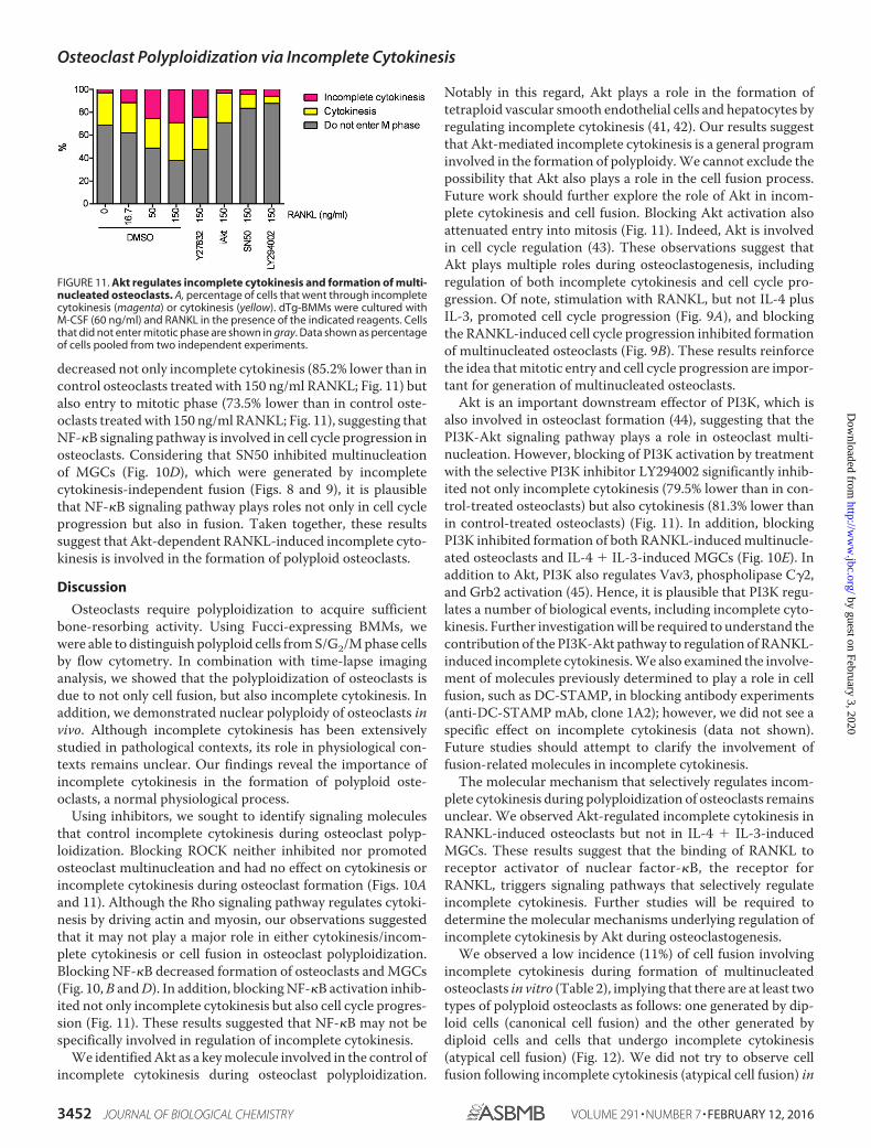
decreased not only incomplete cytokinesis (85.2% lower than incontrol osteoclasts treated with 150 ng/ml RANKL; Fig. 11) butalso entry to mitotic phase (73.5% lower than in control oste-oclasts treated with 150 ng/ml RANKL; Fig. 11), suggesting thatNF-�B signaling pathway is involved in cell cycle progression inosteoclasts. Considering that SN50 inhibited multinucleationof MGCs (Fig. 10D), which were generated by incompletecytokinesis-independent fusion (Figs. 8 and 9), it is plausiblethat NF-�B signaling pathway plays roles not only in cell cycleprogression but also in fusion. Taken together, these resultssuggest that Akt-dependent RANKL-induced incomplete cyto-kinesis is involved in the formation of polyploid osteoclasts.
Discussion
Osteoclasts require polyploidization to acquire sufficientbone-resorbing activity. Using Fucci-expressing BMMs, wewere able to distinguish polyploid cells from S/G2/M phase cellsby flow cytometry. In combination with time-lapse imaginganalysis, we showed that the polyploidization of osteoclasts isdue to not only cell fusion, but also incomplete cytokinesis. Inaddition, we demonstrated nuclear polyploidy of osteoclasts invivo. Although incomplete cytokinesis has been extensivelystudied in pathological contexts, its role in physiological con-texts remains unclear. Our findings reveal the importance ofincomplete cytokinesis in the formation of polyploid oste-oclasts, a normal physiological process.
Using inhibitors, we sought to identify signaling moleculesthat control incomplete cytokinesis during osteoclast polyp-loidization. Blocking ROCK neither inhibited nor promotedosteoclast multinucleation and had no effect on cytokinesis orincomplete cytokinesis during osteoclast formation (Figs. 10Aand 11). Although the Rho signaling pathway regulates cytoki-nesis by driving actin and myosin, our observations suggestedthat it may not play a major role in either cytokinesis/incom-plete cytokinesis or cell fusion in osteoclast polyploidization.Blocking NF-�B decreased formation of osteoclasts and MGCs(Fig. 10, B and D). In addition, blocking NF-�B activation inhib-ited not only incomplete cytokinesis but also cell cycle progres-sion (Fig. 11). These results suggested that NF-�B may not bespecifically involved in regulation of incomplete cytokinesis.
We identified Akt as a key molecule involved in the control ofincomplete cytokinesis during osteoclast polyploidization.
Notably in this regard, Akt plays a role in the formation oftetraploid vascular smooth endothelial cells and hepatocytes byregulating incomplete cytokinesis (41, 42). Our results suggestthat Akt-mediated incomplete cytokinesis is a general programinvolved in the formation of polyploidy. We cannot exclude thepossibility that Akt also plays a role in the cell fusion process.Future work should further explore the role of Akt in incom-plete cytokinesis and cell fusion. Blocking Akt activation alsoattenuated entry into mitosis (Fig. 11). Indeed, Akt is involvedin cell cycle regulation (43). These observations suggest thatAkt plays multiple roles during osteoclastogenesis, includingregulation of both incomplete cytokinesis and cell cycle pro-gression. Of note, stimulation with RANKL, but not IL-4 plusIL-3, promoted cell cycle progression (Fig. 9A), and blockingthe RANKL-induced cell cycle progression inhibited formationof multinucleated osteoclasts (Fig. 9B). These results reinforcethe idea that mitotic entry and cell cycle progression are impor-tant for generation of multinucleated osteoclasts.
Akt is an important downstream effector of PI3K, which isalso involved in osteoclast formation (44), suggesting that thePI3K-Akt signaling pathway plays a role in osteoclast multi-nucleation. However, blocking of PI3K activation by treatmentwith the selective PI3K inhibitor LY294002 significantly inhib-ited not only incomplete cytokinesis (79.5% lower than in con-trol-treated osteoclasts) but also cytokinesis (81.3% lower thanin control-treated osteoclasts) (Fig. 11). In addition, blockingPI3K inhibited formation of both RANKL-induced multinucle-ated osteoclasts and IL-4 � IL-3-induced MGCs (Fig. 10E). Inaddition to Akt, PI3K also regulates Vav3, phospholipase C�2,and Grb2 activation (45). Hence, it is plausible that PI3K regu-lates a number of biological events, including incomplete cyto-kinesis. Further investigation will be required to understand thecontribution of the PI3K-Akt pathway to regulation of RANKL-induced incomplete cytokinesis. We also examined the involve-ment of molecules previously determined to play a role in cellfusion, such as DC-STAMP, in blocking antibody experiments(anti-DC-STAMP mAb, clone 1A2); however, we did not see aspecific effect on incomplete cytokinesis (data not shown).Future studies should attempt to clarify the involvement offusion-related molecules in incomplete cytokinesis.
The molecular mechanism that selectively regulates incom-plete cytokinesis during polyploidization of osteoclasts remainsunclear. We observed Akt-regulated incomplete cytokinesis inRANKL-induced osteoclasts but not in IL-4 � IL-3-inducedMGCs. These results suggest that the binding of RANKL toreceptor activator of nuclear factor-�B, the receptor forRANKL, triggers signaling pathways that selectively regulateincomplete cytokinesis. Further studies will be required todetermine the molecular mechanisms underlying regulation ofincomplete cytokinesis by Akt during osteoclastogenesis.
We observed a low incidence (11%) of cell fusion involvingincomplete cytokinesis during formation of multinucleatedosteoclasts in vitro (Table 2), implying that there are at least twotypes of polyploid osteoclasts as follows: one generated by dip-loid cells (canonical cell fusion) and the other generated bydiploid cells and cells that undergo incomplete cytokinesis(atypical cell fusion) (Fig. 12). We did not try to observe cellfusion following incomplete cytokinesis (atypical cell fusion) in
FIGURE 11. Akt regulates incomplete cytokinesis and formation of multi-nucleated osteoclasts. A, percentage of cells that went through incompletecytokinesis (magenta) or cytokinesis (yellow). dTg-BMMs were cultured withM-CSF (60 ng/ml) and RANKL in the presence of the indicated reagents. Cellsthat did not enter mitotic phase are shown in gray. Data shown as percentageof cells pooled from two independent experiments.
Osteoclast Polyploidization via Incomplete Cytokinesis
3452 JOURNAL OF BIOLOGICAL CHEMISTRY VOLUME 291 • NUMBER 7 • FEBRUARY 12, 2016
by guest on February 3, 2020http://w
ww
.jbc.org/D
ownloaded from

vivo because the phenomenon takes more than a day, surpass-ing the current time limit for intravital imaging. Instead, weused fluorescence in situ hybridization to reveal that osteoclastshave nuclei with more than the diploid complement of chro-mosomes (�2N). In addition, we found circulating osteoclastprecursors (Ly6C� cells) that were G1 � 4C. These results sug-gested that cells that increase ploidy via cell cycle-dependentmechanisms are involved in the formation of multinucleatedosteoclasts in vivo. However, we cannot exclude the possibili-ties that the nuclear polyploidy might also be caused by nuclearfusion, and Ly6C� G1 � 4C cells were merely fusion products.Further studies will be required to clarify this issue.
The proportion of multinucleated osteoclasts generated byatypical cell fusion in vivo, as well as the functional differencesbetween osteoclasts generated by canonical cell fusion andosteoclasts generated by atypical cell fusion, still remainsunclear. FITC gelatin resorption assay showed that the cellsthat underwent incomplete cytokinesis and re-entered S phase(green� binucleated cells) barely degraded FITC gelatin (Fig.6E). These observations suggested that osteoclasts generated byatypical cell fusion might have relatively lower resorption activ-ity than that by canonical cell fusion. However, it cannot beexcluded that the difference of the resorption activity betweencells in G1 phase and cells in S phase merely reflected the dif-ference of their cell cycle phase. To address these issues, it willbe necessary to identify specific markers expressed on cells thatundergo incomplete cytokinesis.
What is the physiological significance of polyploid nucleiwithin a cell? This phenomenon may be just one aspect of thephenotype of terminally differentiated cells, or a consequenceof stress response that preserves cell function. Alternatively,polyploid nuclei may create genetic diversity, which could pro-mote better adaptation to chronic injury or stress (2, 46, 47).Very little is known regarding the physiological function of thepolyploid state, largely due to technical limitations (e.g. therecurrently exist no methods for converting a tissue composed of
polyploid cells into a tissue of the same size composed of diploidcells). A full understanding of the mechanisms of polyploidiza-tion is necessarily to reveal the physiological significance ofosteoclast polyploidization via cell fusion and incompletecytokinesis.
Author Contributions—N. T. and Y. C. designed the study and wrotethe paper. N. T., H. K., and H. M. performed and analyzed the exper-iments. A. S., A. M., M. T., O. K., and M. I. provided reagents anddata analysis. All authors reviewed the results and approved the finalversion of the manuscript.
Acknowledgments—We thank Dr. Matt Walsh for critical discussionand reading the manuscript. We also thank to the Cell and Develop-mental Biology Microscopy Core, Perelman School of Medicine, Uni-versity of Pennsylvania, for technical assistance with microscopy.
References1. Otto, S. P. (2007) The evolutionary consequences of polyploidy. Cell 131,
452– 4622. Pandit, S. K., Westendorp, B., and de Bruin, A. (2013) Physiological signif-
icance of polyploidization in mammalian cells. Trends Cell Biol. 23,556 –566
3. Sakaue-Sawano, A., Hoshida, T., Yo, M., Takahashi, R., Ohtawa, K., Arai,T., Takahashi, E., Noda, S., Miyoshi, H., and Miyawaki, A. (2013) Visual-izing developmentally programmed endoreplication in mammals usingubiquitin oscillators. Development 140, 4624 – 4632
4. Edgar, B. A., Zielke, N., and Gutierrez, C. (2014) Endocycles: a recurrentevolutionary innovation for post-mitotic cell growth. Nat. Rev. Mol. CellBiol. 15, 197–210
5. Lee, H. O., Davidson, J. M., and Duronio, R. J. (2009) Endoreplication:polyploidy with purpose. Genes Dev. 23, 2461–2477
6. Machlus, K. R., and Italiano, J. E., Jr. (2013) The incredible journey: frommegakaryocyte development to platelet formation. J. Cell Biol. 201,785–796
7. Gentric, G., Desdouets, C., and Celton-Morizur, S. (2012) Hepatocytespolyploidization and cell cycle control in liver physiopathology. Int.J. Hepatol. 2012, 282430
8. Gentric, G., Celton-Morizur, S., and Desdouets, C. (2012) Polyploidy and
FIGURE 12. Schematic model depicting the process of osteoclast polyploidization. RANKL stimulation induces a transient increase in cell cycle activity,leading to cell fusion (canonical cell fusion). Along with the transient increase in cell cycle progression, some cells undergo incomplete cytokinesis. Theresultant cells have the potential to undergo cell fusion and are involved in the formation of polyploid osteoclasts (atypical cell fusion).
Osteoclast Polyploidization via Incomplete Cytokinesis
FEBRUARY 12, 2016 • VOLUME 291 • NUMBER 7 JOURNAL OF BIOLOGICAL CHEMISTRY 3453
by guest on February 3, 2020http://w
ww
.jbc.org/D
ownloaded from

liver proliferation. Clin. Res. Hepatol. Gastroenterol. 36, 29 –349. Aguilar, P. S., Baylies, M. K., Fleissner, A., Helming, L., Inoue, N., Podbile-
wicz, B., Wang, H., and Wong, M. (2013) Genetic basis of cell-cell fusionmechanisms. Trends Genet. 29, 427– 437
10. Srinivas, B. P., Woo, J., Leong, W. Y., and Roy, S. (2007) A conservedmolecular pathway mediates myoblast fusion in insects and vertebrates.Nat. Genet. 39, 781–786
11. Teitelbaum, S. L. (2000) Bone resorption by osteoclasts. Science 289,1504 –1508
12. Walsh, M. C., Kim, N., Kadono, Y., Rho, J., Lee, S. Y., Lorenzo, J., and Choi,Y. (2006) Osteoimmunology: interplay between the immune system andbone metabolism. Annu. Rev. Immunol. 24, 33– 63
13. Yagi, M., Miyamoto, T., Sawatani, Y., Iwamoto, K., Hosogane, N., Fujita, N.,Morita, K., Ninomiya, K., Suzuki, T., Miyamoto, K., Oike, Y., Takeya, M.,Toyama, Y., and Suda, T. (2005) DC-STAMP is essential for cell-cell fusion inosteoclasts and foreign body giant cells. J. Exp. Med. 202, 345–351
14. Oren-Suissa, M., and Podbilewicz, B. (2007) Cell fusion during develop-ment. Trends Cell Biol. 17, 537–546
15. Mizoguchi, T., Muto, A., Udagawa, N., Arai, A., Yamashita, T., Hosoya, A.,Ninomiya, T., Nakamura, H., Yamamoto, Y., Kinugawa, S., Nakamura, M.,Nakamichi, Y., Kobayashi, Y., Nagasawa, S., Oda, K., et al. (2009) Identifi-cation of cell cycle-arrested quiescent osteoclast precursors in vivo. J. CellBiol. 184, 541–554
16. Meiyanto, E., Hoshijima, M., Ogawa, T., Ishida, N., and Takeya, T. (2001)Osteoclast differentiation factor modulates cell cycle machinery andcauses a delay in S phase progression in RAW264 cells. Biochem. Biophys.Res. Commun. 282, 278 –283
17. Sankar, U., Patel, K., Rosol, T. J., and Ostrowski, M. C. (2004) RANKLcoordinates cell cycle withdrawal and differentiation in osteoclaststhrough the cyclin-dependent kinase inhibitors p27KIP1 and p21CIP1.J. Bone Miner. Res. 19, 1339 –1348
18. Kim, N. S., Kim, H. J., Koo, B. K., Kwon, M. C., Kim, Y. W., Cho, Y., Yokota,Y., Penninger, J. M., and Kong, Y. Y. (2006) Receptor activator of NF-�Bligand regulates the proliferation of mammary epithelial cells via Id2. Mol.Cell. Biol. 26, 1002–1013
19. Aiba, Y., Kometani, K., Hamadate, M., Moriyama, S., Sakaue-Sawano, A.,Tomura, M., Luche, H., Fehling, H. J., Casellas, R., Kanagawa, O., Mi-yawaki, A., and Kurosaki, T. (2010) Preferential localization of IgG mem-ory B cells adjacent to contracted germinal centers. Proc. Natl. Acad. Sci.U.S.A. 107, 12192–12197
20. Tomura, M., Sakaue-Sawano, A., Mori, Y., Takase-Utsugi, M., Hata, A.,Ohtawa, K., Kanagawa, O., and Miyawaki, A. (2013) Contrasting quiescentG0 phase with mitotic cell cycling in the mouse immune system. PLOSONE 8, e73801
21. Sakaue-Sawano, A., Kurokawa, H., Morimura, T., Hanyu, A., Hama, H.,Osawa, H., Kashiwagi, S., Fukami, K., Miyata, T., Miyoshi, H., Imamura,T., Ogawa, M., Masai, H., and Miyawaki, A. (2008) Visualizing spatiotem-poral dynamics of multicellular cell cycle progression. Cell 132, 487– 498
22. Abe, T., Sakaue-Sawano, A., Kiyonari, H., Shioi, G., Inoue, K., Horiuchi, T.,Nakao, K., Miyawaki, A., Aizawa, S., and Fujimori, T. (2013) Visualizationof cell cycle in mouse embryos with Fucci2 reporter directed by Rosa26promoter. Development 140, 237–246
23. Kikuta, J., Wada, Y., Kowada, T., Wang, Z., Sun-Wada, G. H., Nishiyama,I., Mizukami, S., Maiya, N., Yasuda, H., Kumanogoh, A., Kikuchi, K., Ger-main, R. N., and Ishii, M. (2013) Dynamic visualization of RANKL andTh17-mediated osteoclast function. J. Clin. Invest. 123, 866 – 873
24. Yamaguchi, H., Yoshida, S., Muroi, E., Yoshida, N., Kawamura, M., Kou-chi, Z., Nakamura, Y., Sakai, R., and Fukami, K. (2011) Phosphoinositide3-kinase signaling pathway mediated by p110� regulates invadopodia for-mation. J. Cell Biol. 193, 1275–1288
25. Storchova, Z., and Pellman, D. (2004) From polyploidy to aneuploidy,genome instability and cancer. Nat. Rev. Mol. Cell Biol. 5, 45–54
26. Comai, L. (2005) The advantages and disadvantages of being polyploid.Nat. Rev. Genet. 6, 836 – 846
27. Teitelbaum, S. L., and Ross, F. P. (2003) Genetic regulation of osteoclastdevelopment and function. Nat. Rev. Genet. 4, 638 – 649
28. Takahashi, N., Udagawa, N., Tanaka, S., Murakami, H., Owan, I., Tamura,T., and Suda, T. (1994) Postmitotic osteoclast precursors are mononuclearcells which express macrophage-associated phenotypes. Dev. Biol. 163,212–221
29. Scimeca, J. C., Franchi, A., Trojani, C., Parrinello, H., Grosgeorge, J., Rob-ert, C., Jaillon, O., Poirier, C., Gaudray, P., and Carle, G. F. (2000) The geneencoding the mouse homologue of the human osteoclast-specific 116-kDaV-ATPase subunit bears a deletion in osteosclerotic (oc/oc) mutants.Bone 26, 207–213
30. Jacome-Galarza, C. E., Lee, S. K., Lorenzo, J. A., and Aguila, H. L. (2013)Identification, characterization, and isolation of a common progenitor forosteoclasts, macrophages, and dendritic cells from murine bone marrowand periphery. J. Bone Miner. Res. 28, 1203–1213
31. Ishii, M., Egen, J. G., Klauschen, F., Meier-Schellersheim, M., Saeki, Y.,Vacher, J., Proia, R. L., and Germain, R. N. (2009) Sphingosine-1-phos-phate mobilizes osteoclast precursors and regulates bone homeostasis.Nature 458, 524 –528
32. Brodbeck, W. G., and Anderson, J. M. (2009) Giant cell formation andfunction. Curr. Opin. Hematol. 16, 53–57
33. Vignery, A. (2005) Macrophage fusion: are somatic and cancer cells pos-sible partners? Trends Cell Biol. 15, 188 –193
34. Miyamoto, H., Suzuki, T., Miyauchi, Y., Iwasaki, R., Kobayashi, T., Sato, Y.,Miyamoto, K., Hoshi, H., Hashimoto, K., Yoshida, S., Hao, W., Mori, T.,Kanagawa, H., Katsuyama, E., Fujie, A., et al. (2012) Osteoclast stimulatorytransmembrane protein and dendritic cell-specific transmembrane pro-tein cooperatively modulate cell-cell fusion to form osteoclasts and for-eign body giant cells. J. Bone Miner. Res. 27, 1289 –1297
35. Lee, S. H., Rho, J., Jeong, D., Sul, J. Y., Kim, T., Kim, N., Kang, J. S., Miyamoto,T., Suda, T., Lee, S. K., Pignolo, R. J., Koczon-Jaremko, B., Lorenzo, J., andChoi, Y. (2006) v-ATPase V0 subunit d2-deficient mice exhibit impaired os-teoclast fusion and increased bone formation. Nat. Med. 12, 1403–1409
36. Glotzer, M. (2005) The molecular requirements for cytokinesis. Science307, 1735–1739
37. Etienne-Manneville, S., and Hall, A. (2002) Rho GTPases in cell biology.Nature 420, 629 – 635
38. Moon, J. B., Kim, J. H., Kim, K., Youn, B. U., Ko, A., Lee, S. Y., and Kim, N.(2012) Akt induces osteoclast differentiation through regulating theGSK3beta/NFATc1 signaling cascade. J. Immunol. 188, 163–169
39. Greenblatt, M. B., Park, K. H., Oh, H., Kim, J. M., Shin, D. Y., Lee, J. M., Lee,J. W., Singh, A., Lee, K. Y., Hu, D., Xiao, C., Charles, J. F., Penninger, J. M.,Lotinun, S., Baron, R., et al. (2015) CHMP5 controls bone turnover ratesby dampening NF-�B activity in osteoclasts. J. Exp. Med. 212, 1283–1301
40. Abu-Amer, Y., Darwech, I., and Otero, J. (2008) Role of the NF-�B axis inimmune modulation of osteoclasts and bone loss. Autoimmunity 41,204 –211
41. Celton-Morizur, S., Merlen, G., Couton, D., Margall-Ducos, G., and Des-douets, C. (2009) The insulin/Akt pathway controls a specific cell divisionprogram that leads to generation of binucleated tetraploid liver cells inrodents. J. Clin. Invest. 119, 1880 –1887
42. Hixon, M. L., Muro-Cacho, C., Wagner, M. W., Obejero-Paz, C., Millie, E.,Fujio, Y., Kureishi, Y., Hassold, T., Walsh, K., and Gualberto, A. (2000)Akt1/PKB upregulation leads to vascular smooth muscle cell hypertrophyand polyploidization. J. Clin. Invest. 106, 1011–1020
43. Gonzalez, E., and McGraw, T. E. (2009) The Akt kinases: isoform speci-ficity in metabolism and cancer. Cell Cycle 8, 2502–2508
44. Oikawa, T., Oyama, M., Kozuka-Hata, H., Uehara, S., Udagawa, N., Saya, H.,and Matsuo, K. (2012) Tks5-dependent formation of circumferential podo-somes/invadopodia mediates cell-cell fusion. J. Cell Biol. 197, 553–568
45. Peng, Q., Malhotra, S., Torchia, J. A., Kerr, W. G., Coggeshall, K. M., andHumphrey, M. B. (2010) TREM2- and DAP12-dependent activation ofPI3K requires DAP10 and is inhibited by SHIP1. Sci. Signal. 3, ra38
46. Leevers, S. J., and McNeill, H. (2005) Controlling the size of organs andorganisms. Curr. Opin. Cell Biol. 17, 604 – 609
47. Fox, D. T., and Duronio, R. J. (2013) Endoreplication and polyploidy: in-sights into development and disease. Development 140, 3–12
Osteoclast Polyploidization via Incomplete Cytokinesis
3454 JOURNAL OF BIOLOGICAL CHEMISTRY VOLUME 291 • NUMBER 7 • FEBRUARY 12, 2016
by guest on February 3, 2020http://w
ww
.jbc.org/D
ownloaded from

Miyawaki, Michio Tomura, Osami Kanagawa, Masaru Ishii and Yongwon ChoiNoriko Takegahara, Hyunsoo Kim, Hiroki Mizuno, Asako Sakaue-Sawano, Atsushi
(RANKL)-induced Incomplete Cytokinesis in the Polyploidization of OsteoclastsB LigandκInvolvement of Receptor Activator of Nuclear Factor-
doi: 10.1074/jbc.M115.677427 originally published online December 15, 20152016, 291:3439-3454.J. Biol. Chem.
10.1074/jbc.M115.677427Access the most updated version of this article at doi:
Alerts:
When a correction for this article is posted•
When this article is cited•
to choose from all of JBC's e-mail alertsClick here
Supplemental material:
http://www.jbc.org/content/suppl/2015/12/15/M115.677427.DC1
http://www.jbc.org/content/291/7/3439.full.html#ref-list-1
This article cites 47 references, 16 of which can be accessed free at
by guest on February 3, 2020http://w
ww
.jbc.org/D
ownloaded from