introduction to the medicinal chemistry of schizophrenia · introduction to the medicinal chemistry...
TRANSCRIPT
Introduction to the Medicinal Chemistry of Schizophrenia
1
Introduction to the Medicinal Chemistry
of Schizophrenia
1.1 Schizophrenia
Schizophrenia is one of the most common psychiatric disorders and approximately 1 % of the
worlds population suffer from severe symptoms occupying more than half of the beds in psychiatric
clinics. Schizophrenia is distributed over the whole population independent of sex, location, social
class or color of the skin. A schizophrenic patient is frequently described as a person with a “Dr
Jekyll and Mr. Hyde” personality, but the diagnosis of schizophrenia is more complex than that.
Generally, the symptoms are divided into two classes: positive (reality distortion) and negative
symptoms (psycho-motor poverty syndrome).1 Each patient is different and could suffer from more
or less of positive or negative symptoms. The positive symptoms, e.g., delusions, hallucinations,
grandiosity, excitement, hostility and disorganization are more easily identified, as compared to the
negative symptoms. Examples of negative symptoms are apathy, attentional impairment, affective
blunting, asociality, poverty of speech and anhedonis that may be difficult to distinguish from either
depression or side effects caused by medication with typical antipsychotic drugs.2,3
Approximately half of the schizophrenia patients will experience periods with severe depression
during the course of their illness. Consequently, the detection of depressive symptoms4 is very
important, since, about 7 %–10 % of all schizophrenia patients commit suicide.5
+ Treatment of Schizophrenia
The diagnosis of schizophrenia is just as complex as the medication to suppress the symptoms.
There is no real cure against schizophrenia and most patients are bound to medication for the rest of
their lifes. The drug of choice is more often a trade-off between clinical efficacy and EPS (Extra
Pyramidal Syndrome) or other side-effects. EPS are the side-effects, e.g., major movement disorders
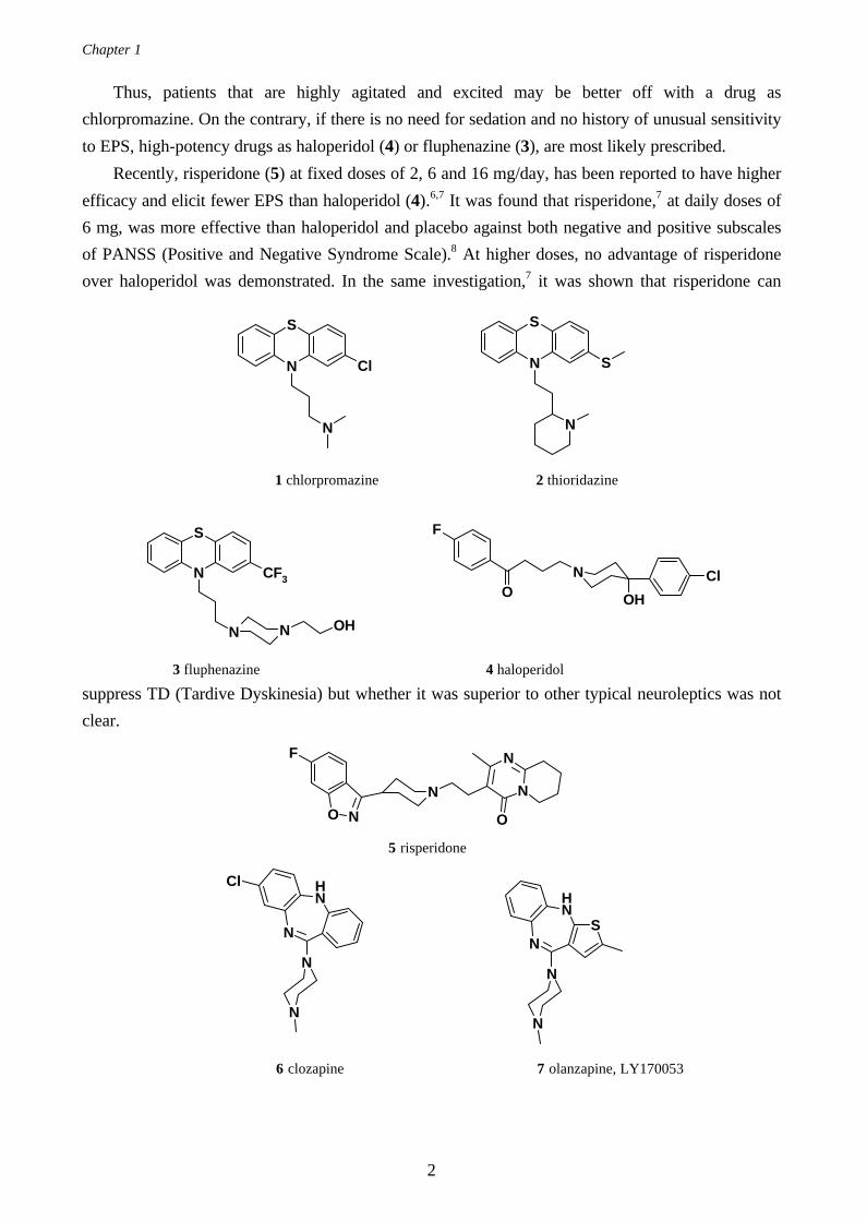
elicited by typical antipsychotic drugs. In general, low-potency drugs, e.g., chlorpromazine (1) or
thioridazine (2), are more sedative and hypotensive than high-potency drugs, e.g., fluphenazine (3)
and haloperidol (4) which, in turn, produce more EPS than low-potency agents.
1
Chapter 1
2
Thus, patients that are highly agitated and excited may be better off with a drug as
chlorpromazine. On the contrary, if there is no need for sedation and no history of unusual sensitivity
to EPS, high-potency drugs as haloperidol (4) or fluphenazine (3), are most likely prescribed.
Recently, risperidone (5) at fixed doses of 2, 6 and 16 mg/day, has been reported to have higher
efficacy and elicit fewer EPS than haloperidol (4).6,7 It was found that risperidone,7 at daily doses of
6 mg, was more effective than haloperidol and placebo against both negative and positive subscales
of PANSS (Positive and Negative Syndrome Scale).8 At higher doses, no advantage of risperidone
over haloperidol was demonstrated. In the same investigation,7 it was shown that risperidone can
suppress TD (Tardive Dyskinesia) but whether it was superior to other typical neuroleptics was not
clear.
N
S
Cl
N
N
S
N
S
1 chlorpromazine 2 thioridazine
ON
OH
Cl
F
N N
N
S
OH
CF3
3 fluphenazine 4 haloperidol
N
O N
F N
N
O
5 risperidone
N
N
NH
N
Cl
N
N
NH
NS
6 clozapine 7 olanzapine, LY170053
Introduction to the Medicinal Chemistry of Schizophrenia
3
Just as risperidone (5), clozapine (6) belongs to the new generation of antipsychotics often
classified as atypical. An atypical antipsychotic drug produce, by definition, fewer EPS than typical
antipsychotics and clozapine is the compound used as reference for new antipsychotics. The
advantages of clozapine (6) over classical antipsychotics are manifold: a) it is effective in the
treatment of both positive and negative symptoms9; b) it is more effective in treatment-refractory
patients10,11 and c) it produces fewer EPS.9,12 However, clozapine is also an example of what often is
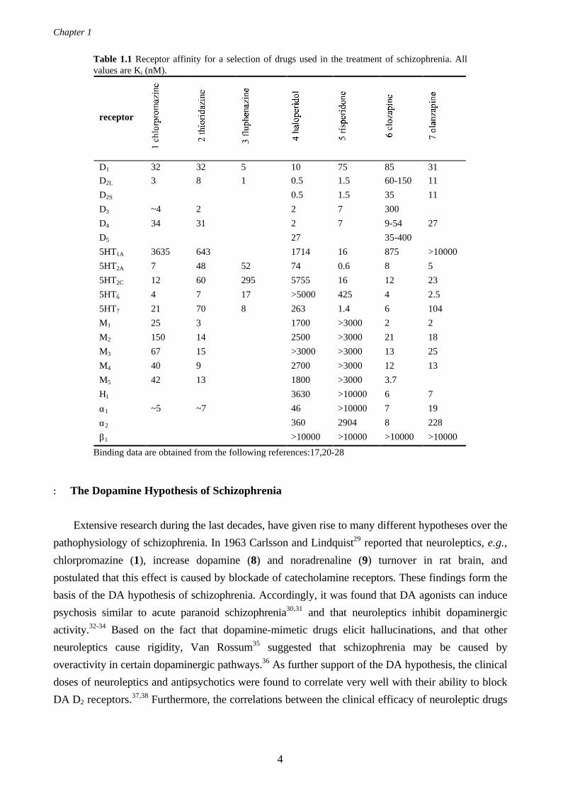
referred to as a “dirty drug” since it has affinity to a large number of different receptors (see Table
1.1). As a consequence, side-effects like hypersalivation (may be due to peripheral drug actions)13
and weight gain14 must be considered. In addition, agranulocytosis,15 a potentially fatal blood
disorder has been observed in patients medicated with clozapine.
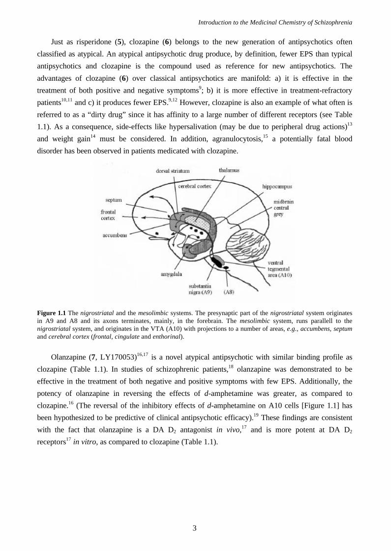
Figure 1.1 The nigrostriatal and the mesolimbic systems. The presynaptic part of the nigrostriatal system originatesin A9 and A8 and its axons terminates, mainly, in the forebrain. The mesolimbic system, runs parallell to thenigrostriatal system, and originates in the VTA (A10) with projections to a number of areas, e.g., accumbens, septumand cerebral cortex (frontal, cingulate and enthorinal).
Olanzapine (7, LY170053)16,17 is a novel atypical antipsychotic with similar binding profile as
clozapine (Table 1.1). In studies of schizophrenic patients,18 olanzapine was demonstrated to be
effective in the treatment of both negative and positive symptoms with few EPS. Additionally, the
potency of olanzapine in reversing the effects of d-amphetamine was greater, as compared to
clozapine.16 (The reversal of the inhibitory effects of d-amphetamine on A10 cells [Figure 1.1] has
been hypothesized to be predictive of clinical antipsychotic efficacy).19 These findings are consistent
with the fact that olanzapine is a DA D2 antagonist in vivo,17 and is more potent at DA D2
receptors17 in vitro, as compared to clozapine (Table 1.1).
Chapter 1
4
Table 1.1 Receptor affinity for a selection of drugs used in the treatment of schizophrenia. Allvalues are Ki (nM).
receptor
D1 32 32 5 10 75 85 31
D2L 3 8 1 0.5 1.5 60-150 11
D2S 0.5 1.5 35 11
D3 ~4 2 2 7 300
D4 34 31 2 7 9-54 27
D5 27 35-400
5HT1A 3635 643 1714 16 875 >10000
5HT2A 7 48 52 74 0.6 8 5
5HT2C 12 60 295 5755 16 12 23
5HT6 4 7 17 >5000 425 4 2.5
5HT7 21 70 8 263 1.4 6 104
M1 25 3 1700 >3000 2 2
M2 150 14 2500 >3000 21 18
M3 67 15 >3000 >3000 13 25
M4 40 9 2700 >3000 12 13
M5 42 13 1800 >3000 3.7
H1 3630 >10000 6 7
α1 ~5 ~7 46 >10000 7 19
α2 360 2904 8 228
β1 >10000 >10000 >10000 >10000
Binding data are obtained from the following references:17,20-28
+ The Dopamine Hypothesis of Schizophrenia
Extensive research during the last decades, have given rise to many different hypotheses over the
pathophysiology of schizophrenia. In 1963 Carlsson and Lindquist29 reported that neuroleptics, e.g.,
chlorpromazine (1), increase dopamine (8) and noradrenaline (9) turnover in rat brain, and
postulated that this effect is caused by blockade of catecholamine receptors. These findings form the
basis of the DA hypothesis of schizophrenia. Accordingly, it was found that DA agonists can induce
psychosis similar to acute paranoid schizophrenia30,31 and that neuroleptics inhibit dopaminergic
activity.32-34 Based on the fact that dopamine-mimetic drugs elicit hallucinations, and that other
neuroleptics cause rigidity, Van Rossum35 suggested that schizophrenia may be caused by
overactivity in certain dopaminergic pathways.36 As further support of the DA hypothesis, the clinical
doses of neuroleptics and antipsychotics were found to correlate very well with their ability to block
DA D2 receptors.37,38 Furthermore, the correlations between the clinical efficacy of neuroleptic drugs
Introduction to the Medicinal Chemistry of Schizophrenia
5
and the in vitro binding affinity of the muscarinic cholinergic, histaminergic (H1), serotonergic (5-
HT2) and α1 receptors, were poor.39
There are two major dopaminergic neuronal systems that project in the forebrain: the
nigrostriatal (A9) and the mesolimbic (A10) systems (Figure 1.1). Parkinsonism is a consequence of
degeneration of neuronal pathways in the A9 system, and EPS induced by treatment with typical
antipsychotics is caused by blockade of dopamine receptors in the same system. Consequently, it was
postulated that the symptoms of schizophrenia originated from hyperactivity in the mesolimbic
dopaminergic systems (A10).34,40
In agreement with the DA hypothesis is the clinical observation that patients with Parkinson’s
disease do not develop schizophrenia.38
+ Serotonin Hypothesis of Schizophrenia
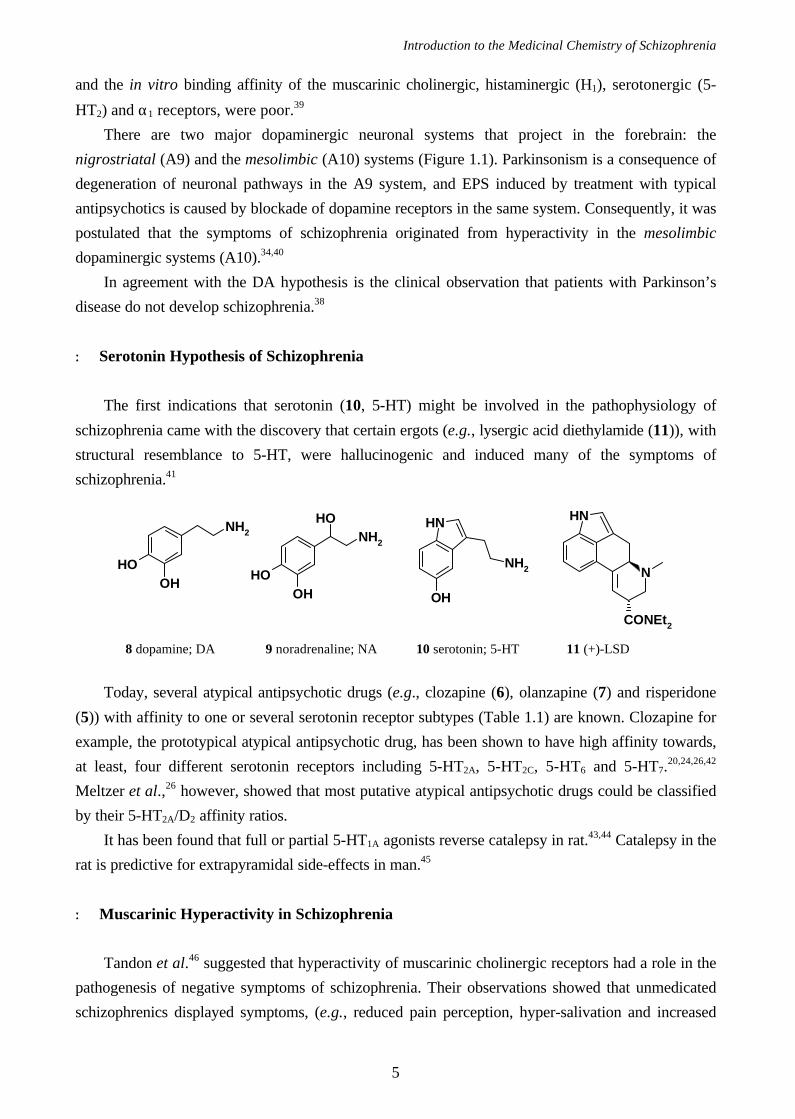
The first indications that serotonin (10, 5-HT) might be involved in the pathophysiology of
schizophrenia came with the discovery that certain ergots (e.g., lysergic acid diethylamide (11)), with
structural resemblance to 5-HT, were hallucinogenic and induced many of the symptoms of
schizophrenia.41
Today, several atypical antipsychotic drugs (e.g., clozapine (6), olanzapine (7) and risperidone
(5)) with affinity to one or several serotonin receptor subtypes (Table 1.1) are known. Clozapine for
example, the prototypical atypical antipsychotic drug, has been shown to have high affinity towards,
at least, four different serotonin receptors including 5-HT2A, 5-HT2C, 5-HT6 and 5-HT7.20,24,26,42
Meltzer et al.,26 however, showed that most putative atypical antipsychotic drugs could be classified
by their 5-HT2A/D2 affinity ratios.
It has been found that full or partial 5-HT1A agonists reverse catalepsy in rat.43,44 Catalepsy in the
rat is predictive for extrapyramidal side-effects in man.45
+ Muscarinic Hyperactivity in Schizophrenia
Tandon et al.46 suggested that hyperactivity of muscarinic cholinergic receptors had a role in the
pathogenesis of negative symptoms of schizophrenia. Their observations showed that unmedicated
schizophrenics displayed symptoms, (e.g., reduced pain perception, hyper-salivation and increased
NH
NH2
OH
NH
N
CONEt2
OH
OHOH
NH2
OHOH
NH2
8 dopamine; DA 9 noradrenaline; NA 10 serotonin; 5-HT 11 (+)-LSD
Chapter 1
6
water intake), which resemble a muscarinic receptor hyperactive state. Occasionally, anti-cholinergic
drugs have been reported effective in treating negative symptoms of schizophrenia.47,48 It was not
clear, however, whether the negative symptoms of schizophrenia or the neuroleptic induced EPS,
were reduced.
Since the atypical antipsychotic drug clozapine (6), has high affinity towards all five muscarinic
receptors (Table 1.1), one may predict that the degree of atypicality is related to its cholinergic
activity. However, Bolden et al.27 could not find any clear pattern in their investigation. Taken
together, according to the investigations of Boldens et al.27 and others,11,49,50 it is not clear whether
anticholinergic activity is essential for an atypical antipsychotic drug or not.
+ The Noradrenaline Receptor
The relationship between noradrenaline (9, NA) and schizophrenia was first studied by Stein et
al.51 in 1971. However, significant and reproducible research established a relationship between NA
levels in the limbic forebrain and the intensity of the schizophrenic symptoms first in 1990.52 Recently
Breier et al.53 demonstrated a direct correlation between the ability of clozapine (6) to elevate plasma
NA levels with its ability to improve positive symptoms of schizophrenia. As yet, no selective drugs
towards the α1, α2 or β receptors with high efficacy in man have been reported.54
1.2 Molecular Biology of Dopamine Receptors
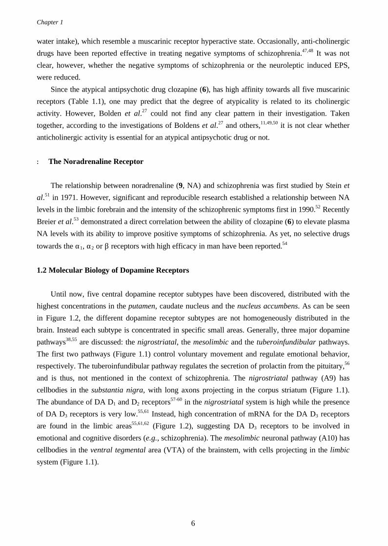
Until now, five central dopamine receptor subtypes have been discovered, distributed with the
highest concentrations in the putamen, caudate nucleus and the nucleus accumbens. As can be seen
in Figure 1.2, the different dopamine receptor subtypes are not homogeneously distributed in the
brain. Instead each subtype is concentrated in specific small areas. Generally, three major dopamine
pathways38,55 are discussed: the nigrostriatal, the mesolimbic and the tuberoinfundibular pathways.
The first two pathways (Figure 1.1) control voluntary movement and regulate emotional behavior,
respectively. The tuberoinfundibular pathway regulates the secretion of prolactin from the pituitary,56
and is thus, not mentioned in the context of schizophrenia. The nigrostriatal pathway (A9) has
cellbodies in the substantia nigra, with long axons projecting in the corpus striatum (Figure 1.1).
The abundance of DA D1 and D2 receptors57-60 in the nigrostriatal system is high while the presence
of DA D3 receptors is very low.55,61 Instead, high concentration of mRNA for the DA D3 receptors
are found in the limbic areas55,61,62 (Figure 1.2), suggesting DA D3 receptors to be involved in
emotional and cognitive disorders (e.g., schizophrenia). The mesolimbic neuronal pathway (A10) has
cellbodies in the ventral tegmental area (VTA) of the brainstem, with cells projecting in the limbic
system (Figure 1.1).
Introduction to the Medicinal Chemistry of Schizophrenia
7
D1 D5 D2 D3 D4
Figure 1.2 The distribution of dopamine receptors in the human brain as determined by the concentrations of mRNAfor the respective receptor subtypes in the different brain areas.
+ G Protein-Coupled Receptors
The dopamine receptors belong to a class of proteins normally referred to as the G protein-
coupled receptor (GPCR) superfamily. To date, no X-ray crystallographic structure of a GPCR is
resolved, but along with molecular cloning and receptor binding studies the amino acid sequence of
all five human DA receptors have been elucidated (Table 1.2). In 1993, Schertler et al.63 provided
evidence that the bovine rhodopsins, G protein-coupled receptors active as the photoreceptors in rod
cells, were arranged in seven α-helices. In 1990, Henderson et al.64 presented a high quality 3D
model of bacteriorhodopsin, also a G protein-coupled receptor, based on cryo-microscopy
experiments. Further refinements of this model have recently been published by Grigorieff et al.65,
Unger et al.66 and Kimura et al.67 The latter group collected structural data from bacteriorhodopsin
crystals at 3.0 Å resolution with 90 % completeness using electron cryo-microscopy. Although the
function of bacteriorhodopsin is different from rhodopsin both proteins bind retinal in a similar way,68
and have similar topology with seven transmembrane helices.
The receptor protein may be folded through the cellular membrane forming seven hydrophobic
trans-membrane α-helices connected, alternately, via intra- and extra-cellular loops. The amino
terminal (N-terminal) and the carboxylic terminal (C-terminal) of the receptor protein reside at the
extra- and the intracellular sides of the cell-membrane, respectively.
The intrinsic activity of a DA agonist is mediated by a signal transduction across the cellular
membrane. That is, the drug-receptor interaction most probably induces a conformational change in
the receptor protein which in turn, activates a G protein coupled to the third intracellular loop.54,69
Accordingly, the activated G protein stimulates (or inhibits) adenylyl cyclase (see Table 1.2) to
Amygdala
HippocampusSubstantianigra
Cerebral cortex
Caudatenucleus
Ventricles
Nucleusaccumbens
Putamen
Island ofCalleja
Olfactorytubercle
Hypothalamus
Chapter 1
8
MAO
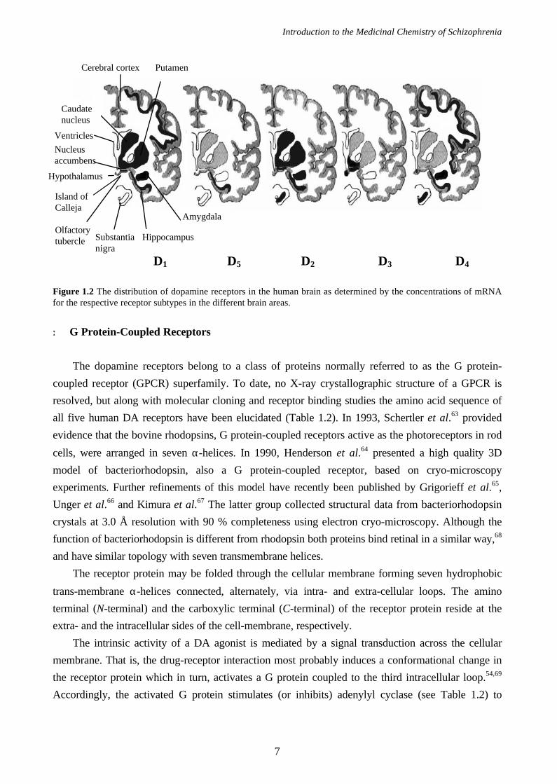
produce cAMP (a so-called second messenger) from AMP (Figure 1.3), which influences various
processes in the cytosol.
Figure 1.3 Schematic representation of a pre and post-synaptic dopaminergic cell.
The third intracellular loop exhibit the largest sequence dissimilarities among the different DA
receptors. The DA D1 and DA D5 receptors have relative short third intracellular loops, are coupled
to Gs proteins and have long C-terminal tails. These two receptors, the D1-like receptors, stimulate
the activity of adenylyl cyclase and the pharmacological functions from known ligands are more or
less, identical.54,61 The D2-like receptors, i.e., D2, D3 and D4 receptors on the other hand, all have
long third intracellular loops with short C-terminal tails, might couple to Gi proteins (or G0 proteins)
and inhibit adenylyl cyclase. Interesting, two forms of the DA D2 receptor have been found, differing
in 29 amino acids in the third intracellular loop,70-72 and seem to have identical pharmacology but
their presence in various cerebral tissues differ.70,73 Hence, a difference in functionality is likely still to
be found. The long and the short forms of the D2 receptor (i.e., D2L and D2S) consist of 443 and 414
amino acid residues (see Table 1.2), respectively. The genes of the dopamine receptor superfamily
can be divided into two different categories: 1) intronless genes that codes for the D1-like receptors
and 2) genes with their coding sequences in discontinuous DNA segments (exons) separated by
sequences (introns) that do not form a part of the mature mRNA. The latter category of genes are
found in the D2-like family of dopamine receptors, which explains the occurrence of a long and a
short form of the DA D2 receptor. In the biosynthesis of mRNA a mechanism called alternative
COMT MAO
(transporter)
Gs
MAO
AC
Dopamine
D2 D3 D4
Tyrosine
Dopa
Dopamine DOPAC
D1 D5
HVA
ATP cAMP
Gs Gi/Go Gi?
Presynaptic neuron
(autoreceptor)
D2, D3 or D4
Postsynaptic neuron
Introduction to the Medicinal Chemistry of Schizophrenia
9
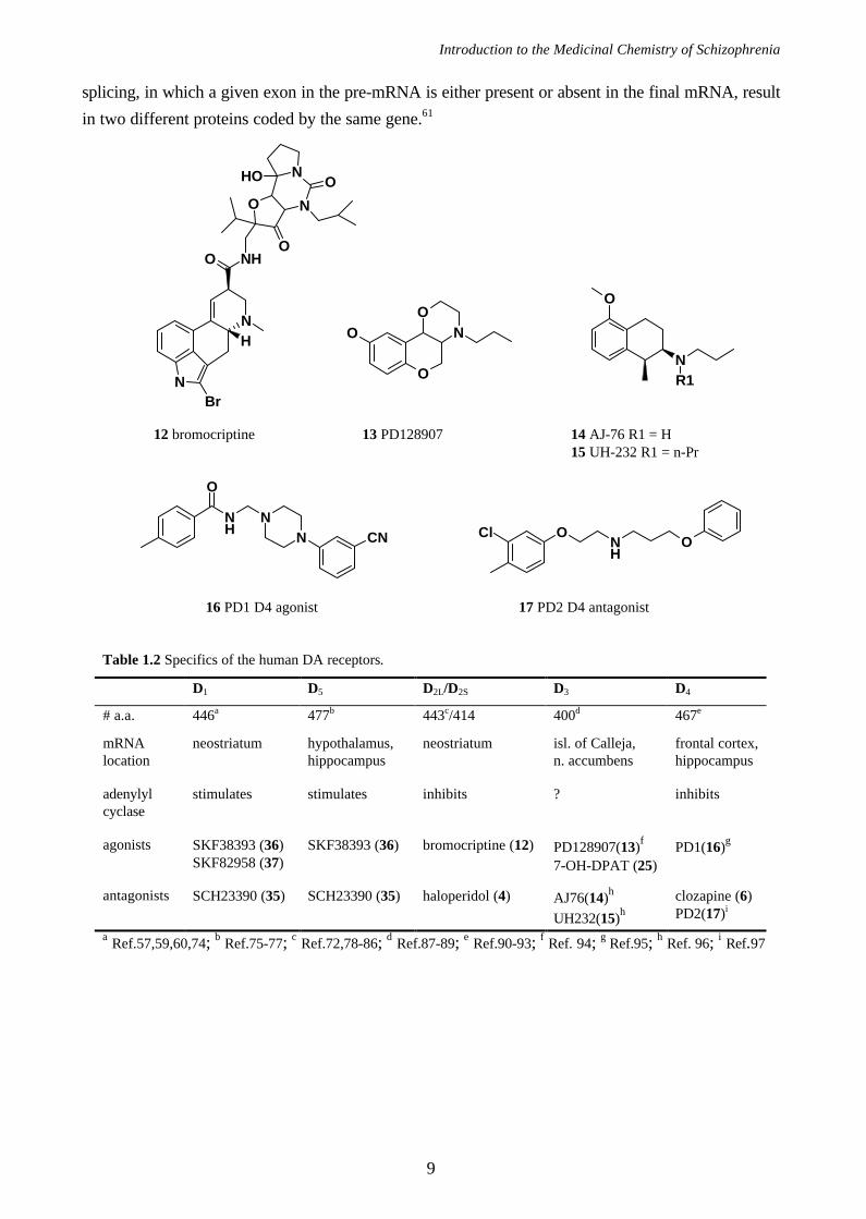
splicing, in which a given exon in the pre-mRNA is either present or absent in the final mRNA, result
in two different proteins coded by the same gene.61
Table 1.2 Specifics of the human DA receptors.
D1 D5 D2L/D2S D3 D4
# a.a. 446a 477b 443c/414 400d 467e
mRNAlocation
neostriatum hypothalamus,hippocampus
neostriatum isl. of Calleja,n. accumbens
frontal cortex,hippocampus
adenylylcyclase
stimulates stimulates inhibits ? inhibits
agonists SKF38393 (36)SKF82958 (37)
SKF38393 (36) bromocriptine (12) PD128907(13)f
7-OH-DPAT (25)PD1(16)g
antagonists SCH23390 (35) SCH23390 (35) haloperidol (4) AJ76(14)h
UH232(15)hclozapine (6)PD2(17)i
a Ref.57,59,60,74; b Ref.75-77; c Ref.72,78-86; d Ref.87-89; e Ref.90-93; f Ref. 94; g Ref.95; h Ref. 96; i Ref.97
O
NO
O
O
NR1
N
NBr
H
NHO
N
N
O
O
OOH
12 bromocriptine 13 PD128907 14 AJ-76 R1 = H15 UH-232 R1 = n-Pr
NH
O
NN CN O
NH
OCl
16 PD1 D4 agonist 17 PD2 D4 antagonist
Chapter 1
10
1.3 Computer-Assisted Molecular Design
The last decade new tools have become available for drug design including computational
chemistry, high-throughput screening98,99 and combinatorial chemistry.100-102 Still, no matter how
advanced our technology have developed or how fast new compounds can be synthesizes and tested,
a medicinal chemist simply has two major questions to answer: Do I understand the structure
activity-relationship for this series of compounds and which compound should I synthesize next? In
order to provide answers to these questions two general work procedures are followed. First, one
may try to build a 3D model, e.g., homology modeling of the target protein and, accordingly, dock
ligands into the active site of the protein. Simply, a potent ligand fits into the receptor while an
inactive ligand does not. The second approach is more basic where ligands, initially, are
superimposed on mutual tentative interaction points (e.g., lone pair of electrons and midpoints of
aromatic rings) with the receptor. Consequently, attempts to explain the potency of the ligands by
comparison of their structures and their relative 3D orientations can be done. This approach is
generally called an “active analogue approach”103 or structure-activity relationship (SAR).
+ Homology Modeling
Until recently, we had no knowledge about the amino acid sequence and less knowledge about
the secondary structure of G protein-coupled receptors (GPCRs). Therefore, computational chemists
have utilized the structure elucidated for bacteriorhodopsin (see above), although the sequence
homology is poor, to create 3D models of GPCRs.92,104-107 Recently, the dopamine D2 receptor was
constructed68 based on the coordinates from bacteriorhodopsin108 itself.
The first problem in GPCR modeling is to determine which amino acid residues that reside in the
transmembrane domains, or stated differently, the alignment of the seven helices. Computer
programs107,109,110 that can perform this kind of alignment, are available. The dopamine receptors
presented in Table 1.3 were downloaded from EMBL in Heidelberg111 and aligned using the ‘whatif’
program.112 The alignments were by no means perfect and refinements were applied manually as
presented in Table 1.3.
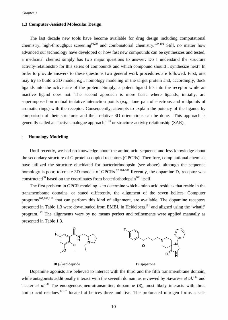
Dopamine agonists are believed to interact with the third and the fifth transmembrane domain,
while antagonists additionally interact with the seventh domain as reviewed by Savarese et al.113 and
Teeter et al.68 The endogenous neurotransmitter, dopamine (8), most likely interacts with three
amino acid residues68,107 located at helices three and five. The protonated nitrogen forms a salt-
NH
O
NO
O
I
NH
NN
O
O
F
18 (S)-epidepride 19 spiperone
Introduction to the Medicinal Chemistry of Schizophrenia
11
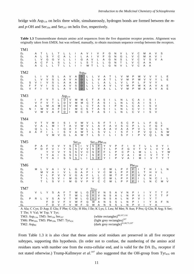
bridge with Asp114 on helix three while, simultaneously, hydrogen bonds are formed between the m-
and p-OH and Ser194 and Ser197 on helix five, respectively.
From Table 1.3 it is also clear that these amino acid residues are preserved in all five receptor
subtypes, supporting this hypothesis. (In order not to confuse, the numbering of the amino acid
residues starts with number one from the extra-cellular end, and is valid for the DA D2L receptor if
not stated otherwise.) Trump-Kallmeyer et al.107 also suggested that the OH-group from Tyr416 on
Table 1.3 Transmembrane domain amino acid sequences from the five dopamine receptor proteins. Alignment wasoriginally taken from EMDL but was refined, manually, to obtain maximum sequence overlap between the receptors.
TM1D2 A T L L T L L I A V I V F G N V L V C M A V SD3 A L S Y C A L I L A I V F G N G L V C M A V ID4 L V G G V L L I G A V L A G N S L V C V S V AD1 A C F L S L L I L S T L L G N T L V C A AD5 A C L L T L L I I W T L L G N V L V C A A
TM2 Asp80
D2 L I V S L A V A D L L V A T L V M P W V V Y L ED3 L V V S L A V A D L L V A T L V M P W V V Y L ED4 S V I V S L A A A D L L L A L L V L P L F V YD1 F F V I S L A V S D L L V A V L V M P W K A V A ED5 V F I V S L A V S D L F V A L L V M P W K A V A E
TM3 Asp114
D2 I F V T L D V M M C T A S I L N L C A I S ID3 V F V T L D V M M C T A S I L N L C A I S ID4 A L M A M D V M L C T A S I F N L C A I S VD1 N I W V A F D I M C S T A S I L N L C V I S VD5 V W V A F D I M C S T A S I L N L C V I S V
TM4D2 V T V M I S I V W V L S F T I S C P L L F G LD3 V A L M I T A V W V L A F A V S C P L L F G FD4 Q L L L I G A T W L L S A A V A A P V L C G L ND1 A A F I L I S V A W T L S V L I S F I P V Q L S WD5 V M V G L A W T L S I L I S F I P V Q L N W
TM5 Ser194 Ser197Phe198
D2 P A F V V Y S S I V S F Y V P F I V T L L V Y ID3 P D F V I Y S S V V S F Y L P F G V T V L V Y AD4 Y V V Y S S V C S F F L P C P L M L L L Y WD1 T Y A I S S S V I S F Y I P V A I M I V T Y TD5 T Y A I S S S L I S F Y I P V A I M I V T Y T
TM6 Phe390
D2 M L V A I V L G V F I I C W L P F F I T H I L ND3 M V A I V L G A F I V C W L P F F L T H V LD4 V L P V V V G A F L L C W T P F F V V H ID1 T L S V I M G V F V C C W L P F F I L N C I LD5 T L S V I M G V F V C C W L P F F I L N C M V
TM7 Tyr416
D2 V L Y S A F T W L G Y V N S A V N P I I Y T T FD3 A T T W L G Y V N S A L N P V I Y T T FD4 P R L V S A V T W L G Y V N S A L N P V I Y TD1 F D V F V W F G W A N S S L N P I I Y A F ND5 F D V F V W F G W A N S S L N P V I Y A
A Ala; C Cys; D Asp; E Glu; F Phe; G Gly; H His; I Ile; K Lys; L Leu; M Met; N Asn; P Pro; Q Gln; R Arg; S Ser;T Thr; V Val; W Trp; Y Tyr;TM3: Asp114, TM5: Ser194 Ser197 (white rectangles)68,107,116
TM6: Phe390, TM5: Phe198, TM7: Tyr416 (light grey rectangles)107
TM2: Asp80 (dark grey rectangle)61,68,73,107
Chapter 1
12
helix seven helps to stabilize the transmitter-receptor complex by interacting with the charged
dopamine nitrogen. Another interesting feature is the narrow aromatic cleft, defined by Phe390 on
helix six and Phe198 on helix five, that may interact with the flat aromatic part of catecholamine
related ligands.107 Finally, site-directed mutagenesis study have shown73,114,115 the importance of
Asp80 in the regulation of D2 affinity for drugs, coupling to adenylate cyclase and sensitivity to Na+
and pH. Sodium decreases the binding of D2 agonists (e.g., dopamine (8))114,115 and increases binding
for some substituted benzamides (e.g., epidepride (18) and sulpiride (40))114,115 but does not affect
binding of other D2 antagonists (e.g., spiperone (19)).68,115 This amino acid residue, Asp80, is
preserved in all five dopamine receptors (see Table 1.3) and Teeter et al.68 called it the ‘sodium site’.
+ Structure-Activity Relationships (SAR)
Although homology modeling is a direct consequence of recent analytical refinements (e.g.,
cloning, crystallization and X-ray techniques) and increased computer efficiency the technique is not,
as yet, able to explain the structure-activity relationship (SAR) for most ligands. Computer models of
receptors cannot, for instance, account for the obvious flexibility of the receptor protein, nor predict
the conformational change of the receptor caused when an agonist binds to the active site. There are,
simply, to many uncertain parameters that we cannot simulate. Therefore, traditional methods where
the activity and inactivity of ligands are explained by superimposition of mutual and possible
interaction points with the receptor, are as valid today as for ten years ago. This approach is,
generally, referred to as “the active analogue approach”,103 upon which many recent computational
methods rely (e.g., CoMFA117). During the last two decades several attempts to construct models
that can explain the SAR of dopamine receptor ligands have been presented. In the following, a
review of the most successful agonist and antagonist models will be given, and the SAR of
dopaminergic compounds will be discussed.
+ The McDermed Dopamine Receptor Concept
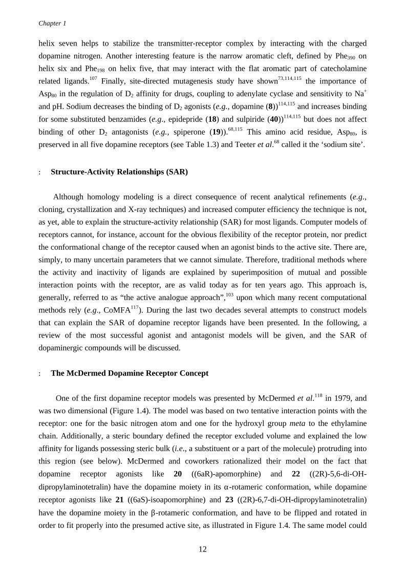
One of the first dopamine receptor models was presented by McDermed et al.118 in 1979, and
was two dimensional (Figure 1.4). The model was based on two tentative interaction points with the
receptor: one for the basic nitrogen atom and one for the hydroxyl group meta to the ethylamine
chain. Additionally, a steric boundary defined the receptor excluded volume and explained the low
affinity for ligands possessing steric bulk (i.e., a substituent or a part of the molecule) protruding into
this region (see below). McDermed and coworkers rationalized their model on the fact that
dopamine receptor agonists like 20 ((6aR)-apomorphine) and 22 ((2R)-5,6-di-OH-
dipropylaminotetralin) have the dopamine moiety in its α-rotameric conformation, while dopamine
receptor agonists like 21 ((6aS)-isoapomorphine) and 23 ((2R)-6,7-di-OH-dipropylaminotetralin)
have the dopamine moiety in the β-rotameric conformation, and have to be flipped and rotated in
order to fit properly into the presumed active site, as illustrated in Figure 1.4. The same model could
Introduction to the Medicinal Chemistry of Schizophrenia
13
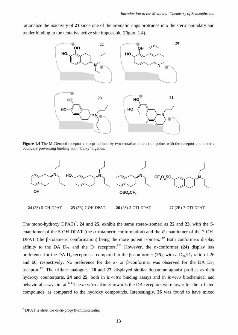
rationalize the inactivity of 21 since one of the aromatic rings protrudes into the steric boundary and
render binding to the tentative active site impossible (Figure 1.4).
OHOH
N
OHOH
N
N
OH
OHN
OH
OH
Figure 1.4 The McDermed receptor concept defined by two tentative interaction points with the receptor and a stericboundary preventing binding with “bulky” ligands.
The mono-hydroxy DPATs∗, 24 and 25, exhibit the same stereo-isomeri as 22 and 23, with the S-
enantiomer of the 5-OH-DPAT (the α-rotameric conformation) and the R-enantiomer of the 7-OH-
DPAT (the β-rotameric conformation) being the more potent isomers.119 Both conformers display
affinity to the DA D2L and the D3 receptors.120 However, the α-conformer (24) display less
preference for the DA D3 receptor as compared to the β-conformer (25), with a D2L/D3 ratio of 26
and 60, respectively. No preference for the α- or β-conformer was observed for the DA D4.2
receptor.120 The triflate analogues, 26 and 27, displayed similar dopamine agonist profiles as their
hydroxy counterparts, 24 and 25, both in in-vitro binding assays and in in-vivo biochemical and
behavioral assays in rat.121 The in vitro affinity towards the DA receptors were lower for the triflated
compounds, as compared to the hydroxy compounds. Interestingly, 26 was found to have mixed
∗ DPAT is short for di-(n-propyl)-aminotetralin.
OH
N NOH
OSO2CF3
N NCF3O2SO
24 (2S)-5-OH-DPAT 25 (2R)-7-OH-DPAT 26 (2S)-5-OTf-DPAT 27 (2R)-7-OTf-DPAT
20
2123
22
Chapter 1
14
DA/5-HT1A properties after oral administration not observed after subcutaneous administration. This
suggest that active metabolite(s) may be formed. (Similar findings were found for 8-OTf-DPAT, a
potent 5-HT1A receptor agonist where the dominating metabolite, the mono-propyl analogue, turned
out to be more potent in vivo than 8-OTf-DPAT itself.121)
NOH
OHNOH
lp
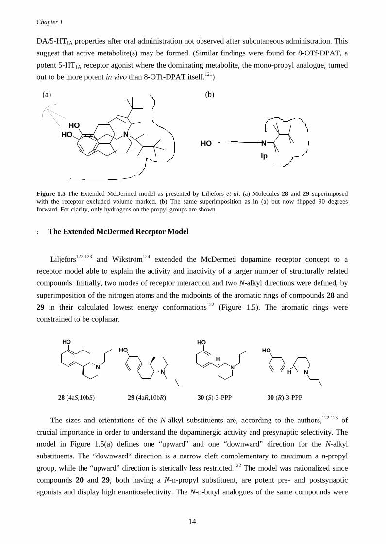
Figure 1.5 The Extended McDermed model as presented by Liljefors et al. (a) Molecules 28 and 29 superimposedwith the receptor excluded volume marked. (b) The same superimposition as in (a) but now flipped 90 degreesforward. For clarity, only hydrogens on the propyl groups are shown.
+ The Extended McDermed Receptor Model
Liljefors122,123 and Wikström124 extended the McDermed dopamine receptor concept to a
receptor model able to explain the activity and inactivity of a larger number of structurally related
compounds. Initially, two modes of receptor interaction and two N-alkyl directions were defined, by
superimposition of the nitrogen atoms and the midpoints of the aromatic rings of compounds 28 and
29 in their calculated lowest energy conformations122 (Figure 1.5). The aromatic rings were
constrained to be coplanar.
The sizes and orientations of the N-alkyl substituents are, according to the authors,122,123 of
crucial importance in order to understand the dopaminergic activity and presynaptic selectivity. The
model in Figure 1.5(a) defines one “upward” and one “downward” direction for the N-alkyl
substituents. The “downward“ direction is a narrow cleft complementary to maximum a n-propyl
group, while the “upward” direction is sterically less restricted.122 The model was rationalized since
compounds 20 and 29, both having a N-n-propyl substituent, are potent pre- and postsynaptic
agonists and display high enantioselectivity. The N-n-butyl analogues of the same compounds were
N
OH
N
OH
N
OH
H
N
OH
H
28 (4aS,10bS) 29 (4aR,10bR) 30 (S)-3-PPP 30 (R)-3-PPP
(a) (b)
Introduction to the Medicinal Chemistry of Schizophrenia
15
found inactive at both pre- and post-synaptic receptors,122,124 most likely due to the fact their N-n-
butyl substituents are too large to fit into the “downward” propyl-cleft. The potency of the N-n-butyl
analogue125 of 28, could be explained since its N-n-butyl protrudes “upwards” in the less sterically
restricted direction when aligned properly into the “active site” (Figure 1.5(a)). In fact, compounds
with as large substituents as phenylethyl or thiopenethyl groups directed “upwards” may still be
active.122,126,127
Liljefors et al.122,123 also investigated the biological active conformations of the enantiomers of
3-PPP, i.e., compounds (S)-30 and (R)-30. Pharmacologically, the (R)-30 enantiomer displays
classical pre- and postsynaptic receptor agonist properties, while the (S)-30 enantiomer is a
presynaptic agonist with postsynaptic antagonistic properties (Table 1.4). In an attempt to explain
the opposed profiles of the enantiomers (R)-30 was fitted in the model with the N-n-propyl directed
in the “downward” propyl-cleft and (S)-30 with the N-n-propyl directed “upwards”. The
superimposition of (R)-30 on (4aR,10bR)-29 exerts an excellent fit explaining the pre- and
postsynaptic properties of (R)-29. The (R)-30 does not fit in its “global energy minimum
conformation” but the N-n-propyl directed in the lipophilic “propyl-cleft” helps to stabilize the
compound in the agonist conformation. Liljefors and coworkers122 found and defined two different
conformations for compound (S)-30 to take: one agonist and one antagonist conformation. The
requirement to activate the postsynaptic receptors seems to be a demand for lipophilicity around the
nitrogen. The (S)-30 has no n-propyl directed into the “propyl-cleft”. Obviously, the methylene
group alfa to the nitrogen in the piperidine ring that is directed into the “propyl-cleft” is not lipophilic
enough to maintain the (S)-30 in a postsynaptic activating conformation. Thus, (S)-30 does not
activate postsynaptic receptors. In
compound (4aR,10bS)-28, however,
the methylene group is maintained in
the “propyl-cleft”, since the
OHB[f]Q skeleton is rigid, and the
postsynaptic receptors can be
activated. The (S)-30 can, as well, assume a low energy conformation that fits into the model in
Figure 1.5 and explain its presynaptic agonist properties.
The model in Figure 1.5(a) cannot explain the inactivity of compounds 31 and 32 since the van
der Waal volume of 30 fits perfectly while the volume from 32 is too large. The orientations of the
propyl groups in Figure 1.5 which can be oriented either in an anti or a gauche conformation with
respect to the nitrogen lone pair of electrons provides an explanation. Liljefors et al.123 concluded
that if one propyl assumes an anti conformation the other one must be gauche and vice versa. If the
“downward” oriented propyl group is oriented in an anti conformation (Figure 1.5(b)) the steric
boundary in front of the nitrogen atom becomes more narrow as compared to in Figure 1.5(a) and
the propyl cleft is located above the plane (Figure 1.5(b)). In this improved model, Figure 1.5(b),
neither of the inactive compounds 31 and 32 fit while the active compounds in Table 1.4 do.
OH
N
OH
N
31 32
Chapter 1
16
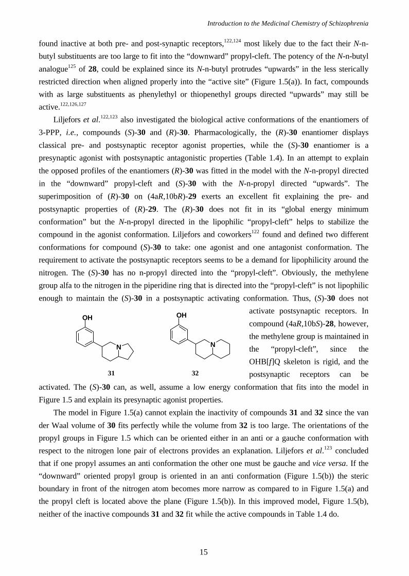
Table 1.4 Intrinsic activity of the ligands discussed
presynaptic agonism
ED50,a nmol/kg
postsynaptic agonism
motor activityb
compound limbic striatum dose µmol/kg sc acc.counts/30 ming
(R)-20c,d 190 220 2.3 361 ± 42
(S)-24d 3.7 3.7 0.31 155 ± 27
(R)-25d 9.5 11 0.31 46 ± 18
(S)-26e 830 1100 12.5 785 ± 123
(R)-27e I I 50 2153 ± 650
(4aS,10bS)-28d 14 14 1.30 62 ± 11
(4aR,10bR)-29c 4 5 1.06 155 ± 32
(S)-30c 800 1700 213 12 ± 2
(R)-30c 1000 1300 13 78 ± 14
33f - - 50 211±63
34f - - 100 290±85a Measured indirectly as inhibition of DA synthesis rate (see ref 124); b Motor activity measured in motility
meters on raserpinized rats (see ref 124); c Data taken from ref 122; d Data taken from ref 124; e Data taken
from ref 128; f Data taken from ref 129; g Values expressed as percentage of saline controls; mean ± SEM.
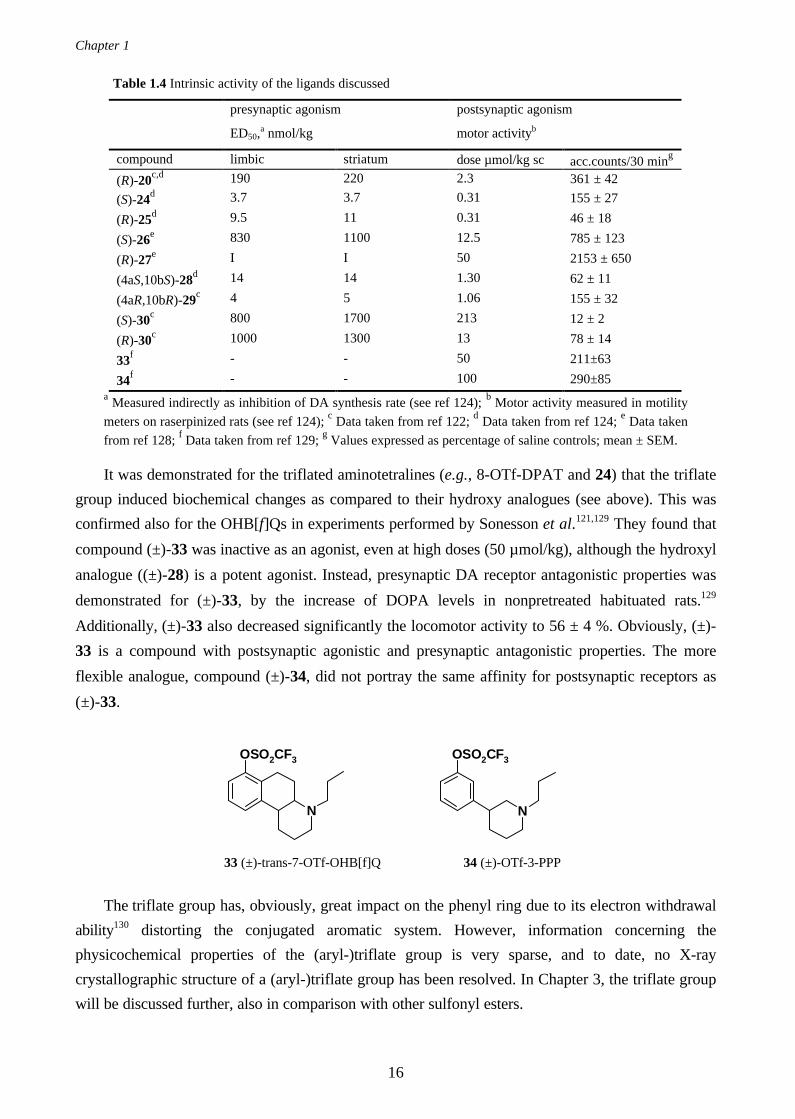
It was demonstrated for the triflated aminotetralines (e.g., 8-OTf-DPAT and 24) that the triflate
group induced biochemical changes as compared to their hydroxy analogues (see above). This was
confirmed also for the OHB[f]Qs in experiments performed by Sonesson et al.121,129 They found that
compound (±)-33 was inactive as an agonist, even at high doses (50 µmol/kg), although the hydroxyl
analogue ((±)-28) is a potent agonist. Instead, presynaptic DA receptor antagonistic properties was
demonstrated for (±)-33, by the increase of DOPA levels in nonpretreated habituated rats.129
Additionally, (±)-33 also decreased significantly the locomotor activity to 56 ± 4 %. Obviously, (±)-
33 is a compound with postsynaptic agonistic and presynaptic antagonistic properties. The more
flexible analogue, compound (±)-34, did not portray the same affinity for postsynaptic receptors as
(±)-33.
The triflate group has, obviously, great impact on the phenyl ring due to its electron withdrawal
ability130 distorting the conjugated aromatic system. However, information concerning the
physicochemical properties of the (aryl-)triflate group is very sparse, and to date, no X-ray
crystallographic structure of a (aryl-)triflate group has been resolved. In Chapter 3, the triflate group
will be discussed further, also in comparison with other sulfonyl esters.
N
OSO2CF3
N
OSO2CF3
33 (±)-trans-7-OTf-OHB[f]Q 34 (±)-OTf-3-PPP
Introduction to the Medicinal Chemistry of Schizophrenia
17
+ Dopamine D1 Agonist and Antagonist Models
The McDermed receptor concept is an example of an early working model of the dopamine
receptor proven useful in the design of new potent ligands. Today, however, with five different
dopamine receptor subtypes known and sophisticated molecular modeling tools available we aim for
models that enable us to explain and understand the structure-activity relationships within as well as
in-between different receptor subtypes.
The SAR of the DA D1 receptor subtype has extensively been scrutinized in the literature during
the last decade.131-133 A challenge has been, and still is, to fully explain the ligand-receptor
interactions of the potent benzazepines. To date, no theory exist that explains why compound 35
(SCH23390) is a selective D1 antagonist while the structurally similar compound, 36 (SKF38393), is
a potent agonist at DA D1 receptors. Compound 35 has a selectivity for the DA D1 over DA D2
receptors with a factor 2093,131 and fully inhibits dopamine stimulated adenylyl cyclase131 (Ki = 0.47
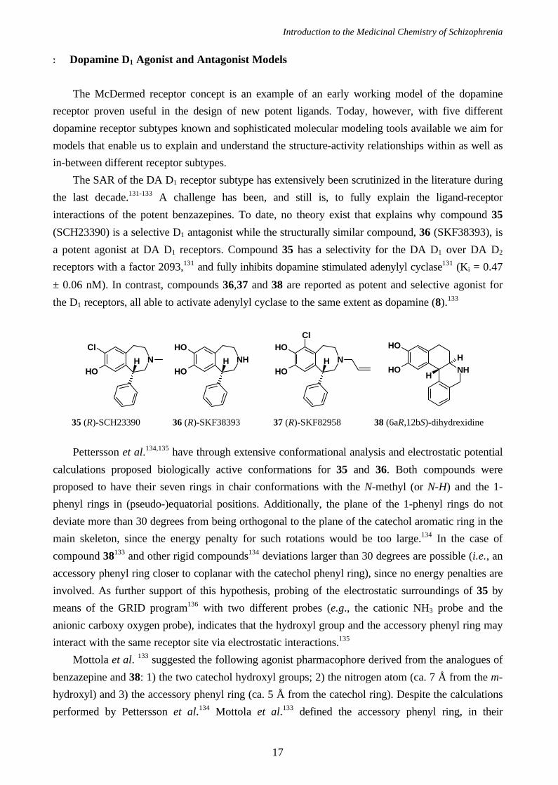
± 0.06 nM). In contrast, compounds 36,37 and 38 are reported as potent and selective agonist for
the D1 receptors, all able to activate adenylyl cyclase to the same extent as dopamine (8).133
Pettersson et al.134,135 have through extensive conformational analysis and electrostatic potential
calculations proposed biologically active conformations for 35 and 36. Both compounds were
proposed to have their seven rings in chair conformations with the N-methyl (or N-H) and the 1-
phenyl rings in (pseudo-)equatorial positions. Additionally, the plane of the 1-phenyl rings do not
deviate more than 30 degrees from being orthogonal to the plane of the catechol aromatic ring in the
main skeleton, since the energy penalty for such rotations would be too large.134 In the case of
compound 38133 and other rigid compounds134 deviations larger than 30 degrees are possible (i.e., an
accessory phenyl ring closer to coplanar with the catechol phenyl ring), since no energy penalties are
involved. As further support of this hypothesis, probing of the electrostatic surroundings of 35 by
means of the GRID program136 with two different probes (e.g., the cationic NH3 probe and the
anionic carboxy oxygen probe), indicates that the hydroxyl group and the accessory phenyl ring may
interact with the same receptor site via electrostatic interactions.135
Mottola et al. 133 suggested the following agonist pharmacophore derived from the analogues of
benzazepine and 38: 1) the two catechol hydroxyl groups; 2) the nitrogen atom (ca. 7 Å from the m-
hydroxyl) and 3) the accessory phenyl ring (ca. 5 Å from the catechol ring). Despite the calculations
performed by Pettersson et al.134 Mottola et al.133 defined the accessory phenyl ring, in their
NHOH
OHHN
Cl
OHH N
OH
OHH
Cl
NH
OH
OHH
H
35 (R)-SCH23390 36 (R)-SKF38393 37 (R)-SKF82958 38 (6aR,12bS)-dihydrexidine
Chapter 1
18
pharmacophore, to be close to coplanar with the catechol ring. Finally, Mottola et al. conclude that
alkylation on the nitrogen diminishes the affinity for DA D1 receptors, defining a steric receptor
boundary in that direction.
Charifson and coworkers131,132 proposed a pharmacophore for DA D1 antagonists, very similar
to the agonist pharmacophore of Mottola et al. but with the second
hydroxyl group replaced by a chlorine atom, derived from analogues
of 35 and the tetrahydroisoquinoline 39. Also in the
tetrahydroisoquinoline series elongation of the nitrogen alkyl (i.e.,
longer than methyl) group was not favorable for DA D1 receptor
binding, although the interatomic distance between the chlorine and
the nitrogen is decreased as compared to the benzazepines.132 Additionally, the stereochemistry for
the tetrahydroisoquinolines is reversed as compared to the benzazepines; both (R)-SCH23390 (35)
and (S)-39 are potent and selective DA D1 antagonists.132
+ Dopamine D2 Antagonist Pharmacophores
Among dopamine antagonist, benzamides are generally characterized by a high selectivity for the
DA D2 receptor subtype with low affinity for the DA D1 receptor and other non-dopamine receptors.
Additionally, the pharmacological profile of benzamides in general is unique, with low propensity to
induce neurological side effects (e.g., extrapyramidal syndromes and tardive dyskinesia)137 and
effective in the treatment of negative symptoms in schizophrenia. Thus, it seems a lot to gain by
learning about the SAR of benzamides. In 1981 Olson et al.138 proposed a pharmacophore based on
N
Cl
OHH
39
O N
NH
OH
O
NH
O
SO2NH2NH
42 (-)-(4aR,8aR)-piquindone
40 (-)-(S)-sulpiride
41 molindone
NNH NH
OF
F
HCOO-
COO-
COO-
NNH
OH
H
H
COO-
43 pimozide
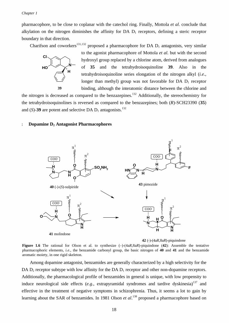
Figure 1.6 The rational for Olson et al. to synthesize (–)-(4aR,8aR)-piquindone (42): Assemble the tentativepharmacophoric elements, i.e., the benzamide carbonyl group, the basic nitrogen of 40 and 41 and the benzamidearomatic moiety, in one rigid skeleton.
π1
π2
π2
π1
π1
π2
π2
π1
Introduction to the Medicinal Chemistry of Schizophrenia
19
potent, but flexible, dopaminergic compound such as sulpiride (40), haloperidol (4), pimozide (43)
and molindone (41). The pharmacophore comprised four different elements as pictured in Figure 1.6
(adopted from Olson et al.138): 1) the benzamide phenyl ring (π-π interaction); 2) the amide carbonyl
oxygen atom (π-π interaction) or, alternatively, an aromatic moiety (π-π interaction); 3) the basic
nitrogen atom (+NH….COO- interaction) and 4) a lipophilic tail corresponding to the heterocyclic
moiety of pimozide (Figure 1.6, 43). Interestingly, the authors introduce the carbonyl oxygen as an
isosteromer to a phenyl ring in the ligand-receptor interaction, supported by a crystal structure where
a similar interaction has been observed.138
In order to validate their model Olsen et al.138 attempted to include the pharmacophoric
elements in one single structure, and the resulting (–)-(4aR,8aR)-piquindone (42) turned out to have
similar properties as haloperidol (4) but with significant decreased cataleptogenic liability. For a
computational chemist a rigid and potent compound (Table 1.5), like 42, is ideal to utilize as a
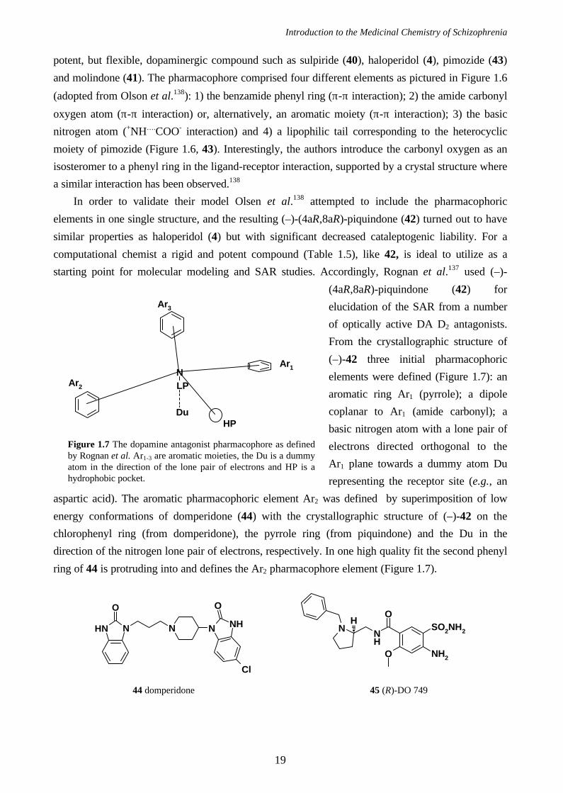
starting point for molecular modeling and SAR studies. Accordingly, Rognan et al.137 used (–)-
(4aR,8aR)-piquindone (42) for
elucidation of the SAR from a number
of optically active DA D2 antagonists.
From the crystallographic structure of
(–)-42 three initial pharmacophoric
elements were defined (Figure 1.7): an
aromatic ring Ar1 (pyrrole); a dipole
coplanar to Ar1 (amide carbonyl); a
basic nitrogen atom with a lone pair of
electrons directed orthogonal to the
Ar1 plane towards a dummy atom Du
representing the receptor site (e.g., an
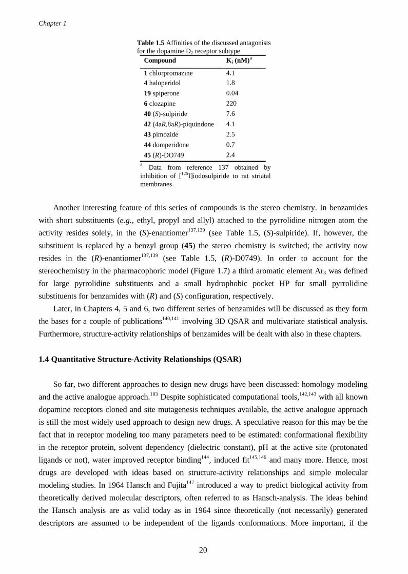
aspartic acid). The aromatic pharmacophoric element Ar2 was defined by superimposition of low
energy conformations of domperidone (44) with the crystallographic structure of (–)-42 on the
chlorophenyl ring (from domperidone), the pyrrole ring (from piquindone) and the Du in the
direction of the nitrogen lone pair of electrons, respectively. In one high quality fit the second phenyl
ring of 44 is protruding into and defines the Ar2 pharmacophore element (Figure 1.7).
NLP
Du
Ar2
Ar1
Ar3
HP
Figure 1.7 The dopamine antagonist pharmacophore as definedby Rognan et al. Ar1-3 are aromatic moieties, the Du is a dummyatom in the direction of the lone pair of electrons and HP is ahydrophobic pocket.
N NHNNNH
O O
Cl
O
NH
N SO2NH2
O NH2
H
44 domperidone 45 (R)-DO 749
Chapter 1
20
Table 1.5 Affinities of the discussed antagonistsfor the dopamine D2 receptor subtype
Compound Ki (nM)a
1 chlorpromazine 4.1
4 haloperidol 1.8
19 spiperone 0.04
6 clozapine 220
40 (S)-sulpiride 7.6
42 (4aR,8aR)-piquindone 4.1
43 pimozide 2.5
44 domperidone 0.7
45 (R)-DO749 2.4a Data from reference 137 obtained byinhibition of [125I]iodosulpiride to rat striatalmembranes.
Another interesting feature of this series of compounds is the stereo chemistry. In benzamides
with short substituents (e.g., ethyl, propyl and allyl) attached to the pyrrolidine nitrogen atom the
activity resides solely, in the (S)-enantiomer137,139 (see Table 1.5, (S)-sulpiride). If, however, the
substituent is replaced by a benzyl group (45) the stereo chemistry is switched; the activity now
resides in the (R)-enantiomer137,139 (see Table 1.5, (R)-D0749). In order to account for the
stereochemistry in the pharmacophoric model (Figure 1.7) a third aromatic element Ar3 was defined
for large pyrrolidine substituents and a small hydrophobic pocket HP for small pyrrolidine
substituents for benzamides with (R) and (S) configuration, respectively.
Later, in Chapters 4, 5 and 6, two different series of benzamides will be discussed as they form
the bases for a couple of publications140,141 involving 3D QSAR and multivariate statistical analysis.
Furthermore, structure-activity relationships of benzamides will be dealt with also in these chapters.
1.4 Quantitative Structure-Activity Relationships (QSAR)
So far, two different approaches to design new drugs have been discussed: homology modeling
and the active analogue approach.103 Despite sophisticated computational tools,142,143 with all known
dopamine receptors cloned and site mutagenesis techniques available, the active analogue approach
is still the most widely used approach to design new drugs. A speculative reason for this may be the
fact that in receptor modeling too many parameters need to be estimated: conformational flexibility
in the receptor protein, solvent dependency (dielectric constant), pH at the active site (protonated
ligands or not), water improved receptor binding144, induced fit145,146 and many more. Hence, most
drugs are developed with ideas based on structure-activity relationships and simple molecular
modeling studies. In 1964 Hansch and Fujita147 introduced a way to predict biological activity from
theoretically derived molecular descriptors, often referred to as Hansch-analysis. The ideas behind
the Hansch analysis are as valid today as in 1964 since theoretically (not necessarily) generated
descriptors are assumed to be independent of the ligands conformations. More important, if the
Introduction to the Medicinal Chemistry of Schizophrenia
21
predictive ability of the model is high enough, valuable time will not be spent synthesizing inactive
compounds.
+ Physicochemical Molecular Descriptors
The applicability of physicochemical descriptors is manifold. First, prediction of the biological
activity (e.g., receptor affinity) as introduced by Hansch et al.147 (see below). Second, they enable us
to search large molecular databases (e.g., Chemical Abstracts structural database) by simply define a
physicochemical profile for the target molecule.148-150 Third, recently developed techniques like
combinatorial chemistry100-102 and high-throughput screening,98,99 call for methods in order to design
the synthesis of the most diverse compounds and for the definition of new specific targets,
respectively.
Examples of commonly used physicochemical descriptors are listed in Table 1.6. which may be
divided into steric, electrostatic and hydrophobic (lipophilic) types of descriptors, although many of
them are a combination of all three types. Some of these descriptors are listed in the literature, while
some may be obtained through analytical experiments151,152 or through computational calculations.142
Steric descriptors like FW, L, B and VdWV (Table 1.6) are simply different attempts to quantify the
molecular structure; electrostatic descriptors like σm, HOMO or LUMO portray the electronic
features of a compound (e.g., the influence of a specific substituent on an aromatic system) and the
hydrophobic descriptors π, logP and logD accounts for a molecules ability to, for instance penetrate
the blood-brain barrier.153,154 Taken together, the array of descriptors collected for a specific
molecule compares to the fingerprint from a human being, hence, it should be unique.
+ Hansch Analysis
In the early 1960s, Hansch and co-workers147 investigated the possibility of expressing a
relationship between structural and physicochemical properties and biological activity, quantitatively.
Typically, properties as logP, σ, or Es representing 1-octanol/water partition coefficient, the well-
known Hammett constant and Tafts steric descriptors (Table 1.6), respectively, were used as
descriptors. In the Hansch analysis, the biological activity log(1/C) were correlated with the
descriptors using Multiple Linear Regression (MLR, see Chapter 2), also called Ordinary Least
Squares (OLS).155,156 The so-called Hansch equation (Equation 1.1) comprise the relationship
established by Hansch et al., where a, b, c, d and e are constants obtained through regression
analysis.
( ) ( )log log log1 2
C a b c d e= − + + + +P P Esσ (1.1)
As was emphasized by Van de Waterbeemd,157 Hansch analysis is a method aiming at describing
the relationship between only a few variables and the biological activity and should not be considered
too much as a predictive model. By employing MLR for the regression analysis a couple of crucial
items need to be considered: 1) keep the ratio of compounds to variables greater than approximately
Chapter 1
22
five and 2) multicollinearity may cause spurious solutions. Today, methods to circumvent these
problems are available, where the original variables are replaced by underlying orthogonal latent
variables (i.e., PCR and PLS in Chapter 2). The predictive ability of a model may then be validated
with crossvalidation155 or by predictions of an external test set.
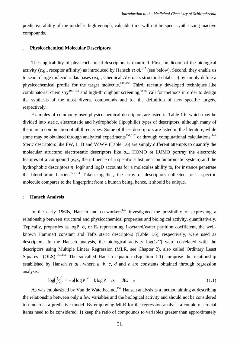
+ Molecular Diversity and Experimental Design
N
R2
R1N
NH
OR2R3
OR5
***
(a) (b)
Figure 1.8 The basic skeleton of trans-OHB[f]Qs (a) and 6-methoxybenzamides (b) used by Nilsson et al. and
Norinder et al., respectively.
In Chapter 3, physicochemical descriptors are employed to guide the selection of which
compounds to synthesize.158 From a library of a 88 tentative OHB[f]Qs (Figure 1.8(a)), only the 15
most diverse compounds were selected to be synthesized by means of a factorial design in the
Principal Properties (PPs). The PPs are score-vectors obtained from a Principal Component Analysis
(PCA, see Chapter 2) of the physicochemical descriptors, and comprise the variation that
significantly discriminate between the compounds. Norinder et al.159 used physicochemical
descriptors in order to increase the understanding of the structure-activity relationships of a series of
benzamides (Figure 1.8(b)). They used a fractional factorial design in the first three PPs, for the
selection of 16 representative compounds, out of 70, for the training set. In the following regression
analysis the original descriptors (not the PPs, thus) from the training set were correlated with the
biological activity (pIC50 for the DA D2 receptor), using PLS. The remaining compounds were
utilized as a test set in order to estimate the predictability of the PLS model. The profound difference
in the approaches used in Chapter 3 and by Norinder et al., should be obvious. Nilsson et al.
generated descriptors for whole molecules, e.g., logP was used rather than the contribution from
single substituents (π). The objective with that investigation was to select the most diverse
compounds to synthesize, not necessarily to create a predictive model. In contrast, Norinder et al.
already had a large data set with compounds tested for biological activity and their aim was to
elucidate also the influence of single substituent positions on the biological activity. Therefore, the
training set was selected such that the diversity in each position R2, R3 and R5, in Figure 1.8(b), was
maximized. The conclusions drawn from this investigation will be discussed further in Chapter 6,
where this data set was analyzed with 3D QSAR (see next section) using multilinear PLS160 as
regression method.
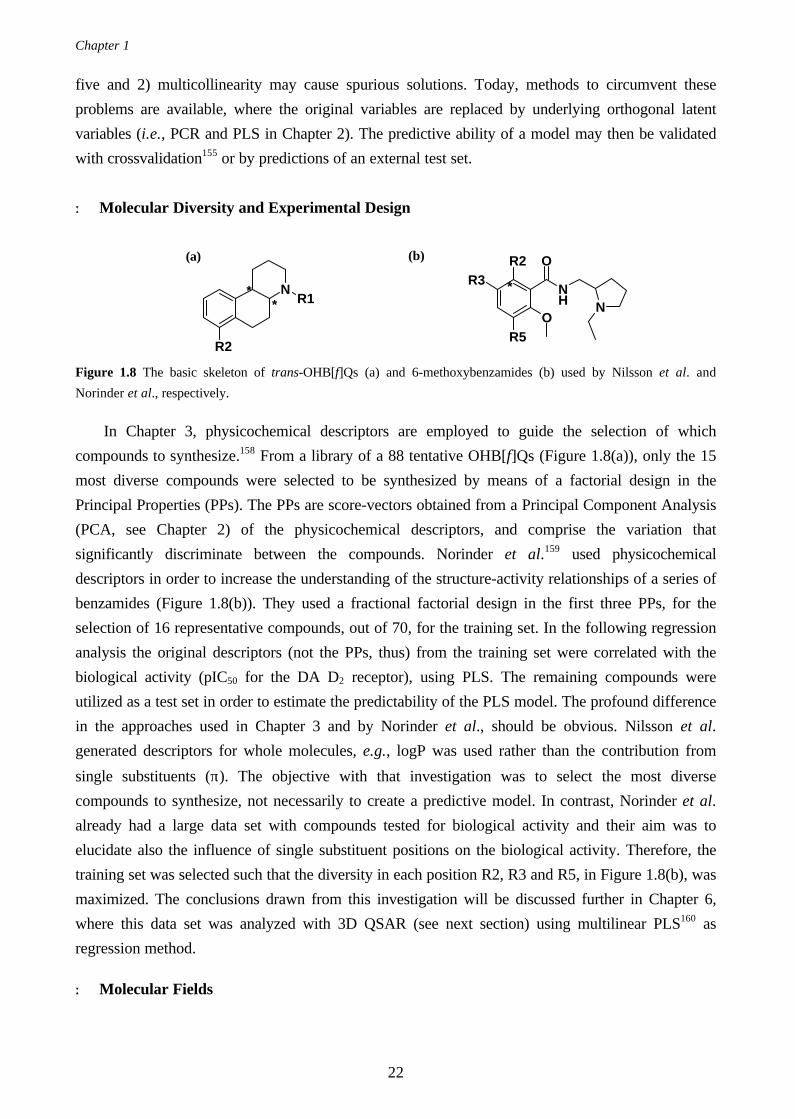
+ Molecular Fields
Introduction to the Medicinal Chemistry of Schizophrenia
23
The obvious extension of the Hansch analysis is modeling where molecular flexibility is
considered. The concept of Comparative Molecular Field Analysis (CoMFA)161 as presented by
Cramer et al.117 in 1988 is such a method, commercially available as a module in the molecular
modeling package SYBYL.142
Figure 1.9 The three dimensional grid used in CoMFA to generate molecular field descriptors. For clarity, grid pointswithin the grid are omitted
1 2 3 4 5 6 7
NNH
O
O
Chapter 1
24
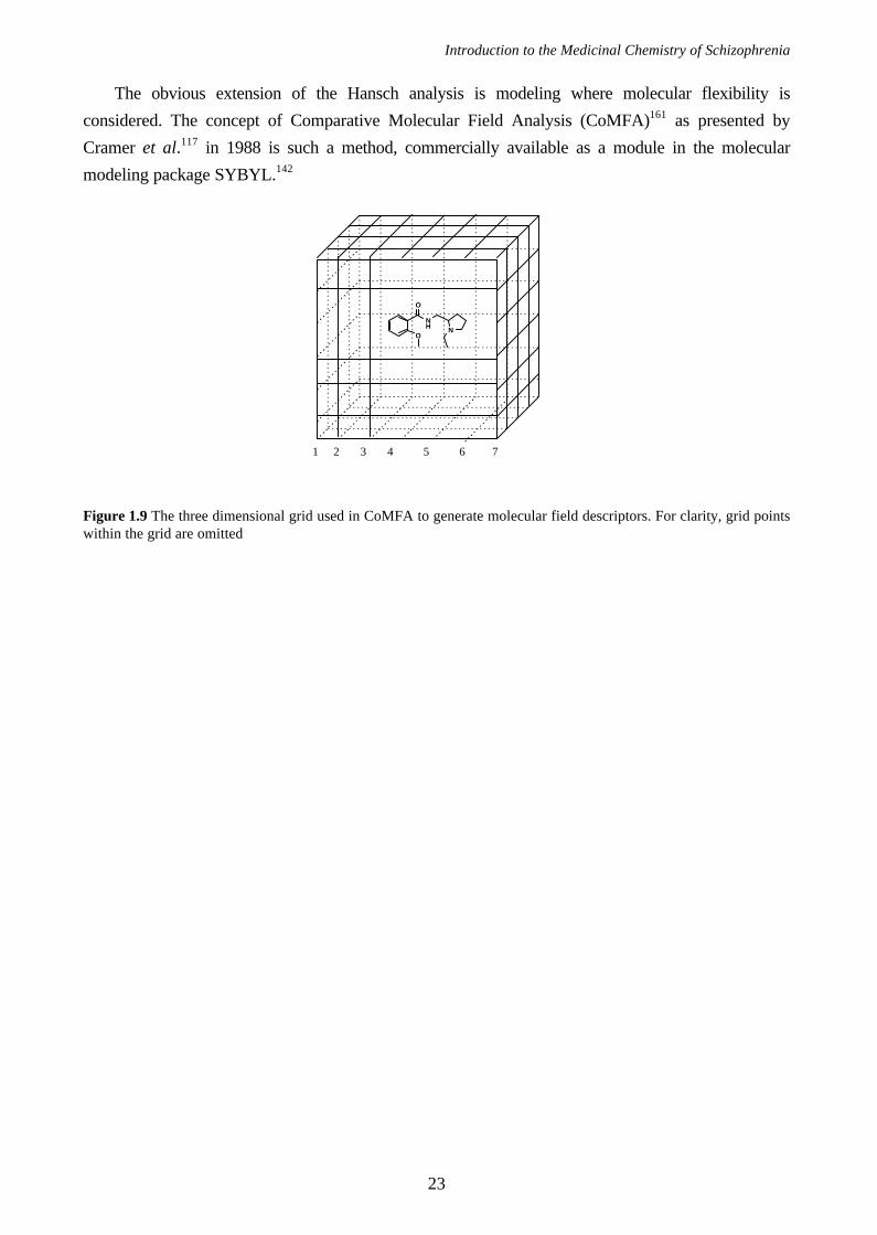
Table 1.6 The most commonly used molecular descriptors available in the literature or throughcomputational calculations.Descriptor Abbr. typea ref. Descriptor short typea ref.
Formula Weight FW ste Ionisation constant pKb 162
Hammett constant σm ele 163 Swain-Lupton field F 163
Hammett constant σp ele 163 Swain-Lupton res. R 163
Tafts polar constant σ* ele 157 VdWaals Volume VdWV ste 142
Tafts steric parameter Es ste 157 VdWaals Area VdWA ste 142
Hansch aromatic fragment π lip 162,163 Connolly Surface Vol. CoVo ste 142
Lipophilicity logP lip 157,162 Connolly Surface Area CoAr ste 142
Lipophilicity (pH=7.4) logD lip 157,162 Electronic Energy ELEC ele 142
Connectivity index (Randic) 1χ ste 157 Core-Core interaction CoCo ste 142
Connectivity index 2χ ste 157 Heat of Formation HoFo ele 142
Molar Refractivity MR ele 164 Ionization potential HOMO ele 142
Verloop Sterimol L ste 157 Electron affinity LUMO ele 142
Verloop Sterimol B ste 157 Dipole moment Dipo ele 142
Eudismic Index EI 165 Point charges chaX ele 142
Ionization constant pKa 162
aele electronic; ste steric; lip lipophilicity
Molecular fields basically are three dimensional representations of the steric, electrostatic and
hydrophobic surroundings of a molecule. A molecular field is generated by enclosing the molecule in
a three dimensional grid (Figure 1.9) and assigning nonbonded interactions between a probe atom
and the molecule in each grid point. Obviously, the difference between two different types of fields is
the algorithm with which the nonbonded interactions are calculated. In SYBYL/CoMFA the steric
field interaction energies (Este) are Lennard-Jones potentials, also referred to as steric 6-12
potentials,166 which are sensitive to changes in the distance between the probe and the atoms (ri) as
can be seen in Equation 1.2. N is the number of atoms in the molecule; A and B are constants
characteristic for the probe atom type and the type of the ith atom in the molecule, respectively.
Er rste = −
=∑ A B
i
i
ii
N
12 61
(1.2)
The electrostatic field interaction energies are less influenced by the distances between the probe and
the atoms but instead the charge of the probe and the point charges of the atoms are important. In
addition, Eele is very sensitive to spatial dielectric behavior of the environment167 and a distance-
dependent dielectric term has been proposed.168 The magnitude of the electrostatic potential (Eele)
between two ions with charges Q and q separated by a distance r is given by Coulomb’s law166:
( ) ( )E
K r r s sele
Q q
= +− +
+
=
∑ i
i
N
ζζ ε ζ ε
12
1
4(1.3)
where N is the number of atoms in the molecule; Q is the charge of the probe atom; qi is the point
charge on the ith atom and K is a constant term. In this formula a homogenous protein phase and a
homogenous solution phase with dielectrics ζ and ε, respectively, are assumed to be present.167 The
depth of each protein atom (sp) in the protein phase is assessed by counting the number of
Introduction to the Medicinal Chemistry of Schizophrenia
25
neighboring protein atoms whose nuclei lie within 4 Å. For the probe atom the depth (sQ) is
calculated similarly. Consequently, Equation 1.3 leads to an effective dielectric of ζ when the
pairwise groups of atoms are so deep in the protein and so close together that the solvent effects can
be neglected. However, when one or both of the atoms approach the surface of the protein the
effective dielectric becomes (ζ + ε)/2 since the term 4sQsq is set to zero.
Prior to calculation of the electrostatic field point charges of the atoms need to be calculated. In
the original CoMFA article117 charges were calculated by the method of Gasteiger and Marsili169 as
was implemented in SYBYL. Today, however, several options are available although most often
point charges are estimated by semi-empirical AM1 single point calculations.170
There are also variations of the above described steric field. Kroemer et al.171 replaced the steric
6-12 potential interactions with atom-based indicator variables. That is, they assigned the values 30
or 0 kcal/mol to a grid point if an atom was present in the adjacent small cube or not. Similar,
Floersheim et at.172 assigned values of either 1 or 0 to a grid point, depending on whether the grid
point was within, or outside, the van der Waals radius of any atom in the target molecule.
In the GRID program136,167 a different and intuitively more appealing (authors comment)
approach is used. Each grid point is assigned with the sum of three different non-bonded
interactions, i.e., Este, Eele and Ehb, as in Equation 1.4. The two former terms are calculated as in
Equations 1.2 and 1.3, while the hydrogen bonding contribution167 is calculated as in Equation 1.5. C
and D are tabulated values for specific atoms; d is the distance between the atoms; m is usually four
but the whole Ehb term is set to zero when θ ≤ 90°. If the probe group donates the hydrogen bond it
is assumed that the probe can orient itself in order to form the most effective hydrogen bond, and the
cos θ is set to unity.
E E E Etot ste ele hb= + +∑ ∑ ∑ (1.4)
[ ]E C d D dhbm= −6 4 cos θ (1.5)
Consequently, and in contrast to other methods (e.g., SYBYL/CoMFA), fields generated in the
GRID program portrays the specifics of certain probes. Thus, it is the users task to select a probe
that best reflects what needs to be investigated. For instance, a water molecule, a carbon atom or a
Ca+2 ion could be chosen to display hydrogen bonding, steric and electrostatic characteristics of the
ligands, respectively. Goodford173 has demonstrated how GRID probes explicitly can reflect
individual properties of specific chemical groups attached to the target molecule.
Fields generated in SYBYL or GRID are the most commonly used since they are commercially
available. However, other molecular descriptors are available, e.g., Molecular Shapes,174-176
Molecular Lipophilicity Potentials,177,178 Molecular Similarity Indices179 (i.e., CoMSIA) and
Molecular Similarity Matrices.180-182
Independent of which program used to generate molecular fields, the following parameters need
to be specified by the user: 1) the grid size; 2) the grid resolution, i.e., grid points per Å; 3) the type
of probes and 4) the probe charge.
+ Comparative Molecular Field Analysis (CoMFA)
Chapter 1
26
As stated above, the logical extension of the Hansch analysis147 is Comparative Molecular Field
Analysis (CoMFA),117 where the physicochemical parameters are replaced or combined with field
descriptors. In analogy with the Hansch analysis, in CoMFA the molecular fields are correlated with
the biological activity. CoMFA takes the three dimensional conformations of the molecules into
consideration, hence, several crucial items need to be considered.
First, in order to find low energy conformations, or rather the global minimum energy
conformation of each compound under investigation, conformational analyses183-186 are conducted.
According to the Boltzman distribution,166 a low energy conformation is more abundant than a high
energy conformation and, consequently, also more likely to be involved in the ligand-receptor
interaction.
Second, all molecules must be aligned in the same coordinate system and several options to
perform this are possible. If a pharmacophore is available, one might choose to superimpose all
molecules on mutual and likely interaction points with the receptor (see Chapter 4). The compounds
could be docked into the active site of a receptor homology model (see Chapter 6) or, alternatively,
the molecular fields could be superimposed in a least square manner.142 Independent of which
alignment approach that is employed, in CoMFA the differences between the aligned molecular fields
are correlated with the biological activity. Therefore, an alignment procedure where the global
overlap between structurally related compounds140,187 (Chapter 4) are maximized, is likely to perform
just as good as a more elaborated and rational alignment140 (Chapter 6). This is the case when the
ligands are flexible and the rationale is pure statistical: If the alignment is not performed with
maximized overlap between the molecules an increased level of insignificant variation, i.e., noise is
inevitable. Noise may, or may not, affect the predictability detrimentally.
Third, molecular fields are generated first when the molecules are properly aligned.
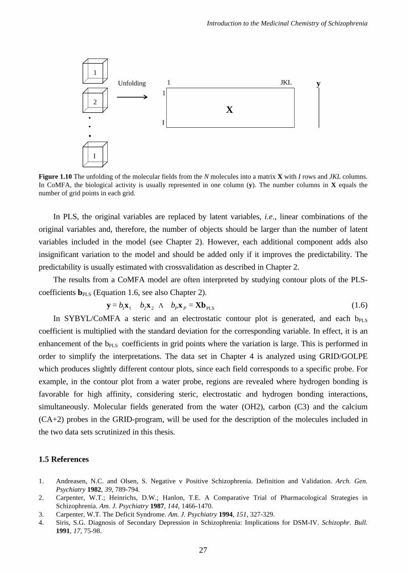
In Figure 1.10, a typical CoMFA data set consisting of I molecules characterized with a single
molecular field is shown. In order to perform the PLS analysis each grid, with the dimensions J, K
and L, is unfolded to form a row with JKL number of columns. Traditionally, only one response
variable is considered in CoMFA, e.g., the affinity for a central dopamine receptor and, therefore, a
bilinear PLS1 algorithm188-190 is used for the regression analysis. The theory covering the basics of
PLS analysis is discussed in Chapter 2, but specific details important for CoMFA are pointed out in
the following.
Introduction to the Medicinal Chemistry of Schizophrenia
27
Figure 1.10 The unfolding of the molecular fields from the N molecules into a matrix X with I rows and JKL columns.In CoMFA, the biological activity is usually represented in one column (y). The number columns in X equals thenumber of grid points in each grid.
In PLS, the original variables are replaced by latent variables, i.e., linear combinations of the
original variables and, therefore, the number of objects should be larger than the number of latent
variables included in the model (see Chapter 2). However, each additional component adds also
insignificant variation to the model and should be added only if it improves the predictability. The
predictability is usually estimated with crossvalidation as described in Chapter 2.
The results from a CoMFA model are often interpreted by studying contour plots of the PLS-
coefficients bPLS (Equation 1.6, see also Chapter 2).
∃y x x x Xb= + + + =b b bP P1 1 2 2 Λ PLS (1.6)
In SYBYL/CoMFA a steric and an electrostatic contour plot is generated, and each bPLS
coefficient is multiplied with the standard deviation for the corresponding variable. In effect, it is an
enhancement of the bPLS coefficients in grid points where the variation is large. This is performed in
order to simplify the interpretations. The data set in Chapter 4 is analyzed using GRID/GOLPE
which produces slightly different contour plots, since each field corresponds to a specific probe. For
example, in the contour plot from a water probe, regions are revealed where hydrogen bonding is
favorable for high affinity, considering steric, electrostatic and hydrogen bonding interactions,
simultaneously. Molecular fields generated from the water (OH2), carbon (C3) and the calcium
(CA+2) probes in the GRID-program, will be used for the description of the molecules included in
the two data sets scrutinized in this thesis.
1.5 References
1. Andreasen, N.C. and Olsen, S. Negative v Positive Schizophrenia. Definition and Validation. Arch. Gen.Psychiatry 1982, 39, 789-794.
2. Carpenter, W.T.; Heinrichs, D.W.; Hanlon, T.E. A Comparative Trial of Pharmacological Strategies inSchizophrenia. Am. J. Psychiatry 1987, 144, 1466-1470.
3. Carpenter, W.T. The Deficit Syndrome. Am. J. Psychiatry 1994, 151, 327-329.4. Siris, S.G. Diagnosis of Secondary Depression in Schizophrenia: Implications for DSM-IV. Schizophr. Bull.
1991, 17, 75-98.
•••
1
2
I
Unfolding
X
1
I
1 JKL y
Chapter 1
28
5. Roy, A. Suicide in Chronic Schizophrenia. Brit. J. Psychiat. 1982, 141, 171-177.6. Borison, R.L.; Diamond, B.I.; Pathiraja, A.; Meibach, R.C. Novel Antipsychotic Drugs. Meltzer, H.Y. Ed.;
Raven: New York, 1992; pp. 2237. Chouinard, G.; Jones, B.; Remington, G.; Bloom, D.; Addington, D.; MacEwan, G.W.; Labelle, A.; Beauclair,
L.; Arnott, W. A Canadian Multicenter Placebo-Controlled Study of Fixed Doses of Risperidone andHaloperidol in the Treatment of Chronic Schizophrenic Patients. J. Clin. Psychopharmacol 1993, 13, 25-40.
8. Kay, S.R.; Fisbein, A.; Opler, L.A. The Positive and Negative Syndrome Scale (PANSS) for Schizophrenia.Schizophr. Bull. 1987, 13, 261-276.
9. Claghorn, J.; Honingfeld, G.; Abuzzahab, F.S.; Wang, R.; Steinbook, R.; Tuason, V.; Klerman, G. The Risksand Benefits of Clozapine versus Chlorpromazine. J. Clin. Psychopharmacol 1987, 7, 377-384.
10. Kane, J.; Honigfield, G.; Singer, J.; Meltzer, H.Y. Clozapine for the Treatment-Resistant Schizophrenic. Arch.Gen. Psychiatry 1988, 45, 789-796.
11. Meltzer, H.Y.; Bastani, B.; Kwon, K.Y.; Ramirez, L.F.; Burnett, S.; Sharpe, J. A Prospective Study ofClozapine in Treatment-Resistant Schizophrenic Patients. I. Preliminary report. Psychopharmacology 1989, 99,S68-S72.
12. Juul Povisen, U.; Norig, U.; Fog, R.; Gerlach, J. Tolerability and Therapeutic Effect of Clozapine. ActaPsychiatr. Scand. 1985, 71, 176-185.
13. Casey, D.E. Clozapine: Neuroleptic-Induced EPS and Tardive Dyskinesia. Psychopharmacology 1989, 99, S47-S53.
14. Peacock, L.; Solgaard, T.; Lublin, H.; Gerlach, J. Clozapine versus Typical Antipsychotics. Aretro- andProspective Study of Extrapyramidal Side Effects. Psychopharmacology 1996, 124, 188-196.
15. Lieberman, J.A.; Johns, C.A.; Kane, J.M.; Rai, K.R.; Pisciotta, A.V.; Saltz, B.L.; Howard, A. Clozapine-Induced Agranulocitosis: Non-Cross-Reactivity With Other Psychotropic Drugs. J. Clin. Psychiatry 1988, 49,271-277.
16. Stockton, M.E. and Rasmussen, K. Olanzapine, a Novel Atypical Antipsychotic, Reverses d-Amphetamine-induced Inhibition of Midbrain Dopamine cells. Psychopharmacology 1996, 124, 50-56.
17. Moore, N.A.; Calligaro, D.O.; Wong, D.T.; Bymaster, F.; Tye, M.C. The Pharmacology of Olanzapine andOther New Antipsychotic Agents. Curr. Opin. Invest. Drugs 1993, 2, 281-293.
18. Beasley, C.M.; Tollefson, G.; Tran, P.; Satterlee, W.; Sanger, T.; Hamilton, S. The Olanzapine HGAD StudyQroup (1995) Olanzapine versus Placebo and Haloperidol: Acute Phase Result of the North American Doubleblind Olanzapine trial. Neuropsychopharmacology 1996, 14, 105-118.
19. Bunney, B.S. Animal Models in Psychology and Neurology. Hanin and Usdin, Eds.; Pergamon Press: NewYork, 1977; pp. 91-104.
20. Roth, B.L.; Craigo, S.C.; Choudhary, M.S.; Uluer, A.; Monsma, F.J.; Shein, Y.; Meltzer, H.Y. Binding ofTypical and Atypical Antipsychotic Agents to 5-Hydroxytryptamine-6 and 5-Hydroxytryptamine-7 Receptors. J.Pharmacol. Exp. Ther. 1994, 268, 1403-1410.
21. Koehler, K.F.; Radesater, A.C.; Karlsson-Boethius, G.; Bryske, B.; Widman, M. Regional Distribution and invivo Binding of the Typical Antipsychotic Drug remoxipride. J. Neural Transm. 1992, 87, 49-62.
22. Walker, J.M.; Bowen, W.D.; Walker, F.O.; Matsumoto, R.R.; De Costa, B.; Rice, K.C. Sigma Receptors:Biology and Function. Pharmacol. Rev. 1990, 42, 355-390.
23. Assie, M.-B.; Sleight, A.J.; Koek, W. Biphasic Displacement of [3H]YM-09151-2 Binding in the Rat Brain byThioridazine, Risperidone and Clozapine, but not by Other Antpsychotics. Eur. J. Pharmacol. 1993, 237, 183-189.
24. Canton, H.; Verriele, L.; Colpaert, F.C. Binding of Typical and Atypical Antipsychotics to 5HT1C and 5HT2
Sites: Clozapine Potently Interacts with 5HT1C Sites. Eur. J. Pharmacol. 1990, 191, 93-96.25. Watling, K.J.; Beer, M.S.; Stanton, J.A.; Newberry, N.R. Interaction of the Atypical Neuroleptic Clozapine with
5-HT3 Receptors in the Cerbral Cortex and Superior Cervical Ganglion of the Rat. Eur. J. Pharmacol. 1990,182, 465-471.
26. Meltzer, H.Y.; Matsubar, S.; Lee, J.-C. Classification of Typical and Atypical Antipsychotic Drugs on the Basicof Dopamine D-1, D-2 and Serotonin2 pKi Values. J. Pharmacol. Exp. Ther. 1989, 251, 238-246.
27. Bolden, C.; Cusack, B.; Richelson, E. Antagonism by Antimuscarinic and Neuroleptic Compounds at the FiveCloned Human Muscarinic Cholinergic Receptors Expressed in Chinese Hamster Ovary Cells. J. Pharmacol.Exp. Ther. 1992, 260, 576-580.
28. Sonesson, C. PhD Thesis. Arylpiperidine and Arylpyrrolidine Derivatives with Potential Antipsychotic Efficacy.Synthesis and Quantitative Structure-Activity Relationships Faculty of Pharmacy, Uppsala University, Sweden.1995
29. Carlsson, A. and Lindquist, M. Effect of Chlorpromazine or Haloperidol on Formation of 3-Methoxythyramineand Normethanephrine in Mouse Brain. Acta Pharmacol. Toxicol. 1963, 20, 140-144.
30. Randrup, A. and Munkvad, I. Special Antagonism of Amphetamine-Induced Abnormal Behavior: Inhibition ofStereotyped Activity with Increase of some Normal Activities. Psycopharmacologia 1965, 7, 416-422.
Introduction to the Medicinal Chemistry of Schizophrenia
29
31. Snyder, L.A. Science 1974, 184, 1243-1253.32. Seeman, P. Fed. Proc. 1974, 33, 24633. Carlsson, A. Antipsychotic Drugs, Neurotransmitters, and Schizophrenia. Am. J. Psychiatry 1978, 135, 164-
173.34. Carlsson, A. Does Dopamine Have a Role in Schizophrenia? Biol Psychiatry 1978, 13, 3-21.35. Van Rossum, J.M. The Significance of Dopamine-Receptor Blockade for the mechanism of Action of
Neuroleptic Drugs. Arch. Int. Pharmacodyn. Ther. 1966, 160, 492-494.36. Matthysse, S. Antipsychotic Drug Actions: a Clue to the Neuropathology of Schizophrenia? Fed. Proc. 1973,
32, 200-205.37. Seeman, P. Antipsychotic Drugs: Direct Correlation Between Clinical Potency and Presynaptic Action on
Dopamine Neurons. Science 1975, 188, 1217-1219.38. Seeman, P. Dopamine Receptors and the Dopamine Hypothesis of Schizophrenia. Synapse 1987, 1, 133-152.39. Peroutka, S.J. and Snyder, S.H. Relationship of Neuroleptic Drug Effects at Brain Dopamine, Serotonin, alfa-
Adrenergic, and Histamine Receptors to Clinical Potency. Am. J. Psychiatry 1980, 137, 1518-1522.40. Chiodo, L.A. and Bunney, B.S. Typical and Atypical neuroleptics: Differential Effects of Chronic
Administration on the Activity of A9 and A10 Midbrain Dopaminergic Neurons. J. Neurosci. 1983, 3, 1607-1619.
41. Wooley Proc. Natl. Acad. Sci. USA 1954, 40, 228-231.42. Roth, B.L.; Ciaranello, R.D.; Meltzer, H.Y. Binding of Typical and Atypical Antipsychotic Agents to
Transiently Expressed 5-HT1C Receptors. J. Pharmacol. Exp. Ther. 1992, 260, 1361-1365.43. Hicks, P.B. The Efffect of Serotonergic Agents on Haloperidol Induced Catalepsy. Life Sci. 1990, 47, 1609-
1615.44. Neal-Beliveau, B.S.; Joyce, J.N.; Lucki, I. Serotonergic Involvement in Haloperidol-Induced Catalepsy. J.
Pharmacol. Exp. Ther. 1993, 265, 207-217.45. Hornykiewicz, O. Dopamine in the Basala Ganglia. Its Role and Therapeutic Implications (Including the
Clinical Use of L-DOPA). Br. Med. Bul. 1973, 29, 172-178.46. Tandon, R. and Greden, J.F. Cholinergic Hyperactivity and Negative Schizophrenic Symptoms. Arch. Gen.
Psychiatry 1989, 46, 745-753.47. Tandon, R.; Greden, J.F.; Silk, K.R. Treatment of Negative Schizophrenic Symptoms with Trihexyphenidyl. J.
Clin. Psychopharmacol 1988, 8, 212-215.48. Tandon, R. and Greden, J.F. Trihexyphenidyl Treatment of Negative Schizophrenic Symptoms. Acta Psychiatr.
Scand. 1987, 76, 73249. Leysen, J.E.; Janssen, P.M.F.; Schotte, A.; Luyten, W.H.M.L.; Megens, A.A.H.P. Interaction of Antipsychotic
Drugs with Neurotransmitter Receptor Sites in vitro and in vivo in Relation to Pharmacological and ClinicalEffects: Role of 5-HT2 Receptors. Psychopharmacology 1993, 112, S40-S54.
50. Rivest, R. and Marsden, C.A. Muscarinic Antagonists Attenuate the Increase in Accumbens and StriatumDopamine Metabolism Produced by Clozapine but not by Haloperidol. Br. J. Pharmacol. 1991, 104, 234-238.
51. Stein, L. and Wise, C.D. Possible Etiology of Schizophrenia: Progressive Damage to the Noradrenergic RewardSystem by 5-Hydroxydopamine. Science 1971, 171, 1032-1036.
52. Van Kammen, D.P.; Peters, J.; Yao, J.; Van Kammen, W.B.; Neylen, T.; Shaw, D.; Linnoila, M.Norepinephrine in Acute Exacerbations of Chronic Schizophrenia. Arch. Gen. Psychiatry 1990, 47, 161-168.
53. Breier, A.; Buchanan, R.W.; Waltrip, R.W.; Liswak, S.; Holmes, C.; Goldstein, D.S. The Effect of Clozapine onPlasma Norepinephrine: Relationship to Clinical Efficacy. Neuropsychopharmacology 1994, 10, 1-7.
54. Csernansky, J.G. Antipsychotics. Ed.; Springer: Berlin, 199655. Seeman P. Dopamine Receptors and Psychosis. Scientific American. 1995, 28-37.56. Civelli, O.; Bunzow, J.; Albert, P.; Van Tol, H.; Grandy, D. Molecular Biology of the Dopamine D2 Receptor.
NIDA. Res. Monogr. 1991, 111, 45-53.57. Dearry, A.; Gingrich, J.A.; Falardeau, P.; Fremeau, R.T.; Bates, M.D.; Caron, M.G. Molecular Cloning and
Expression of the Gene for a Human D1 Dopamine Receptor. Nature 1990, 347, 72-76.58. Monsma, F.J. Molecular Cloning and Expression of a D1 Dopamine Receptor Linked to Adenylyl Cyclase
Activation. Proc. Natl. Acad. Sci. USA 1990, 87, 6723-6727.59. Sunahara, R.K.; Niznik, H.B.; Weiner, D.M.; Stormann, T.M.; Brann, M.R.; Kennedy, J.L.; Gelernter, J.E.;
Rozmahel, R.; Yang, Y.; Israel, Y.; Seeman, P.; O'Dowd, B.F. Human Dopamine D1 Receptor Encoded by anIntronless Gene on Chromosome 5. Nature 1990, 347, 80-83.
60. Zhou, Q.-Y.; Grandy, D.K.; Thambi, L.; Kushner, J.A.; Van Tol, H.; Cone, R.; Pribnow, D.; Salon, J.; Bunzow,J.R.; Civelli, O. Cloning and Expression of Human and Rat D1 Dopamine Receptors. Nature 1990, 347, 76-80.
61. Schwartz, J.-C.; Giros, B.; Martres, M.-P.; Sokoloff, P. The Dopaminergic Receptor Family: Molecular Biologyand Pharmacology. The Neurosciences 1992, 4, 99-108.
Chapter 1
30
62. Bouthenet, M.L.; Souil, E.; Martres, M.P.; Sokoloff, P.; Giros, B.; Schwartz, J.C. Localization of Dopamine D3
Receptor mRNA in the Rat Brain Using in situ Hybridization Histochemistry: Comparison with Dopamine D2
Receptor mRNA. Brain Res. 1991, 564, 203-219.63. Schertler, G.F.X.; Villa, C.; Henderson, R. Projection Structure of Rhodopsin. Nature 1993, 362, 770-772.64. Henderson, R.; Baldwin, J.M.; Ceska, T.A.; Zemlin, F.; Beeleman, E.; Downing, K.H. Model for the Structure
of Bacteriorhodopsin Based on High Resolution Electron Cryo-Microscopy. J. Mol. Biol. 1990, 213, 899-929.65. Grigorieff, N.; Ceska, T.A.; Downing, K.H.; Baldwin, J.M.; Henderson, R. Electron-Crystallographic
Refinement of the Structure of Bacteriorhodopsin. J. Mol. Biol. 1996, 259, 393-421.66. Unger, V.M.; Hargrave, P.A.; Baldwin, J.M.; Schertler, G.F.X. Arrangement of Rhodopsin Transmembrane α-
Helices. Nature 1997, 389, 203-206.67. Kimura, Y.; Vassylyev, D.G.; Miyazawa, A.; Kidera, A.; Matsushima, M.; Mitsuoka, K.; Murata, K.; Hirai, T.;
Fujiyoshi, Y. Surface of Bacteriorhodopsin Revealed by High-Resolution Electron Crystallography. Nature1997, 389, 206-211.
68. Teeter, M.M.; Froimowitz, M.; Stec, B.; DuRand, C.J. Homology Modeling of the Dopamine D2 Receptor andits Testing by Docking of Agonists and Tricyclic Antagonists. J. Med. Chem. 1994, 37, 2874-2888.
69. Rippman, F. and Böttcher, H. Molecular Modelling of G Protein-Coupled Receptors- A New Approach toStudying Structure Activity Relationships. Kontakte 1994, 1, 30-36.
70. Giros, B.; Sokoloff, P.; Martres, M.P.; Riou, J.F.; Emorine, L.J.; Schwartz, J.C. Alternative Splicing Directs theExpression of two D2 Dopamine Receptor Isoforms. Nature 1989, 342, 923-926.
71. Monsma, F.J. Multiple D2 Dopamine Receptors Produced by Alternative RNA Splicing. Nature 1989, 342, 926-929.
72. Dal Toso, R.; Sommer, B.; Ewert, M.; Herb, A.; Pritchett, D.B.; Bach, A.; Shivers, B.D.; Seeburg, P.H. TheDopamine D2 Receptor: Two Molecular Forms Generated by Alternative Splicing. EMBO J. 1989, 8, 4025-4034.
73. Schwartz, J.-C.; Levesque, D.; Martres, M.-P.; Sokoloff, P. Dopamine D3 Receptor: Basic and Clinical Aspects.Clinical Neuropharmacology 1993, 16, 295-314.
74. Ohara, K.; Ulpian, C.; Seeman, P.; Sunahara, R.K.; Van Tol, H.; Niznik, H.B. Schizophrenia: Dopamine D1
Receptor Sequence is Normal, but has DNA Polymorphisms. Neuropsychopharmacology 1993, 8, 131-135.75. Sunahara, R.K.; Guan, H.-C.; O'Dowd, B.F.; Seeman, P.; Laurier, L.G.; NG, G.; George, S.R.; Torchia, J.; Van
Tol, H.; Niznik, H.B. Cloning of the Gene for a Human Dopamine D5 Receptor with Higher Affinity forDopamine than D1. Nature 1991, 350, 614-619.
76. Grandy, D.K.; Zhang, Y.; Bouvier, C.; Zhou, Q.-Y.; Johnson, R.A.; Allen, L.; Buck, K.; Bunzow, J.R.; Salon,J.; Civelli, O. Multiple Human D5 Dopamine Receptor Genes: a Functional Receptor and Two Pseudogenes.Proc. Natl. Acad. Sci. USA 1991, 88, 9175-9179.
77. Sobell, J.L.; Lind, T.J.; Sigurdson, D.C.; Zald, D.H.; Snitz, B.E.; Grove, W.M.; Heston, L.L.; Sommer, S.S. TheD5 Dopamine Receptor Gene in Schizophrenia: Identification of a Nonsense Change and Multiple MissenseChanges but Lack of Association with Disease. Hum. Mol. Genet. 1995, 4, 507-514.
78. Selbie, L.A.; Hayes, G.; Shine, J. The Major Dopamine D2 Receptor: Molecular Analysis of the Human D2A
Subtype. DNA 1989, 8, 683-689.79. Robakis, N.K.; Mohamadi, M.; Fu, D.J.; Sambamurit, K.; Refolo, L.M. Human Retina D2 Receptor cDNAs have
Multiple Polyadenylation Sites and Differ from a Pituitary Clone at the 5' Non-Coding Region. Nucleic AcidsRes. 1990, 18, 1299.
80. Grandy, D.K.; Marchionni, M.A.; Makam, H.; Stofko, R.E.; Alfano, M.; Frothingham, L.; Fisher, J.B.; Burke-Howie, K.J.; Bunzow, J.R.; Server, A.C.; Civelli, O. Cloning of the cDNA and Gene for a Human D2 DopamineReceptor. Proc. Natl. Acad. Sci. USA 1989, 86, 9762-9766.
81. Stormann, T.M.; Gdula, D.C.; Weiner, D.M.; Brann, M.R. Molecular Cloning and Expression of a DopamineD2 Receptor from Human Retina. Mol. Pharmacol. 1990, 37, 1-6.
82. Selbie, L.A.; Hayes, G.; Shine, J. DNA Homology Screening: Isolation and Characterization of the Human D2A
Dopamine Receptor Subtype. Adv. Second Messenger Phosphoprotein Res. 1990, 24, 9-14.83. Araki, K.; Kuwano, R.; Morii, K.; Hayashi, S.; Minoshima, S.; Shimizu, N.; Katagiri, T.; Usui, H.; Kumanishi,
T.; Takahashi, Y. Structure and Expression of Human and rat D2 Dopamine Receptor Genes [published erratumappears in Neurochem Int 1992 Oct;21(3):465]. Neurochem. Int. 1992, 21, 91-98.
84. Dearry, A.; Falardeau, P.; Shores, C.; Caron, M.G. D2 Dopamine Receptors in the Human Retina: Cloning ofcDNA and Localization of mRNA. Cell. Mol. Neurobiol. 1991, 11, 437-453.
85. Seeman, P.; Ohara, K.; Ulpian, C.; Seeman, M.V.; Jellinger, K.; Tol, H.H.; Niznik, H.B. Schizophrenia:Normal Sequence in the Dopamine D2 Receptor Region that Couples to G-proteins. DNA Polymorphisms in D2.Neuropsychopharmacology 1993, 8, 137-142.
86. Itokawa, M.; Arinami, T.; Futamura, N.; Hamaguchi, H.; Toru, M. A Structural Polymorphism of HumanDopamine D2 receptor, D2(Ser311→Cys). Biochem. Biophys. Res. Commun. 1993, 196, 1369-1375.
Introduction to the Medicinal Chemistry of Schizophrenia
31
87. Giros, B.; Martres, M.-P.; Sokoloff, P.; Schwartz, J.-C. Gene Cloning of Human Dopaminergic D3 Receptor andIdentification of its Chromosome. C. R. Acad. Sci. , III, Sci, Vie 1990, 311, 501-508.
88. Schmauss, C.; Haroutunian, V.; Davis, K.L.; Davidson, M. Selective Loss of Dopamine D3-type ReceptormRNA Expression in Parietal and Motor Cortices of Patients with Chronic Schizophrenia. Proc. Natl. Acad.Sci. USA 1993, 90, 8942-8946.
89. Liu, K.; Bergson, C.; Levenson, R.; Schmauss, C. On the Origin of mRNA Encoding the Truncated DopamineD3-type Receptor D3nf and Detection of D3nf-like Immunoreactivity in Human Brain. J. Biol. Chem. 1994, 269,29220-29226.
90. Van Tol, H.; Wu, C.M.; Guan, H.C.; Ohara, K.; Bunzow, J.R.; Civelli, O.; Kennedy, J.; Seeman, P.; Niznik,H.B.; Jovanovic, V. Multiple Dopamine D4 Receptor Variants in the Human Population [see comments]. Nature1992, 358, 149-152.
91. Van Tol, H.; Bunzow, J.R.; Guan, H.-C.; Sunahara, R.K.; Seeman, P.; Niznik, H.B.; Civelli, O. Cloning of theGene for a Human Dopamine D4 Receptor with High Affinity for the Antipsychotic Clozapine. Nature 1991,350, 610-614.
92. Livingstone, D.J.; Strange, P.G.; Naylor, L.H. Molecular Modelling of D2-like Dopamine Receptors. Biochem.J. 1992, 287, 277-282.
93. Seeman, P.; Ulpian, C.; Chouinard, G.; Van Tol, H.; Dwosh, H.; Lieberman, J.A.; Siminovitch, K.; Liu, I.S.C.;Waye, J.; Voruganti, P.; Hudson, C.; Serjant, G.R.; Masibay, A.S.; Seeman, M.V. Dopamine D4 ReceptorVariant, D4GLYCINE194, in Africans, but Not in Caucasians: no Association with Schizophrenia. Am. J. Med.Genet. 1994, 54, 384-390.
94. Dijkstra, D.; Mulder, T.B.; Rollema, H.; Tepper, P.G.; Van der Weide, J.; Horn, A.S. Synthesis andPharmacology of trans-4-n-Propyl-3,4,4a,10b-tetrahydro-2H,5H-1-benzopyrano[4,3-b]-1,4-oxazin-7- and -9-ols:the Significance of Nitrogen pKa Values for Central Dopamine Receptor Activation. J. Med. Chem. 1988, 31,2178-2182.
95. Glase, S.A.; Akunne, H.C.; Georgic, L.M.; Heffner, T.G.; MacKenzie, R.G.; Manley, P.J.; Pugsley, T.A.; Wise,L.D. Substituted [(4-Phenylpierazinyl)-methyl]benzamides: Selective Dopamine D4 Agonists. J. Med. Chem.1997, 40, 1771-1772.
96. Johansson, A.M.; Fredriksson, K.; Hacksell, U.; Grol, C.J.; Svensson, K.; Carlsson, A.; Sundell, S. Synthesisand Pharmacology of the Enantiomers of cis-7-hydroxy-3-methyl-2-(dipropylamino)tetralin. J. Med. Chem.1990, 33, 2925-2929.
97. Unangst, P.C.; Capiris, T.; Connor, D.T.; Doubleday, R.; Heffner, T.G.; MacKenzie, R.G.; Miller, S.R.;Pugsley, T.A.; Wise, L.D. (Aryloxy)alkylamines as Selective Human Dopamine D4 Receptor Antagonists:Potential Antipsychotic Agents. J. Med. Chem. 1997, 40, 4026-4029.
98. Chapman, D. The Measurement of Molecular Diversity: a Three-Dimensional Approach. J. Comput. -AidedMol. Design 1996, 10, 501-512.
99. Broach, J.R. and Thorner, J. High-Throughput Screening for Drug Discovery. Nature. 1996, 384, 14-16.100. Verdine, G.L. The Combinatorial Chemistry of Nature. Nature. 1996, 384, 11-13.101. Murray, C.W.; Clark, D.E.; Auton, T.R.; Firth, M.A.; Li, J.; Sykes, R.A.; Waszkowycz, B.; Westhead, D.R.;
Young, S.C. PRO_SELECT: Combining Structure-Based Drug Design and Combinatorial Chemistry for RapidLead Discovery. 1. Technology. J. Comput. Aided. Mol. Des. 1997, 11, 193-207.
102. Lam, K.S. Application of Combinatorial Library Methods in Cancer Research and Drug Discovery. Anticancer.Drug. Des. 1997, 12, 145-167.
103. Marshall, G.R.; Barry, C.D.; Bosshard, H.E.; Dammkoehler, R.A.; Dunn, D.A. The Conformational Parameterin Drug Design: the Active Analogue Approach. ACS Symp. Ser. 1979, 112, 205-226.
104. Dahl, S.G.; Edvardsen, O.; Sylte, I. Molecular Dynamics of Dopamine at the D2 Receptor. Proc. Natl. Acad.Sci. USA 1991, 88, 8111-8115.
105. Findlay, J. and Eliopoulos, E. Three-dimensional Modeling of G protein-linked Receptors. TIPS 1990, 11, 492-499.
106. Hibert, M.; Trumpp-Kallmeyer, S.; Bruinvels, A.; Hoflack, J. Three-Dimensional Models of NeurotransmitterG-Binding Protein-Coupled Receptors. Mol. Pharmacol. 1991, 40, 8-15.
107. Trumpp-Kallmeyer, S.; Hoflack, J.; Bruinvels, A.; Hibert, M. Modeling of G-Protein-Coupled Receptors:Application to Dopamine, Adrenaline, Serotonin, Acetylcholine, and Mammalian Opsin Receptors. J. Med.Chem. 1992, 35, 3448-3462.
108. Shertler, G.F.X.; Villa, C.; Henderson, R. Projection Structure of Rhodopsin. Nature 1993, 362, 770-772.109. Needleman, S.B. and Wunsch, C.D. A General Method to Applicable to the Search for Similarity in Amino
Acid Sequence of Two Proteins. J. Mol. Biol. 1970, 48, 443-453.110. Dayhoff, H.O.; Schwartz, R.M.; Orcott, B.C. Atlas of Protein Sequence and Structure. NBFR: Washington DC,
1978;111. EMBL in Heidelberg. http://swift.embl-heidelberg.de/7tm.112. The Whatif Program. http://www.sander.embl-heidelberg.de/whatif.
Chapter 1
32
113. Savarese, T.M. and Frazer, C.M. In Vitro Mutagenesis and the Search for Structure-Function RelationshipsAmong G Protein-Coupled Receptors. Biochem. J. 1992, 283, 1-19.
114. Neve, K.A. Regulation of Dopamine D2 Receptors by Sodium and pH. Mol. Pharmacol. 1991, 39, 570-578.115. Neve, K.A.; Cox, B.A.; Henningsen, R.A.; Spannoyannis, A.; Neve, R.L. Pivotal Role for Aspartate-80 in the
Regulation of Dopamine D2 Receptor Affinity for Drugs and Inhibition of Adenyl Cylase. Mol. Pharmacol.1991, 39, 733-739.
116. Strader, C.D.; Candelore, M.R.; Hill, W.S.; Sigal, I.S.; Dixon, R.A. Identification of Two Serine ResiduesInvolved in Agonist Activation of the β-Adrenergic Receptor. J. Biol Chem. 1989, 264, 13572-13578.
117. Cramer III, R.D.; Patterson, D.E.; Bunce, J.D. Comparative Molecular Field Analyses (CoMFA). 1. Effect ofShape on Binding of Steroids to Carrier Proteins. J. Am. Chem. Soc. 1988, 110, 5959-5967.
118. McDermed, J.D.; Freeman, H.S.; Ferris, R.M. Catecholamines: Basic and Clinical Frontiers. Usdin, E.; Kopin,I.J.; Barchas, J. Eds.; Pergamon Press: New York, 1979; pp. 568-577.
119. Copinga, S. PhD Thesis. The 2-Aminotetralin System as a Structural Base for New Dopamine- and Melatonin-Receptor Agents, Dept. of Medicinal Chemistry, University of Groningen, The Netherlands. 1994
120. Van Vliet, L.A.; Tepper, P.G.; Dijkstra, D.; Damsma, G.; Wikström, H.; Pugsley, T.A.; Akunne, H.C.; Heffner,T.G.; Glase, S.A.; Wise, L.D. Affinity for Dopamine D2, D3 and D4 Receptors of 2-Aminotetralins. Relevance ofD2 Agonist Binding for Determination of Receptor Subtype Selectivity. J. Med. Chem. 1996, 39, 4233-4237.
121. Sonesson, C.; Boije, M.; Svensson, K.; Ekman, A.; Carlsson, A.; Romero, A.G.; Martin, I.J.; Duncan, J.N.;King, L.J.; Wikström, H. Orally Active Central Dopamine and Serotonin Receptor Ligands: 5-, 6-, 7-, and 8-[[Trifluoromethyl)sulfonyl]oxy]-2-(di-n-propylamino)tetralins and the Formation of Active Metabolites in vivo.J. Med. Chem. 1993, 36, 3409-3416.
122. Liljefors, T. and Wikström, H. A Molecular Mechanics Approach to the Understanding of PresynapticSelectivity for Centrally Acting Dopamine Receptor Agonists of the Phenylpiperidine Series. J. Med. Chem.1986, 29, 1896-1904.
123. Liljefors, T.; Bøgesø, K.P.; Hyttel, J.; Wikström, H.; Svensson, K.; Carlsson, A. Pre- and PostsynapticDopaminergic Activities of Indolizidine and Quinolizidine Derivatives of 3-(3-Hydroxyphenyl)-N-(n-propyl)piperidine (3-PPP). Further Developments of a Dopamine Receptor Model. J. Med. Chem. 1990, 33,1015-1022.
124. Wikström, H.; Andersson, B.; Sanchez, D.; Lindberg, P.; Arvidsson, L.E.; Johansson, A.M.; Nilsson, J.L.;Svensson, K.; Hjorth, S.; Carlsson, A. Resolved Monophenolic 2-Aminotetralins and 1,2,3,4,4a,5,6,10b-octahydrobenzo[f]quinolines: Structural and Stereochemical Considerations for Centrally Acting Pre- andPostsynaptic Dopamine-Receptor Agonists. J. Med. Chem. 1985, 28, 215-225.
125. Wikström, H.; Sanchez, D.; Lindberg, P.; Arvidsson, L.; Hacksell, U.; Johansson, A.M.; Nilsson, J.L.; Hjorth,S.; Carlsson, A. Monophenolic Octahydrobenzo[f]quinolines: Central Dopamine- and Serotonin-ReceptorStimulating Activity. J. Med. Chem. 1982, 25, 925-931.
126. Froimowitz, M.; Deng, Y.; Jacob, J.N.; Li, N.; Cody, V. Drug Design and Discovery. Dopaminergic (4aR,10bS)-cis- and (4aS,10bS)-trans-Octahydrobenzoquinolines have Similiar Pharmacophores. Harward AcademicPublishers GmbH.: 1995; pp. 73-81.
127. Horn, A.S.; Tepper, P.G.; Kebabian, J.W.; Beart, P.M. N-O434, a Very Potent and Specific New D2 DopamineReceptor Agonist. Eur. J. Pharmacol. 1984, 99, 125-126.
128. Sonesson, C.; Barf, T.; Nilsson, J.; Dijkstra, D.; Carlsson, A.; Svensson, K.; Smith, M.W.; Martin, I.J.; Duncan,J.N.; King, L.J.; Wikström, H. Synthesis and Evaluation of Pharmacological and Pharmacokinetic Properties ofMonopropyl Analogs of 5-, 7-, and 8-[[(Trifluoromethyl)sulfonyl]oxy]-2-aminotetralins: Central Dopamine andSerotonin Receptor Activity. J. Med. Chem. 1995, 38, 1319-1329.
129. Sonesson, C.; Lin, C.H.; Hansson, L.; Waters, N.; Svensson, K.; Carlsson, A.; Smith, M.W.; Wikström, H.Substituted (S)-Phenylpiperidines and Rigid Congeners as Preferential Dopamine Autoreceptor Antagonists:Synthesis and Structure-Activity Relationships. J. Med. Chem. 1994, 37, 2735-2753.
130. Sheppard, W.A. The Effect on Fluorine Substitution on the Electronic Properties of Alkoxy, Alkylthio andAlkylsulfonyl Groups. J. Am. Chem. Soc. 1962, 85, 1314-1318.
131. Charifson, P.S.; Wyrick, S.D.; Hoffman, A.J.; Simmons, R.M.; Bowen, J.P.; McDougald, D.L.; Mailman, R.B.Synthesis and Pharmacological Characterization of 1-phenyl-, 4-phenyl-, and 1-benzyl-1,2,3,4-tetrahydroisoquinolines as Dopamine Receptor Ligands. J. Med. Chem. 1988, 31, 1941-1946.
132. Charifson, P.S.; Bowen, J.P.; Wyrick, S.D.; Hoffman, A.J.; Cory, M.; McPhail, A.T.; Mailman, R.B.Conformational Analysis and Molecular Modeling of 1-phenyl-, 4-phenyl-, and 1-benzyl-1,2,3,4-tetrahydroisoquinolines as D1 Dopamine Receptor Ligands. J. Med. Chem. 1989, 32, 2050-2058.
133. Mottola, D.M.; Laiter, S.; Watts, V.J.; Tropscha, A.; Wyrick, S.D.; Nichols, D.E.; Mailman, R.B.Conformational Analysis of D1 Dopamine Receptor Agonists: Pharmacophore Assessment and ReceptorMapping. J. Med. Chem. 1996, 39, 285-296.
Introduction to the Medicinal Chemistry of Schizophrenia
33
134. Pettersson, I.; Liljefors, T.; Bøgesø, K. Conformational Analysis and Structure-Activity Relationships ofSelective Dopamine D1 Receptor Agonists and Antagonists of the Benzazepine Series. J. Med. Chem. 1990, 33,2197-2204.
135. Pettersson, I.; Gundertofte, K.; Palm, J.; Liljefors, T. A Study on the Contribution of the 1-phenyl Substituent tothe Molecular Electrostatic Potentials of some Penzazepines in Relation to Selective Dopamine D1 ReceptorActivity. J. Med. Chem. 1992, 35, 502-507.
136. GRID, Goodford, P.J. Molecular Discovery Ltd, University of Oxford, England, SGI.137. Rognan, D.; Sokoloff, P.; Mann, A.; Martres, M.P.; Schwartz, J.C.; Costentin, J.; Wermuth, C.G. Optically
Active Benzamides as Predictive Tools for Mapping the Dopamine D2 Receptor. Eur. J. Pharmacol 1990, 189,59-70.
138. Olson, G.L.; Cheung, H.C.; Morgan, K.D.; Blount, J.F.; Todaro, L.; Berger, L.; Davidson, A.B.; Boff, E. ADopamine Receptor Model and its Application in the Design of a New Class of Rigid Pyrrolo[2,3-g]isoquinolineAntipsychotics. J. Med. Chem. 1981, 24, 1026-1034.
139. Högberg, T. Novel Substituted Salicylamides and Benzamides as Selective D2-Receptor Antagonists. Drugs Fut.1991, 16, 333-357.
140. Nilsson, J.; Wikström, H.; Smilde, A.K.; Glase, S.; Pugsley, T.A.; Cruciani, G.; Pastor, M.; Clementi, S. AGRID/GOLPE 3D-QSAR Study on a Set of Benzamides and Naphthamides, with Affinity for the Dopamine D3
Receptor Subtype. J. Med. Chem. 1997, 40, 833-840.141. Nilsson, J.; De Jong, S.; Smilde, A.K. Multiway Calibration in 3D QSAR. J. of Chemometrics 1997, 11, 511-
524.142. SYBYL- Molecular Modeling Software, 6.3, Tripos Incorporated, 1699 S. Hanley Rd, St. Louis, Missouri
63144-2913, USA,143. MacroModel-Interactive Molecular Modeling System, 4.5, Mohamadi, F.; Richards, N.G.J.; Guida, W.C.;
Liskamp, R.; Caufield, C.; Chang, G.; Hendrickson, T.; Still, W.C. New York, Macromodel Program v.4.5144. Computer-Assisted Lead Finding and Optimization. Current Tools for Medicinal Chemistry. van de
Waterbeemd, H.; Testa, B.; Folkers, G. Eds.; VHCA and Wiley: Weinheim, 1997145. Gschwend, D.A.; Good, A.C.; Kuntz, I.D. Molecular Docking Towards Drug Discovery. J. Mol. Recognit.
1996, 9, 175-186.146. Fersht, A.R.; Knill Jones, J.W.; Bedouelle, H.; Winter, G. Reconstruction by Site-Directed Mutagenesis of the
Transition State for the Activation of Tyrosine by the Tyrosyl-tRNA Synthetase: a Mobile Loop Envelopes theTransition State in an Induced-Fit Mechanism. Biochemistry. 1988, 27, 1581-1587.
147. Hansch, C. and Fujita, T. rho-sigma-pi Analysis. A Method for the Correlation of Biological Activity andChemical Structure. J. Am. Chem. Soc. 1964, 86, 1616-1626.
148. Boyd, S.M.; Beverley, M.; Norskov, L.; Hubbard, R.E. Characterising the Geometric Diversity of FunctionalGroups in Chemical Databases. J. Comput. -Aided Mol. Design 1995, 9, 417-424.
149. Shemetulskis, N.E.; Dunbar Jr, J.B.; Dunbar, B.W.; Moreland, D.W.; Humblet, C. Enahancing the Diversity ofa Corporate Database Using Chemical Database Clustering and Analysis. J. Comput. -Aided Mol. Design 1995,9, 407-416.
150. Albano, C.; Lundstedt, T.; Carlsson, R. Screening of Suitable Solvents in Organic Synthesis. Strategies forSolvent Selection. Acta Chem. Scand. 1985, 79-91.
151. Collin, S.; Tayar, N.A.; Van de Waterbeemd, H.; Moureau, F.; Vercauteren, D.P.; Durant, F.; Langlois, M.;Testa, B. QSAR of Nortropane-Substituted Benzamides: Use of Lipophilic (RP-HPLC) and Electronic (1H-NMR) Parameters. Eur. J. Med. Chem 1989, 24, 163-169.
152. Tayar, N.e.; Kilpatrick, G.J.; Van de Waterbeemd, H.; Testa, B.; Jenner, P.; Marsden, C.D. Interaction ofNeuroleptic drugs with Rat Striatal D1 and D2 Dopamine Receptors: a Quantitaive Structure-ActivityRelationship Study. Eur. J. Med. Chem 1988, 23, 173-182.
153. Hansch, C. and Leo, A. Substituent Constants for Correlation Analysis in Chemistry and Biology. Wiley: NewYork, 1979
154. Kessler, R.M.; Sib Ansari, M.; De Paulis, T.; Schmidt, D.E.; Clanton, J.A.; Smith, H.E.; Manning, R.G.;Gillespie, D.; Ebert, M.H. High Affinty Dopamine D2 Receptor Radioligands. 1. Regional Rat BrainDistribution of Iodinated Benzamides. J. Nuc. Med. 1991, 32, 1593-1600.
155. Geladi, P. and Kowalski, B.R. Partial Least Squares: A Tutorial. Anal. Chem. Acta 1986, 185, 1-17.156. Eriksson, L. and Johansson, E. Multivariate Design and Modeling in QSAR. Chemom. and Intell. Lab. Syst.
1996, 34, 1-19.157. Chemometric Methods in Molecular Design. van de Waterbeemd, H. Ed.; VCH: Weinheim, 1995158. Nilsson, J.; Selditz, U.; Pugsley, T.A.; Sundell, S.; Lundmark, M.; Smilde, A.K.; Wikström, H. Design,
Syntheses and QSAR of a Series trans-1,2,3,4,4a,5,6,10b-Octahydrobenzo[f]quinolines with DopaminergicAffinity. submitted 1997,
159. Norinder, U. and Högberg, T. A Quantitative Structure-Activity Relationship for some Dopamine D2
Antagonists of Benzamide Type. Acta Pharm. Nord. 1992, 4, 73-78.
Chapter 1
34
160. Bro, R. Multiway Calibration. Multilinear PLS. J. of Chemometrics 1996, 10, 47-61.161. Cramer III, R.D. and Wold, S. inventors. Comparative Molecular Field Analyses (COMFA). 5025388. United
States. Date Filed: 1988/08/26.162. Pallas 1.2, 1.2, CompuDrug Chemistry Ltd. Program for calculation of logP, logD, and pKa, MS-Windows 3.1.163. Hansch, C.; Leo, A.; Taft, R.W. A Survey of Hammett Substituent Constants and Resonance and Field
Parameters. Chem. Rev. 1991, 91, 165-195.164. Ong, V.S. and Hites, R.A. Relationship Between Gas Chromatographic Retention Indexes and Computer-
Calculated Physical Properties of Four Compound Classes. Anal. Chem. 1991, 63, 2829-2834.165. Van de Waterbeemd, H.; el Tayar, N.; Testa, B.; Wikström, H.; Largent, B. Quantitative Structure-Activity
Relationships and Eudismic Analyses of the Presynaptic Dopaminergic Activity and Dopamine D2 and sigmaReceptor Affinities of 3-(3-hydroxyphenyl)piperidines and octahydrobenzo[f]quinolines. J. Med. Chem. 1987,30, 2175-2181.
166. Moore, W.J. Basic Physical Chemistry. Prentice-Hall: London, 1983167. Goodford, P.J. A Computational Procedure for Determining Energetically Favorable Binding Sites on
Biologically Important Macromolecules. J. Med. Chem. 1985, 28, 849-857.168. Hopfinger, A.J. Conformational Properties of Macromolecules. 2, Chapter 2. Academic Press.: New York, 1973169. Gasteiger, J. and Marsili, M. Tetrahedron 1980, 36, 3219170. Dewar, M.J.S.; Zoebisch, E.G.; Healy, E.F.; Stewart, J.J.P. AM1: A New General Purpose Quantum Mechanical
Molecular Model. J. Am. Chem. Soc. 1985, 107, 3902-3909.171. Kroemer, R.T. and Hecht, P. Replacement of Steric 6-12 Potential-Derived Interaction Energies by Atom-based
Indicator Variables in CoMFA Leads to Models of Higher Consistency. J. Comput. -Aided Mol. Design 1995, 9,205-212.
172. Floersheim, P. Trends in QSAR and Molecular Modelling 92. Escom: Leiden, 1993; pp. 227173. Goodford, P.J. Multivariate Characterization of Molecules for QSAR Analyses. J. of Chemometrics 1996, 10,
107-117.174. Green, S.M. and Marshall, G.R. 3D-QSAR: A Current Perspective. TIPS 1995, 16, 285175. Dunn III, W.J.; Hopfinger, A.J.; Catana, C.; Duraiswami, C. Solution of Conformation and Alignment Tensors
for the Binding of Trimethoprim and Its Analogs to Dihyrofolate Reductase: 3D-Quantitative Structure-ActivityRelationship Study Using Molecular Shape analysis, 3-Way Partial Least-Squares Regression, and 3-Way FactorAnalysis. J. Med. Chem. 1996, 39, 4825-4832.
176. Hopfinger, A.J. Theory and Application of Molecular Potential Energy Fields in Molecular Shape Analysis: AQuantitative Structure-Activity Relationship of 2,4-Diamino-5-Benzylpyrimidines as Dihydrofolate ReductaseInhibitiors. J. Med. Chem. 1983, 26, 990-996.
177. Gaillard, P.; Carrupt, P.A.; Testa, B.; Boudon, A. Molecular Lipophilicity Potential, a Tool in 3D QSAR:Method and Applications. J. Comput. Aided. Mol. Des. 1994, 8, 83-96.
178. Gaillard, P.; Carrupt, P.A.; Testa, B.; Schambel, P. Binding of Arylpiperazines, (Aryloxy)propanolamines, andTetrahydropyridylindoles to the 5-HT1A Receptor: Contribution of the Molecular Lipophilicity Potential toThree-Dimensional Quantitative Structure-Affinity Relationship Models. J. Med. Chem. 1996, 39, 126-134.
179. Klebe, G.; Abraham, U.; Mietzner, T. Molecular Similarity Indices in a Comparative Analysis (CoMSIA) ofDrug Molecules to Correlate and Predict their Biological Activity. J. Med. Chem. 1994, 37, 4130-4146.
180. Good, A.C.; So, S.S.; Richards, W.G. Structure-Activity Relationships from Molecular Similarity Matrices. J.Med. Chem. 1993, 36, 433-438.
181. Good, A.C.; Peterson, S.J.; Richards, W.G. QSAR's from Similarity Matrices. Technique Validation andApplication in the Comparison of Different Similarity Evaluation Methods. J. Med. Chem. 1993, 36, 2929-2937.
182. Good, A.C.; Hodgkin, E.E.; Richards, W.G. Similarity Screening of Molecular Data Sets. J. Comput. Aided.Mol. Des. 1992, 6, 513-520.
183. Howard, A.E. and Kollman, P.A. An Analysis of Current Methodologies for Conformational Searching ofComplex Molecules. J. Med. Chem. 1988, 31, 1669-1675.
184. Saunders, M.; Houk, K.N.; Wu, Y.-D.; Still, W.C.; Lipton, M.; Chang, G.; Guida, W.C. Conformations ofCycloheptadecane. A Comparison of Methods for Conformational Searching. J. Am. Chem. Soc. 1990, 112,1419-1427.
185. Lipton, M. and Still, W.C. The Multiple Minimum Problem in Molecular Modeling. Tree Searching InternalCoordinate Conformational Space. J. Comp. Chem. 1988, 9, 343-355.
186. Chang, G.; Guida, W.C.; Still, W.C. An Internal Coordinate Monte Carlo Method for SearchingConformational Space. J. Am. Chem. Soc. 1989, 111, 4379-4386.
187. Davis, A.M.; Gensmantel, N.P.; Johansson, E.; Marriott, D.P. The Use of the GRID Program in the 3-D QSARAnalysis of a Series of Calcium-Channel Agonists. J. Med. Chem. 1994, 37, 963-972.
188. Wold, H. Systems Under Indirect Observation. The Basic Design and Some Extensions. Jöreskog, K. and Wold,H. Eds. North-Holland.: Amsterdam, 1982

Introduction to the Medicinal Chemistry of Schizophrenia
35
189. Wold, S.; Ruhe, A.; Wold, H.; Dunn III, W.J. J. Sci. Stat. Comput. 1984, 5, 735-743.190. Martens, H. and Næs, T. Multivariate Calibration. John Wiley & Sons: New York, 1989