intersection of diverse neuronal ... · intersection of diverse neuronal...
TRANSCRIPT
REVIEW SUMMARY◥
NEURODEVELOPMENT
Intersection of diverse neuronalgenomes and neuropsychiatric disease:The Brain Somatic Mosaicism NetworkMichael J. McConnell,*† John V. Moran,*† Alexej Abyzov, Schahram Akbarian,Taejeong Bae, Isidro Cortes-Ciriano, Jennifer A. Erwin, Liana Fasching, Diane A. Flasch,Donald Freed, Javier Ganz, Andrew E. Jaffe, Kenneth Y. Kwan, Minseok Kwon,Michael A. Lodato, Ryan E. Mills, Apua C. M. Paquola, Rachel E. Rodin,Chaggai Rosenbluh, Nenad Sestan, Maxwell A. Sherman, Joo Heon Shin, Saera Song,Richard E. Straub, Jeremy Thorpe, Daniel R. Weinberger, Alexander E. Urban, Bo Zhou,Fred H. Gage, Thomas Lehner, Geetha Senthil, Christopher A. Walsh, Andrew Chess,Eric Courchesne, Joseph G. Gleeson, Jeffrey M. Kidd, Peter J. Park, Jonathan Pevsner,Flora M. Vaccarino, Brain Somatic Mosaicism Network‡
BACKGROUND:Elucidating the genetic archi-tecture of neuropsychiatric disorders remainsa major scientific and medical challenge.Emerging genomic technologies now permitthe analysis of somatic mosaicism in humantissues. The measured frequencies ofsingle-nucleotide variants (SNVs), smallinsertion/deletion (indel) mutations, struc-tural variants [including copy numbervariants (CNVs), inversions, transloca-tions, and whole-chromosome gains orlosses], and mobile genetic element in-sertions (MEIs) indicate that each neu-ron may harbor hundreds of somaticmutations. Given the long life span ofneurons and their central role in neuralcircuits and behavior, somatic mosa-icism represents a potential mechanismthat may contribute to neuronal diver-sity and the etiology of numerous neu-ropsychiatric disorders.
ADVANCES: Somatic mutations thatconfer cellular proliferative or cellularsurvival phenotypes have been identi-fied in patients with cortical malfor-mations. These data have led to thehypothesis that somaticmutationsmayalso confer phenotypes to subsets ofneurons, which could increase the riskof developing certain neuropsychiatricdisorders. Genomic technologies, includingadvances in long-read, next-generation DNAsequencing technologies, single-cell genomics,and cutting-edge bioinformatics, can nowmake it possible to determine the types andfrequencies of somatic mutations within thehuman brain. However, a comprehensive un-derstanding of the contribution of somaticmosaicism to neurotypical brain development
and neuropsychiatric disease requires a co-ordinated, multi-institutional effort.The National Institute of Mental Health
(NIMH) has formed a network of 18 investi-gative teams representing 15 institutions called
the Brain Somatic Mosaicism Network (BSMN).Each research teamwill use an array of genomictechnologies to exploit well-curated humantissue repositories in an effort to define thefrequency and pattern of somatic mutations inneurotypical individuals and in schizophrenia,autism spectrum disorder, bipolar disorder,Tourette syndrome, and epilepsy patient pop-ulations. Collectively, these efforts are estimated
to generate a community resource of more than10,000 DNA-sequencing data sets and will enablea cross-platform integrated analysis with otherNIMH initiatives, such as the PsychENCODEproject and the CommonMind Consortium.
OUTLOOK: A fundamental open question inneurodevelopmental genetics is whether andhow somatic mosaicism may contribute toneuronal diversity within the neurotypical spec-trum and in diseased brains. Healthy individ-uals may harbor known pathogenic somaticmutations at subclinical frequencies, and
the local composition ofneural cell types may bealtered by mutations con-ferring prosurvival phe-notypes in subsets ofneurons. By extension,the neurotypical archi-
tecture of somatic mutations may confercircuit-level differences that would not bepresent if every neuron had an identicalgenome. Given the apparent abundance ofsomatic mutations within neurons, an in-depth understanding of how different typesof somatic mosaicism affect neural functioncould yield mechanistic insight into the eti-ology of neurodevelopmental and neuropsy-
chiatric disorders.The BSMN will examine large col-
lections of postmortem brain tissuefrom neurotypical individuals andpatients with neuropsychiatric dis-orders. By sequencing brain DNA andsingle neuronal genomes directly, ra-ther than genomic DNA derived fromperipheral blood or other somatictissues, the BSMN will test the hy-pothesis that brain somatic variantscontribute to neuropsychiatric dis-ease. Notably, it is also possible thatsome inherited germline variants con-fer susceptibility to disease, whichis later exacerbated by somatic muta-tions. Confirming such a scenario couldincrease our understanding of the ge-netic risk architecture of neuropsy-chiatric disease and may, in part,explain discordant neuropsychiatricphenotypes between identical twins.Results from these studies may leadto the discovery of biomarkers andgenetic targets to improve the treat-ment of neuropsychiatric disease and
may offer hope for improving the lives ofpatients and their families.▪
RESEARCH
McConnell et al., Science 356, 395 (2017) 28 April 2017 1 of 1
Author affiliations are available in the full article online.*These authors contributed equally to this work.†Corresponding author. Email: [email protected] (M.J.M.);[email protected] (J.V.M.)‡Full membership of the Brain Somatic Mosaicism Networkis listed in the supplementary materials.Cite this article as M. J. McConnell et al., Science 356,eaal1641 (2017). DOI: 10.1126/science.aal1641
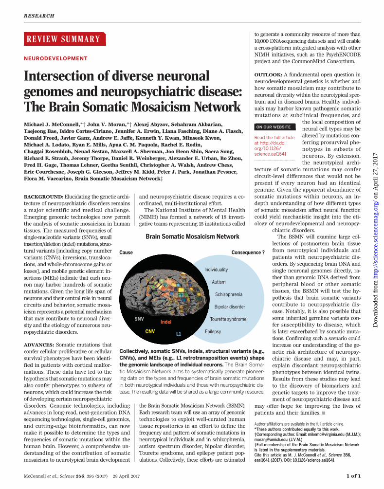
Collectively, somatic SNVs, indels, structural variants (e.g.,CNVs), and MEIs (e.g., L1 retrotransposition events) shapethe genomic landscape of individual neurons.The Brain Soma-tic Mosaicism Network aims to systematically generate pioneer-ing data on the types and frequencies of brain somatic mutationsin both neurotypical individuals and those with neuropsychiatric dis-ease.The resulting data will be shared as a large community resource.
ON OUR WEBSITE◥
Read the full articleat http://dx.doi.org/10.1126/science.aal1641..................................................
on
Apr
il 27
, 201
7ht
tp://
scie
nce.
scie
ncem
ag.o
rg/
Dow
nloa
ded
from
REVIEW◥
NEURODEVELOPMENT
Intersection of diverse neuronalgenomes and neuropsychiatric disease:The Brain Somatic Mosaicism NetworkMichael J. McConnell,1*† John V. Moran,2,3*† Alexej Abyzov,4 Schahram Akbarian,5
Taejeong Bae,4 Isidro Cortes-Ciriano,6 Jennifer A. Erwin,7 Liana Fasching,8
DianeA. Flasch,2 Donald Freed,9,10 JavierGanz,11,12 AndrewE. Jaffe,13 Kenneth Y.Kwan,2,14
Minseok Kwon,6 Michael A. Lodato,11,12 Ryan E. Mills,2,15 Apua C. M. Paquola,7
Rachel E. Rodin,11,12 Chaggai Rosenbluh,16 Nenad Sestan,17 Maxwell A. Sherman,6
Joo Heon Shin,13 Saera Song,18,19 Richard E. Straub,13 Jeremy Thorpe,9,10
Daniel R. Weinberger,13,20,21 Alexander E. Urban,22 Bo Zhou,22 Fred H. Gage,7
Thomas Lehner,23 Geetha Senthil,23 Christopher A. Walsh,11,12 Andrew Chess,16
Eric Courchesne,24 Joseph G. Gleeson,18,19 Jeffrey M. Kidd,2,15 Peter J. Park,6
Jonathan Pevsner,9,10 Flora M. Vaccarino,8,25 Brain Somatic Mosaicism Network‡
Neuropsychiatric disorders have a complex genetic architecture. Human genetic population-based studies have identified numerous heritable sequence and structural genomic variantsassociatedwith susceptibility to neuropsychiatric disease.However, these germline variants donot fully account fordisease risk. During brain development, progenitor cells undergobillionsofcell divisions to generate the ~80 billion neurons in the brain.The failure to accurately repairDNA damage arising during replication, transcription, and cellular metabolism amid thisdramatic cellular expansion can lead to somatic mutations. Somatic mutations that altersubsets of neuronal transcriptomes and proteomes can, in turn, affect cell proliferation andsurvival and lead to neurodevelopmental disorders.The long life span of individual neurons andthe direct relationship between neural circuits and behavior suggest that somaticmutations insmall populations of neurons can significantly affect individual neurodevelopment.The BrainSomatic Mosaicism Network has been founded to study somatic mosaicism both inneurotypical human brains and in the context of complex neuropsychiatric disorders.
The human body reaches a steady-state levelof approximately 1014 cells in adulthood.Because DNA replication and DNA repairare imperfect processes (estimated at ~0.27to 0.99 errors in ~109 nucleotides per cell
division) (1), somatic cells within an individualmust differ in the presence of single-nucleotidevariants (SNVs) and/or small insertion/deletion
(indel) mutations (2–4). In addition to SNVsand indels (5), subsets of neurons also harborstructural variants [which include large (>1 Mb)copy number variants (CNVs), inversions, trans-locations, andwhole-chromosome gains or losses(6–10)] and smaller mobile genetic element in-sertions (MEIs) (11–16). Here, we define somaticmosaicism as the existence of different genomes
within the cells of amonozygotic individual.Well-known examples of somatic mosaicism includeichthyosis with confetti and lines of Blaschko (4).Healthy neuronal development requires that
neural stem cells and progenitor cells (NPCs)undergo tens of billions of cell divisions, bothbefore birth and during the first years of life, togenerate the ~80 billion neurons in the fullydeveloped human brain (17). Because neuronsare among the longest-lived cells in the body, theaccumulation of somatic mutations (i.e., SNVs,indels, structural variants, andMEIs)withinNPCs,or perhaps postmitotic neurons (18), could influ-ence neuronal development, complexity, and func-tion (19, 20). Indeed, mounting evidence indicatesthat somatic mutations in small populations ofneurons contribute to various neurodevelopmentaldisorders (Table 1).Genomic studies implicitly assume that every
cell within an individual has the same genome.Family-based genetic studies, genome-wide asso-ciation studies (GWAS), and exome sequencinganalyses have identified numerous common, rare,and de novo germline SNVs and CNVs associatedwith an increased risk of autism spectrumdisorder(ASD),schizophrenia,andbipolardisorder,buteachvariant only represents a minor component ofpopulation-level disease risk (21–24). In general,these approaches sequence the DNA from avail-able clinical samples (e.g., peripheral blood) to in-terrogate an individual’s germline genome; theydo not account for any additional disease riskbrought about by somatic mutations that occurduringbraindevelopment.Toaddress thisknowl-edge gap, theNational Institute ofMentalHealth(NIMH) supported the formation of the Brain So-matic Mosaicism Network (BSMN). Notably, sev-eral outstanding reviews have recently discussedhow somatic mutations within the brain may con-tribute to neurological disease [e.g., (2, 25, 26)].Here,webuild on thesediscussions andhighlighthowsomaticmutationswithin thebrainmaycon-tribute to neuronal diversity.We also evaluateemerging genomic approaches to measure andvalidate somaticmosaicismand summarize BSMNefforts to generate a large publicly available re-source to evaluate the contribution of somaticmo-saicism to neuropsychiatric disease (Fig. 1).
RESEARCH
McConnell et al., Science 356, eaal1641 (2017) 28 April 2017 1 of 9
1Department of Biochemistry and Molecular Genetics, Department of Neuroscience, Center for Brain Immunology and Glia, Children’s Health Research Center, and Center for Public Health Genomics,University of Virginia School of Medicine, 1340 Jefferson Park Avenue, Charlottesville, VA 22908, USA. 2Department of Human Genetics, University of Michigan Medical School, 1241 East Catherine Street,Ann Arbor, MI 48109, USA. 3Department of Internal Medicine, University of Michigan, 1500 EastMedical Center Drive, Ann Arbor, MI 48109, USA. 4Department of Health Sciences Research, Center forIndividualized Medicine, Mayo Clinic, 200 1st Street S.W., Rochester, MN 55905, USA. 5Department of Psychiatry, Friedman Brain Institute, Icahn School ofMedicine at Mount Sinai Hess Center for Science andMedicine, 1470 Madison Avenue, New York, NY 10029, USA. 6Department of Biomedical Informatics, HarvardMedical School, 10 Shattuck Street, Boston, MA 02115, USA. 7The Salk Institute for BiologicalStudies, 10010 North Torrey Pines Road, La Jolla, CA 92037, USA. 8Child Study Center, Yale School of Medicine, 333 Cedar Street, NewHaven, CT 06520, USA. 9Department of Neurology, Kennedy KriegerInstitute, 707 North Broadway, Baltimore, MD 21205. 10Program in Biochemistry, Cellular and Molecular Biology, Johns Hopkins School of Medicine, 725 North Wolfe Street, Baltimore, MD 21205, USA.11Division of Genetics and Genomics, Manton Center for Orphan Disease, and Howard Hughes Medical Institute, Boston Children’s Hospital, 3 Blackfan Circle, Boston, MA 02115, USA. 12Departments ofNeurology and Pediatrics, Harvard Medical School, 25 Shattuck Street, Boston, MA 02115, USA. Broad Institute of MIT and Harvard, 415 Main Street, Cambridge, MA 02142, USA. 13Lieber Institute for BrainDevelopment, 855 North Wolfe Street, Baltimore, MD 21205, USA. 14Molecular and Behavioral Neuroscience Institute, University of MichiganMedical School, 109 Zina Pitcher Place, Ann Arbor, MI 48109, USA.15Department of Computational Medicine and Bioinformatics, University of Michigan Medical School, 100Washtenaw Avenue, Ann Arbor, MI 48109, USA. 16Department of Cell, Developmental and RegenerativeBiology, Department of Genetics and Genomic Sciences, Department of Neuroscience, Friedman Brain Institute, Icahn Institute for Genomics and Multiscale Biology, Icahn School ofMedicine at Mount Sinai,One Gustave L. Levy Place, New York, NY 10029, USA. 17Department of Neuroscience and Kavli Institute for Neuroscience, Yale School ofMedicine, 333 Cedar Street, New Haven, CT 06510, USA. 18HowardHughesMedical Institute, Laboratory of Pediatric Brain Disease, The Rockefeller University, 1230 York Avenue, New York, NY 10065. 19Rady Institute of GenomicMedicine, University of California, 9500 GilmanDrive, San Diego, La Jolla, CA 92093, USA. 20Departments of Psychiatry and Behavioral Sciences and Neuroscience, 600NorthWolfe Street, Baltimore, MD 21287, USA. 21McKusick-Nathans Institute ofGenetic Medicine, Johns Hopkins School of Medicine, 733 North Broadway, Baltimore, MD 21230, USA. 22Department of Psychiatry and Behavioral Sciences and Department of Genetics, Stanford UniversitySchool of Medicine, 3165 Porter Drive, Palo Alto, CA, 94304, USA. 23Office of Genomics Research Coordination, National Institute of Mental Health, National Institutes of Health, 6001 Executive Boulevard,Rockville, MD 20852, USA. 24AutismCenter of Excellence, Department of Neuroscience, School of Medicine, University of California San Diego, 8110 La Jolla Shores Drive, La Jolla, CA 92037, USA.25Department of Neuroscience, Yale School of Medicine, 333 Cedar Street, New Haven, CT 06520, USA.*These authors contributed equally to this work.†Corresponding author. Email: [email protected] (M.J.M.); [email protected] (J.V.M.)‡Full membership of the Brain Somatic Mosaicism Network is listed in the supplementary materials.
on
Apr
il 27
, 201
7ht
tp://
scie
nce.
scie
ncem
ag.o
rg/
Dow
nloa
ded
from
McConnell et al., Science 356, eaal1641 (2017) 28 April 2017 2 of 9
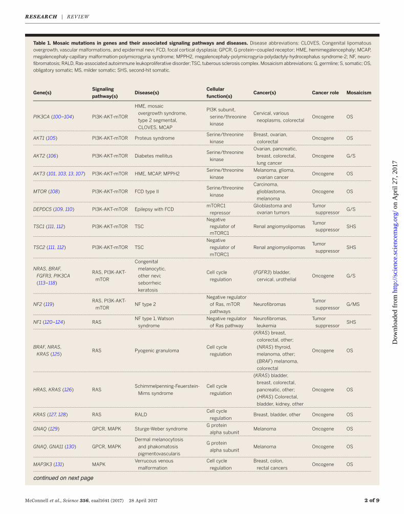
Table 1. Mosaic mutations in genes and their associated signaling pathways and diseases. Disease abbreviations: CLOVES, Congenital lipomatous
overgrowth, vascular malformations, and epidermal nevi; FCD, focal cortical dysplasia; GPCR, G protein–coupled receptor; HME, hemimegalencephaly; MCAP,
megalencephaly-capillary malformation-polymicrogyria syndrome; MPPH2, megalencephaly-polymicrogyria-polydactyly-hydrocephalus syndrome-2; NF, neuro-fibromatosis; RALD, Ras-associated autoimmune leukoproliferative disorder;TSC, tuberous sclerosis complex.Mosaicism abbreviations: G, germline; S, somatic; OS,
obligatory somatic; MS, milder somatic; SHS, second-hit somatic.
Gene(s)Signaling
pathway(s)Disease(s)
Cellular
function(s)Cancer(s) Cancer role Mosaicism
PIK3CA (100–104) PI3K-AKT-mTOR
HME, mosaic
overgrowth syndrome,
type 2 segmental,
CLOVES, MCAP
PI3K subunit,
serine/threonine
kinase
Cervical, various
neoplasms, colorectalOncogene OS
.. .. ... ... .. ... .. ... ... .. ... ... .. ... .. ... ... .. ... ... .. ... ... .. ... .. ... ... .. ... ... .. ... .. ... ... .. ... ... .. ... ... .. ... .. ... ... .. ... ... .. ... .. ... ... .. ... ... .. ... ... .. ... .. ... ... .. ... ... .. ... .. ... ... .. ... ... .. ... ... .. ... .. ... ... .. ... ... .. ... .. ... ... .. ... ... .. ... ... .. ... .. ... ... .. ... ... .. ... .. ... ... .. ... ... .. ... ... .. ... .. ... ... .. ... ... .
AKT1 (105) PI3K-AKT-mTOR Proteus syndromeSerine/threonine
kinase
Breast, ovarian,
colorectalOncogene OS
.. .. ... ... .. ... .. ... ... .. ... ... .. ... .. ... ... .. ... ... .. ... ... .. ... .. ... ... .. ... ... .. ... .. ... ... .. ... ... .. ... ... .. ... .. ... ... .. ... ... .. ... .. ... ... .. ... ... .. ... ... .. ... .. ... ... .. ... ... .. ... .. ... ... .. ... ... .. ... ... .. ... .. ... ... .. ... ... .. ... .. ... ... .. ... ... .. ... ... .. ... .. ... ... .. ... ... .. ... .. ... ... .. ... ... .. ... ... .. ... .. ... ... .. ... ... .
AKT2 (106) PI3K-AKT-mTOR Diabetes mellitusSerine/threonine
kinase
Ovarian, pancreatic,
breast, colorectal,
lung cancer
Oncogene G/S
.. .. ... ... .. ... .. ... ... .. ... ... .. ... .. ... ... .. ... ... .. ... ... .. ... .. ... ... .. ... ... .. ... .. ... ... .. ... ... .. ... ... .. ... .. ... ... .. ... ... .. ... .. ... ... .. ... ... .. ... ... .. ... .. ... ... .. ... ... .. ... .. ... ... .. ... ... .. ... ... .. ... .. ... ... .. ... ... .. ... .. ... ... .. ... ... .. ... ... .. ... .. ... ... .. ... ... .. ... .. ... ... .. ... ... .. ... ... .. ... .. ... ... .. ... ... .
AKT3 (101, 103, 13, 107) PI3K-AKT-mTOR HME, MCAP, MPPH2Serine/threonine
kinase
Melanoma, glioma,
ovarian cancerOncogene OS
.. .. ... ... .. ... .. ... ... .. ... ... .. ... .. ... ... .. ... ... .. ... ... .. ... .. ... ... .. ... ... .. ... .. ... ... .. ... ... .. ... ... .. ... .. ... ... .. ... ... .. ... .. ... ... .. ... ... .. ... ... .. ... .. ... ... .. ... ... .. ... .. ... ... .. ... ... .. ... ... .. ... .. ... ... .. ... ... .. ... .. ... ... .. ... ... .. ... ... .. ... .. ... ... .. ... ... .. ... .. ... ... .. ... ... .. ... ... .. ... .. ... ... .. ... ... .
MTOR (108) PI3K-AKT-mTOR FCD type IISerine/threonine
kinase
Carcinoma,
glioblastoma,
melanoma
Oncogene OS
.. .. ... ... .. ... .. ... ... .. ... ... .. ... .. ... ... .. ... ... .. ... ... .. ... .. ... ... .. ... ... .. ... .. ... ... .. ... ... .. ... ... .. ... .. ... ... .. ... ... .. ... .. ... ... .. ... ... .. ... ... .. ... .. ... ... .. ... ... .. ... .. ... ... .. ... ... .. ... ... .. ... .. ... ... .. ... ... .. ... .. ... ... .. ... ... .. ... ... .. ... .. ... ... .. ... ... .. ... .. ... ... .. ... ... .. ... ... .. ... .. ... ... .. ... ... .
DEPDC5 (109, 110) PI3K-AKT-mTOR Epilepsy with FCDmTORC1
repressor
Glioblastoma andovarian tumors
Tumorsuppressor
G/S.. .. ... ... .. ... .. ... ... .. ... ... .. ... .. ... ... .. ... ... .. ... ... .. ... .. ... ... .. ... ... .. ... .. ... ... .. ... ... .. ... ... .. ... .. ... ... .. ... ... .. ... .. ... ... .. ... ... .. ... ... .. ... .. ... ... .. ... ... .. ... .. ... ... .. ... ... .. ... ... .. ... .. ... ... .. ... ... .. ... .. ... ... .. ... ... .. ... ... .. ... .. ... ... .. ... ... .. ... .. ... ... .. ... ... .. ... ... .. ... .. ... ... .. ... ... .
TSC1 (111, 112) PI3K-AKT-mTOR TSC
Negative
regulator ofmTORC1
Renal angiomyolipomasTumorsuppressor
SHS
.. .. ... ... .. ... .. ... ... .. ... ... .. ... .. ... ... .. ... ... .. ... ... .. ... .. ... ... .. ... ... .. ... .. ... ... .. ... ... .. ... ... .. ... .. ... ... .. ... ... .. ... .. ... ... .. ... ... .. ... ... .. ... .. ... ... .. ... ... .. ... .. ... ... .. ... ... .. ... ... .. ... .. ... ... .. ... ... .. ... .. ... ... .. ... ... .. ... ... .. ... .. ... ... .. ... ... .. ... .. ... ... .. ... ... .. ... ... .. ... .. ... ... .. ... ... .
TSC2 (111, 112) PI3K-AKT-mTOR TSC
Negative
regulator of
mTORC1
Renal angiomyolipomasTumor
suppressorSHS
.. .. ... ... .. ... .. ... ... .. ... ... .. ... .. ... ... .. ... ... .. ... ... .. ... .. ... ... .. ... ... .. ... .. ... ... .. ... ... .. ... ... .. ... .. ... ... .. ... ... .. ... .. ... ... .. ... ... .. ... ... .. ... .. ... ... .. ... ... .. ... .. ... ... .. ... ... .. ... ... .. ... .. ... ... .. ... ... .. ... .. ... ... .. ... ... .. ... ... .. ... .. ... ... .. ... ... .. ... .. ... ... .. ... ... .. ... ... .. ... .. ... ... .. ... ... .
NRAS, BRAF,
FGFR3, PIK3CA
(113–118)
RAS, PI3K-AKT-
mTOR
Congenital
melanocytic,
other nevi;
seborrheic
keratosis
Cell cycle
regulation
(FGFR3) bladder,
cervical, urothelialOncogene G/S
.. .. ... ... .. ... .. ... ... .. ... ... .. ... .. ... ... .. ... ... .. ... ... .. ... .. ... ... .. ... ... .. ... .. ... ... .. ... ... .. ... ... .. ... .. ... ... .. ... ... .. ... .. ... ... .. ... ... .. ... ... .. ... .. ... ... .. ... ... .. ... .. ... ... .. ... ... .. ... ... .. ... .. ... ... .. ... ... .. ... .. ... ... .. ... ... .. ... ... .. ... .. ... ... .. ... ... .. ... .. ... ... .. ... ... .. ... ... .. ... .. ... ... .. ... ... .
NF2 (119)RAS, PI3K-AKT-
mTORNF type 2
Negative regulator
of Ras, mTOR
pathways
NeurofibromasTumor
suppressorG/MS
.. .. ... ... .. ... .. ... ... .. ... ... .. ... .. ... ... .. ... ... .. ... ... .. ... .. ... ... .. ... ... .. ... .. ... ... .. ... ... .. ... ... .. ... .. ... ... .. ... ... .. ... .. ... ... .. ... ... .. ... ... .. ... .. ... ... .. ... ... .. ... .. ... ... .. ... ... .. ... ... .. ... .. ... ... .. ... ... .. ... .. ... ... .. ... ... .. ... ... .. ... .. ... ... .. ... ... .. ... .. ... ... .. ... ... .. ... ... .. ... .. ... ... .. ... ... .
NF1 (120–124) RASNF type 1, Watson
syndrome
Negative regulator
of Ras pathway
Neurofibromas,
leukemia
Tumor
suppressorSHS
.. .. ... ... .. ... .. ... ... .. ... ... .. ... .. ... ... .. ... ... .. ... ... .. ... .. ... ... .. ... ... .. ... .. ... ... .. ... ... .. ... ... .. ... .. ... ... .. ... ... .. ... .. ... ... .. ... ... .. ... ... .. ... .. ... ... .. ... ... .. ... .. ... ... .. ... ... .. ... ... .. ... .. ... ... .. ... ... .. ... .. ... ... .. ... ... .. ... ... .. ... .. ... ... .. ... ... .. ... .. ... ... .. ... ... .. ... ... .. ... .. ... ... .. ... ... .
BRAF, NRAS,
KRAS (125)RAS Pyogenic granuloma
Cell cycle
regulation
(KRAS) breast,
colorectal, other;
(NRAS) thyroid,
melanoma, other;
(BRAF) melanoma,
colorectal
Oncogene OS
.. .. ... ... .. ... .. ... ... .. ... ... .. ... .. ... ... .. ... ... .. ... ... .. ... .. ... ... .. ... ... .. ... .. ... ... .. ... ... .. ... ... .. ... .. ... ... .. ... ... .. ... .. ... ... .. ... ... .. ... ... .. ... .. ... ... .. ... ... .. ... .. ... ... .. ... ... .. ... ... .. ... .. ... ... .. ... ... .. ... .. ... ... .. ... ... .. ... ... .. ... .. ... ... .. ... ... .. ... .. ... ... .. ... ... .. ... ... .. ... .. ... ... .. ... ... .
HRAS, KRAS (126) RASSchimmelpenning-Feuerstein-
Mims syndrome
Cell cycle
regulation
(KRAS) bladder,
breast, colorectal,
pancreatic, other;
(HRAS) Colorectal,
bladder, kidney, other
Oncogene OS
.. .. ... ... .. ... .. ... ... .. ... ... .. ... .. ... ... .. ... ... .. ... ... .. ... .. ... ... .. ... ... .. ... .. ... ... .. ... ... .. ... ... .. ... .. ... ... .. ... ... .. ... .. ... ... .. ... ... .. ... ... .. ... .. ... ... .. ... ... .. ... .. ... ... .. ... ... .. ... ... .. ... .. ... ... .. ... ... .. ... .. ... ... .. ... ... .. ... ... .. ... .. ... ... .. ... ... .. ... .. ... ... .. ... ... .. ... ... .. ... .. ... ... .. ... ... .
KRAS (127, 128) RAS RALDCell cycle
regulationBreast, bladder, other Oncogene OS
.. .. ... ... .. ... .. ... ... .. ... ... .. ... .. ... ... .. ... ... .. ... ... .. ... .. ... ... .. ... ... .. ... .. ... ... .. ... ... .. ... ... .. ... .. ... ... .. ... ... .. ... .. ... ... .. ... ... .. ... ... .. ... .. ... ... .. ... ... .. ... .. ... ... .. ... ... .. ... ... .. ... .. ... ... .. ... ... .. ... .. ... ... .. ... ... .. ... ... .. ... .. ... ... .. ... ... .. ... .. ... ... .. ... ... .. ... ... .. ... .. ... ... .. ... ... .
GNAQ (129) GPCR, MAPK Sturge-Weber syndromeG protein
alpha subunitMelanoma Oncogene OS
.. .. ... ... .. ... .. ... ... .. ... ... .. ... .. ... ... .. ... ... .. ... ... .. ... .. ... ... .. ... ... .. ... .. ... ... .. ... ... .. ... ... .. ... .. ... ... .. ... ... .. ... .. ... ... .. ... ... .. ... ... .. ... .. ... ... .. ... ... .. ... .. ... ... .. ... ... .. ... ... .. ... .. ... ... .. ... ... .. ... .. ... ... .. ... ... .. ... ... .. ... .. ... ... .. ... ... .. ... .. ... ... .. ... ... .. ... ... .. ... .. ... ... .. ... ... .
GNAQ, GNA11 (130) GPCR, MAPK
Dermal melanocytosis
and phakomatosis
pigmentovascularis
G protein
alpha subunitMelanoma Oncogene OS
.. .. ... ... .. ... .. ... ... .. ... ... .. ... .. ... ... .. ... ... .. ... ... .. ... .. ... ... .. ... ... .. ... .. ... ... .. ... ... .. ... ... .. ... .. ... ... .. ... ... .. ... .. ... ... .. ... ... .. ... ... .. ... .. ... ... .. ... ... .. ... .. ... ... .. ... ... .. ... ... .. ... .. ... ... .. ... ... .. ... .. ... ... .. ... ... .. ... ... .. ... .. ... ... .. ... ... .. ... .. ... ... .. ... ... .. ... ... .. ... .. ... ... .. ... ... .
MAP3K3 (131) MAPKVerrucous venous
malformation
Cell cycle
regulation
Breast, colon,
rectal cancersOncogene OS
.. .. ... ... .. ... .. ... ... .. ... ... .. ... .. ... ... .. ... ... .. ... ... .. ... .. ... ... .. ... ... .. ... .. ... ... .. ... ... .. ... ... .. ... .. ... ... .. ... ... .. ... .. ... ... .. ... ... .. ... ... .. ... .. ... ... .. ... ... .. ... .. ... ... .. ... ... .. ... ... .. ... .. ... ... .. ... ... .. ... .. ... ... .. ... ... .. ... ... .. ... .. ... ... .. ... ... .. ... .. ... ... .. ... ... .. ... ... .. ... .. ... ... .. ... ... .
continued on next page
RESEARCH | REVIEW
on
Apr
il 27
, 201
7ht
tp://
scie
nce.
scie
ncem
ag.o
rg/
Dow
nloa
ded
from
Mechanisms of somatic mosaicismDNA damage occurs constantly in every cell inour bodies, and many components of the DNAdamage response are essential for neurodevel-opment. Single-strand and double-strand DNAbreaks, as well as base mutations, arise as aconsequence of DNA replication, transcription,epigenetic modification, cellular respiration, andenvironmental stressors. If the resultant damageis not accurately repaired, DNA mutations canoccur that can lead to somatic variation amongneurons and other cell types.The nonhomologous end-joining (NHEJ) path-
way of DNA repair is required for neurodevelop-ment. Mice deficient in NHEJ proteins exhibitextensive NPC apoptosis and often die prenatally(27). Intriguingly, the embryonic lethality andNPC apoptosis phenotypes are rescued in a p53-null mouse background, suggesting that genotoxicstress contributes to lethality (28). Consistent withthese data, compound heterozygous mutations inDNA damage response genes [e.g., ataxia telan-
giectasia mutated (ATM), ataxia telangiectasia-related (ATR), and ATR-interacting protein (ATRIP)]can lead to increased mutational loads, neuro-developmental brain defects, and neuronal de-generation (29–31). More broadly, deficits in otherDNA repair pathways, such as transcription-coupled repair, homologous recombination, andnucleotide excision repair, also can lead to hu-man neurodevelopmental phenotypes (32, 33).Defects in different DNA repair pathways are
associated with distinct somatic mutation profiles.For example, SNVs and indels can arise fromerrors during base excision repair, nucleotideexcision repair, and transcription-coupled repair(33). Moreover, the action of the apolipoproteinB mRNA editing enzyme, catalytic polypeptide-like-3 (APOBEC3) family of cytosine deaminaseproteins can lead to cytidine-to-uridine transitionmutations on single-strand DNA that, upon rep-lication, lead to guanosine-to-adenosine muta-tions on the opposing DNA strand (34). Errorsmade during DNAmismatch repair also can lead
to either interspersed SNVs or indels within mi-crosatellite repeat sequences, whereas errors madeduring double-strand break repair by homolo-gous recombination, NHEJ, or alternative-NHEJcan lead to CNVs (35, 36).Errors incurred during DNA replication or
transcription also can lead to the formation ofCNVs. Large, actively transcribed genes that un-dergo replication during late S-phase correspondto chromosomal fragile sites and are hot spotsfor the generation of genomic variants and trans-locations (37, 38). Because neuronal genes areoverrepresented among the longest genes in thehuman genome, transcription may predisposethese genes to somatic CNVs (39). Indeed, in-tragenic deletions within large, neuronally ex-pressed genes (e.g., AUTS2, IMMP2L,NXRN1, andCNTNAP2) are associated with ASD, intellectualdisability, and other neurodevelopmental disor-ders (40, 41). Thus, if individuals harbor somaticCNVs at these loci in many neurons or in neu-rons within specific functional brain regions, theymay be susceptible to neurological disease.Long interspersed element-1s (LINE-1s or L1s)
canmobilize (i.e., retrotranspose)within the brain,leading to another form of somatic variation(42). Active L1s encode two proteins, ORF1p andORF2p, which are required for retrotransposition.ORF2p contains endonuclease and reverse tran-scriptase activities that are needed to “copy-and-paste” L1 sequences into a new genomic locationby amechanism termed target-site primed reversetranscription (TPRT) (42,43). In addition to canon-ical TPRT, L1s occasionally can integrate intoendogenous DNA lesions (44). Moreover, recom-bination events that arise either during (15, 45–47)or after L1 retrotransposition (48) can lead to theformation of structural variants.
Somatic mutations in human diseaseMosaicism and structural brainabnormalities
One of the most common causes of medicallyrefractory pediatric epilepsy is focal dysplasia ofthe cerebral cortex. Until recently, the basis of
McConnell et al., Science 356, eaal1641 (2017) 28 April 2017 3 of 9
Gene(s)Signaling
pathway(s)Disease(s)
Cellular
function(s)Cancer(s) Cancer role Mosaicism
.. .. ... ... .. ... .. ... ... .. ... ... .. ... .. ... ... .. ... ... .. ... ... .. ... .. ... ... .. ... ... .. ... .. ... ... .. ... ... .. ... ... .. ... .. ... ... .. ... ... .. ... .. ... ... .. ... ... .. ... ... .. ... .. ... ... .. ... ... .. ... .. ... ... .. ... ... .. ... ... .. ... .. ... ... .. ... ... .. ... .. ... ... .. ... ... .. ... ... .. ... .. ... ... .. ... ... .. ... .. ... ... .. ... ... .. ... ... .. ... .. ... ... .. ... ... .
GNAS (132, 133) GPCRMcCune-Albright
syndrome
G protein alpha
subunit
Adenomas,
carcinomas,
ovarian neoplasms
Oncogene OS
.. .. ... ... .. ... .. ... ... .. ... ... .. ... .. ... ... .. ... ... .. ... ... .. ... .. ... ... .. ... ... .. ... .. ... ... .. ... ... .. ... ... .. ... .. ... ... .. ... ... .. ... .. ... ... .. ... ... .. ... ... .. ... .. ... ... .. ... ... .. ... .. ... ... .. ... ... .. ... ... .. ... .. ... ... .. ... ... .. ... .. ... ... .. ... ... .. ... ... .. ... .. ... ... .. ... ... .. ... .. ... ... .. ... ... .. ... ... .. ... .. ... ... .. ... ... .
JAK2 (134, 135) JAK-STAT
Myelofibrosis,
polycythemia vera,
and essential
thrombocythemia
Cell cycle
regulationLeukemia Oncogene SHS
.. .. ... ... .. ... .. ... ... .. ... ... .. ... .. ... ... .. ... ... .. ... ... .. ... .. ... ... .. ... ... .. ... .. ... ... .. ... ... .. ... ... .. ... .. ... ... .. ... ... .. ... .. ... ... .. ... ... .. ... ... .. ... .. ... ... .. ... ... .. ... .. ... ... .. ... ... .. ... ... .. ... .. ... ... .. ... ... .. ... .. ... ... .. ... ... .. ... ... .. ... .. ... ... .. ... ... .. ... .. ... ... .. ... ... .. ... ... .. ... .. ... ... .. ... ... .
SCN1A (136) Sodium channel Dravet syndrome Neural excitation – – G/MS.. .. ... ... .. ... .. ... ... .. ... ... .. ... .. ... ... .. ... ... .. ... ... .. ... .. ... ... .. ... ... .. ... .. ... ... .. ... ... .. ... ... .. ... .. ... ... .. ... ... .. ... .. ... ... .. ... ... .. ... ... .. ... .. ... ... .. ... ... .. ... .. ... ... .. ... ... .. ... ... .. ... .. ... ... .. ... ... .. ... .. ... ... .. ... ... .. ... ... .. ... .. ... ... .. ... ... .. ... .. ... ... .. ... ... .. ... ... .. ... .. ... ... .. ... ... .
NLRP3 (137)Caspase/
inflammasomeCINCA syndrome
Inflammasome
subunit– – G/MS
.. .. ... ... .. ... .. ... ... .. ... ... .. ... .. ... ... .. ... ... .. ... ... .. ... .. ... ... .. ... ... .. ... .. ... ... .. ... ... .. ... ... .. ... .. ... ... .. ... ... .. ... .. ... ... .. ... ... .. ... ... .. ... .. ... ... .. ... ... .. ... .. ... ... .. ... ... .. ... ... .. ... .. ... ... .. ... ... .. ... .. ... ... .. ... ... .. ... ... .. ... .. ... ... .. ... ... .. ... .. ... ... .. ... ... .. ... ... .. ... .. ... ... .. ... ... .
PORCN (138) WntFocal dermal
hypoplasiaO-acyltransferase – – G/MS
.. .. ... ... .. ... .. ... ... .. ... ... .. ... .. ... ... .. ... ... .. ... ... .. ... .. ... ... .. ... ... .. ... .. ... ... .. ... ... .. ... ... .. ... .. ... ... .. ... ... .. ... .. ... ... .. ... ... .. ... ... .. ... .. ... ... .. ... ... .. ... .. ... ... .. ... ... .. ... ... .. ... .. ... ... .. ... ... .. ... .. ... ... .. ... ... .. ... ... .. ... .. ... ... .. ... ... .. ... .. ... ... .. ... ... .. ... ... .. ... .. ... ... .. ... ... .
PIGA (139) Hematopoiesis
Paroxysmal
nocturnal
hemoglobinuria
ER protein
processingLeukemia – OS
.. .. ... ... .. ... .. ... ... .. ... ... .. ... .. ... ... .. ... ... .. ... ... .. ... .. ... ... .. ... ... .. ... .. ... ... .. ... ... .. ... ... .. ... .. ... ... .. ... ... .. ... .. ... ... .. ... ... .. ... ... .. ... .. ... ... .. ... ... .. ... .. ... ... .. ... ... .. ... ... .. ... .. ... ... .. ... ... .. ... .. ... ... .. ... ... .. ... ... .. ... .. ... ... .. ... ... .. ... .. ... ... .. ... ... .. ... ... .. ... .. ... ... .. ... ... .
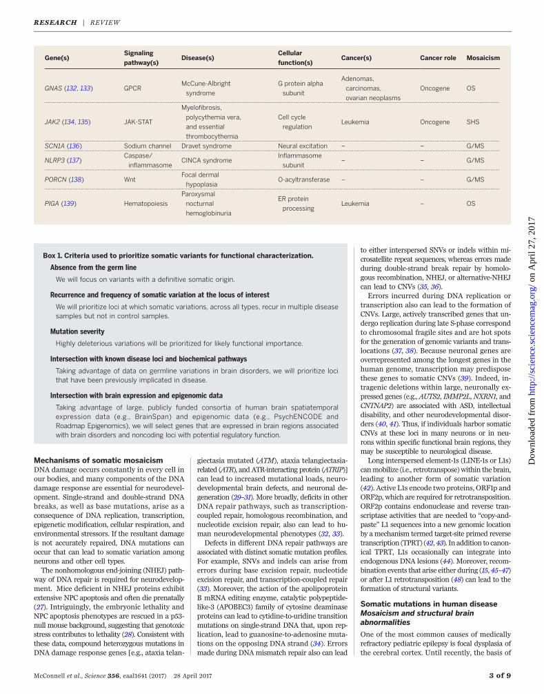
Box 1. Criteria used to prioritize somatic variants for functional characterization.
Absence from the germ line
We will focus on variants with a definitive somatic origin.
Recurrence and frequency of somatic variation at the locus of interest
We will prioritize loci at which somatic variations, across all types, recur in multiple diseasesamples but not in control samples.
Mutation severity
Highly deleterious variations will be prioritized for likely functional importance.
Intersection with known disease loci and biochemical pathways
Taking advantage of data on germline variations in brain disorders, we will prioritize locithat have been previously implicated in disease.
Intersection with brain expression and epigenomic data
Taking advantage of large, publicly funded consortia of human brain spatiatemporalexpression data (e.g., BrainSpan) and epigenomic data (e.g., PsychENCODE andRoadmap Epigenomics), we will select genes that are expressed in brain regions associatedwith brain disorders and noncoding loci with potential regulatory function.
RESEARCH | REVIEW
on
Apr
il 27
, 201
7ht
tp://
scie
nce.
scie
ncem
ag.o
rg/
Dow
nloa
ded
from
this disorder remained a medical mystery. Geneticstudies of the most severe form of focal dysplasia,hemimegalencephaly, in which one entire cere-bral hemisphere is enlarged in size, led to theidentification of gain-of-function somatic muta-tions in the phosphatidylinositol-3-kinase (PI3K)–protein kinase B (Akt) and mammalian target ofrapamycin (mTOR) signaling pathways (Table 1,Fig. 2). We now know that mutations in mTORare the single largest contributor to focal dyspla-sia in pediatric epilepsy (49–51). Similarly, germ-line mutations in one allele of the TSC1 or TSC2gene confer susceptibility to tuberous sclerosis, adisease characterized by facial and skin lesions,seizures, intellectual disability, cardiac and renaltumors, and cortical tubers (52). Because the Tsc1and Tsc2 proteins are negative regulators of themTOR-signaling pathway, a second somaticallyacquired mutation is required for disease onset.Somatic mutations that mildly activate the mTOR-signaling pathway also cause symmetrical over-growth syndromes such asmegalencephaly-capillarymalformation syndrome, megalencephaly, andcertain forms of polymicrogyria (49–51). Com-mon to all of these phenotypes is the presence ofhypertrophic neural-like “balloon” cells, whichcarry the somatic mutation yet fail to transform toamalignant cell type (52).Somatic mutations that inappropriately acti-
vate Ras signaling or related signaling pathwayscan likewise confer proliferation and survival phe-notypes to subsets of cells and cause neurologicaldisease. For example, a gain-of-function somaticmutation in GNAQ, encoding G protein subunitalpha q, can lead to Sturge-Weber syndrome, adisease characterized by vascular anomaly in thebrain, glaucoma, seizures, stroke, and intellectualdisability (53). The same GNAQ mutation, occur-ring in a different somatic cell type later in de-velopment, can cause uveal melanoma (54). Because
mutations in certain neurodevelopmental dis-orders (e.g., neurofibromatosis, tuberous sclero-sis, Proteus syndrome, and other neurocutaneousdisorders) either activate proto-oncogenes or in-activate tumor suppressor genes, it is not sur-prising that similar mutations in non-neuronalcell types manifest as cancers. Intriguingly, post-mitotic neurons are rarely the source of braintumors, suggesting that postmitotic neurons mayhave safeguards that ensure against dediffer-entiation and further proliferation.Relative to germline mutations, somatic mu-
tations can lead to milder cases of heritable neu-rodevelopmental disorders. For example, somaticmutations in genes involved in neuronal migra-tion are estimated to represent 5 to 10% of de novomutations and are detected more frequently inpatients with unexplained brain malformationswhen studied with sensitive high-throughput se-quencing methods (55). Moreover, somatic muta-tions within the LIS1 or DCX genes can lead togross disruptions of neuronal migration, whereasgermline mutations in LIS1 or DCX result inlissencephaly (56, 57). Results from several ex-periments also suggest that somatic mutationsthat lead to a reduction of gene copy number inmigrating neurons can lead to cell-autonomousdefects in neuronalmigration, with severe epilepsyand intellectual disability as a consequence (56, 57).
ASD and other commonneuropsychiatric diseases
Genetic approaches have not yet fully explainedthe etiology of ASD, bipolar disorder, schizo-phrenia, or Tourette syndrome. Although gene-by-gene and gene-by-environment interactionscould, in principle, account for additional dis-ease risk, somatic mosaicism is another potentialmechanism that warrants exploration as a con-tributor to neuropsychiatric diseases (58).
De novo SNVs and CNVs, particularly loss-of-function mutations, are significant contributorsto ASD risk (21, 59–62). In addition to de novogermline mutations, a substantial number of denovo somatic mutations (i.e., ~5.4% of de novoevents) are detected in the blood of ASD patientsand are enriched in ASD probands (22). Somaticmosaic mutations also have been identifiedthroughout postmortem ASD brains or, in someinstances, in more localized areas in ASD brains(59). Evidence of continuous, widespread corti-cal mismigration, as seen in some mutant mice,has not been reported in the postmortem ASDbrain (63, 64). However, NPCs from a subset ofASD patients with enlarged brain volumes areinherently more proliferative and display abnor-mal neurogenesis when compared to controls(65, 66). Other ASD patients have focal corticalabnormalities, including disorganized neuronsand lamina, polymicrogyria, and other local sur-face malformations (67). Thus, in addition to spe-cific mutations, additional cell cycles may furtheraffect somatic mutational loads in patients.Prenatal challenges to the immune system in
animals (i.e., maternal immune activation) (68)can also lead to many features like those presentin ASDbrains.Maternal immune activation leadsto increased cellular proliferation, brain size, andASD-like behaviors in animal models (69–72).Intriguingly, an elevated prevalence of MEIs wasobserved in a primatemodel ofmaternal immuneactivation (73). Elevated MEI levels likewise areobserved in schizophrenia (73) andRett syndromepatients (74), suggesting that somaticMEI burdenmay play a role in the etiology of some neuro-developmental and neuropsychiatric diseases.
Methods to detect somatic mutations
The difficulty in detecting a somatic mutationdepends on its frequency within a cell population.
McConnell et al., Science 356, eaal1641 (2017) 28 April 2017 4 of 9
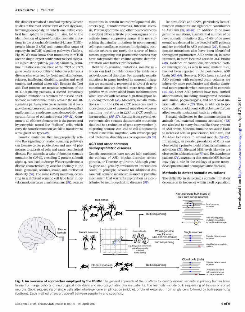
Fig. 1. An overview of approaches employed by the BSMN. The general approach of the BSMN is to identify mosaic variants in primary human braintissue from large cohorts of neurotypical individuals and neuropsychiatric disease patients. The methods include bulk sequencing of tissues or sortedneurons (top), sequencing of single cells after whole-genome amplification (middle), or clonal expansion from single cells followed by bulk sequencing(bottom). Each method offers a trade-off between sensitivity and specificity.
RESEARCH | REVIEW
on
Apr
il 27
, 201
7ht
tp://
scie
nce.
scie
ncem
ag.o
rg/
Dow
nloa
ded
from
Whereas mutations affecting a large fraction(e.g., 50%) of cells are readily detected in bulktissue sequencing experiments and generally re-sult in high-confidence calls, mutations affect-ing one or a few cells are unlikely to be detectedwith bulk tissue sequencing approaches. The iden-tification and validation of rare somatic mutationsrequires sequencing DNA derived from smallpools of cells, single cells, or clonally repro-grammed cells followed by robust computationaldata analyses (Fig. 1).
Bulk tissue approaches
Whole-genomesequencing (WGS) orwhole-exomesequencing (WES) of DNAderived frombulk braintissue allows a straightforward approach to dis-covering somatic mosaicism (26). WGS andWESminimize sequencing artifacts that can confounddownstream analyses and, in the case of WGS,provide an opportunity for identifying a widerange of structural rearrangements, includinginversions and translocations. However, WGSandWES using standard sequencing depths havereduced statistical power to detect mutationsthat occur at low frequencies (i.e., <10% of cells ina population at 30 to 100x coverage). Althoughincreasing sequence coverage allows detection ofsomatic variants at lower frequencies, it quicklybecomes cost prohibitive. Moreover, WGS andWES do not provide information on how somaticvariants are distributed across individual cell lin-eages within a bulk tissue sample.
Sorted-pools approaches
Fluorescence-activated cell or nuclei sorting (FACS/FANS) can be used to isolate specific neural pop-ulations (e.g., NeuN+ neurons versus NeuN– cells
or cortical inhibitory interneurons versus excit-atory principal neurons). Analysis of sorted nu-clei populations (e.g., 5000 or 500,000 cells) fromspecific brain regions increases the power to de-tect somatic mosaicism that arises in one lineage,because these genomes are no longer diluted bygenomesderived fromother lineages. Independentpools of sorted nuclei can then be subjected toRNA sequencing (RNA-seq) and quantitative re-verse transcription polymerase chain reaction(qRT-PCR) to confirm cell type–specific gene ex-pression profiles (75). In addition to increasingthe power for detecting a somatic mutation, cellsorting before DNA extraction could yield infor-mation about the embryological origin and de-velopmental trajectory of somatic variation acrossthe brain. Large pools of sorted cells can yieldenoughDNA for the direct examination of somaticvariants by WGS or WES. However, smaller poolsizes will only generate small amounts of DNA;thus, they are best suited for generating PCRamplicon libraries (e.g., as used inMEI detectionand other targeted sequencing) or for subsequentwhole-genome amplification (WGA).
Single-cell approaches
WGA can be used to analyze the genomes ofsingle neurons (26). The spectrum of mutationsidentified from the genomes of single neuronscan then be compared to germline variants inbulk tissue data derived from a non-neuronalcontrol (e.g., brain dural fibroblasts or heart) toidentify candidate somatic mutations (5). WGAapproaches already are used in pre-implantationgenetic screening of embryos (76, 77) and include(i) degenerate-oligonucleotide-primed PCR (DOP-PCR), (ii) multiple displacement amplification
(MDA), and (iii) multiple annealing and looping-based amplification (MALBAC). Each methodhas its advantages and drawbacks. In general,DOP-PCR provides coverage evenly across thegenome, which facilitates the detection of largeCNVs and chromosomal aneuploidies. However,DOP-PCR has a higher read duplication rate, lowermapping rate, and lower recovery rate whencompared with MDA and MALBAC (78) and iscost prohibitive for SNV, indel, and MEI detec-tion. By comparison, MDA yields a high rate ofartificial chimeric DNA molecules that can leadto false-positive calls in downstream analyses(79), whereas MALBAC exhibits reduced coverageof certain genomic regions (14, 16, 80), especiallythose rich in repetitive sequences (78). Consider-able advances have recently been made in detect-ing SNVs (81, 82), CNVs (83), and MEIs (16) inWGA samples; however, best practices necessi-tate evaluating each WGA approach for the de-tection of specific types of somatic mosaicism.Clonal expansion of single cells using human-
induced pluripotent stem cell (hiPSC) technologyor somatic cell nuclear transfer (SCNT) providesa biological alternative to WGA (80, 84). Anyvariant uniformly identified in the clonal line,but not in controls, represents a candidate so-matic mutation that requires confirmation in thetissue of origin. In contrast, mutations introducedduring cell culture will be present in a lowerfrequency of cells within a clonal cell line andcan be discriminated from bona fide somaticmutations in downstream computational analy-sis. Although the clonal isolation and expansionof primary human neural stem and progenitorcells is possible, the analysis of human neuronalgenomes using clonal reprogramming has severallimitations. Foremost among these is the avail-ability of live human neurons. Moreover, neitherclonal reprogramming nor SCNT have been re-ported using human neurons; SCNT is furtherlimited by the expense and availability of humanoocytes. Finally, reprogramming approaches cur-rently are only successful in ~10% of cells; thus,any neurons harboring highly aberrant genomesmay be refractory to reprogramming. Despite thesecaveats, clonal reprogramming of human neuronsis theoretically possible. In addition, it is note-worthy that mouse neurons reprogrammed bySCNT contain genomic rearrangements (e.g.,kataegis and chromothripsis) that would be verychallenging to validate using current WGA ap-proaches (84).
Computational methods formutation detection
WGS and WES have been used successfully todetect somatic SNVs in family-based studiesof Mendelian disease and large-scale sequencingstudies of human patient cohorts (2). To identifySNVs, most computational approaches comparecall sets generated from an affected sample tothose generated from amatched healthy/unaffectedsample and/or a control population. These com-parisons allow the identification and subsequentexclusion of germline polymorphisms from down-stream analyses; however, care must be taken to
McConnell et al., Science 356, eaal1641 (2017) 28 April 2017 5 of 9
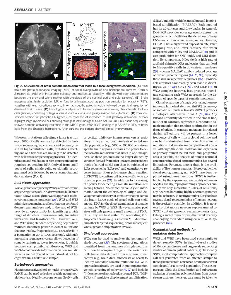
Fig. 2. An example of brain somatic mosaicism that leads to a focal overgrowth condition. (A) Axialbrain magnetic resonance imaging (MRI) of focal overgrowth of one hemisphere (arrows) from a2-month-old child with intractable epilepsy and intellectual disability. MRI showed poor differentiationbetween the gray and white matter with dysplasia of the cortical gyri and sulci (arrows). (B) Brainmapping using high-resolution MRI or functional imaging such as positron emission tomography (PET),together with electrocorticography to fine-map specific epileptic foci, is followed by surgical resection ofdiseased brain tissue. (C) Histological analysis with hematoxylin/eosin showing characteristic ballooncells (arrows) consisting of large nuclei, distinct nucleoli, and glassy eosinophilic cytoplasm. (D) Immuno-stained section for phospho-S6 (green), as evidence of increased mTOR pathway activation. Arrowshighlight large dysplastic cell showing strongest immunosignal. Scale bar, 50 mm. Bulk tissue sequencingshowed somatic activating mutation in the MTOR gene c.6644C>T leading to p.S2215F in 15% of braincells from the diseased hemisphere. After surgery, the patient showed clinical improvement.
RESEARCH | REVIEW
on
Apr
il 27
, 201
7ht
tp://
scie
nce.
scie
ncem
ag.o
rg/
Dow
nloa
ded
from
ensure that any candidate somatic mutations arenot germline variants that were missed in thematched control. In general, variant callers ini-tially developed to detect mutations in canceroffer higher sensitivity for detecting mosaicSNVs when compared with standard approachesused to detect germline variants (85, 86).Somatic CNVs can be detected by identify-
ing deviations either from the expected depthof sequence or in the expected distances be-tween paired-end sequencing reads. Similarly,inversions can be identified through differencesin the orientations of paired-end sequencing reads.Numerous approaches have been developed toidentify CNVs fromWGS (7, 87–89), and most canbe applied directly to identify somatic mutations.For example, recent studies using WGA in con-junction withWGS have identified megabase-scalede novo CNVs in human and mouse neuronsbased on differences in read-depth across genomicbins (6–9). CNVs are more difficult to identifyusing WES due to the biases encountered duringthe capture of target exons (90).Somatic MEIs can be detected from bulk tis-
sue, PCR amplicons generated from sorted-cellfractions, or single-cell WGA DNA using split-readand paired-end information (e.g., one paired-endread may map to the reference genome, whereasanother may map to a MEI) (91, 92). Detectinglow-frequency MEIs with fewer supporting readsrequires careful bioinformatic analyses that candistinguish signal from noise, followed by ex-perimental validation with orthogonal methods(14, 93). The analysis of single-cell data remainschallenging due to the presence of chimeras gen-erated during WGA (14, 16, 94); thus, care mustbe taken in calling MEIs.
Validation of somatic mutations
It is essential to validate all candidate somaticmutations. False-positive calls can arise from DNAsequencing errors, contamination with germlinevariants, chimeric molecules generated duringsingle-cell WGA, PCR-induced nucleotide sub-stitutions, and the failure to amplify certain ge-nomic regions. False-negative calls are dependenton the allele frequency of the somatic mutationwithin the sample, the type of mutation, and themethod of detection. Orthologous experimentalmethods are required to eliminate false-positivesand to calibrate the confidence of detection fordifferent types of somatic mutations. Validationexperiments can then be performed on either thetissue of origin or amplified material used to dis-cover the variant. The first approach represents abiological validation, which establishes the pres-ence of a variant call in unamplified DNA fromthe source sample. The second approach repre-sents a technical validation, which establishesthe presence/absence of variant calls in the DNAsource material used for discovery.
Biological/primary validation in thetissue of origin
Validation on unamplified DNA from the tissueof origin provides confirmation that a candidatecall is a genuine somatic variant and rules out
the possibility that it corresponds to a DNA am-plification artifact or a mutation that occurredduring clonal expansion. Biological validation re-quires a variant to be present in multiple cells inthe tissue of origin at a frequency above experi-mental detection limits. As such, the failure tovalidate a variant in the tissue of origin does notnecessarily represent a false call. For example,only ~50% of CNVs manifested in hiPSC clonescould be directly confirmed in the primary fibro-blast cells used to derive hiPSCs (80).Somatic variants can be confirmed in unam-
plified cell source material by (i) targeted DNAcapture followed by high-coverage (>100x) DNAresequencing, (ii) high-coverage sequencing ofmultiplexed PCR amplicons, and (iii) droplet dig-ital PCR (ddPCR). These approaches vary inthroughput and sensitivity. Targeted DNA cap-ture and resequencing can require the creationof several thousand custom oligonucleotides de-signed to capture the genomic DNA either includ-ing or surrounding the putative variants. Thecaptured DNA then is subjected to high-coveragepaired-end DNA sequencing, yielding a typicalsensitivity of variant detection in greater than1% of cells. Amplicon sequencing involves PCRamplification of candidate loci followed by high-coverage paired-end DNA sequencing, yieldinga typical sensitivity of variant detection in greaterthan 0.1% of cells. Finally, ddPCR involves par-titioning a DNA sample into large numbers ofindividual droplets that generally contain onecopy of template DNA. PCR takes place withinthese droplets, leading to the production of afluorescent readout, either through the use of anintercalating dye or a fluorescent oligomer probe,to indicate the presence or absence of the PCRtarget of interest. Subsequent quantification ofthe fluorescent droplets allows a determinationof the number of copies of the target locus presentin the sample, yielding a typical sensitivity ofvariant detection in greater than 0.001% of cells(95). Although extremely sensitive, ddPCR requiresthe optimization of primers, probes, and am-
plification conditions, which is time-consumingand limits throughput.The goal when employing biological valida-
tion procedures is to detect putative somaticvariants and to assess, as precisely as possible,the frequency of each variant in that tissue oforigin. Biological validation can (i) determinewhether certain individuals in the populationare more prone to somatic variation than others,(ii) investigate whether different areas of the brainand/or specific brain cell types have varyingamounts and types of particular forms of somaticvariation, (iii) assess whether developmentaltiming contributes to somatic variation, and (iv)reveal whether somatic variations increase as afunction of the number of cell divisions and/ora function of age in postmitotic neurons.
Technical validation onsource/amplified material
If a somatic variant is only present in a single cell,it will be impossible to validate in bulk tissue.Likewise, a variant present in very few cells maybe difficult to validate in the tissue of origin. Thus,technical validation in the source DNA used to dis-cover a putative variant can be used to determinewhether a call is true or false. Technical validationtypically employs PCR, qPCR, and Sanger sequenc-ing of the locus in the DNA source material (e.g.,WGA DNA or DNA from a clonal cell population).Multiple true/false verdicts form the basis forestimating false-discovery and false-negative ratesin the resultant call sets.
Present understanding of theprevalence of somatic mutation inneurotypical individuals
Recent studies revealed that mosaic neuronalgenomes are the rule, rather than the exception;every neuron probably has a different genomethan the neurons with which it forms synapses.Not unexpectedly, SNVs are the most prevalentsomatic mutations. A “triple calling” strategy wasused to identify and validate clonal SNVs in
McConnell et al., Science 356, eaal1641 (2017) 28 April 2017 6 of 9
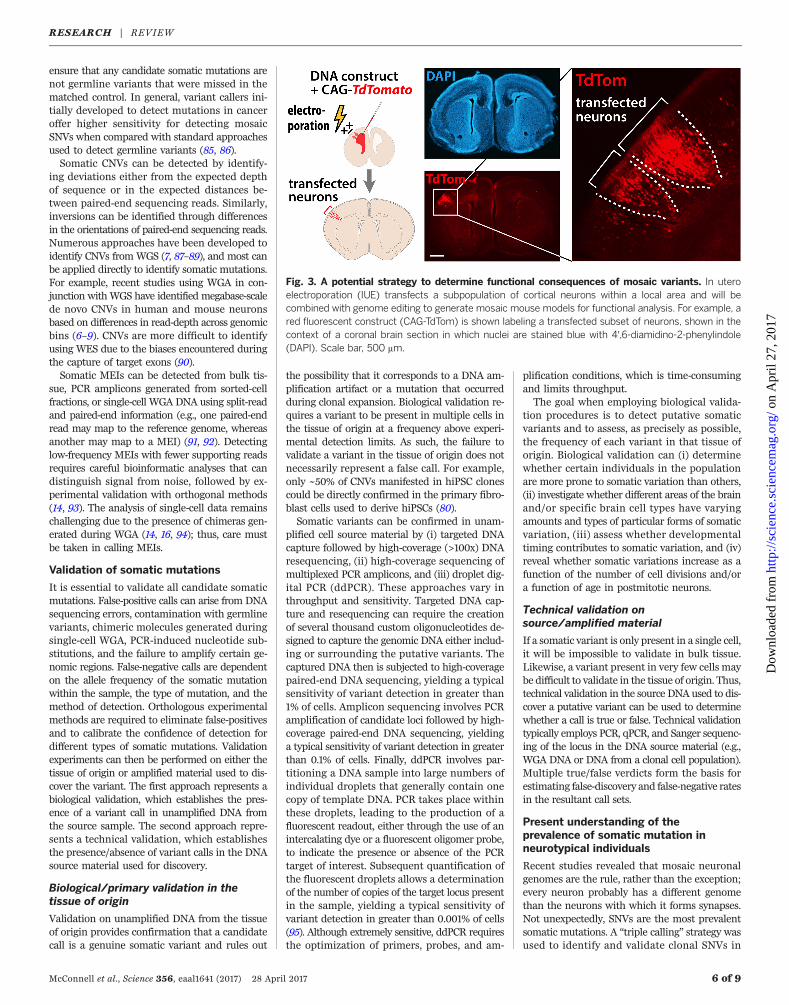
Fig. 3. A potential strategy to determine functional consequences of mosaic variants. In uteroelectroporation (IUE) transfects a subpopulation of cortical neurons within a local area and will becombined with genome editing to generate mosaic mouse models for functional analysis. For example, ared fluorescent construct (CAG-TdTom) is shown labeling a transfected subset of neurons, shown in thecontext of a coronal brain section in which nuclei are stained blue with 4′,6-diamidino-2-phenylindole(DAPI). Scale bar, 500 mm.
RESEARCH | REVIEW
on
Apr
il 27
, 201
7ht
tp://
scie
nce.
scie
ncem
ag.o
rg/
Dow
nloa
ded
from
MDA-amplified DNA from single neurons iso-lated from a neurotypical brain, leading to es-timates of ~1000 to 1500 SNVs per neuronalgenome (5). By comparison to human corticalneurons, a SCNT experiment in reprogrammedmouse olfactory neurons detected hundreds ofSNVs per neuron and a lower proportion of C-to-Ttransition mutations (84). Although the divergentSNV rates between these two studies may arisefrom technical differences (as discussed above),both approaches establish that SNVs representan important form of somatic mutation in bothhuman and mouse neurons.Brain somatic CNVs initially were identified
by comparing the sequences of bulk DNA derivedfrom multicellular samples of different brain re-gions to the sequences of DNA derived from so-matic tissues (96, 97). The first single-cell studyof neuronal CNVs analyzed 110 human frontalcortex neurons and found that 13 to 41% of theneurons contained at least one megabase-scalede novo CNV (6). Additional studies, which an-alyzed fewer neuronal genomes, confirmed thatde novo CNVs occur in at least 10% of neurons(7, 8). CNVs can be shared by multiple neuronsand inherited in a clonal manner (8). Further-more, megabase-scale CNVs typically alter thecopy number of 10 or more genes in individualneurons. In addition to expression-level differ-ences that can accompany gene copy numberchanges, mosaic neuronal CNVs also are expectedto reveal or abate pernicious alleles on a neuron-by-neuron basis in every individual.L1 retrotransposon insertions alter the tran-
scriptional regulation of genes in myriad ways(42). Initial studies used engineered L1s containinga retrotransposition indicator cassette to discoverMEI activity in mouse brain (98) and in humanNPCs in vitro (99). Studies of MDA-amplifiedNeuN-positive nuclei isolated from a neuro-typical human brain, followed by L1-transposonprofiling (13) or WGS (15, 16), have since suggestedthat 0.2 to 1 L1 insertion occur per neuronalgenome. Another report, which employedMALBACWGA in conjunction with L1 capture technology(RC-seq), reported an average of 13 L1 insertionsin every neuronal genome (11), although a sub-sequent study suggested a high false-positive ratein these data (14). By comparison, SCNT experi-ments in mouse olfactory neurons reported ≤1.3MEI per neuronal genome (84). An extrapolationof these data indicates that potentially billionsof neurons in the neurotypical brain containde novo MEIs. Additional studies are requiredto determine whether L1s retrotranspose at vary-ing rates in different brain regions, in differentindividuals, or preferentially insert into expressedgenes, and whether other mobile elements [e.g.,Alu retrotransposons (42)] also contribute to intra-individual neuronal genetic diversity.
Generation of a community resource
The BSMNwill generate comprehensive maps ofsomatic genomic variation in neurotypical anddiseased human brains, including a prioritizedcall set of confirmed somatic variants (Box 1) thatmay contribute to neuropsychiatric disease and
epilepsy. Functional validation experiments willbeperformedusingCRISPR/Cas9-mediatedgenomeengineering, hiPSC-based neurogenesis, and mosaicmouse models generated by in utero electropora-tion (Fig. 3). The BSMN is initially determiningconcordance among disparate sequencing andbioinformatic approaches by performing a “com-mon experiment” in which pulverized tissue fromone neurotypical individual in the Lieber brainrepository has been distributed to all of theworking groups for independent assessment ofmosaicism.The BSMN will generate an estimated 10,000
sequencing data sets that comprise >600 terabytesof data and facilitate data-sharing through theBSMNKnowledge Portal (www.synapse.org/bsmn)and the NIMH Data Archive (https://data-archive.nimh.nih.gov). Coordinated analyses with dataderived from some of the same brain samplesby the CommonMind (www.synapse.org/cmc) andPsychENCODE (www.synapse.org/pec) initiativesmay elucidate the effect of somatic mosaicismon tissue-wide gene expression. Data generatedthough the BSMN initiative will be released tothe broader research community on an ongoingbasis through a controlled-access mechanism thatfollows NIH policies and regulatory requirements.
REFERENCES AND NOTES
1. M. Lynch, Rate, molecular spectrum, and consequences ofhuman mutation. Proc. Natl. Acad. Sci. U.S.A. 107, 961–968(2010). doi: 10.1073/pnas.0912629107; pmid: 20080596
2. D. Freed, E. L. Stevens, J. Pevsner, Somatic mosaicism in thehuman genome. Genes (Basel) 5, 1064–1094 (2014).doi: 10.3390/genes5041064; pmid: 25513881
3. S. A. Frank, Evolution in health and medicine Sacklercolloquium: Somatic evolutionary genomics: Mutationsduring development cause highly variable genetic mosaicismwith risk of cancer and neurodegeneration. Proc. Natl. Acad.Sci. U.S.A. 107 (suppl. 1), 1725–1730 (2010). doi: 10.1073/pnas.0909343106; pmid: 19805033
4. J. R. Lupski, Genome mosaicism—One human, multiplegenomes. Science 341, 358–359 (2013). doi: 10.1126/science.1239503; pmid: 23888031
5. M. A. Lodato et al., Somatic mutation in single humanneurons tracks developmental and transcriptional history.Science 350, 94–98 (2015). doi: 10.1126/science.aab1785;pmid: 26430121
6. M. J. McConnell et al., Mosaic copy number variation inhuman neurons. Science 342, 632–637 (2013).doi: 10.1126/science.1243472; pmid: 24179226
7. K. A. Knouse, J. Wu, A. Amon, Assessment of megabase-scale somatic copy number variation using single-cellsequencing. Genome Res. 26, 376–384 (2016). doi: 10.1101/gr.198937.115; pmid: 26772196
8. X. Cai et al., Single-cell, genome-wide sequencing identifiesclonal somatic copy-number variation in the human brain.Cell Reports 8, 1280–1289 (2014). doi: 10.1016/j.celrep.2014.07.043; pmid: 25159146
9. S. K. Rehen et al., Constitutional aneuploidy in the normalhuman brain. J. Neurosci. 25, 2176–2180 (2005).doi: 10.1523/JNEUROSCI.4560-04.2005; pmid: 15745943
10. Y. B. Yurov et al., Aneuploidy and confined chromosomalmosaicism in the developing human brain. PLOS ONE 2,e558 (2007). doi: 10.1371/journal.pone.0000558;pmid: 17593959
11. K. R. Upton et al., Ubiquitous L1 mosaicism in hippocampalneurons. Cell 161, 228–239 (2015). doi: 10.1016/j.cell.2015.03.026; pmid: 25860606
12. J. K. Baillie et al., Somatic retrotransposition alters thegenetic landscape of the human brain. Nature 479, 534–537(2011). doi: 10.1038/nature10531; pmid: 22037309
13. G. D. Evrony et al., Single-neuron sequencing analysis of L1retrotransposition and somatic mutation in the human brain.Cell 151, 483–496 (2012). doi: 10.1016/j.cell.2012.09.035;pmid: 23101622
14. G. D. Evrony, E. Lee, P. J. Park, C. A. Walsh, Resolving rates ofmutation in the brain using single-neuron genomics. eLife 5,e12966 (2016). doi: 10.7554/eLife.12966; pmid: 26901440
15. J. A. Erwin et al., L1-associated genomic regions are deletedin somatic cells of the healthy human brain. Nat. Neurosci.19, 1583–1591 (2016).
16. G. D. Evrony et al., Cell lineage analysis in human brain usingendogenous retroelements. Neuron 85, 49–59 (2015).doi: 10.1016/j.neuron.2014.12.028; pmid: 25569347
17. J. H. Lui, D. V. Hansen, A. R. Kriegstein, Development andevolution of the human neocortex. Cell 146, 18–36 (2011).doi: 10.1016/j.cell.2011.06.030; pmid: 21729779
18. A. Macia et al., Engineered LINE-1 retrotransposition innondividing human neurons. Genome Res. 27, 335–348 (2017).
19. A. R. Muotri, F. H. Gage, Generation of neuronal variabilityand complexity. Nature 441, 1087–1093 (2006). doi:10.1038/nature04959; pmid: 16810244
20. D. M. Bushman, J. Chun, The genomically mosaic brain:Aneuploidy and more in neural diversity and disease. Semin.Cell Dev. Biol. 24, 357–369 (2013). doi: 10.1016/j.semcdb.2013.02.003; pmid: 23466288
21. I. Iossifov et al., The contribution of de novo codingmutations to autism spectrum disorder. Nature 515, 216–221(2014). doi: 10.1038/nature13908; pmid: 25363768
22. D. Freed, J. Pevsner, The contribution of mosaic variants toautism spectrum disorder. PLOS Genet. 12, e1006245 (2016).doi: 10.1371/journal.pgen.1006245; pmid: 27632392
23. M. Fromer et al., De novo mutations in schizophreniaimplicate synaptic networks. Nature 506, 179–184 (2014).doi: 10.1038/nature12929; pmid: 24463507
24. D. Malhotra, J. Sebat, CNVs: Harbingers of a rare variantrevolution in psychiatric genetics. Cell 148, 1223–1241(2012). doi: 10.1016/j.cell.2012.02.039; pmid: 22424231
25. A. Poduri, G. D. Evrony, X. Cai, C. A. Walsh, Somatic mutation,genomic variation, and neurological disease. Science 341,1237758 (2013). doi: 10.1126/science.1237758;pmid: 23828942
26. J. H. Lee, Somatic mutations in disorders with disruptedbrain connectivity. Exp. Mol. Med. 48, e239 (2016).
27. J. M. Sekiguchi et al., Nonhomologous end-joining proteinsare required for V(D)J recombination, normal growth, andneurogenesis. Cold Spring Harb. Symp. Quant. Biol. 64,169–181 (1999). doi: 10.1101/sqb.1999.64.169;pmid: 11232282
28. K. M. Frank et al., DNA ligase IV deficiency in mice leads todefective neurogenesis and embryonic lethality via the p53pathway. Mol. Cell 5, 993–1002 (2000). doi: 10.1016/S1097-2765(00)80264-6; pmid: 10911993
29. M. J. McConnell et al., Failed clearance of aneuploidembryonic neural progenitor cells leads to excess aneuploidyin the Atm-deficient but not the Trp53-deficient adultcerebral cortex. J. Neurosci. 24, 8090–8096 (2004).doi: 10.1523/JNEUROSCI.2263-04.2004; pmid: 15371510
30. N. G. Coufal et al., Ataxia telangiectasia mutated (ATM)modulates long interspersed element-1 (L1)retrotransposition in human neural stem cells. Proc. Natl.Acad. Sci. U.S.A. 108, 20382–20387 (2011). doi: 10.1073/pnas.1100273108; pmid: 22159035
31. I. Y. Iourov, S. G. Vorsanova, T. Liehr, A. D. Kolotii, Y. B. Yurov,Increased chromosome instability dramatically disruptsneural genome integrity and mediates cerebellardegeneration in the ataxia-telangiectasia brain. Hum. Mol.Genet. 18, 2656–2669 (2009). doi: 10.1093/hmg/ddp207;pmid: 19414482
32. J. Shen et al., Mutations in PNKP cause microcephaly,seizures and defects in DNA repair. Nat. Genet. 42, 245–249(2010). doi: 10.1038/ng.526; pmid: 20118933
33. C. M. Carvalho, J. R. Lupski, Mechanisms underlyingstructural variant formation in genomic disorders. Nat. Rev.Genet. 17, 224–238 (2016). doi: 10.1038/nrg.2015.25;pmid: 26924765
34. Y. L. Chiu, W. C. Greene, The APOBEC3 cytidine deaminases:An innate defensive network opposing exogenousretroviruses and endogenous retroelements. Annu. Rev.Immunol. 26, 317–353 (2008). doi: 10.1146/annurev.immunol.26.021607.090350; pmid: 18304004
35. M. F. Arlt, T. E. Wilson, T. W. Glover, Replication stressand mechanisms of CNV formation. Curr. Opin. Genet. Dev.22, 204–210 (2012). doi: 10.1016/j.gde.2012.01.009;pmid: 22365495
36. P. J. Hastings, J. R. Lupski, S. M. Rosenberg, G. Ira,Mechanisms of change in gene copy number. Nat. Rev.Genet. 10, 551–564 (2009). doi: 10.1038/nrg2593;pmid: 19597530
McConnell et al., Science 356, eaal1641 (2017) 28 April 2017 7 of 9
RESEARCH | REVIEW
on
Apr
il 27
, 201
7ht
tp://
scie
nce.
scie
ncem
ag.o
rg/
Dow
nloa
ded
from
37. P. C. Wei et al., Long neural genes harbor recurrent DNAbreak clusters in neural stem/progenitor cells. Cell 164,644–655 (2016). doi: 10.1016/j.cell.2015.12.039;pmid: 26871630
38. T. E. Wilson et al., Large transcription units unify copynumber variants and common fragile sites arising underreplication stress. Genome Res. 25, 189–200 (2015).doi: 10.1101/gr.177121.114; pmid: 25373142
39. I. F. King et al., Topoisomerases facilitate transcription oflong genes linked to autism. Nature 501, 58–62 (2013).doi: 10.1038/nature12504; pmid: 23995680
40. M. J. Zylka, J. M. Simon, B. D. Philpot, Gene length matters inneurons. Neuron 86, 353–355 (2015). doi: 10.1016/j.neuron.2015.03.059; pmid: 25905808
41. H. W. Gabel et al., Disruption of DNA-methylation-dependentlong gene repression in Rett syndrome. Nature 522, 89–93(2015). doi: 10.1038/nature14319; pmid: 25762136
42. S. R. Richardson et al., The influence of LINE-1 and SINEretrotransposons on mammalian genomes. Microbiol. Spectr.3, MDNA3-0061-2014 (2015). doi: 10.1128/microbiolspec.MDNA3-0061-2014
43. D. D. Luan, M. H. Korman, J. L. Jakubczak, T. H. Eickbush,Reverse transcription of R2Bm RNA is primed by a nick atthe chromosomal target site: A mechanism for non-LTRretrotransposition. Cell 72, 595–605 (1993). doi: 10.1016/0092-8674(93)90078-5; pmid: 7679954
44. T. A. Morrish et al., DNA repair mediated by endonuclease-independent LINE-1 retrotransposition. Nat. Genet. 31,159–165 (2002). doi: 10.1038/ng898; pmid: 12006980
45. N. Gilbert, S. Lutz, T. A. Morrish, J. V. Moran, Multiple fates ofL1 retrotransposition intermediates in cultured human cells.Mol. Cell. Biol. 25, 7780–7795 (2005). doi: 10.1128/MCB.25.17.7780-7795.2005; pmid: 16107723
46. D. E. Symer et al., Human l1 retrotransposition is associatedwith genetic instability in vivo. Cell 110, 327–338 (2002).doi: 10.1016/S0092-8674(02)00839-5; pmid: 12176320
47. N. Gilbert, S. Lutz-Prigge, J. V. Moran, Genomic deletionscreated upon LINE-1 retrotransposition. Cell 110, 315–325(2002). doi: 10.1016/S0092-8674(02)00828-0;pmid: 12176319
48. J. M. Chen, P. D. Stenson, D. N. Cooper, C. Férec, Asystematic analysis of LINE-1 endonuclease-dependentretrotranspositional events causing human genetic disease.Hum. Genet. 117, 411–427 (2005). doi: 10.1007/s00439-005-1321-0; pmid: 15983781
49. G. M. Mirzaa et al., Association of MTOR mutations withdevelopmental brain disorders, including megalencephaly,focal cortical dysplasia, and pigmentary mosaicism.JAMA Neurol. 73, 836–845 (2016). doi: 10.1001/jamaneurol.2016.0363; pmid: 27159400
50. G. M. Mirzaa et al., Characterisation of mutations of thephosphoinositide-3-kinase regulatory subunit, PIK3R2, inperisylvian polymicrogyria: A next-generation sequencingstudy. Lancet Neurol. 14, 1182–1195 (2015). doi: 10.1016/S1474-4422(15)00278-1; pmid: 26520804
51. J. B. Rivière et al., De novo germline and postzygoticmutations in AKT3, PIK3R2 and PIK3CA cause a spectrum ofrelated megalencephaly syndromes. Nat. Genet. 44,934–940 (2012). doi: 10.1038/ng.2331; pmid: 22729224
52. E. P. Henske, S. Jóźwiak, J. C. Kingswood, J. R. Sampson,E. A. Thiele, Tuberous sclerosis complex. Nat. Rev. Dis. Primers2, 16035 (2016). doi: 10.1038/nrdp.2016.35; pmid: 27226234
53. M. D. Shirley et al., Sturge-Weber syndrome and port-winestains caused by somatic mutation in GNAQ. N. Engl. J. Med.368, 1971–1979 (2013). doi: 10.1056/NEJMoa1213507;pmid: 23656586
54. C. D. Van Raamsdonk et al., Frequent somatic mutations ofGNAQ in uveal melanoma and blue naevi. Nature 457,599–602 (2009). doi: 10.1038/nature07586;pmid: 19078957
55. S. S. Jamuar et al., Somatic mutations in cerebral corticalmalformations. N. Engl. J. Med. 371, 733–743 (2014).doi: 10.1056/NEJMoa1314432; pmid: 25140959
56. F. Sicca et al., Mosaic mutations of the LIS1 gene causesubcortical band heterotopia. Neurology 61, 1042–1046(2003). doi: 10.1212/WNL.61.8.1042; pmid: 14581661
57. J. G. Gleeson, Classical lissencephaly and double cortex(subcortical band heterotopia): LIS1 and doublecortin.Curr. Opin. Neurol. 13, 121–125 (2000). doi: 10.1097/00019052-200004000-00002; pmid: 10987567
58. T. R. Insel, Brain somatic mutations: The dark matter ofpsychiatric genetics? Mol. Psychiatry 19, 156–158 (2014).doi: 10.1038/mp.2013.168; pmid: 24342990
59. A. M. D’Gama et al., Targeted DNA sequencing from autismspectrum disorder brains implicates multiple geneticmechanisms. Neuron 88, 910–917 (2015). doi: 10.1016/j.neuron.2015.11.009; pmid: 26637798
60. I. Iossifov et al., De novo gene disruptions in children on theautistic spectrum. Neuron 74, 285–299 (2012). doi: 10.1016/j.neuron.2012.04.009; pmid: 22542183
61. B. M. Neale et al., Patterns and rates of exonic de novomutations in autism spectrum disorders. Nature 485,242–245 (2012). doi: 10.1038/nature11011; pmid: 22495311
62. J. Sebat et al., Strong association of de novo copy numbermutations with autism. Science 316, 445–449 (2007).doi: 10.1126/science.1138659; pmid: 17363630
63. J. J. Hutsler, M. F. Casanova, Review: Cortical construction inautism spectrum disorder: Columns, connectivity and thesubplate. Neuropathol. Appl. Neurobiol. 42, 115–134 (2016).doi: 10.1111/nan.12227; pmid: 25630827
64. A. P. Donovan, M. A. Basson, The neuroanatomy of autism - Adevelopmental perspective. J. Anat. 230, 4–15 (2017).doi: 10.1111/joa.12542; pmid: 27620360
65. M. C. Marchetto et al., Altered proliferation and networks inneural cells derived from idiopathic autistic individuals.Mol. Psychiatry, 10.1038/mp.2016.95 (2016).
66. J. Mariani et al., FOXG1-dependent dysregulation of GABA/glutamate neuron differentiation in autism spectrumdisorders. Cell 162, 375–390 (2015). doi: 10.1016/j.cell.2015.06.034; pmid: 26186191
67. R. Stoner et al., Patches of disorganization in the neocortexof children with autism. N. Engl. J. Med. 370, 1209–1219(2014). doi: 10.1056/NEJMoa1307491; pmid: 24670167
68. P. H. Patterson, Maternal infection and immune involvementin autism. Trends Mol. Med. 17, 389–394 (2011).doi: 10.1016/j.molmed.2011.03.001; pmid: 21482187
69. G. B. Choi et al., The maternal interleukin-17a pathway inmice promotes autism-like phenotypes in offspring.Science 351, 933–939 (2016). doi: 10.1126/science.aad0314; pmid: 26822608
70. S. E. Smith, R. M. Elliott, M. P. Anderson, Maternal immuneactivation increases neonatal mouse cortex thickness andcell density. J. Neuroimmune Pharmacol. 7, 529–532 (2012).doi: 10.1007/s11481-012-9372-1; pmid: 22570011
71. N. V. Malkova, C. Z. Yu, E. Y. Hsiao, M. J. Moore,P. H. Patterson, Maternal immune activation yields offspringdisplaying mouse versions of the three core symptoms ofautism. Brain Behav. Immun. 26, 607–616 (2012).doi: 10.1016/j.bbi.2012.01.011; pmid: 22310922
72. H. Soumiya, H. Fukumitsu, S. Furukawa, Prenatal immunechallenge compromises development of upper-layer but notdeeper-layer neurons of the mouse cerebral cortex.J. Neurosci. Res. 89, 1342–1350 (2011). doi: 10.1002/jnr.22636; pmid: 21674566
73. M. Bundo et al., Increased l1 retrotransposition in theneuronal genome in schizophrenia. Neuron 81, 306–313(2014). doi: 10.1016/j.neuron.2013.10.053; pmid: 24389010
74. A. R. Muotri et al., L1 retrotransposition in neurons ismodulated by MeCP2. Nature 468, 443–446 (2010).doi: 10.1038/nature09544; pmid: 21085180
75. B. B. Lake et al., Neuronal subtypes and diversity revealedby single-nucleus RNA sequencing of the human brain.Science 352, 1586–1590 (2016). doi: 10.1126/science.aaf1204; pmid: 27339989
76. J. R. Vermeesch, T. Voet, K. Devriendt, Prenatal and pre-implantation genetic diagnosis. Nat. Rev. Genet. 17, 643–656(2016). doi: 10.1038/nrg.2016.97; pmid: 27629932
77. L. Yan et al., Live births after simultaneous avoidance ofmonogenic diseases and chromosome abnormality by next-generation sequencing with linkage analyses. Proc. Natl.Acad. Sci. U.S.A. 112, 15964–15969 (2015). doi: 10.1073/pnas.1523297113; pmid: 26712022
78. Y. Hou et al., Comparison of variations detection betweenwhole-genome amplification methods used in single-cellresequencing. Gigascience 4, 37 (2015). doi: 10.1186/s13742-015-0068-3; pmid: 26251698
79. R. S. Lasken, T. B. Stockwell, Mechanism of chimeraformation during the multiple displacement amplificationreaction. BMC Biotechnol. 7, 19 (2007). doi: 10.1186/1472-6750-7-19; pmid: 17430586
80. A. Abyzov et al., Somatic copy number mosaicism inhuman skin revealed by induced pluripotent stem cells.Nature 492, 438–442 (2012). doi: 10.1038/nature11629;pmid: 23160490
81. A. Roth et al., Clonal genotype and population structureinference from single-cell tumor sequencing. Nat. Methods
13, 573–576 (2016). doi: 10.1038/nmeth.3867;pmid: 27183439
82. H. Zafar, Y. Wang, L. Nakhleh, N. Navin, K. Chen, Monovar:Single-nucleotide variant detection in single cells. Nat.Methods 13, 505–507 (2016). doi: 10.1038/nmeth.3835;pmid: 27088313
83. T. Garvin et al., Interactive analysis and assessment of single-cell copy-number variations. Nat. Methods 12, 1058–1060(2015). doi: 10.1038/nmeth.3578; pmid: 26344043
84. J. L. Hazen et al., The complete genome sequences, uniquemutational spectra, and developmental potency of adultneurons revealed by cloning. Neuron 89, 1223–1236 (2016).doi: 10.1016/j.neuron.2016.02.004; pmid: 26948891
85. K. Cibulskis et al., Sensitive detection of somatic pointmutations in impure and heterogeneous cancer samples.Nat. Biotechnol. 31, 213–219 (2013). doi: 10.1038/nbt.2514; pmid: 23396013
86. D. C. Koboldt et al., VarScan: Variant detection in massivelyparallel sequencing of individual and pooled samples.Bioinformatics 25, 2283–2285 (2009). doi: 10.1093/bioinformatics/btp373; pmid: 19542151
87. X. Zhao, S. B. Emery, B. Myers, J. M. Kidd, R. E. Mills,Resolving complex structural genomic rearrangements usinga randomized approach. Genome Biol. 17, 126 (2016).doi: 10.1186/s13059-016-0993-1; pmid: 27287201
88. R. Xi, S. Lee, Y. Xia, T.-M. Kim, P. J. Park, Copy numberanalysis of whole-genome data using BIC-seq2 and itsapplication to detection of cancer susceptibility variants.Nucleic Acids Res. 44, 6274–6286 (2016). doi: 10.1093/nar/gkw491; pmid: 27260798
89. R. M. Layer, C. Chiang, A. R. Quinlan, I. M. Hall, LUMPY: Aprobabilistic framework for structural variant discovery.Genome Biol. 15, R84 (2014). doi: 10.1186/gb-2014-15-6-r84;pmid: 24970577
90. A. M. Meynert, M. Ansari, D. R. FitzPatrick, M. S. Taylor,Variant detection sensitivity and biases in whole genome andexome sequencing. BMC Bioinformatics 15, 247 (2014).doi: 10.1186/1471-2105-15-247; pmid: 25038816
91. E. Lee et al., Landscape of somatic retrotransposition inhuman cancers. Science 337, 967–971 (2012). doi: 10.1126/science.1222077; pmid: 22745252
92. C. Stewart et al., A comprehensive map of mobile elementinsertion polymorphisms in humans. PLOS Genet. 7, e1002236(2011). doi: 10.1371/journal.pgen.1002236; pmid: 21876680
93. S. R. Richardson, S. Morell, G. J. Faulkner, L1retrotransposons and somatic mosaicism in the brain. Annu.Rev. Genet. 48, 1–27 (2014). doi: 10.1146/annurev-genet-120213-092412; pmid: 25036377
94. C. Z. Zhang et al., Calibrating genomic and allelic coveragebias in single-cell sequencing. Nat. Commun. 6, 6822 (2015).doi: 10.1038/ncomms7822; pmid: 25879913
95. B. J. Hindson et al., High-throughput droplet digital PCRsystem for absolute quantitation of DNA copy number.Anal. Chem. 83, 8604–8610 (2011). doi: 10.1021/ac202028g;pmid: 22035192
96. A. Piotrowski et al., Somatic mosaicism for copy numbervariation in differentiated human tissues. Hum. Mutat. 29,1118–1124 (2008). doi: 10.1002/humu.20815;pmid: 18570184
97. M. O’Huallachain, K. J. Karczewski, S. M. Weissman,A. E. Urban, M. P. Snyder, Extensive genetic variation insomatic human tissues. Proc. Natl. Acad. Sci. U.S.A. 109,18018–18023 (2012). doi: 10.1073/pnas.1213736109;pmid: 23043118
98. A. R. Muotri et al., Somatic mosaicism in neuronal precursorcells mediated by L1 retrotransposition. Nature 435,903–910 (2005). doi: 10.1038/nature03663;pmid: 15959507
99. N. G. Coufal et al., L1 retrotransposition in human neuralprogenitor cells. Nature 460, 1127–1131 (2009). doi: 10.1038/nature08248; pmid: 19657334
100. K. C. Kurek et al., Somatic mosaic activating mutations inPIK3CA cause CLOVES syndrome. Am. J. Hum. Genet. 90,1108–1115 (2012). doi: 10.1016/j.ajhg.2012.05.006;pmid: 22658544
101. J. H. Lee et al., De novo somatic mutations incomponents of the PI3K-AKT3-mTOR pathway causehemimegalencephaly. Nat. Genet. 44, 941–945 (2012).doi: 10.1038/ng.2329; pmid: 22729223
102. M. J. Lindhurst et al., Mosaic overgrowth with fibroadiposehyperplasia is caused by somatic activating mutations inPIK3CA. Nat. Genet. 44, 928–933 (2012). doi: 10.1038/ng.2332; pmid: 22729222
McConnell et al., Science 356, eaal1641 (2017) 28 April 2017 8 of 9
RESEARCH | REVIEW
on
Apr
il 27
, 201
7ht
tp://
scie
nce.
scie
ncem
ag.o
rg/
Dow
nloa
ded
from
103. J. B. Rivière et al., De novo germline and postzygoticmutations in AKT3, PIK3R2 and PIK3CA cause a spectrumof related megalencephaly syndromes. Nat. Genet. 44,934–940 (2012). doi: 10.1038/ng.2331; pmid: 22729224
104. Y. Samuels, V. E. Velculescu, Oncogenic mutations of PIK3CAin human cancers. Cell Cycle 3, 1221–1224 (2004).doi: 10.4161/cc.3.10.1164; pmid: 15467468
105. M. J. Lindhurst et al., A mosaic activating mutation in AKT1associated with the Proteus syndrome. N. Engl. J. Med. 365,611–619 (2011). doi: 10.1056/NEJMoa1104017;pmid: 21793738
106. K. Hussain et al., An activating mutation of AKT2 and humanhypoglycemia. Science 334, 474 (2011). doi: 10.1126/science.1210878; pmid: 21979934
107. A. Poduri et al., Somatic activation of AKT3 causeshemispheric developmental brain malformations. Neuron 74,41–48 (2012). doi: 10.1016/j.neuron.2012.03.010;pmid: 22500628
108. J. S. Lim et al., Brain somatic mutations in MTOR cause focalcortical dysplasia type II leading to intractable epilepsy.Nat. Med. 21, 395–400 (2015). doi: 10.1038/nm.3824;pmid: 25799227
109. L. Bar-Peled et al., A Tumor suppressor complex with GAPactivity for the Rag GTPases that signal amino acidsufficiency to mTORC1. Science 340, 1100–1106 (2013).doi: 10.1126/science.1232044; pmid: 23723238
110. S. Baulac et al., Familial focal epilepsy with focal corticaldysplasia due to DEPDC5 mutations. Ann. Neurol. 77,675–683 (2015). doi: 10.1002/ana.24368; pmid: 25623524
111. European Chromosome 16 Tuberous Sclerosis Consortium,Identification and characterization of the tuberous sclerosisgene on chromosome 16. Cell 75, 1305–1315 (1993).doi: 10.1016/0092-8674(93)90618-Z; pmid: 8269512
112. M. van Slegtenhorst et al., Identification of the tuberoussclerosis gene TSC1 on chromosome 9q34. Science 277,805–808 (1997). doi: 10.1126/science.277.5327.805;pmid: 9242607
113. F. Bourdeaut et al., Mosaicism for oncogenic G12D KRASmutation associated with epidermal nevus, polycystic kidneysand rhabdomyosarcoma. J. Med. Genet. 47, 859–862 (2010).doi: 10.1136/jmg.2009.075374; pmid: 20805368
114. S. Gantner et al., Absence of BRAF and HRAS mutations ineruptive Spitz naevi. Br. J. Dermatol. 164, 873–877 (2011).doi: 10.1111/j.1365-2133.2011.10210.x; pmid: 21418173
115. C. Hafner et al., Oncogenic PIK3CA mutations occur inepidermal nevi and seborrheic keratoses with a characteristicmutation pattern. Proc. Natl. Acad. Sci. U.S.A. 104,13450–13454 (2007). doi: 10.1073/pnas.0705218104;pmid: 17673550
116. C. Hafner et al., Mosaicism of activating FGFR3 mutations inhuman skin causes epidermal nevi. J. Clin. Invest. 116,2201–2207 (2006). doi: 10.1172/JCI28163; pmid: 16841094
117. T. Papp et al., Mutational analysis of the N-ras, p53,p16INK4a, CDK4, and MC1R genes in human congenitalmelanocytic naevi. J. Med. Genet. 36, 610–614 (1999).pmid: 10465111
118. P. M. Pollock et al., High frequency of BRAF mutations innevi. Nat. Genet. 33, 19–20 (2003). doi: 10.1038/ng1054;pmid: 12447372
119. G. A. Rouleau et al., Alteration in a new gene encoding aputative membrane-organizing protein causes neuro-fibromatosis type 2. Nature 363, 515–521 (1993).doi: 10.1038/363515a0; pmid: 8379998
120. R. M. Cawthon et al., A major segment of theneurofibromatosis type 1 gene: cDNA sequence, genomicstructure, and point mutations. Cell 62, 193–201 (1990).doi: 10.1016/0092-8674(90)90253-B; pmid: 2114220
121. C. Garcia-Linares et al., Dissecting loss of heterozygosity(LOH) in neurofibromatosis type 1-associated neurofibromas:Importance of copy neutral LOH. Hum. Mutat. 32, 78–90(2011). doi: 10.1002/humu.21387; pmid: 21031597
122. L. Messiaen et al., Mosaic type-1 NF1 microdeletions as acause of both generalized and segmental neurofibromatosistype-1 (NF1). Hum. Mutat. 32, 213–219 (2011). doi: 10.1002/humu.21418; pmid: 21280148
123. S. A. Rasmussen, J. M. Friedman, NF1 gene andneurofibromatosis 1. Am. J. Epidemiol. 151, 33–40 (2000).doi: 10.1093/oxfordjournals.aje.a010118; pmid: 10625171
124. M. R. Wallace et al., Type 1 neurofibromatosis gene:Identification of a large transcript disrupted in three NF1patients. Science 249, 181–186 (1990). doi: 10.1126/science.2134734; pmid: 2134734
125. L. Groesser et al., BRAF and RAS mutations in sporadic andsecondary pyogenic granuloma. J. Invest. Dermatol. 136,481–486 (2016). doi: 10.1038/JID.2015.376; pmid: 26802240
126. L. Groesser et al., KRAS, HRAS and egfr mutations insporadic sebaceous gland hyperplasia. Acta Derm. Venereol.96, 737–741 (2016). pmid: 26804118
127. J. E. Niemela et al., Somatic KRAS mutations associatedwith a human nonmalignant syndrome of autoimmunityand abnormal leukocyte homeostasis. Blood 117,2883–2886 (2011). doi: 10.1182/blood-2010-07-295501;pmid: 21079152
128. M. Takagi et al., Autoimmune lymphoproliferative syndrome-like disease with somatic KRAS mutation. Blood 117,2887–2890 (2011). doi: 10.1182/blood-2010-08-301515;pmid: 21063026
129. M. D. Shirley et al., Sturge-Weber syndrome and port-winestains caused by somatic mutation in GNAQ. N. Engl. J. Med.368, 1971–1979 (2013). doi: 10.1056/NEJMoa1213507;pmid: 23656586
130. A. C. Thomas et al., Mosaic activating mutations in GNA11and GNAQ are associated with phakomatosispigmentovascularis and extensive dermal melanocytosis.J. Invest. Dermatol. 136, 770–778 (2016). doi: 10.1016/j.jid.2015.11.027; pmid: 26778290
131. J. A. Couto et al., A somatic MAP3K3 mutation is associatedwith verrucous venous malformation. Am. J. Hum. Genet. 96,480–486 (2015). doi: 10.1016/j.ajhg.2015.01.007;pmid: 25728774
132. J. L. Patten et al., Mutation in the gene encoding thestimulatory G protein of adenylate cyclase in Albright’s
hereditary osteodystrophy. N. Engl. J. Med. 322, 1412–1419(1990). doi: 10.1056/NEJM199005173222002; pmid: 2109828
133. W. F. Schwindinger, C. A. Francomano, M. A. Levine,Identification of a mutation in the gene encoding the alphasubunit of the stimulatory G protein of adenylyl cyclase inMcCune-Albright syndrome. Proc. Natl. Acad. Sci. U.S.A.89, 5152–5156 (1992). doi: 10.1073/pnas.89.11.5152;pmid: 1594625
134. E. J. Baxter et al., Acquired mutation of the tyrosine kinaseJAK2 in human myeloproliferative disorders. Lancet 365,1054–1061 (2005). doi: 10.1016/S0140-6736(05)74230-6;pmid: 15781101
135. R. Kralovics et al., A gain-of-function mutation of JAK2 inmyeloproliferative disorders. N. Engl. J. Med. 352, 1779–1790(2005). doi: 10.1056/NEJMoa051113; pmid: 15858187
136. C. Depienne et al., Mechanisms for variable expressivity ofinherited SCN1A mutations causing Dravet syndrome.J. Med. Genet. 47, 404–410 (2010). doi: 10.1136/jmg.2009.074328; pmid: 20522430
137. N. Tanaka et al., High incidence of NLRP3 somatic mosaicismin patients with chronic infantile neurologic, cutaneous,articular syndrome: Results of an International MulticenterCollaborative Study. Arthritis Rheum. 63, 3625–3632 (2011).doi: 10.1002/art.30512; pmid: 21702021
138. K. H. Grzeschik et al., Deficiency of PORCN, a regulator ofWnt signaling, is associated with focal dermal hypoplasia.Nat. Genet. 39, 833–835 (2007). doi: 10.1038/ng2052;pmid: 17546031
139. J. Takeda et al., Deficiency of the GPI anchor caused by asomatic mutation of the PIG-A gene in paroxysmal nocturnalhemoglobinuria. Cell 73, 703–711 (1993). doi: 10.1016/0092-8674(93)90250-T; pmid: 8500164
ACKNOWLEDGMENTS
We thank T. Insel for initiating this project, L. Bingaman forongoing administrative assistance, and N. Leff and M. L. Gage forcopyediting assistance. J.M.K acknowledges support provided bythe Pew Biomedical Scholars Award. J.V.M. is an inventor onpatent application 6150160, held by the John Hopkins Universityand the Trustees of the University of Pennsylvania, which coversthe compositions and methods of use of mammalianretrotransposons. We also acknowledge the support of NIH R01MH100914, Genomic mosaicism in developing human brain(F.M.V.). Some figures use images from the Servier Medical ArtPowerPoint Image Bank. All the work was supported byU01MH106883, U01MH106874, U01MH106893, U01MH106892,U01MH106882, U01MH106876, U01MH1068898, U01MH106891,and U01MH106884. We regret that space constraints limit thenumber of references and apologize to many colleagues whosevery valuable contributions to this field are not cited.
SUPPLEMENTARY MATERIALS
www.sciencemag.org/content/356/6336/eaal1641/suppl/DC1Brain Somatic Mosaicism Network Listing
10.1126/science.aal1641
McConnell et al., Science 356, eaal1641 (2017) 28 April 2017 9 of 9
RESEARCH | REVIEW
on
Apr
il 27
, 201
7ht
tp://
scie
nce.
scie
ncem
ag.o
rg/
Dow
nloa
ded
from
(6336), . [doi: 10.1126/science.aal1641]356Science Mosaicism Network (April 27, 2017) Jonathan Pevsner, Flora M. Vaccarino and Brain SomaticCourchesne, Joseph G. Gleeson, Jeffrey M. Kidd, Peter J. Park, Lehner, Geetha Senthil, Christopher A. Walsh, Andrew Chess, EricWeinberger, Alexander E. Urban, Bo Zhou, Fred H. Gage, Thomas Shin, Saera Song, Richard E. Straub, Jeremy Thorpe, Daniel R.Chaggai Rosenbluh, Nenad Sestan, Maxwell A. Sherman, Joo Heon Lodato, Ryan E. Mills, Apua C. M. Paquola, Rachel E. Rodin,Andrew E. Jaffe, Kenneth Y. Kwan, Minseok Kwon, Michael A. Liana Fasching, Diane A. Flasch, Donald Freed, Javier Ganz,Akbarian, Taejeong Bae, Isidro Cortes-Ciriano, Jennifer A. Erwin, Michael J. McConnell, John V. Moran, Alexej Abyzov, Schahramdisease: The Brain Somatic Mosaicism NetworkIntersection of diverse neuronal genomes and neuropsychiatric
Editor's Summary
, this issue p. eaal1641Scienceneuropsychiatric disorders.Somatic mutations caused by mobile genetic elements or errors in DNA repair may underlie certain
review the implications and causes of single-cell genomic diversity for brain function.et al.McConnell that somewhere along the line of development, an organism's individual cellular genomes diverge.
The cells that make up an organism may all start from one genome, but somatic mutations meanSingle-cell diversity in the brain
This copy is for your personal, non-commercial use only.
Article Tools
http://science.sciencemag.org/content/356/6336/eaal1641article tools: Visit the online version of this article to access the personalization and
Permissionshttp://www.sciencemag.org/about/permissions.dtlObtain information about reproducing this article:
is a registered trademark of AAAS. ScienceAdvancement of Science; all rights reserved. The title Avenue NW, Washington, DC 20005. Copyright 2016 by the American Association for thein December, by the American Association for the Advancement of Science, 1200 New York
(print ISSN 0036-8075; online ISSN 1095-9203) is published weekly, except the last weekScience
on
Apr
il 27
, 201
7ht
tp://
scie
nce.
scie
ncem
ag.o
rg/
Dow
nloa
ded
from