international consensus on histologic diagnosis of early hepatocellular neoplasia
TRANSCRIPT
International consensus on histologic diagnosis of earlyhepatocellular neoplasia
Ian R. Wanless
Department of Pathology, Dalhousie University, Queen Elizabeth II Health Sciences Center, Halifax, Canada
1. The precursor lesions for the development of hepatocellu-lar carcinoma are believed to be high-grade dysplasticnodules. These lesions have atypical and proliferative fea-tures that distinguish them from normal or cirrhotic liver butare not sufficient for the diagnosis of carcinoma.2. Individual HGDN are often heterogeneous and completesampling may reveal regions of carcinoma within these oth-erwise benign lesions.3. Invasion of stroma is considered a definitive feature ofHCC. However, this feature is not always present in early HCCand is seldom found in needle biopsies.
4. Accurate diagnosis of dysplastic nodules and well-differentiated HCC requires skill and experience. However,accurate diagnosis with needle biopsies may be impossible ifthe highest grade of atypia is not sampled. Fine needle aspi-ration is not appropriate for small lesions that are expected tobe early hepatic neoplasia. This technique should be reservedfor suspected moderate- or poorly differentiated HCC.
Key words: Diagnosis, histology, biopsy, hepatocellularcarcinoma, dysplastic nodule, dysplasia, Laennec
Advances in imaging have led to the discovery of manysmall hepatic nodules that test the pathologist’s acumen.These lesions are difficult because they tend to be well-differentiated with subtle deviation from normal.
There is a clear sequence of lesions that precede theemergence of hepatocellular carcinoma (HCC).1–5 Theearliest lesion is multifocal low-grade dysplasia in whichclusters of hepatocytes are found with similar pheno-type but differing from hepatocytes in adjacent tissue.The nuclear atypia is either minimal or not evident,although the uniformity of the nuclei and the exclusionof older polyploid nuclei is a clue to the presence of thisform of dysplasia. The cytoplasm is uniform in stainingproperties and is often more eosinophilic than normal.These changes evolve into expanding nodules calledhigh-grade dysplastic nodules (HGDN). HGDN haveatypical features that distinguish them from normal orcirrhotic liver but are not sufficient for the diagnosis ofcarcinoma. Individual HGDN are often heterogeneousand complete sampling may reveal regions of carcinomawithin these otherwise benign lesions.
Accurate diagnosis of dysplastic nodules and well-differentiated HCC is difficult even with the entire
lesion available for examination. Accurate diagnosiswith needle biopsies may be impossible if the highestgrade of atypia is not sampled. In addition, invasion ofstroma, a useful criterion of carcinoma,6–8 is seldomfound on needle biopsies. The main difficulties are:1. Many histologic criteria require quantitative assess-
ment and can not be easily summarized as present orabsent.
2. The number of criteria suggested in the literature aretoo numerous to achieve interobserver consensus.The weighting of individual criteria is subjective.
3. Many criteria of neoplasia, such as widened platesand mitotic activity, are also found in reactive states,and thus, are not specific for malignancy. Clinicalhistory needs to be taken into consideration to avoiderrors.
4. There is no consensus on the reliability of new fea-tures such as stromal invasion and CD34 positivityof endothelial cells.
5. Because of sampling error, some criteria are of valueonly in large resection specimens.
6. Intralesional variability limits the accuracy of smallbiopsies.
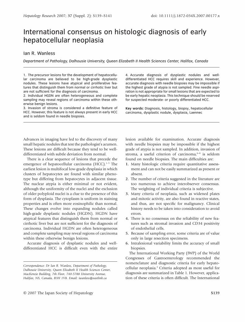
The International Working Party (IWP) of the WorldCongresses of Gastroenterology recommended thenomenclature and diagnostic criteria for early hepato-cellular neoplasia.3 Criteria adopted as most useful fordiagnosis are summarized in Table 1. However, applica-tion of these criteria is often difficult. The International
Correspondence: Dr Ian R. Wanless, Department of Pathology,Dalhousie University, Queen Elizabeth II Health Sciences Center,MacKenzie Building, 7th Floor, 740-5788 University Avenue,Halifax, NS, Canada, B3H 1V8. Email: [email protected]
Hepatology Research 2007; 37 (Suppl. 2): S139–S141 doi: 10.1111/j.1872-034X.2007.00177.x
© 2007 The Japan Society of Hepatology S139
Consensus Group for Hepatic Neoplasia (ICGHN) iscurrently assessing the reliability of histologic diagnosisof early HCC and dysplastic nodules. This group,comprised of two dozen hepatopathologists from tencountries, met in 2002, 2004, 2005, and 2006. Earlyresults from these meetings indicate that refinementsof the diagnostic criteria and a teaching aid for generalpathologists are needed.
A new diagnostic approach is being developed, to beknown as the Laennec Classification of HepatocellularNeoplasia (LHN). The proposed LHN system will bedefined in detail elsewhere. The system can be appliedto premalignant lesions and all grades of hepatocellularcarcinoma.
The group decided that the best criteria for differen-tiation of HCC from dysplastic nodules on needlebiopsies are: (i) liver cell plates more than two cellsin width or atypical plate structure; (ii) high nuclearto cytoplasmic (N/C) ratio; and (iii) nuclear atypia.The LHN system may assist the standardization andinterpretation of these criteria. A standard set ofphotographs is available free of charge at http://laennecliverpathology.com.
In this system, nuclear atypia, N/C ratio, and archi-tectural atypia are graded 1–4 or 1–5. The scores of
these three components are summed, giving a possiblerange of 3–13. When two or more distinct regions arenoted, more than one Laennec score is reported alongwith a comment on the percent of the lesion occupiedby each score. Stromal invasion, mitoses, and arterial-ization are not considered in this system because theyare highly dependent on sample size. However, whenpresent, they are very important for the diagnosis ofHCC.
The advantages of this system are: (i) it can be appliedto small and large samples; (ii) complexity is reduced;
Table 1 International Working Party histologic criteria to distinguish hepatocellular nodules
Histologic feature Largeregenerativenodule
Dysplasticnodule,low grade
Dysplasticnodule,high grade
Well-differentiatedHCC
ModeratelydifferentiatedHCC
Clone-like populations - + + + +Plates >3 cells wide - - - - +Mitotic figures >5/10 HPF - - - - +Cell density more than twice normal† - - - + +Invasion of stroma or portal tracts - - - + +Irregular nuclear contour, at least moderate - - - + +Absence of portal tracts (arterial supply) - - +/– + +Mitotic figures, occasional (1–5/10 HPF)‡ - - + + +Cell density >1.3 times normal - - + + +Nuclear hyperchromasia - - + + +Irregular nuclear contour, at least mild - - + + +Pseudogland formation +§ - + + +Cytoplasmic basophilia - - + + +Cytoplasmic clear cell change - - + + +Reticulin less than normal - - - - +Increased or decreased iron accumulation - + + + +
†As a measure of increased nuclear to cytoplasmic ratio.‡Mitoses may occur in any lesion in the presence of cholestasis or recent necrosis.§Especially when cholestasis present.HCC, hepatocellular carcinoma; HPF, high power field (10 ¥ 40).Table is modified after the International Working Party.3
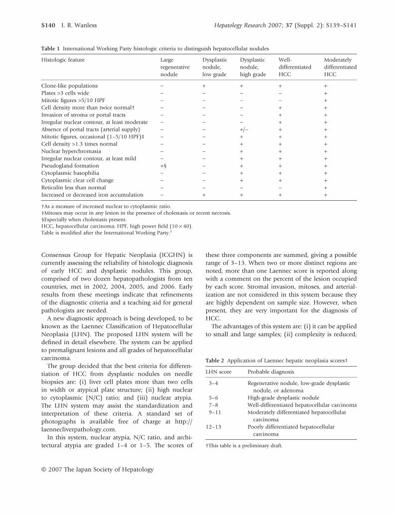
Table 2 Application of Laennec hepatic neoplasia scores†
LHN score Probable diagnosis
3–4 Regenerative nodule, low-grade dysplasticnodule, or adenoma
5–6 High-grade dysplastic nodule7–8 Well-differentiated hepatocellular carcinoma9–11 Moderately differentiated hepatocellular
carcinoma12–13 Poorly differentiated hepatocellular
carcinoma
†This table is a preliminary draft.
S140 I. R. Wanless Hepatology Research 2007; 37 (Suppl. 2): S139–S141
© 2007 The Japan Society of Hepatology

(iii) criteria are standardized; and (iv) the use of anumerical score reinforces the fact that early neoplasticfeatures form a spectrum not easily divided into benignand malignant diagnostic entities. The clinical signifi-cance of a given Laennec score is currently being evalu-ated. Early results suggest that a score can be interpretedas listed in Table 2.
CONFLICT OF INTEREST
NO CONFLICT OF interest statement has beenreceived from the author.
REFERENCES
1 Wada K, Kondo F, Kondo Y. Large regenerative nodules anddysplastic nodules in cirrhotic livers: a histopathologicstudy. Hepatology 1988; 8: 1684–8.
2 Theise ND, Park YN, Kojiro M. Dysplastic nodules and hepa-tocarcinogenesis. Clin Liver Dis 2002; 6: 497–512.
3 International Working Party. Terminology of nodular hepa-tocellular lesions. Hepatology 1995; 22: 983–93.
4 Sakabe K, Yamamoto T, Kubo S et al. Correlation betweendynamic computed tomographic and histopathologicalfindings in the diagnosis of small hepatocellular carcinoma.Dig Surg 2004; 21: 413–20.
5 Kojiro M, Roskams T. Early hepatocellular carcinomaand dysplastic nodules. Semin Liver Dis 2005; 25: 133–42.
6 Kondo F, Kondo Y, Nagato Y, Tomizawa M, Wada K. Inter-stitial tumour cell invasion in small hepatocellular carci-noma. Evaluation in microscopic and low magnificationviews. J Gastroenterol Hepatol 1994; 9: 604–12.
7 Nakano M, Saito A, Yamamoto M, Doi M, Takasaki K.Stromal and blood vessel wall invasion in well-differentiatedhepatocellular carcinoma. Liver 1997; 17: 41–6.
8 Park YN, Kojiro M, Di Tommaso L et al. Ductular reaction ishelpful in defining early stromal invasion, small hepatocel-lular carcinomas, and dysplastic nodules. Cancer 2007; 109:915–23.
Hepatology Research 2007; 37 (Suppl. 2): S139–S141 Histologic diagnosis of early hepatocellular neoplasia S141
© 2007 The Japan Society of Hepatology