insulin crystallization depends on zinc transporter znt8 ... · pdf fileand preserved...
TRANSCRIPT
Insulin crystallization depends on zinc transporterZnT8 expression, but is not required for normalglucose homeostasis in miceK. Lemairea,1, M. A. Ravierb,c,1, A. Schraenena, J. W. M. Creemersd, R. Van de Plase, M. Granvika, L. Van Lommela,E. Waelkensf, F. Chimientig, G. A. Rutterh, P. Gilonb, P. A. in’t Veldi, and F. C. Schuita,2
aGene Expression Unit, Department Molecular Cell Biology, Katholieke Universiteit Leuven, Herestraat 49, 3000 Leuven, Belgium; bUnit of Endocrinologyand Metabolism, University of Louvain, Faculty of Medicine, Université Catholique de Louvain 55.30, 1200 Brussels, Belgium; cInstitut National de la Sante etde la Recherche Medicale, U661, Equipe avenir, Centre National de la Recherche Scientifique, Unite Mixte de Recherche 5203, Universite Montpellier (IFR3),Institut de Genomique Fonctionnelle, 34090 Montpellier, France; dDepartment of Human Genetics, Katholieke Universiteit Leuven, Herestraat 49,3000 Leuven, Belgium; eDepartment of Electrical Engineering, Katholieke Universiteit Leuven, Kasteelpark Arenberg 10, 3001 Leuven, Belgium;fPrometa, Department of Molecular Cell Biology, Katholieke Universiteit Leuven, Herestraat 49, 3000 Leuven, Belgium; gMellitech, SAS, Commissariat al’Energie Atomique, 17 rue des Martyrs, 38054 Grenoble, France; hDepartment of Cell Biology Division of Medicine Sir Alexander Fleming Building ImperialCollege, London Exhibition Road, London SW7 2AZ, United Kingdom; and iDepartment of Pathology, Vrije Universiteit Brussel, Laarbeeklaan 103,1090 Brussels, Belgium
Edited by Donald F. Steiner, University of Chicago, Chicago, IL and approved July 16, 2009 (received for review June 15, 2009)
Zinc co-crystallizes with insulin in dense core secretory granules,but its role in insulin biosynthesis, storage and secretion is un-known. In this study we assessed the role of the zinc transporterZnT8 using ZnT8-knockout (ZnT8�/�) mice. Absence of ZnT8 ex-pression caused loss of zinc release upon stimulation of exocytosis,but normal rates of insulin biosynthesis, normal insulin contentand preserved glucose-induced insulin release. Ultrastructurally,mature dense core insulin granules were rare in ZnT8�/� beta cellsand were replaced by immature, pale insulin ‘‘progranules,’’ whichwere larger than in ZnT8�/� islets. When mice were fed a controldiet, glucose tolerance and insulin sensitivity were normal. How-ever, after high-fat diet feeding, the ZnT8�/� mice became glucoseintolerant or diabetic, and islets became less responsive to glucose.Our data show that the ZnT8 transporter is essential for theformation of insulin crystals in beta cells, contributing to thepackaging efficiency of stored insulin. Interaction betweenthe ZnT8�/� genotype and diet to induce diabetes is a model forfurther studies of the mechanism of disease of human ZNT8 genemutations.
dense core granule � diabetes � zinc
Z inc plays a crucial role in many cell functions; as a result, bothzinc deficiency (1) and excess of free zinc (2) are toxic to
mammalian cells. The abundance of zinc per cell is tissue-dependent and the zinc content of pancreatic beta cells is amongthe highest in the body. In beta cells, zinc was proposed to berequired for multiple steps in insulin synthesis and release (3–5),but conclusive evidence is lacking. After synthesis in the ER,pro-insulin is transported into the Golgi apparatus where im-mature, pale secretory ‘‘progranules’’ are formed (6). Thesegranules contain pro-insulin-zinc hexamers which are furtherprocessed into mature insulin and C-peptide by the prohormoneconvertases PC1/3 and PC2 (7). After maturation, the zinc-insulin hexamers form water-insoluble crystals (3). It has beensuggested that crystal formation increases the degree of con-version of soluble pro-insulin to insoluble insulin, but nearlynormal pro-insulin processing occurs in patients with mutatedhistidine-B10 insulin, which cannot crystallize (5). Futhermore,in several animal species such as guinea pig and hagfish, insulindoes not have a histidine at position B10, so that no zinc-insulincrystals form; in these species, insulin is processed, and glucosehomeostasis is normal (8, 9).
Two large zinc transporter families exist [reviewed in (10)].Influx of zinc from the extracellular space into the cytosol ismediated by members of the, Slc39 (ZIP, Zrt/Irt-like protein)protein family, which has 14 isoforms. Efflux of zinc into the
extracellular space as well as transport of zinc from the cytosolinto intracellular organelles is mediated by 10 known isoforms ofthe Slc30 (ZnT, zinc transporters) family. Members of bothtransporter families exhibit tissue-specific expression, where theyfulfil unique functions (10, 11). For example, ZnT8 (Slc30a8) wasidentified as a pancreatic beta cell-specific secretory granule zinctransporter (11, 12).
The interest in ZnT8 was aroused by the observation thatepitopes of the ZnT8 protein are recognized by autoantibodiesof patients with type 1 diabetes (T1D) (13). Furthermore, in agenome-wide association study (14), which was replicated inindependent cohorts (15–18), an association was made betweena SNP marker within the human SLC30A8 gene and geneticsusceptibility to type 2 diabetes (T2D). The exact pathogenicsignificance of this polymorphism is still unknown.
To better understand the role of ZnT8 in pancreatic beta cells,we studied the phenotype of mice that are deficient in ZnT8expression (ZnT8�/�). Our data show that (i) ZnT8 is essentialfor insulin crystal formation that shapes the dense core ofsecretory granules, (ii) that ZnT8 is the only transporter in thebeta cell that provides the required zinc ions for this process, and(iii) that ZnT8 is only essential for normal glucose homeostasisafter the prolonged environmental � cell stress of a high fat diet.
ResultsIslets from ZnT8�/� Mice Do Not Express the ZnT8 Protein, Have NoDithizone Staining, and Do Not Release Zinc. To assess the role of thebeta cell zinc transporter ZnT8 in glucose homeostasis, westudied ZnT8�/� mice that were generated by Cre recombinasemediated deletion of the first exon, including the transcriptionstart site (Fig. S1). Mice were born in Mendelian proportions andequal male/female ratio. No differences in growth and bodyweight were observed. ZnT8�/� mice did not express ZnT8mRNA (Fig. 1A) and protein (Fig. 1B), and ZnT8 immunostain-ing of islet sections was negative (Fig. 1C). ZnT8 deficiency wasnot compensated by overexpression of one of the other zinc
Author contributions: K.L., M.A.R., P.G., and F.C.S. designed research; K.L., M.A.R., A.S.,J.W.M.C., R.V.d.P., M.G., L.V.L., P.G., and P.A.i.V. performed research; E.W., F.C., and G.A.R.contributed new reagents/analytic tools; K.L., M.A.R., P.G., P.A.i.V., and F.C.S. analyzeddata; and K.L. and F.C.S. wrote the paper.
The authors declare no conflict of interest.
This article is a PNAS Direct Submission.
1K.L. and M.A.R. contributed equally to this work.
2To whom correspondence should be addressed. E-mail: [email protected].
This article contains supporting information online at www.pnas.org/cgi/content/full/0906587106/DCSupplemental.
www.pnas.org�cgi�doi�10.1073�pnas.0906587106 PNAS Early Edition � 1 of 6
CELL
BIO
LOG
Y

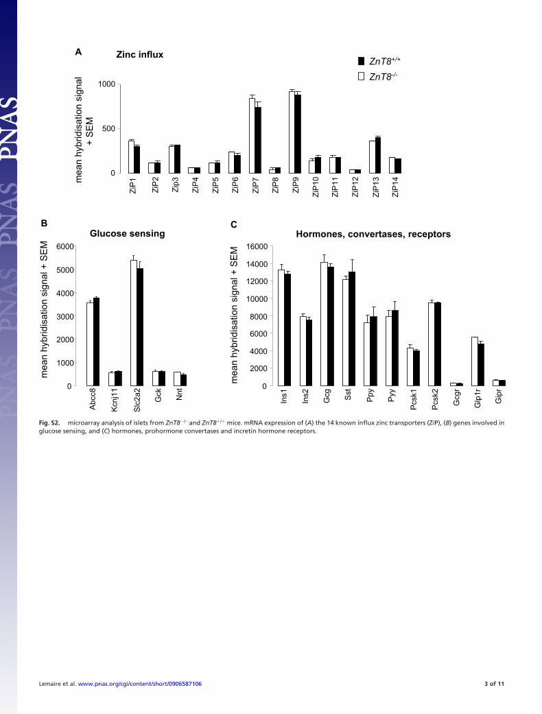
transporters (Fig. 1 A). Furthermore, the mRNA expressionprofiles of ZnT8�/� and ZnT8�/� mice were very similar; as wefound no differences in expression of mRNA’s encoding otherimportant beta-cell proteins (Fig. S2).
To investigate the consequence of beta-cell ZnT8 deficiencyon islet zinc content, we performed in vivo and in vitro isletdithizone staining, a technique to distinguish pancreatic isletsfrom exocrine cells (19), for instance used in vitro for human isletisolations (20) or in situ to visualise islets (21). ZnT8�/� isletswere negative with this staining, both in situ (Fig. 2A), and inisolated islets (Fig. 2B). These results indicate that there is a
direct connection between beta cell expression of the ZnT8transporter, which allows zinc influx in insulin secretory granulesand islet reactivity to dithizone.
Zinc release from secretory granules from small clusters ofZnT8�/� and ZnT8�/� islet cells stimulated by 15 mM glucoseand 1 �M forskolin was imaged with total internal f luorescencemicroscopy (TIRF) using FluoZin-3 (22). A strong differencebetween ZnT8�/� and ZnT8�/� beta cells was found (6.3 � 1.1vs. 0.07 � 0.03 zinc exocytotic events/min, respectively, P �0.0001) (Fig. 2C and D). A general defect of exocytosis inZnT8�/� beta cells was excluded by measuring release of NPY-
Fig. 1. ZnT8 expression is absent in ZnT8�/� mice. (A) mRNA expression of the 10 known efflux zinc transporters (Slc30a family) analyzed via microarray (n �3). (B) ZnT8 protein expression was analyzed via western blots. Please note complete absence of the expected MW for ZnT8 monomers (M) and SDS-resistantdimers (D), * � non-specific protein. (C) Immunohistochemistry of pancreatic sections from ZnT8�/� and ZnT8�/� mice. In ZnT8�/� mice ZnT8 protein is stronglyexpressed in all insulin-positive cells and weakly expressed in a minority of glucagon cells. Nuclei are stained blue by 4�,6-diamidino-2-phenylindole (DAPI). (Scalebar, 10 �m.)
Fig. 2. ZnT8 is required for islet dithizone staining and glucose-regulated granular zinc release from beta cells. (A) Dithizone staining of pancreata from ZnT8�/�
and ZnT8�/� mice, 15 min after i.p. injection of the dye. Islets can be seen at the pancreatic surface of ZnT8�/� mice but not of ZnT8�/� mice. (B) Dithizone stainingof isolated islets is positive in ZnT8�/� mice (red color) but negative in ZnT8�/� mice. (C) Zinc release from clusters of islet cells imaged by TIRF microscopy during7 min. Cluster size was 3.7 � 0.4 vs. 3.8 � 0.3 cells, for ZnT8�/� and ZnT8�/�, respectively (ns). Medium contained 8 �M FluoZin-3 and exocytosis was stimulatedby 15 mM glucose and 1 �M forskolin. One zinc exocytotic event from a ZnT8�/� four-cell cluster is illustrated, boundaries between cells are shown as dottedlines (left panel). Images were taken every 30 ms in the region delimited by a square (middle panel), and the intensity of fluorescence for this release event isplotted on the right panel. Arrows show fluorescence intensity for the indicated time points illustrated in the middle panel, (D) Quantification of zinc exocytoticevents in clusters of ZnT8�/� and ZnT8�/� islet cells. The horizontal line shows the average of 18 clusters from three individual mice.
2 of 6 � www.pnas.org�cgi�doi�10.1073�pnas.0906587106 Lemaire et al.
pHluorin, a pH sensitive probe (23) (Fig. S3 A and B) and byinsulin release experiments (see following paragraph).
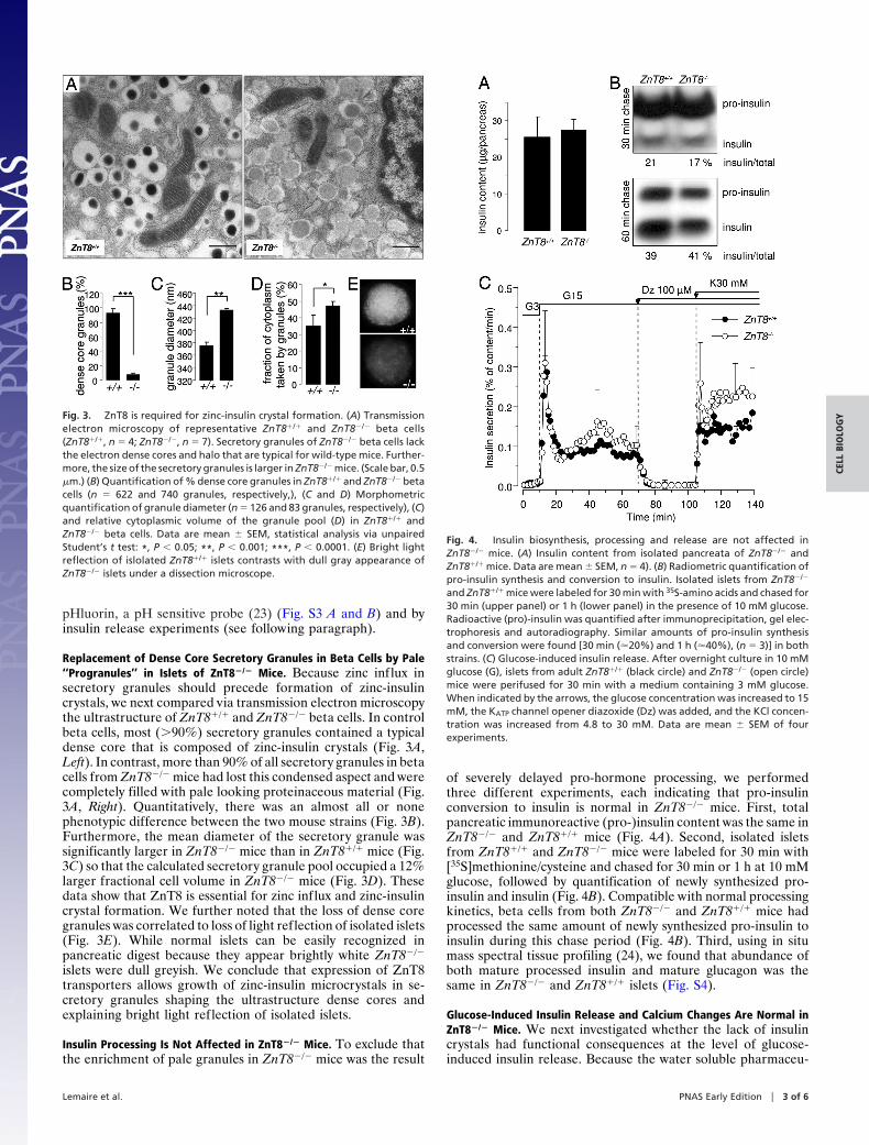
Replacement of Dense Core Secretory Granules in Beta Cells by Pale‘‘Progranules’’ in Islets of ZnT8�/� Mice. Because zinc influx insecretory granules should precede formation of zinc-insulincrystals, we next compared via transmission electron microscopythe ultrastructure of ZnT8�/� and ZnT8�/� beta cells. In controlbeta cells, most (�90%) secretory granules contained a typicaldense core that is composed of zinc-insulin crystals (Fig. 3A,Left). In contrast, more than 90% of all secretory granules in betacells from ZnT8�/� mice had lost this condensed aspect and werecompletely filled with pale looking proteinaceous material (Fig.3A, Right). Quantitatively, there was an almost all or nonephenotypic difference between the two mouse strains (Fig. 3B).Furthermore, the mean diameter of the secretory granule wassignificantly larger in ZnT8�/� mice than in ZnT8�/� mice (Fig.3C) so that the calculated secretory granule pool occupied a 12%larger fractional cell volume in ZnT8�/� mice (Fig. 3D). Thesedata show that ZnT8 is essential for zinc influx and zinc-insulincrystal formation. We further noted that the loss of dense coregranules was correlated to loss of light reflection of isolated islets(Fig. 3E). While normal islets can be easily recognized inpancreatic digest because they appear brightly white ZnT8�/�
islets were dull greyish. We conclude that expression of ZnT8transporters allows growth of zinc-insulin microcrystals in se-cretory granules shaping the ultrastructure dense cores andexplaining bright light reflection of isolated islets.
Insulin Processing Is Not Affected in ZnT8�/� Mice. To exclude thatthe enrichment of pale granules in ZnT8�/� mice was the result
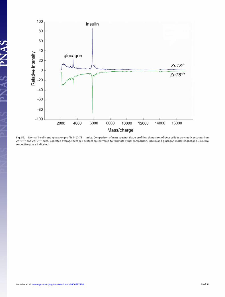
of severely delayed pro-hormone processing, we performedthree different experiments, each indicating that pro-insulinconversion to insulin is normal in ZnT8�/� mice. First, totalpancreatic immunoreactive (pro-)insulin content was the same inZnT8�/� and ZnT8�/� mice (Fig. 4A). Second, isolated isletsfrom ZnT8�/� and ZnT8�/� mice were labeled for 30 min with[35S]methionine/cysteine and chased for 30 min or 1 h at 10 mMglucose, followed by quantification of newly synthesized pro-insulin and insulin (Fig. 4B). Compatible with normal processingkinetics, beta cells from both ZnT8�/� and ZnT8�/� mice hadprocessed the same amount of newly synthesized pro-insulin toinsulin during this chase period (Fig. 4B). Third, using in situmass spectral tissue profiling (24), we found that abundance ofboth mature processed insulin and mature glucagon was thesame in ZnT8�/� and ZnT8�/� islets (Fig. S4).
Glucose-Induced Insulin Release and Calcium Changes Are Normal inZnT8�/� Mice. We next investigated whether the lack of insulincrystals had functional consequences at the level of glucose-induced insulin release. Because the water soluble pharmaceu-
Fig. 3. ZnT8 is required for zinc-insulin crystal formation. (A) Transmissionelectron microscopy of representative ZnT8�/� and ZnT8�/� beta cells(ZnT8�/�, n � 4; ZnT8�/�, n � 7). Secretory granules of ZnT8�/� beta cells lackthe electron dense cores and halo that are typical for wild-type mice. Further-more, the size of the secretory granules is larger in ZnT8�/� mice. (Scale bar, 0.5�m.) (B) Quantification of % dense core granules in ZnT8�/� and ZnT8�/� betacells (n � 622 and 740 granules, respectively,), (C and D) Morphometricquantification of granule diameter (n � 126 and 83 granules, respectively), (C)and relative cytoplasmic volume of the granule pool (D) in ZnT8�/� andZnT8�/� beta cells. Data are mean � SEM, statistical analysis via unpairedStudent’s t test: *, P � 0.05; **, P � 0.001; ***, P � 0.0001. (E) Bright lightreflection of islolated ZnT8�/� islets contrasts with dull gray appearance ofZnT8�/� islets under a dissection microscope.
Fig. 4. Insulin biosynthesis, processing and release are not affected inZnT8�/� mice. (A) Insulin content from isolated pancreata of ZnT8�/� andZnT8�/� mice. Data are mean � SEM, n � 4). (B) Radiometric quantification ofpro-insulin synthesis and conversion to insulin. Isolated islets from ZnT8�/�
and ZnT8�/� mice were labeled for 30 min with 35S-amino acids and chased for30 min (upper panel) or 1 h (lower panel) in the presence of 10 mM glucose.Radioactive (pro)-insulin was quantified after immunoprecipitation, gel elec-trophoresis and autoradiography. Similar amounts of pro-insulin synthesisand conversion were found [30 min (�20%) and 1 h (�40%), (n � 3)] in bothstrains. (C) Glucose-induced insulin release. After overnight culture in 10 mMglucose (G), islets from adult ZnT8�/� (black circle) and ZnT8�/� (open circle)mice were perifused for 30 min with a medium containing 3 mM glucose.When indicated by the arrows, the glucose concentration was increased to 15mM, the KATP channel opener diazoxide (Dz) was added, and the KCl concen-tration was increased from 4.8 to 30 mM. Data are mean � SEM of fourexperiments.
Lemaire et al. PNAS Early Edition � 3 of 6
CELL
BIO
LOG
Y
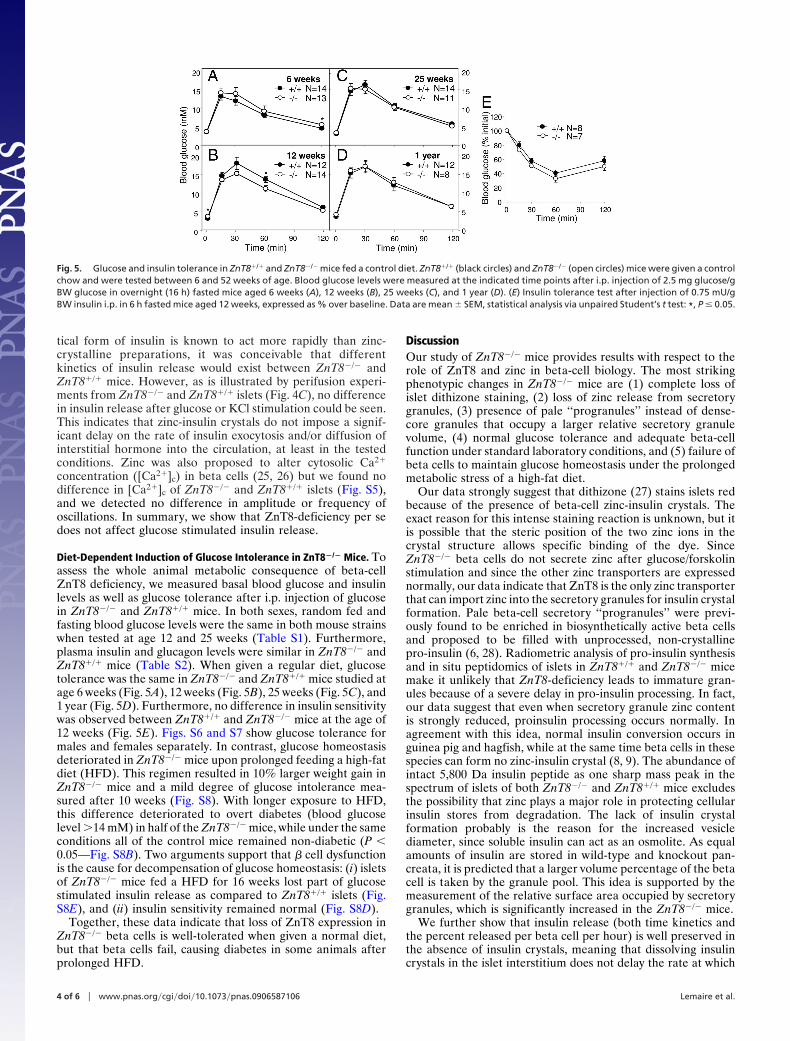
tical form of insulin is known to act more rapidly than zinc-crystalline preparations, it was conceivable that differentkinetics of insulin release would exist between ZnT8�/� andZnT8�/� mice. However, as is illustrated by perifusion experi-ments from ZnT8�/� and ZnT8�/� islets (Fig. 4C), no differencein insulin release after glucose or KCl stimulation could be seen.This indicates that zinc-insulin crystals do not impose a signif-icant delay on the rate of insulin exocytosis and/or diffusion ofinterstitial hormone into the circulation, at least in the testedconditions. Zinc was also proposed to alter cytosolic Ca2�
concentration ([Ca2�]c) in beta cells (25, 26) but we found nodifference in [Ca2�]c of ZnT8�/� and ZnT8�/� islets (Fig. S5),and we detected no difference in amplitude or frequency ofoscillations. In summary, we show that ZnT8-deficiency per sedoes not affect glucose stimulated insulin release.
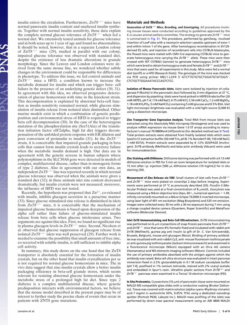
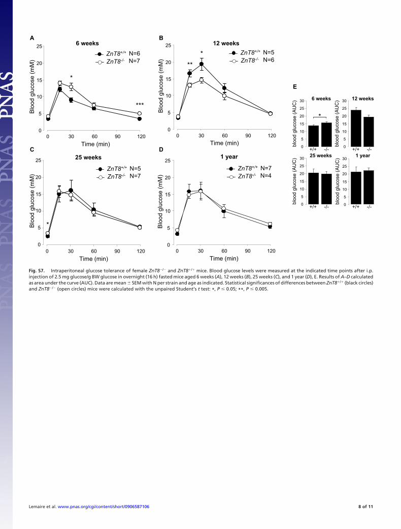
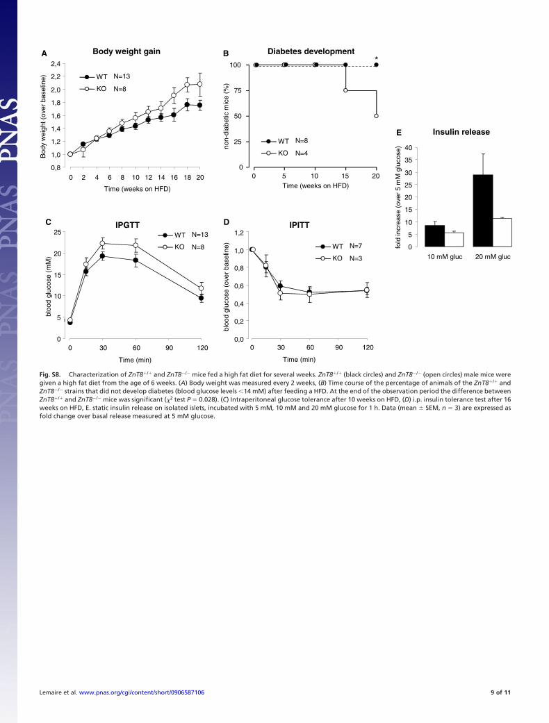
Diet-Dependent Induction of Glucose Intolerance in ZnT8�/� Mice. Toassess the whole animal metabolic consequence of beta-cellZnT8 deficiency, we measured basal blood glucose and insulinlevels as well as glucose tolerance after i.p. injection of glucosein ZnT8�/� and ZnT8�/� mice. In both sexes, random fed andfasting blood glucose levels were the same in both mouse strainswhen tested at age 12 and 25 weeks (Table S1). Furthermore,plasma insulin and glucagon levels were similar in ZnT8�/� andZnT8�/� mice (Table S2). When given a regular diet, glucosetolerance was the same in ZnT8�/� and ZnT8�/� mice studied atage 6 weeks (Fig. 5A), 12 weeks (Fig. 5B), 25 weeks (Fig. 5C), and1 year (Fig. 5D). Furthermore, no difference in insulin sensitivitywas observed between ZnT8�/� and ZnT8�/� mice at the age of12 weeks (Fig. 5E). Figs. S6 and S7 show glucose tolerance formales and females separately. In contrast, glucose homeostasisdeteriorated in ZnT8�/� mice upon prolonged feeding a high-fatdiet (HFD). This regimen resulted in 10% larger weight gain inZnT8�/� mice and a mild degree of glucose intolerance mea-sured after 10 weeks (Fig. S8). With longer exposure to HFD,this difference deteriorated to overt diabetes (blood glucoselevel �14 mM) in half of the ZnT8�/� mice, while under the sameconditions all of the control mice remained non-diabetic (P �0.05—Fig. S8B). Two arguments support that � cell dysfunctionis the cause for decompensation of glucose homeostasis: (i) isletsof ZnT8�/� mice fed a HFD for 16 weeks lost part of glucosestimulated insulin release as compared to ZnT8�/� islets (Fig.S8E), and (ii) insulin sensitivity remained normal (Fig. S8D).
Together, these data indicate that loss of ZnT8 expression inZnT8�/� beta cells is well-tolerated when given a normal diet,but that beta cells fail, causing diabetes in some animals afterprolonged HFD.
DiscussionOur study of ZnT8�/� mice provides results with respect to therole of ZnT8 and zinc in beta-cell biology. The most strikingphenotypic changes in ZnT8�/� mice are (1) complete loss ofislet dithizone staining, (2) loss of zinc release from secretorygranules, (3) presence of pale ‘‘progranules’’ instead of dense-core granules that occupy a larger relative secretory granulevolume, (4) normal glucose tolerance and adequate beta-cellfunction under standard laboratory conditions, and (5) failure ofbeta cells to maintain glucose homeostasis under the prolongedmetabolic stress of a high-fat diet.
Our data strongly suggest that dithizone (27) stains islets redbecause of the presence of beta-cell zinc-insulin crystals. Theexact reason for this intense staining reaction is unknown, but itis possible that the steric position of the two zinc ions in thecrystal structure allows specific binding of the dye. SinceZnT8�/� beta cells do not secrete zinc after glucose/forskolinstimulation and since the other zinc transporters are expressednormally, our data indicate that ZnT8 is the only zinc transporterthat can import zinc into the secretory granules for insulin crystalformation. Pale beta-cell secretory ‘‘progranules’’ were previ-ously found to be enriched in biosynthetically active beta cellsand proposed to be filled with unprocessed, non-crystallinepro-insulin (6, 28). Radiometric analysis of pro-insulin synthesisand in situ peptidomics of islets in ZnT8�/� and ZnT8�/� micemake it unlikely that ZnT8-deficiency leads to immature gran-ules because of a severe delay in pro-insulin processing. In fact,our data suggest that even when secretory granule zinc contentis strongly reduced, proinsulin processing occurs normally. Inagreement with this idea, normal insulin conversion occurs inguinea pig and hagfish, while at the same time beta cells in thesespecies can form no zinc-insulin crystal (8, 9). The abundance ofintact 5,800 Da insulin peptide as one sharp mass peak in thespectrum of islets of both ZnT8�/� and ZnT8�/� mice excludesthe possibility that zinc plays a major role in protecting cellularinsulin stores from degradation. The lack of insulin crystalformation probably is the reason for the increased vesiclediameter, since soluble insulin can act as an osmolite. As equalamounts of insulin are stored in wild-type and knockout pan-creata, it is predicted that a larger volume percentage of the betacell is taken by the granule pool. This idea is supported by themeasurement of the relative surface area occupied by secretorygranules, which is significantly increased in the ZnT8�/� mice.
We further show that insulin release (both time kinetics andthe percent released per beta cell per hour) is well preserved inthe absence of insulin crystals, meaning that dissolving insulincrystals in the islet interstitium does not delay the rate at which
Fig. 5. Glucose and insulin tolerance in ZnT8�/� and ZnT8�/� mice fed a control diet. ZnT8�/� (black circles) and ZnT8�/� (open circles) mice were given a controlchow and were tested between 6 and 52 weeks of age. Blood glucose levels were measured at the indicated time points after i.p. injection of 2.5 mg glucose/gBW glucose in overnight (16 h) fasted mice aged 6 weeks (A), 12 weeks (B), 25 weeks (C), and 1 year (D). (E) Insulin tolerance test after injection of 0.75 mU/gBW insulin i.p. in 6 h fasted mice aged 12 weeks, expressed as % over baseline. Data are mean � SEM, statistical analysis via unpaired Student’s t test: *, P � 0.05.
4 of 6 � www.pnas.org�cgi�doi�10.1073�pnas.0906587106 Lemaire et al.
insulin enters the circulation. Furthermore, ZnT8�/� mice havenormal pancreatic insulin content and unaltered insulin synthe-sis. Together with normal insulin sensitivity, these data explainthe complete normal glucose tolerance of ZnT8�/� when fed anormal diet. We extensively tested animals for glucose toleranceand in both sexes up to 1 year of age and found no abnormalities.It should be noted, however, that in a separate London colonyof ZnT8�/� mice (29), studied in parallel with our colony,impaired glucose tolerance was observed with standard diet,despite the existence of less dramatic alterations in granulemorphology. Since the Leuven and London colonies were de-rived from the same mouse line, we wondered whether subtlechanges in the environment could be responsible for differencesin phenotype. To address this issue, we fed control animals andZnT8�/� mice a HFD, a condition known to increase themetabolic demand for insulin and which can trigger beta- cellfailure in the presence of an underlying genetic defect (30, 31).In agreement with this idea, we observed progressive deterio-ration of glucose homeostasis with time in the knockout strain.This decompensation is explained by abnormal beta-cell func-tion as insulin sensitivity remained normal, while glucose stim-ulation of insulin release from isolated islets diminished. Othermurine models exist in which a combination of genetic predis-position and environmental stress of HFD is required to triggerbeta cell decompensation (30). In the case of the heterozygousmutation of the phosphorylation site (Ser51Ala) of the transla-tion initiation factor eIF2alpha, high fat diet triggers decom-pensation of the unfolded protein response with ER dilation andpoor conversion of proinsulin to insulin (31). In the ZnT8�/�
strain, it is conceivable that impaired granule packaging in betacells that cannot form insulin crystals leads to secretory failurewhen the metabolic insulin demand is high. Our data are inagreement with the genetic screens in human subjects wherepolymorphisms in the SLC30A8 gene were detected in models ofcomplex, multifactorial disease, rather than in monogenic formsof type 2 diabetes. Also in agreement with our own data, anindependent ZnT8�/� line was reported recently in which normalglucose tolerance was observed when the animals were given astandard diet (32); in these animals islet zinc content decreaseddramatically, but insulin crystals were not measured; moreover,the influence of HFD was not tested.
Recently, the hypothesis was proposed that Zn2�, co-releasedwith insulin, inhibits glucagon secretion in a paracrine manner(33). Since glucose stimulated zinc release is diminished in isletsfrom ZnT8�/� mice, it is conceivable that the mechanism ofimpaired glucose homeostasis is based upon derepression of thealpha cell rather than failure of glucose-stimulated insulinrelease from beta cells when glucose intolerance arises. Twoarguments are against this idea. First, we found no significant risein plasma glucagon levels in ZnT8�/� mice. Second, Nicolson etal. observed that glucose suppression of glucagon release fromisolated ZnT8�/� islets was well preserved (29). Further work isneeded to examine the possibility that small amounts of free zinc,co-secreted with soluble insulin, is still sufficient to inhibit alphacell activity.
In summary, this study shows on the one hand that the ZnT8transporter is absolutely essential for the formation of insulincrystals, but on the other hand that insulin crystallization per seis not required for normal glucose homeostasis in mice. The invivo data suggest that zinc-insulin crystals allow a better insulinpackaging efficiency in beta-cell granule stores, which seemsrelevant for resisting abnormal glucose homeostasis under themetabolic stress of a prolonged high fat diet. Since type 2diabetes is a complex multifactorial disease, where geneticpredisposition interacts with environmental factors, we believethat the mouse model described in the present paper may be ofinterest to further study the precise chain of events that occur inpatients with ZNT8 gene mutations.
Materials and MethodsGeneration of ZnT8�/� Mice, Breeding, and Genotyping. All procedures involv-ing mouse tissues were conducted according to guidelines approved by theK.U.Leuven animal welfare committee. The strategy to generate ZnT8�/� miceis summarized in Fig. S1. This procedure, performed by genOway, involvedinsertion of LoxP sites with a neomycin selection cassette upstream of exon 1and within intron 1 of the gene. After homologous recombination in SV129-derived ES cells, and injection of recombinant cells into C57BL/6 blastocytes,the floxed mice were mated with CMV-Cre-expressing C57BL/6J mice to gen-erate heterozygous mice carrying the ZnT8�/� allele. These mice were thencrossed with WT C57Bl6/J (Janvier) to generate heterozygous ZnT8�/� micewhich were bred to obtain homozygous male and female ZnT8�/� and ZnT8�/�
mice that were used for all experiments in this study. Mice were fed a normaldiet (ssniff) or a HFD (Research Diets). The genotype of the mice was checkedvia PCR using primer MEL1-L2/F4 5�-GTCTTGTGTGCTGGATGTATGG/5�-CTCTGCTTGGAAATACCCAGTCTCC.
Isolation of Mouse Pancreatic Islets. Islets were isolated by injection of colla-genase P (Roche) in the pancreatic duct followed by 3-min digestion at 37 °C,and dispersion by pipetting. Islets were hand-picked in HEPES Krebs buffer (20mM HEPES, pH 7.4; 119 mM NaCl; 4.75 mM KCl; 2.54 mM CaCl2; 1.2 mM MgSO4;1.18 mM KH2PO4; 5 mM NaHCO3) containing 5 mM glucose and 0.5% BSA. Isletlight microscopic brightness was evaluated with a cold light source (3,000 K)using a dissection microscope.
Zinc Transporter Gene Expression Analysis. Total RNA from mouse islets wasextracted using the Absolutely RNA microprep (Stratagene) and was used toanalyze mRNA expression via mouse Gene 1.0 ST arrays according to manu-facturer’s manual 701880Rev4 (Affymetrix) (for detailed method see SI Text).Total protein extracts were obtained from freshly isolated islets which werelysed in S1 extraction buffer (50 mM Tris, pH 8; 1% Nonidet P-40; 150 mM NaCl;1 mM EDTA). Protein extracts were separated by 4–12% SDS/PAGE (Invitro-gen). ZnT8 antibody (Mellitech) and beta-actin antibody (Abcam) were usedfor immunodetection.
Zinc Staining with Dithizone. Dithizone staining was performed with a 0.13 mMdithizone solution in PBS for 5 min at room temperature for isolated islets or15 min after i.p. injection [500 �L dithizone solution (10 mg/mL)] for in situstaining (34).
Measurment of Zinc Release via TIRF. Small clusters of islet cells from ZnT8�/�
and ZnT8�/� mice were plated on coverslips 2 days before imaging. Experi-ments were performed at 37 °C as previously described (35). FluoZin-3 (Mo-lecular Probes) was used at a final concentration of 8 �mol/L. Exocytosis wasmeasured using a Nikon objective lens (Apo, 60, numerical aperture � 1.49,infinity corrected) mounted on a Nikon Eclipse TE2000-E inverted microscope,using laser light of 491 nm excitation (Mag Biosystems) and 535 nm emission.Images were collected every 30 ms with a 30 ms-exposure during 7 min usinga charge coupled device camera (Quantem, Roper Scientific) and MetaFluorsoftware (Molecular Devices).
Islet ZnT8 Immunostaining and Beta Cell Ultrastructure. ZnT8 immunostainingwas performed on 6-�m cryosections of snap-frozen pancreata from ZnT8�/�
and ZnT8�/� mice that were 4% formalin fixed and incubated with rabbit antiZnT8 (Mellitech), guinea pig anti insulin (a gift of Dr. C. Van Schravendijk,Brussels, Belgium), mouse anti glucagon (Novo). Binding of primary antibod-ies was visualized with anti-rabbit Cy3, anti- mouse fluorescein isothiocyanateor anti-guinea pig isothiocyanate (Jackson Immunoresearch) and examined ina fluorescence microscope (Nikon) equipped with an Orca AG camera(Hamamatsu) and NIS-elements imaging software (Nikon). Controls includedthe use of primary antibodies absorbed with the antigen against which theantibody was raised. Beta cell ultra-structure was evaluated in intact pancreasimmersion-fixed in 2.5% glutaraldehyde in 0.1 M sodium cacodylate buffer(pH 7.4), postfixed in 1% osmium tetroxide, stained with 2% uranyl acetateand embedded in Spurr’s resin. Ultrathin plastic sections from ZnT8�/� andZnT8�/� pancreas were examined in a Tecnai 10 electron microscope (FEI).
In Situ Proteomics. Cryosections (10 �m) of pancreatic tissue were mounted onMALDI-MS compatible glass slides with a conductive coating (Bruker Dalton-ics). Tissue was covered with matrix solution (alpha-cyano-4hydroxy-cinnamicacid 7 mg/ml in acetonitrile 50%, 0.05% TFA) using a dedicated nanodropspotter (Portrait P630, Labcyte Inc.). MALDI mass profiling of the islets wasperformed by direct mass spectral measurement using an ABI 4800 MALDI
Lemaire et al. PNAS Early Edition � 5 of 6
CELL
BIO
LOG
Y

TOF/TOF Analyzer (Applied Biosystems) running in linear mode. The selectedmass over charge (m/z) range was 2,000–17,000 (24).
Measurement of Pro-Insulin Biosynthesis, Insulin Secretion, and Dynamics of[Ca2�]c. Insulin biosynthesis was performed as described earlier (31), withminor changes see SI Text. Insulin release was measured as described before(35, 36) in batches of 25 overnight cultured ZnT8�/� and ZnT8�/� islets peri-fused at 37 °C with test solutions of which the composition is indicated at thetop of the figure. At the end of the experiments, the islets were recovered, andtheir insulin content was determined after extraction in acid-ethanol. Cyto-solic calcium was measured as described before (37) in overnight culturedZnT8�/� and ZnT8�/� islets loaded at 37 °C for 2 h with 2 �mol/L fura PE3-AM(MoBiTec).
Glucose/Insulin Tolerance Tests and Insulin/Glucagon Assays. For the glucosetolerance tests, mice of 6, 12, and 25 weeks of age were fasted overnight (16h). Glucose (2.5 mg/g BW) was administered i.p. and blood glucose levels werefollowed during 2 h using AccuCheck glucostrips. For insulin tolerance test, 6-h
fasted mice were injected i.p. with insulin (0.75 mU/g BW) and blood glucoselevels were measured as described above. Plasma insulin was measured fromblood taken via the vena cava or the tail vein and centrifuged for 10 min at1,100 g at 4 °C. For glucagon measurements, aprotinin (250 IU/�L) wasadded to blood before centrifugation. Insulin content was measured in thetotal pancreas after dissection, snap freezing in liquid nitrogen, and homog-enization in 75% ethanol; 0.1% Triton-X-100; 1.2% 12 N HCl using a Douncer.(Pro-)insulin was quantified using ELISA (Crystal Chem) and glucagon wasquantified using RIA (Linco).
ACKNOWLEDGMENTS. We thank Marleen Bergmans, Nicole Buelens, andJulien Carpent for excellent technical assistance and D. Daro for help con-structing the adenovirus expressing pHluorin. This work was supported bypostdoctoral fellowships from the FWO Vlaanderen to K.L., and from theFNRS, Brussels to M.A.R. Research was further supported by grants of theEuropean Union (EURODIA FP6–518153), the Belgian Ministry for SciencePolicy (IUAP P6/40), SymBioSys (the K.U. Leuven center of excellence of SystemsBiology), the K.U. Leuven (GOA 2004/11 and GOA 2008/16), and ARC (05/10–328) from the General Direction of Scientific Research of the French Commu-nity of Belgium. P.G. is research director from the FNRS, Brussels.
1. Sekler I, Sensi SL, Hershfinkel M, Silverman WF (2007) Mechanism and regulation ofcellular zinc transport. Mol Med 13:337–343.
2. Frederickson CJ, Koh JY, Bush AI (2005) The neurobiology of zinc in health and disease.Nat Rev Neurosci 6:449–462.
3. Dodson G, Steiner D (1998) The role of assembly in insulin’s biosynthesis. Curr OpinStruct Biol 8:189–194.
4. Huang XF, Arvan P (1995) Intracellular transport of proinsulin in pancreatic beta-cells.Structural maturation probed by disulfide accessibility. J Biol Chem 270:20417–20423.
5. Carroll RJ, et al. (1988) A mutant human proinsulin is secreted from islets of Langerhansin increased amounts via an unregulated pathway. Proc Natl Acad Sci USA 85:8943–8947.
6. Orci L, et al. (1985) Direct identification of prohormone conversion site in insulin-secreting cells. Cell 42:671–681.
7. Creemers JW, Jackson RS, Hutton JC (1998) Molecular and cellular regulation ofprohormone processing Semin Cell Dev Biol 9:3–10.
8. Emdin SO, Dodson GG, Cutfield JM, Cutfield SM (1980) Role of zinc in insulin biosyn-thesis. Some possible zinc-insulin interactions in the pancreatic B-cell. Diabetologia19:174–182.
9. Smith LF (1966) Species variation in the amino acid sequence of insulin. Am J Med40:662–666.
10. Liuzzi JP, Cousins RJ (2004) Mammalian zinc transporters. Annu Rev Nutr 24:151–172.11. Chimienti F, Devergnas S, Favier A, Seve M (2004) Identification and cloning of a
beta-cell-specific zinc transporter, ZnT-8, localized into insulin secretory granules.Diabetes 53:2330–2337.
12. Chimienti F, Favier A, Seve M (2005) ZnT-8, a pancreatic beta-cell-specific zinc trans-porter. Biometals 18:313–317.
13. Wenzlau JM, et al. (2007) The cation efflux transporter ZnT8 (Slc30A8) is a majorautoantigen in human type 1 diabetes. Proc Natl Acad Sci USA 104:17040–17045.
14. Sladek R, et al. (2007) A genome-wide association study identifies novel risk loci fortype 2 diabetes. Nature 445:881–885.
15. Wenzlau JM, et al. (2008) A common non-synonymous single nucleotide polymorphismin the SLC30A8 gene determines ZnT8 autoantibody specificity in type 1 diabetes.Diabetes 57:2693–2697.
16. Saxena R, et al. (2007) Genome-wide association analysis identifies loci for type 2diabetes and triglyceride levels. Science 316:1331–1336.
17. Scott LJ, et al. (2007) A genome-wide association study of type 2 diabetes in Finnsdetects multiple susceptibility variants. Science 316:1341–1345.
18. Zeggini E, et al. (2007) Replication of genome-wide association signals in UK samplesreveals risk loci for type 2 diabetes. Science 316:1336–1341.
19. Maske H (1957) Interaction between insulin and zinc in the islets of Langerhans.Diabetes 6:335–341.
20. Latif ZA, Noel J, Alejandro R (1988) A simple method of staining fresh and culturedislets. Transplantation 45:827–830.
21. Shima C, et al. (1996) Enhancement and stabilization of dithizone vital staining for zincin rat organs using adduct formation. Anal Biochem 236:173–175.
22. Qian WJ, Gee KR, Kennedy RT (2003) Imaging of Zn2� release from pancreaticbeta-cells at the level of single exocytotic events. Anal Chem 75:3468–3475.
23. Miesenbock G, De Angelis DA, Rothman JE (1998) Visualizing secretion and synaptictransmission with pH-sensitive green fluorescent proteins. Nature 394:192–195.
24. Chaurand P, Stoeckli M, Caprioli RM (1999) Direct profiling of proteins in biologicaltissue sections by MALDI mass spectrometry. Anal Chem 71:5263–5270.
25. Bloc A, Cens T, Cruz H, Dunant Y (2000) Zinc-induced changes in ionic currents of clonalrat pancreatic -cells: Activation of ATP-sensitive K� channels. J Physiol 529 Pt 3:723–734.
26. Bancila V, et al. (2005) Two SUR1-specific histidine residues mandatory for zinc-inducedactivation of the rat KATP channel. J Biol Chem 280:8793–8799.
27. Niclauss N, et al. (2008) Computer-assisted digital image analysis to quantify the massand purity of isolated human islets before transplantation. Transplantation 86:1603–1609.
28. Kiekens R, et al. (1992) Differences in glucose recognition by individual rat pancreaticB cells are associated with intercellular differences in glucose-induced biosyntheticactivity. J Clin Invest 89:117–125.
29. Nicolson TJ, et al. (In press) The type 2 diabetes-associated vesicular zinc transporterSLC30A8/ZnT8 is critical for beta-cell Zn2� handling and normal glucose homeostasis.Diabetes, in press.
30. Cnop M, et al. (2008) An update on lipotoxic endoplasmic reticulum stress in pancreaticbeta-cells. Biochem Soc Trans 36:909–915.
31. Scheuner D, et al. (2005) Control of mRNA translation preserves endoplasmic reticulumfunction in beta cells and maintains glucose homeostasis. Nat Med 11:757–764.
32. Pound LD, et al. (2009) Deletion of the mouse Slc30a8 gene encoding zinc transporter-8results in impaired insulin secretion. Biochem J 421:371–376.
33. Ishihara H, et al. (2003) Islet beta-cell secretion determines glucagon release fromneighbouring alpha-cells. Nat Cell Biol 5:330–335.
34. Louagie E, et al. (2008) Role of furin in granular acidification in the endocrine pancreas:identification of the V-ATPase subunit Ac45 as a candidate substrate. Proc Natl AcadSci USA 105:12319–12324.
35. Ravier MA, Gilon P, Henquin JC (1999) Oscillations of insulin secretion can be triggeredby imposed oscillations of cytoplasmic Ca2� or metabolism in normal mouse islets.Diabetes 48:2374–2382.
36. Michael DJ, et al. (2004) Fluorescent cargo proteins in pancreatic beta-cells: Designdetermines secretion kinetics at exocytosis. Biophys J 87:L03–L05.
37. Arredouani A, et al. (2002) SERCA3 ablation does not impair insulin secretion butsuggests distinct roles of different sarcoendoplasmic reticulum Ca(2�) pumps forCa(2�) homeostasis in pancreatic beta-cells. Diabetes 51:3245–3253.
6 of 6 � www.pnas.org�cgi�doi�10.1073�pnas.0906587106 Lemaire et al.

Supporting InformationLemaire et al. 10.1073/pnas.0906587106SI TextZinc Transporter Gene Expression Analysis. Total RNA from mouseislets was extracted using the Absolutely RNA microprep (Strat-agene). Total RNA quantity and quality was determined andRNA profiles of all tested samples were similar with sharp 18Sand 28S rRNA peaks on a flat baseline. Total RNA (100 ng) wasused to analyze mRNA expression via mouse Gene 1.0 ST arraysaccording to manufacturer’s manual 701880Rev4 (Affymetrix).Briefly, double-stranded cDNA was synthesized with randomhexamers tagged with a T7 promoter sequence. Double-strandedcDNA was subsequently amplified by T7 RNA polymerase intoantisense cRNA using a Genechip WT synthesis and Amplifi-cation kit (Affymetrix). In the second cycle of cDNA synthesis,random hexamers and dUTP were used to prime reverse tran-scription of cRNA to sense single stranded DNA (ssDNA). Next,cRNA was hydrolyzed and the quality and relative abundance ofthe ssDNA preparations were assessed. Using uracil DNAglycosylase and apurinic/apyrimidinic endonuclease1, ssDNA(5.5 �g) was fragmented with a GeneChip WT terminal labelingkit (Affymetrix). The fragmented DNA was biotin labeled usingAffymetrix DNA Labeling reagent and hybridized to AffymetrixMouse Gene 1.0 arrays during 17 h at 45 °C in an Affymetrixhybridization oven. Arrays were washed and stained in anAffymetrix f luidics station and scanned using an Affymetrix3000 GeneScanner. All image files were analyzed using Af-fymetrix Expression Console™ software and quality controls ofthe arrays were according to manufacturer’s criteria. Raw datawere analyzed with RMA sketch using standard settings forGene 1.0 ST arrays of Expression Console in the AGCC software(Affymetrix).
Vector and Adenovirus Generation. To generate NPY-pHluorin,venus was removed from pNPY-Venus-N1 (kindly provided byA. Miyawaki) by digestion with BamHI and NotI. cDNA encod-
ing pHluorin was amplified by PCR from the vector pCI-Vamp2-pHluorin (kindly provided by G. Miesenbock) using the follow-ing primers: primer1, 5�- CGGGATCCACGTAGTAAAGGA-GAACTTTTCACTGG-3� (BamHI site underlined); primer 2,5�-ATTAGCGGCCGCTTAACCGGTTTTGTATAG-3� (NotIsite underlined), and was then ligated into the same BamHI andNotI site of pNPY-Venus-NI. The adenovirus was constructedand amplified, using the pAdEasy system (1). Briefly, NPY-pHluorin plasmid was digested using BglII and NotI and trans-ferred into pShuttle-CMV. Recombination with pAdEasy-1,transfection into HEK 293 cells, and viral amplification wereperformed as described (1, 2).
Measurements of Pro-Insulin Biosynthesis. Insulin biosynthesis wasperformed as described earlier, with minor changes (3). Briefly,15 islets per condition were preincubated for 1h in HEPES Krebsbuffer at 5 mM glucose and then labeled with 200 �Ci [35S]me-thionine/cysteine (Perkin-Elmer Easytag express protein label-ing mix, specific activity 1,175 Ci/mmol) for 30 min at 37 °C at10 mM glucose, followed by a 30 min or 1 h chase at 37 °C at 10mM glucose. The islet lysates were immunoprecipitated using(pro)-insulin antiserum (kindly provided by J. Hutton) andbound to protein A/G Sepharose (GE Healthcare). After elu-tion, newly synthesized pro-insulin and insulin were separated bySDS/PAGE and quantified for autoradiographic signals usingScion Image densitometry software.
Static Insulin Release. Insulin release was measured from staticallyincubated, size matched, freshly isolated islets (n � 7 per tube).Incubations were for 1 h at 37 °C in HEPES-buffered Krebssolution (� 0.5% BSA), supplemented with 5 mM, 10 mM or 20mM glucose. After incubation of the islets, the supernatant wasremoved immediately and frozen for insulin release measure-ment, using a commercially availabe ELISA kit (Mercodia). Theislet pellets were resuspended in ice-cold acid-EtOH (75%EtOH, 0.1 N HCl) to measure insulin content.
1. He TC, et al. (1998) A simplified system for generating recombinant adenoviruses. ProcNatl Acad Sci USA 95:2509–2514.
2. Ravier MA, Rutter GA (2005) Glucose or insulin, but not zinc ions, inhibit glucagonsecretion from mouse pancreatic alpha-cells. Diabetes 54:1789–1797.
3. Scheuner D, et al. (2005) Control of mRNA translation preserves endoplasmic reticulumfunction in beta cells and maintains glucose homeostasis. Nat Med 11:757–764.
Lemaire et al. www.pnas.org/cgi/content/short/0906587106 1 of 11

A B
ZnT8+/+ZnT8-/- ZnT8+/-
3000
1000
40003500
2500
500
M
42952593
ZnT8 endogenous locus
ZnT8 targetedlocus
ZnT8 Cre-mediatedexcised locus
MEL1-L2 MEL1-F44295 bp
Exon 2Exon 1
Exon 2Exon 1FRT
NEO
MEL1-L2 MEL1-F42593 bp
LoxP
Exon 2
LoxP
LoxP
FRT
Exon 3
Exon 3
Exon 3
FZ PZ RZ
FZ PZ RZ
Fig. S1. ZnT8 knock out strategy. (A) ZnT8 knockout mice were generated by insertion of a LoxP site together with an FRT flanked neomycin selection cassettewithin intron 1 and a proximal LoxP site within the upstream exon 1, resulting in a recombination event that removes control elements of transcription and thestart codon for translation (red arrow). MEL1-L2 and MEL1-F4 are the primers used for genotyping. Fz, Pz adn Rz are the primers used for QPCR. (B) PCR examplesof genotyping of ZnT8�/�, ZnT8�/� and ZnT8�/� mice. M, marker.
Lemaire et al. www.pnas.org/cgi/content/short/0906587106 2 of 11
A
ZiP1
ZiP2
Zip3
ZiP4
ZiP5
ZiP6
ZiP7
ZiP8
ZiP9
ZiP1
0
ZiP1
1
ZiP1
2
ZiP1
3
ZiP1
4
Zinc influx
B
0
1000
2000
3000
4000
5000
6000
Abcc
8
Kcnj
11
Slc2
a2
Gck Nnt
0
2000
4000
6000
8000
10000
12000
14000
16000
Ins1
Ins2
Gcg Ss
t
Ppy
Pyy
Pcsk
1
Pcsk
2
Gcg
r
Glp
1r
Gip
r
CGlucose sensing Hormones, convertases, receptors
0
500
1000m
ean
hybr
idis
atio
n si
gnal
+ S
EM
mea
n hy
brid
isat
ion
sign
al +
SE
M
mea
n hy
brid
isat
ion
sign
al +
SE
M
ZnT8+/+
ZnT8-/-
Fig. S2. microarray analysis of islets from ZnT8�/�and ZnT8�/� mice. mRNA expression of (A) the 14 known influx zinc transporters (ZiP), (B) genes involved inglucose sensing, and (C) hormones, prohormone convertases and incretin hormone receptors.
Lemaire et al. www.pnas.org/cgi/content/short/0906587106 3 of 11

Fig. S3. Exocytosis in islets from ZnT8�/�and ZnT8�/� beta cells. (A) Insulin exocytosis stimulated by 15 mM glucose and 1 �M forskolin was imaged during 7min by total internal reflection fluorescence microscopy after adenoviral expression of NPY-pHluorin. One representative exocytosis event from a cell within aZnT8�/� three-cell cluster is illustrated. Delimitation between cells is shown by dotted lines on the left panel, single images taken every 30 ms in the regiondelimited by a square from ‘‘cell a’’ are illustrated in the middle, and the intensity of fluorescence for this release event is plotted on the right panel. Arrowsshow fluorescence intensity for the indicated time points illustrated in the middle panel. (B) Number of flashes counted manually in clusters of ZnT8�/� andZnT8�/� islet cells in similar experiments as that illustrated in A. The horizontal line shows the average of 12 clusters from three individual mice.
Lemaire et al. www.pnas.org/cgi/content/short/0906587106 4 of 11
ZnT8+/+
ZnT8-/-
Mass/charge
Rel
ativ
e in
tens
ity
100
80
60
40
20
0
-20
-100
-80
-60
-40
2000 4000 6000 8000 10000 12000 14000 16000
insulin
glucagon
Fig. S4. Normal insulin and glucagon profile in ZnT8�/� mice. Comparison of mass spectral tissue profiling signatures of beta cells in pancreatic sections fromZnT8�/� and ZnT8�/� mice. Collected average beta cell profiles are mirrored to facilitate visual comparison. Insulin and glucagon masses (5,800 and 3,483 Da,respectively) are indicated.
Lemaire et al. www.pnas.org/cgi/content/short/0906587106 5 of 11

[Ca2+
] c (n
mol
/l)
100
150
200
250
ZnT8+/+
ZnT8-/-
K30 mMDz 100 µM
G3G15
0 4 8 12 16 20 24 28 32 36 40
Time (min)Fig. S5. Changes in free cytosolic calcium concentration are normal in ZnT8�/� mice. After overnight culture in 10 mM glucose (G), islets from adult ZnT8�/�
(solid line) and ZnT8�/� (dotted line) mice were perifused for 30 min with a medium containing 3 mM glucose. Only the last period of perifusion in G3 is shown.When indicated by the arrows, the glucose concentration was increased to 15 mM, the KATP channel opener diazoxide (Dz) was added, and the KCl concentrationwas increased from 4.8 to 30 mM. Data are mean � SEM of 35 ZnT8�/� and 42 ZnT8�/� islets.
Lemaire et al. www.pnas.org/cgi/content/short/0906587106 6 of 11

0
5
10
15
20
25
0 30 60 90 120
0
5
10
15
20
25
0 30 60 90 120
Blo
od g
luco
se (m
M)
0 30 60 90 1200
5
10
15
20
25
Blo
od g
luco
se (m
M)
Blo
od g
luco
se (m
M)
Time (min)Time (min)
Time (min)
*
6 weeks
25 weeks
12 weeks
0
5
10
15
20
25
0 30 60 90 120
Blo
od g
luco
se (m
M)
Time (min)
1 year
ZnT8+/+ZnT8-/-
N=7N=8
ZnT8+/+ZnT8-/-
N=5N=4
ZnT8+/+ZnT8-/-
N=9N=4
ZnT8+/+ZnT8-/-
N=8N=6
0
5
10
15
20
25
30
bloo
d gl
ucos
e (A
UC
)
0
5
10
15
20
25
30
bloo
d gl
ucos
e (A
UC
)
0
5
10
15
20
25
30
bloo
d gl
ucos
e (A
UC
)
0
5
10
15
20
25
30
bloo
d gl
ucos
e (A
UC
)6 weeks 12 weeks
25 weeks 1 year-/-+/+ -/-+/+
-/-+/+ -/-+/+
A B
C D
E
Fig. S6. Intraperitoneal glucose tolerance of male ZnT8�/� and ZnT8�/� mice. Blood glucose levels were measured at the indicated time points after i.p. injectionof 2.5 mg glucose/g BW glucose in overnight (16 h) fasted mice aged 6 weeks (A), 12 weeks (B), 25 weeks (C), and 1 year (D), (E) Results of A–D calculated as areaunder the curve (AUC). ZnT8�/� (black circles) and ZnT8�/� (open circles), data are mean � SEM with N per strain and age as indicated. Statistical significancesof differences between ZnT8�/� and ZnT8�/� mice were calculated with the unpaired Student’s t test: *, P � 0.05.
Lemaire et al. www.pnas.org/cgi/content/short/0906587106 7 of 11
0
5
10
15
20
25
0 30 60 90 120
0
5
10
15
20
25
0 30 60 90 120
Blo
od g
luco
se (m
M)
0 30 60 90 1200
5
10
15
20
25
Blo
od g
luco
se (m
M)
Blo
od g
luco
se (m
M)
Time (min)Time (min)
Time (min)
*
***
*
***
6 weeks
25 weeks
12 weeks
0
5
10
15
20
25
0 30 60 90 120
Blo
od g
luco
se (m
M)
Time (min)
1 year
ZnT8+/+ZnT8-/-
N=5N=6
ZnT8+/+ZnT8-/-
N=7N=4
ZnT8+/+ZnT8-/-
N=5N=7
ZnT8+/+ZnT8-/-
N=6N=7
0
5
10
15
20
25
30
bloo
d gl
ucos
e (A
UC
)
0
5
10
15
20
25
30
bloo
d gl
ucos
e (A
UC
)
0
5
10
15
20
25
30
bloo
d gl
ucos
e (A
UC
)
0
5
10
15
20
25
30
bloo
d gl
ucos
e (A
UC
)6 weeks 12 weeks
25 weeks 1 year-/-+/+ -/-+/+
-/-+/+ -/-+/+
A B
C D
E
*
Fig. S7. Intraperitoneal glucose tolerance of female ZnT8�/� and ZnT8�/� mice. Blood glucose levels were measured at the indicated time points after i.p.injection of 2.5 mg glucose/g BW glucose in overnight (16 h) fasted mice aged 6 weeks (A), 12 weeks (B), 25 weeks (C), and 1 year (D), E. Results of A–D calculatedas area under the curve (AUC). Data are mean � SEM with N per strain and age as indicated. Statistical significances of differences between ZnT8�/� (black circles)and ZnT8�/� (open circles) mice were calculated with the unpaired Student’s t test: *, P � 0.05; **, P � 0.005.
Lemaire et al. www.pnas.org/cgi/content/short/0906587106 8 of 11
bloo
d gl
ucos
e (o
ver
base
line)
0,0
0,2
0,4
0,6
0,8
1,0
1,2
0 30 60 90 120
IPITT
Time (min)
WT
KO
N=7
N=3
0,8
1,0
1,2
1,4
1,6
1,8
2,0
2,2
2,4
0 2 4 6 8 10 12 14 16 18 20
Bod
y w
eigh
t (ov
er b
asel
ine)
Body weight gain
Time (weeks on HFD)
0
5
10
15
20
25
0 30 60 90 120
bloo
d gl
ucos
e (m
M)
IPGTT
Time (min)
0
5
10
15
20
25
30
35
40
10 mM gluc 20 mM gluc
fold
incr
ease
(ov
er 5
mM
glu
cose
)
Insulin release
0 5 10 15 200
25
50
75
100
Time (weeks on HFD)
non-
diab
etic
mic
e (%
)
Diabetes developmentA B
C D
E
WT
KO
N=13
N=8
WT
KO
N=8
N=4
WT
KO
N=13
N=8
*
Fig. S8. Characterization of ZnT8�/� and ZnT8�/� mice fed a high fat diet for several weeks. ZnT8�/� (black circles) and ZnT8�/� (open circles) male mice weregiven a high fat diet from the age of 6 weeks. (A) Body weight was measured every 2 weeks, (B) Time course of the percentage of animals of the ZnT8�/� andZnT8�/� strains that did not develop diabetes (blood glucose levels �14 mM) after feeding a HFD. At the end of the observation period the difference betweenZnT8�/� and ZnT8�/� mice was significant (�2 test P � 0.028). (C) Intraperitoneal glucose tolerance after 10 weeks on HFD, (D) i.p. insulin tolerance test after 16weeks on HFD, E. static insulin release on isolated islets, incubated with 5 mM, 10 mM and 20 mM glucose for 1 h. Data (mean � SEM, n � 3) are expressed asfold change over basal release measured at 5 mM glucose.
Lemaire et al. www.pnas.org/cgi/content/short/0906587106 9 of 11

Table S1. Blood glucose levels in ZnT8�/� and ZnT8�/� mice of 12 and 25 weeks old
ZnT8�/� ZnT8�/�
12 weeks 25 weeks 12 weeks 25 weeks
AllBlood glucose, mM 6.44 � 0.17 6.74 � 0.28 6.40 � 0.26 6.43 � 0.24n 26 16 31 15Fasted blood glucose, mM 3.31 � 0.24 3.12 � 0.25 3.73 � 0.15 3.34 � 0.18n 14 14 14 11
MaleBlood glucose, mM 6.59 � 0.20 7.33 � 0.37 6.50 � 0.39 7.12 � 0.26n 19 9 19 7Fasted blood glucose, mM 3.32 � 0.35 3.51 � 0.29 3.78 � 0.18 3.43 � 0.41n 9 9 8 4
FemaleBlood glucose, mM 6.03 � 0.28 5.98 � 0.22 6.25 � 0.31 5.83 � 0.23n 7 7 12 8Fasted blood glucose, mM 3.28 � 0.29 2.11 � 0.07 3.65 � 0.28 3.29 � 0.20n 5 5 6 7*
Data are means � SEM. *, P � 0.05.
Lemaire et al. www.pnas.org/cgi/content/short/0906587106 10 of 11

Table S2. Insulin and glucagon levels in male ZnT8�/� and ZnT8�/� mice
ZnT8�/� ZnT8�/�
Plasma insulin, ng/mL 0.78 � 0.21 0.76 � 0.18n 10 13Plasma glucagon, pg/mL 47 � 3.51 66 � 10.59n 7 5
Data are means � SEM, average age 12 weeks, insulin and 23 weeks, glucagon
Lemaire et al. www.pnas.org/cgi/content/short/0906587106 11 of 11