induction of apoptosis in leukemia cell lines by new ...371892/uq371892_oa.pdf · induction of...
TRANSCRIPT

�������� ����� ��
Induction of apoptosis in leukemia cell lines by new copper(II) complexescontaining naphthyl groups via interaction with death receptors
Christiane Fernandes, Adolfo Horn Jr, Bruna F. Lopes, Erika S. Bull,Nathalia F.B. Azeredo, Milton M. Kanashiro, Franz V. Borges, Adailton J.Bortoluzzi, Bruno Szpoganicz, Anderson B. Pires, Roberto W.A. Franco, JoaoCarlos de A. Almeida, Leide L.F. Maciel, Jackson A.L.C. Resende, GerhardSchenk
PII: S0162-0134(15)30087-8DOI: doi: 10.1016/j.jinorgbio.2015.09.014Reference: JIB 9815
To appear in: Journal of Inorganic Biochemistry
Received date: 28 May 2015Revised date: 10 September 2015Accepted date: 30 September 2015
Please cite this article as: Christiane Fernandes, Adolfo Horn Jr, Bruna F. Lopes, ErikaS. Bull, Nathalia F.B. Azeredo, Milton M. Kanashiro, Franz V. Borges, Adailton J.Bortoluzzi, Bruno Szpoganicz, Anderson B. Pires, Roberto W.A. Franco, Joao Carlosde A. Almeida, Leide L.F. Maciel, Jackson A.L.C. Resende, Gerhard Schenk, Inductionof apoptosis in leukemia cell lines by new copper(II) complexes containing naphthylgroups via interaction with death receptors, Journal of Inorganic Biochemistry (2015), doi:10.1016/j.jinorgbio.2015.09.014
This is a PDF file of an unedited manuscript that has been accepted for publication.As a service to our customers we are providing this early version of the manuscript.The manuscript will undergo copyediting, typesetting, and review of the resulting proofbefore it is published in its final form. Please note that during the production processerrors may be discovered which could affect the content, and all legal disclaimers thatapply to the journal pertain.

ACC
EPTE
D M
ANU
SCR
IPT
ACCEPTED MANUSCRIPT1
Induction of apoptosis in leukemia cell lines by new copper(II) complexes containing naphthyl groups
via interaction with death receptors
Christiane Fernandes a,*
, Adolfo Horn Jr a,*
, Bruna F. Lopes a, Erika S. Bull
a, Nathália F. B. Azeredo
a,
Milton M. Kanashiro b, Franz V. Borges
b, Adailton J. Bortoluzzi
c, Bruno Szpoganicz
c, Anderson B.
Pires c, Roberto W. A. Franco
d, João Carlos de A. Almeida
e, Leide L. F. Maciel
e, Jackson A. L. C.
Resende f and Gerhard Schenk
g
a Laboratório de Ciências Químicas, Universidade Estadual do Norte Fluminense, 28013-602, Campos
dos Goytacazes/RJ, Brazil
b Laboratório de Biologia do Reconhecer, Universidade Estadual do Norte Fluminense, 28013-602,
Campos dos Goytacazes/RJ, Brazil
c Departamento de Química, Universidade Federal de Santa Catarina, 88040-900, Florianópolis/SC,
Brazil
d Laboratório de Ciências Físicas, Universidade Estadual do Norte Fluminense, 28013-602, Campos
dos Goytacazes/RJ, Brazil
eLaboratório de Fisiologia e Bioquímica de Microrganismos, Universidade Estadual do Norte
Fluminense Darcy Ribeiro, 28013-602, Campos dos Goytacazes/RJ, Brazil
fLaboratório de Difração de Raios X, Universidade Federal Fluminense, 24020-150, Niterói/RJ, Brazil
g School of Chemistry and Molecular Biosciences, The University of Queensland, Brisbane, QLD 4072,
Australia
Dedicated to Prof. Graeme R. Hanson in memoriam.
*corresponding authors. E-mail: [email protected] (C. Fernandes), [email protected] (A. Horn Jr).

ACC
EPTE
D M
ANU
SCR
IPT
ACCEPTED MANUSCRIPT2
ABSTRACT: The synthesis, physico-chemical characterization and cytotoxicity of four new ligands
and their respective copper(II) complexes toward two human leukemia cell lines (THP-1 and U937) are
reported (i.e. [(HL1)Cu(μ-Cl)2Cu(HL1)]Cl2. H2O (1), [(H2L2)Cu(μ-Cl)2Cu(H2L2)]Cl2.5H2O (2),
[(HL3)Cu(μ-Cl)2Cu(HL3)]Cl2.4H2O (3), [(H2L4)Cu(μ-Cl)2Cu(H2L4)]Cl2.6H2O (4)). Ligands HL1 and
HL3 contain two pyridines, amine and alcohol moieties with a naphthyl pendant unit yielding a N3O
coordination metal environment. Ligands H2L2 and H2L4 have pyridine, phenol, amine and
alcohol groups with a naphthyl pendant unit providing a N2O2 coordination metal environment. These
compounds are likely to be dinuclear in the solid state but form mononuclear species in solution. The
complexes have an antiproliferative effect against both leukemia cell lines; complex (2) exhibits higher
activity than cisplatin against U937 (8.20 vs 16.25 µmol dm-3
) and a comparable one against THP-1.
These human neoplastic cells are also more susceptible than peripheral blood mononuclear cells
(PBMCs) toward the tested compounds. Using C57BL/6 mice an LD50 of 55 mg kg-1
was determined
for complex (2), suggesting that this compound is almost four times less toxic than cisplatin (LD50=
14.5 mg kg-1
). The mechanism of cell death promoted by ligand H2L2 and by complexes (2) and (4)
was investigated by a range of techniques demonstrating that the apoptosis signal triggered at least by
complex (2) starts from an extrinsic pathway involving the activation of caspases 4 and 8. This signal is
amplified by mitochondria with the concomitant release of cytochrome c and the activation of caspase
9.
Keywords: copper(II) complexes; X-ray diffraction studies; apoptosis; THP-1, U937 and PBMC cells;
Caspases; mechanism of cell death.
ACC
EPTE
D M
ANU
SCR
IPT
ACCEPTED MANUSCRIPT3
1. Introduction
Leukemia is a broad term covering a spectrum of diseases. It is part of a group of diseases
affecting the blood, bone marrow and lymphoid system, and which are all known as hematological
neoplasms. An estimated combined total of 156,420 people in the US were diagnosed with leukemia,
lymphoma or myeloma in 2014. New cases of leukemia, lymphoma and myeloma accounted for 9
percent of the estimated 1,665,540 new cancer cases diagnosed in the US in 2014.1
About 90% of all
leukemia cases are diagnosed in adults. While the treatment of childhood leukemia has improved
significantly (with an average 85-95% complete response (CR) rate and 80-90% long–term survival),
the prognoses for adult leukemic patients are still unsatisfactory; the average CR rate is 70-80% but
long-term survival is only 20-30%.2-4
The development of Gleevec® (imatinib mesylate, a tyrosine kinase inhibitor), frequently
referred to as the "leukemia pill", has revolutionized CML (cronic myelogeneous leukemia) treatment.
This oral drug was approved by the FDA for use in 2001.5 In 2011, Sprycel (dasatinib, another tyrosine
kinase inhibitor) was developed to treat CML, a drug that induces hematologic and cytogenic responses
in patients who cannot tolerate or are resistant to imatinib mesylate. Its major side effect is reversible
myelosuppression, and it was thus suggested that a successful long-term treatment of CML may require
a cocktail of kinase inhibitors.6
Due to its biological relevance and the role copper plays in tumor angiogenesis,7 especially in
the early stage, a large number of copper(II) complexes have been synthesized and screened for
anticancer activity; many of these derivatives display prominent in vitro cytotoxic activity.8-10
However,
very few in vitro studies on copper(II) complexes have been translated into preclinical in vivo
models.11-15
In 2014, Xu and co-workers reported anti-tumor activity of a copper complex containing
disulfiram in its structure. This complex targets lymphoid malignant cell lines (Raji and Molt4 cells), in
vitro and in vivo.16
In 2015, Erxleben and co-workers reported results for a dinuclear copper(II)

ACC
EPTE
D M
ANU
SCR
IPT
ACCEPTED MANUSCRIPT4
complex, containing the ligand 2,6-bis(1,4,7-triazacyclonon- 1-ylmethyl)-4-methylphenol), which
displays low toxicity against a human non-tumor cell line and IC50 values comparable or even lower
than those of cisplatin against various cancer cell lines. Their complex interacts with DNA and
activates the p53-dependent pathway of apoptosis.17
Zhu and co-workers reported potent antitumoral
activity against human cell lines (A549, Eca109 and SGC7901) for three mononuclear complexes
containing the ligand H2TBHP (2-(3,5-di-tert-butyl-2- hydroxybenzylamino)-2-benzyl-acetic acid). The
associated apoptosis is related to the effect of these copper(II) complexes on the expression of p53, Bax
and Bcl-2.18
In an earlier study we reported that the complex [Cu(HPClNOL)Cl]Cl. MeOH (HPClNOL= [1-
(bis-pyridin-2-ylmethyl-amino)-3-chloropropan-2-ol) was able to promote DNA cleavage in vitro and
was toxic to the THP-1 cell line, inducing cell death by apoptosis.19
In a subsequent study we reported
that the cytotoxic activity of the iron(III) complex [Cl(HPClNOL)Fe(III)(μ-
O)Fe(III)(HPClNOL)Cl]Cl2.2H2O, employing the same HPClNOL ligand, takes place in the
mitochondria, also involving so-called death receptors, which suggested that both pathways (intrinsic
and extrinsic) may be involved in the apoptotic stimuli.20
Attempting to search for more active
compounds as well as to expand on our understanding of the events occurring within cells that were
exposed to a cytotoxic agent, we have synthesized a family of new ligands (Scheme 1) and their copper
complexes (Scheme 2).
HL1-H2L4 (see Scheme 1) are derived from the ligands HPClNOL and H2BPClNOL (= 1-(bis-
pyridin-2-ylmethyl-amino)-3-chloropropan-2-ol), which have previously led to the synthesis of
compounds with interesting biological properties (e.g. antitumoral, antibacterial and antioxidant).19-23
The main difference between those previously reported ligands (HPClNOL and H2BPClNOL) and
HL1-H2L4, is the presence of a naphthyl group in the latter. This modification was based on the
demonstrated relevance of naphthalene derivatives as valuable pharmacologic compounds possessing

ACC
EPTE
D M
ANU
SCR
IPT
ACCEPTED MANUSCRIPT5
various important biological properties such as anti-inflammatory, antibacterial, hypotensive,
bradycardiac and antitumoral activities.24,25
Ligands HL1 and HL3 are isomers (possessing two
pyridinic groups), as are H2L2 and H2L4 (possessing one phenolic and one pyridinic group). The
biological activity of the corresponding copper complexes was assessed both in vitro and in vivo
through the evaluation of IC50 (on human leukemia cell lines normal cells) and DL50 (mice) values.
The interaction of the copper complexes with cancer cells was monitored via fluorescence and electron
microscopies, cell cycle analysis, annexin staining, mitochondrial membrane potential (ΔΨm) analysis,
release of cytocrome c and the activation of caspases.
2. Experimental
2.1. Materials.
The ligands and their respective copper complexes were synthesized using analytical grade
reagents. UV-Vis, electrochemical and MS investigations were carried out employing spectroscopic,
HPLC or MS quality solvents. All chemicals and reagents were purchased from Aldrich (Sigma-
Aldrich) and used as such.
2.2. Spectroscopic Measurements.
1H and
13C NMR spectra were recorded on a JEOL eclipse 400+ spectrometer. Chemical shifts
(δ) are given in ppm, and the spectra were recorded in appropriate, deuterated solvents, as indicated.
TMS (0 ppm) was employed as standard. Infrared spectra were recorded on a Shimadzu FT-IR 8300
spectrophotometer. The solid sample was prepared in a KBr pellet and spectra were recorded over the
frequency range of 400-4000 cm-1
. UV-Vis spectra for all the ligands and copper complexes were
recorded in methanol on a UV-Vis Varian, Cary 50 Bio. Full scan mass spectra (MS mode) were
obtained on a MicroTOF LC Bruker Daltonics spectrometer equipped with an electrospray source

ACC
EPTE
D M
ANU
SCR
IPT
ACCEPTED MANUSCRIPT6
operating in positive ion mode. Samples were dissolved in a MeOH/H2O (50/50) solution and were
injected in the apparatus by direct infusion. EPR spectra were obtained using a Bruker E500
spectrometer with a high sensitive cylindrical cavity, operating at X-band (9 GHz) at 100 K. The
following experimental settings were used: central magnetic field 3,200 G; sweep width 2,000 G;
microwave power 2 mW; modulation frequency 100 kHz; modulation amplitude 10 G; receiver gain
60 dB; sweep time 180 s; time constant 20.48 ms; number of scans 1. The Qpow program26
was used to
simulate EPR spectra. Calibration of g-values was based on the g = 1.9797 signal of a MgO:Cr3+
marker.
2.3. X-ray crystallography.
A blue plate-like crystal with dimensions of 0.08 x 0.18 x 0.33 mm3 was selected from a crystalline
sample of complex (2) for crystallographic analysis. X-ray diffraction data were measured on a Kappa
APEXII Duo diffractometer. Intensities were collected with and scans and were corrected for
Lorentz and polarization effects. An empirical absorption correction (multi-scan) was also applied to all
measured intensities with maximum and minimum transmission factors of 0.925 and 0.737,
respectively. 27
A green plate-like crystal with dimensions of 0.11 × 0.23 × 0.28.mm3 was selected from
a crystalline sample of complex (4) for crystallographic analysis. X-ray diffraction data were measured
on a Bruker D8 Venture diffractometer System with CMOS Photon 100 detector. Intensities were
collected with and scans and were corrected for Lorentz and polarization effects. An empirical
absorption correction (multi-scan) was also applied to all measured intensities with maximum and
minimum transmission factors of 0.925 and 0.737, respectively. An empirical absorption correction
(multi-scan) was also applied to all measured intensities with maximum and minimum transmission
factors of 0.925 and 0.737, respectively. The structures were solved by direct methods and refined by
full-matrix least-squares on F2 using Shelx package.
28 The structures were solved by direct methods and
ACC
EPTE
D M
ANU
SCR
IPT
ACCEPTED MANUSCRIPT7
refined by full-matrix least-squares on F2. All non-hydrogen atoms were refined with anisotropic
displacement parameters. Hydrogen atoms bonded to C atoms were placed at their idealized positions
using standard geometric criteria. H atoms of the coordinated alcohol group were located from Fourier
difference maps. To complex (2), the contribution to the scattering of highly disordered molecules of
solvate (probably water or methanol) was removed from the refinement using the squeeze procedure in
the PLATON software.29
The formula weight and density were calculated from the atom list. Potential
solvent accessible voids of 244 Å3 are observed in the crystal structure, which would contain 72
electrons per unit cell.Potential solvent accessible voids of 244 Å3 are observed in the crystal structure.
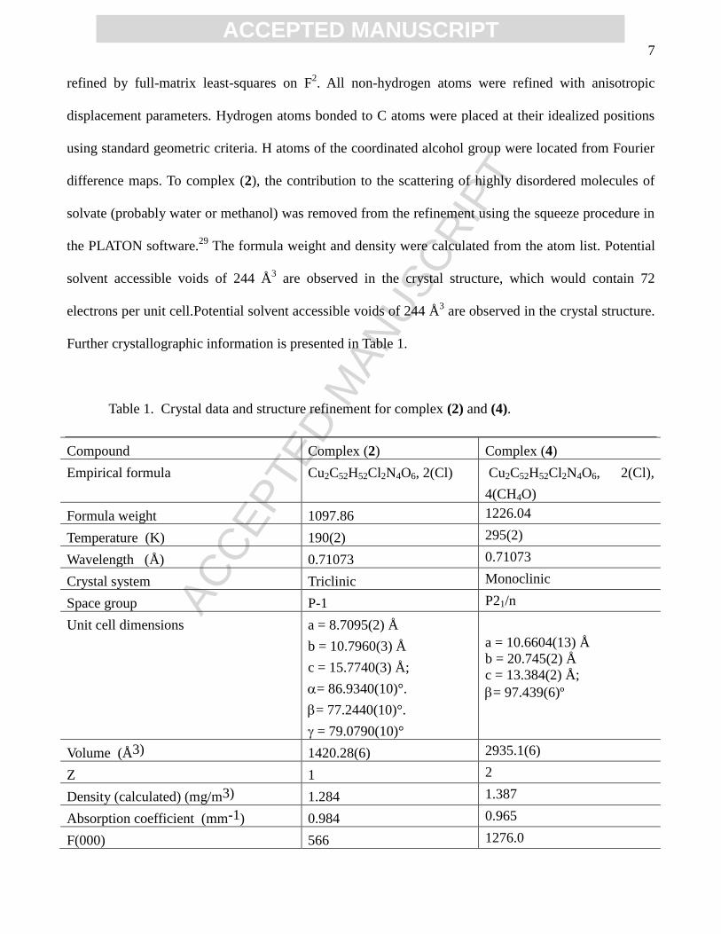
Further crystallographic information is presented in Table 1.
Table 1. Crystal data and structure refinement for complex (2) and (4).
Compound Complex (2) Complex (4)
Empirical formula Cu2C52H52Cl2N4O6, 2(Cl) Cu2C52H52Cl2N4O6, 2(Cl),
4(CH4O)
Formula weight 1097.86 1226.04
Temperature (K) 190(2) 295(2)
Wavelength (Å) 0.71073 0.71073
Crystal system Triclinic Monoclinic
Space group P-1 P21/n
Unit cell dimensions a = 8.7095(2) Å
b = 10.7960(3) Å
c = 15.7740(3) Å;
= 86.9340(10)°.
= 77.2440(10)°.
= 79.0790(10)°
a = 10.6604(13) Å
b = 20.745(2) Å
c = 13.384(2) Å;
= 97.439(6)º
Volume (Å3) 1420.28(6) 2935.1(6)
Z 1 2
Density (calculated) (mg/m3) 1.284 1.387
Absorption coefficient (mm-1) 0.984 0.965
F(000) 566 1276.0

ACC
EPTE
D M
ANU
SCR
IPT
ACCEPTED MANUSCRIPT8
Theta range for data collection (degree) 1.92 to 31.48 2.2 to 25.4
Index ranges -12 h 12, -15 k 15, -
23 l 23
-11 ≤ h ≤ 12, -24 ≤ k ≤ 24, -
16 ≤ l ≤ 15
Reflections collected 32822 14227
Independent reflections 9416 5334
Observed reflections 7540 3967
Rint 0.0269 0.0331
Restraints 0 0
Parameters 312 348
Goodness-of-fit on F2 1.068 1.033
Final R indices [I>2(I)] R1 = 0.0385, wR2 = 0.1053 R1 = 0.0372, wR2 = 0.0838
R indices (all data) R1 = 0.0503, wR2 = 0.1100 R1 = 0.0626, wR2 = 0.0961
Largest diff. peak and hole (e.Å-3) 1.337 and -1.012 0.52 and -0.31
2.4. Conductivity Measurements and Melting Point Determination.
The electrical conductivity of a 1x10⁻³
mol dm-3
solution of each complex was measured with a
Biocristal conductometer. Melting points were measured on a Microquimica MQAPF-301 apparatus.
2.5. Elemental Analyses.
The purity of tested compounds was determined by combustion elemental analyses conducted
with a Thermo Scientific FLASH 2000 CHNS/O analyzer.
2.6. Cyclic Voltammetry Analyses.
Cyclic voltammograms (CVs) were carried out with an Autolab PGSTAT 10
potentiostat/galvanostat in acetonitrile containing 0.1 mol dm-3
tetrabutylammonium perchlorate
(TBAClO4) as the supporting electrolyte under argon atmosphere at room temperature. The
electrochemical cell employed was a standard three-electrode configuration: a glassy carbon working
electrode, a platinum-wire auxiliary electrode and a commercial Ag/AgCl electrode immersed in a salt
bridge containing 0.1 mol dm-3
TBAClO4. The formal potential of the ferrocenium/ferrocene couple

ACC
EPTE
D M
ANU
SCR
IPT
ACCEPTED MANUSCRIPT9
was 0.426 V vs the reference electrode Ag/AgCl, being established as 0.400 V vs NHE.30
2.7. Potentiometric Titration.
The potentiometric studies were carried out with a Corning 350 digital pH meter fitted with a
blue-glass and Ag/AgCl reference electrodes calibrated to read -log[H+] directly, designated as pH.
Bidistilled water in the presence of KMnO4 was used to prepare the solutions. The electrode was
calibrated using the data obtained from the potentiometric titration of a known volume of a standard
0.100 mol dm-3
HCl solution with a standard 0.100 mol dm-3
KOH solution. The ionic strength of the
HCl solution was maintained at 0.100 mol dm-3
by addition of KCl. The measurements were carried out
in a thermostated cell containing the compound or ligand solution (0.5 mmol/50 cm3) with ionic
strength adjusted to 0.100 mol dm-3
by addition of KCl, at 25.00ºC. The experiments were performed
under argon flow to eliminate the presence of CO2. The samples were titrated by addition of fixed
volumes of a standard CO2-free KOH solution (0.100 mol dm-3
). Computations were carried out with
the BEST7 program, and species distribution diagrams were obtained with SPE and SPEPLOT
programs.
2.8. Syntheses of Ligands.
2.8.1.Synthesis of HL1.
Ligand HL1 (Scheme 1) was prepared by the reaction between bis-(pyridin-2-ylmethyl)amine31
(0.025 mol, 4.98g) and 2-(1-naphthylmethyl)oxirane (0.025 mol, 5g), in ethanol. The mixture was
stirred for 5 days at room temperature, which was follow by TLC, using ethanol as eluent.
Subsequently, the solvent was removed under reduced pressure and the residue was to 50 cm3 of water.
The compound was extracted with five 50 cm3 portion of CHCl3 and the extracts were combined,
washed with brine, dried over anhydrous MgSO4, filtered and concentrated under reduced pressure. A

ACC
EPTE
D M
ANU
SCR
IPT
ACCEPTED MANUSCRIPT10
brown oil was obtained. Yield: 6.5g (65%).1H NMR (400 MHz, CDCl3) δ/ppm: 2.96 (dd, 1H), 3.12 (dd,
1H), 3.97 (d, 2H), 4.05-4.09 (m, 3H), 4.19 (dd, 1H), 4.28- 4.35 (m, 1H), 6.77 (d, 1H), 7.12-7.16 (m,
4H), 7.32-7.36 (m. 1H), 7.39 (dd, 1H), 7.46 (t, 2H), 7.58 (t, 2H), 7.76 (d, 1H), 8.09 (d, 1H), 8.56 (m,
2H). 13
C NMR (100 MHz, CDCl3) δ/ppm: 58.20, 60.78 (2C), 67.99, 70.27, 104.87, 120.50, 122.26,
122.42 (2C), 123.43 (2C), 125.26, 125.78, 126.07, 126.29, 127.59, 134.64, 136.88 (2C), 149.21 (2C),
154.65, 157.28(2C).IR (cm-1) ν: 3700-3100, 3051, 2928, 2839, 1628, 1589, 1508, 1435, 1269, 791,
771.UV-Vis λ(nm) data (ε, dm3 mol
-1 cm
-1): 210 (7.39x10
4), 230 (4.79x10
4), 262 (1.29x10
4), 267
(1.24x10⁴), 292 (9.64x10
3), 305 (6.35x10
3), 320 (3.7x10
3).
2.8.2. Synthesis of H2L2.
The Ligand H2L2 (See Scheme 1) was prepared employing a similar method, but using 2-
(hydroxybenzyl)(2-pyridylmethyl)amine)32
(0.025 mol, 5.35g) in place of bis-(pyridin-2-
ylmethyl)amine. The mixture was stirred at room temperature and after one day a white solid was
obtained, which was removed by filtration. The solid was recrystallized in hot hexane. After standing
the solution for a few days a crystalline white solid was filtered off and dried with ether. Yield: 6.6g
(64%).m.p. 140ºC. Anal. Calcd for C26H26N2O3; MW = 414.5 g mol-1
: C, 75.34; H, 6.32; N, 6.76.
Found: C, 75.65; H, 6.59; N, 6.64%. 1H NMR (400 MHz, CDCl3) δ/ppm: 3.00 (dd, 2H), 3.79-3.84(m,
1H), 3.91-4.00 (m, 1H), 4.03-4.08 (m, 2H), 4.13 (d, 1H), 4.15-4.19 (m, 1H), 4.35-4.39 (m, 1H), 6.77 (d,
1H), 6.81 (t, 1H), 6.89 (d, 1H), 7.06 (d, 1 H), 7.17-7.25 (m, 3H), 7.34 (t, 1H), 7.41-7.44 (m, 1H), 7.48
(t, 2H), 7.69 (t, 1H), 7.78 (d, 1H), 8.03 (d, 1H), 8.63 (d, 1H).
13C NMR (100 MHz, CDCl3) δ/ppm: 56.90, 58.61, 58.72, 67.72, 70.09, 104.71, 116.67, 119.14, 120.43,
121.80, 122.32, 122.54, 123.26, 125.12, 125.43, 125.80, 126.29, 127.39, 129.21, 129.60, 134.39,
137.18, 148.81, 154.14, 157.28, 157.45. IR (cm-1) ν: 3300-2800, 2927, 2831, 1581, 1489, 1400, 1269,
1242, 791, 756. UV-Vis λ(nm) data (ε, dm3 mol
-1 cm
-1): 209 (1.21x10
5), 231 (7.47x10
4), 268

ACC
EPTE
D M
ANU
SCR
IPT
ACCEPTED MANUSCRIPT11
(1.89x10⁴), 280 (1.99x10
4), 293 (1.64x10
4), 306 (1.11x10
4), 319 (6.8x10
3).
2.8.3. Synthesis of HL3.
The same methodology employed for the synthesis of HL1 was employed here, but now using
2-(2-naphthylmethyl)oxirane (0.025 mol, 5g) (See Scheme 1). A brown oil was obtained. Yield: 7.1g
(71%). 1H NMR (400 MHz, CDCl3) δ/ppm: 2.89 (m, 1H), 3.04 (dd, 1H), 3.92-4.12 (m, 6H), 4.21-4.26
(m, 1H), 7.11 (d, 4H), 7.26-7.31 (m, 3H), 7.40 (t, 2H), 7.68-7.74 (m, 2H), 7.56 (t, 2H), 8.54 (d, 2H).
13C NMR (100 MHz, CDCl3) δ/ppm: 58.41, 60.73 (2C), 67.94, 70.42, 106.88, 119.21, 122.42 (2C),
123.41, 123.82 (2C), 126.52, 127.00, 127.83, 129.19, 129.50, 134.71, 136.88 (2C), 149.18 (2C),
156.99, 159.25 (2C). IR (cm-1
) ν: 2800-3300, 3055, 2928, 2835, 1628, 1597, 1512, 1469, 1258, 837,
752, 771. UV-Vis λ(nm) data (ε, dm3 mol
-1 cm
-1): 225 (9.06x10
4), 262 (1.5x10
4), 270 (1.26x10
4), 281
(6.12x10³).
2.8.4. Synthesis of H2L4.
The ligand H2L4 (See Scheme 1) was prepared employing similar method described for H2L2.
A brown-redish oil was obtained. Yield: 7.0g (67%). 1H NMR (400 MHz, CDCl3) δ/ppm: 2.91 (m, 2H),
3.78 (d, 1H), 3.92-4.00 (m, 2H), 4.02-4.04 (m, 1H), 4.25-4.28 (m, 1H), 6.79 (t, 1H), 6.87 (dd, 1H),
7.02-7.04 (m, 2H), 7.07-7.10 (m, 2H), 7.17 (d, 2H), 7.22 (t, 1H), 7.32 (t, 1H), 7.42 (t, 1H), 7.65 (dd,
1H), 7.68-7.71 (m, 1H), 7.74 (d, 1H), 8.61 (m, 1H). 13
C NMR (100 MHz, CDCl3) δ/ppm: 57.09, 58.71,
58.99, 67.75, 70.34, 106.97, 116.67, 118.81, 119.27, 122.50, 122.68, 123.37, 123.77, 126.42, 126.92,
127.69, 129.19, 129.26, 129.42, 129.74, 134.29, 137.37, 148.92, 156.58, 157.40, 157.57.
IR (cm-1
) ν: 3455, 3053, 2949, 2845, 1628, 1599, 1508, 1477, 1395, 1267, 1258, 839, 763, 748. UV-
Vis λ(nm) data (ε, dm3 mol
-1 cm
-1): 225 (1.02x10
5), 262 (1.1x10
4), 270 (1.07x10
4), 281 (6.46x10
3). See
Scheme 1.

ACC
EPTE
D M
ANU
SCR
IPT
ACCEPTED MANUSCRIPT12
NNH
N
O
O
MeOH
NN
N
O
HO
HL1
O
O
MeOH
NN
N
O
HO
HL3
NNH
OH
O
O
MeOH
NN
O
HO
OH
H2L2
O
O
MeOH
NN
O
HO
OH
H2L4
Scheme 1. Scheme of synthesis for ligands HL1-H2L4.
2.9. Syntheses of copper complexes.
2.9.1. Synthesis of [(HL1)Cu(μ-Cl)2Cu(HL1)]Cl2.H2O (1).
Complex (1) (Scheme 2) was prepared in a reaction between HL1 (1.0 mmol, 0.398g) and
CuCl2.6H2O (1.0 mmol, 0.1705 g) in ethyl acetate, with constant stirring at room temperature. After
allowing the blue solution to stand for a few days a microcrystalline blue solid was filtered off and
dried with ether. Yield: 0.34g (62%). m.p. 197ºC. IR (cm-1
): ν(OH), 3425-3483 (s); ν(CH), 3047 and
3008 (s); ν(CH2), 2936(s); ν(CH2), 2881 (s); (C=C and C=N), 1609 (s), 1578 (s), 1443 (s) and 1396 (s);
ν(C-O-C), 1269 (s); (CH), 798 (s) and 775 (s). Anal. Calcd for [(HL1)Cu(μ-Cl)2Cu(HL1)]Cl2. H2O
(1) (C50H52Cu2Cl4N6O5, MW= 1086 g mol-1
): C, 55.30; H, 4.83; N, 7.74. Found: C, 55.49; H, 4.64; N,
7.85%. UV-Vis λ(nm) data (ε, dm3 mol
-1 cm
-1): 209 (1.54x10
4), 230 (1.01x10
4), 262 (9.49x10
4), 267
(5.99x10⁴), 292 (1.92x10
4), 305 (1.69x10
4), 320 (5.66x10
3), 707 (77.91). Ω= 83 μS cm
-1 (1:1

ACC
EPTE
D M
ANU
SCR
IPT
ACCEPTED MANUSCRIPT13
electrolyte- MeOH). MS (ESI) in H2O/MeOH 1:1: [M]+
= 497: [Cu(HL1)Cl]+. E1/2= -0.324 V vs NHE
(CuII/Cu
I).
2.9.2. Synthesis of [(H2L2)Cu(μ-Cl)2Cu(H2L2)]Cl2.5H2O (2).
Complex (2) (Scheme 2) was obtained using H2L2 (1.0 mmol, 0.414g) in place of HL1, using
mixture of ethylacetate/methanol (3:2) as solvent. After allowing the green solution to stand for a few
days a microcrystalline green solid was filtered off and dried with ether. Yield: 0.52g (90%). m.p.
150ºC. IR (cm-1
): ν(OH), 3348 (b); ν(CH), 3047 (s); ν(CH2), 2931(s); ν(CH2), 2712 (s); (C=C and
C=N), 1613 (s), 1578 (s), 1447 (s) and 1400 (s); ν(C-O-C), 1269 (s); ν(C-O), 1242 (s); (CH), 798 (s)
and 748 (s). Anal. Calcd for [(H2L2)Cu(μ-Cl)2Cu(H2L2)]Cl2.5H2O (2) (C52H62Cu2Cl4N4O11, MW=
1187.91 g/mol): C, 53.38; H, 5.17; N, 4.79. Found: C, 53.22; H, 5.52; N, 4.77 %. UV-Vis λ(nm) data (ε,
dm3 mol
-1 cm
-1): 208 (6.09x10
4), 230 (3.73x10
4), 268 (9.37x10
4), 288 (1.11x10
4), 306 (6.29x10
3), 320
(3.19x10³), 444 (18.21), 722 (72.07). Ω= 111 μS cm
-1 (1:1 electrolyte- MeOH). MS (ESI): [M]
+ = 512
[Cu(H2L2)Cl]+. Epa= 0.164 and Epc= -0.588 V vs NHE. The respective complex could be obtained as
single crystals suitable for x-ray diffraction when ethanol is employed in place of
ethylacetate/methanol, under heating for 10 h. Anal. Calcd for [(H2L2)Cu(μ-Cl)2Cu(H2L2)]Cl2 (2)
C52H52Cu2Cl4N4O6, MW= 1097 g mol-1
): C, 52.57; H, 5.26; N, 4.72. Found: C, 52.73; H, 5.39; N,
4.94%. m.p. 150ºC. Ω= 111 μS cm-1
(1:1 electrolyte- MeOH).
2.9.3.Synthesis of [(HL3)Cu(μ-Cl)2Cu(HL3)]Cl2.4H2O (3).
Complex (3) (Scheme 2) was obtained using ethyl acetate and HL3 (1.0 mmol, 0.398g) in place
of HL1. After allowing the green solution to stand for a few days a microcrystalline dark green solid
was filtered off and dried with ether. Yield: 0.25g (45%). m.p. 136ºC. IR (cm-1
): ν(OH), 2750-3605 (b);
ν(CH), 3103, 3057 and 3021 (s); ν(CH2), 2951(s); 2932(s) 2878 (s); (C=C and C=N), 1627 (s),

ACC
EPTE
D M
ANU
SCR
IPT
ACCEPTED MANUSCRIPT14
1609(s), 1508 (s), 1468 (s) and 1442 (s); ν(C-O-C), 1259 (s); (CH), 834 (s) and 769 (s). Anal. Calcd
for [(HL3)Cu(μ-Cl)2Cu(HL3)]Cl2.4H2O (3) C50H58Cu2Cl4N6O8, MW= 1140 g mol-1
): C, 52.68; H, 5.13;
N, 7.37. Found: C, 52.51; H, 5.58; N, 7.54%. UV-Vis λ(nm) data (ε, dm3 mol
-1 cm
-1): 225 (9.74x10
4),
259 (1.72x10⁴), 270 (1.23x10
4), 281 (7.16x10
3), 702 (68.69). Ω= 82 μS cm
-1(1:1 electrolyte- MeOH).
MS (ESI): [M]+
= 497 [Cu(HL3)Cl]+. E1/2= -0.348 V vs NHE (Cu
II/Cu
I).
2.9.4.Synthesis of [(H2L4)Cu(μ-Cl)2Cu(H2L4)]Cl2.6H2O (4).
Complex (4) (Scheme 2) was obtained using H2L4 (1.0 mmol, 0.414g) in place of HL1, using
an ethyl acetate/methanol mixture (3:2). After allowing the green solution to stand for a few days a
microcrystalline green solid was filtered off and dried with ether. Yield: 0.58g (97%). m.p. 105ºC.
IR(cm-1
): ν(OH), 3350-3000 (b); ν(CH), 3059 (s); ν(CH2), 2931(s); ν(CH2), 2873 (s); (C=C and C=N),
1628 (s), 1597 (s), 1508 (s), 1458 (s) and 1399 (s); ν(C-O-C), 1257 (s); ν(C-O), 1247 (s); (CH), 844
(s) and 760 (s). Anal. Calcd for [(H2L4)Cu(μ-Cl)2Cu(H2L4)]Cl2.6H2O (4) (C52H64Cu2Cl4N4O12, MW=
1206 g mol-1
): C, 51.79; H, 5.35; N, 4.65. Found: C, 51.96; H, 5.66; N, 4.70%. UV-Vis λ(nm) data (ε,
dm3 mol
-1 cm
-1): 225.94 (9.68x10
3), 262.22 (1.4x10
3), 270.04 (1.34x10
3), 281.97 (1.07x10
3), 440.32
(29.58), 731.93 (68.34). Ω= 108 μS cm-1
(1:1 electrolyte, MeOH). MS (ESI): [M]+= 512
[Cu(H2L4)Cl]+. Epa= 0.10 and Epc= -0.548 V vs NHE. The respective complex could be obtained as
single crystals suitable for x-ray diffraction when methanol is employed in place of
ethylacetate/methanol, under heating for 10 h, resulting in the complex [(H2L4)Cu(μ-
Cl)2Cu(H2L4)]Cl2.4CH3OH.

ACC
EPTE
D M
ANU
SCR
IPT
ACCEPTED MANUSCRIPT15
2+
N
Cu
N
N Cl
OH
O
N
Cu
N
NCl
OH
O
(1)
N
Cu
N
OH Cl
OH
O
N
Cu
N
OHCl
OH
O
2+
(2)
N
Cu
N
N Cl
OH
O
N
Cu
N
NCl
OH
O
2+
(3)
2+
N
Cu
N
OHCl
OH
O
N
Cu
N
OH Cl
OH
O
(4)
Scheme 2. Structures of complexes (1)-(4). The proposed structures are based on X-ray diffraction data
performed for complexes (2) and (4).
2.10. Culture of leukemia cell lines.
Human leukemia cell lines THP-1 (acute monocytic leukemia cell line) and U937 (histiocytic
lymphoma cell line) were cultured routinely in DMEM-F12 medium (Gibco, BRL), supplemented
with 10% fetal calf serum and gentamicyn (20mg/ml, Gibco, BRL) at 37 °C in a humidified
atmosphere containing 5% CO2 in air. Culture media were changed every 2–3 days.

ACC
EPTE
D M
ANU
SCR
IPT
ACCEPTED MANUSCRIPT16
2.11. Isolation of normal human peripheral blood mononuclear cells (PBMC).
Blood samples were collected from health donors in Sodium Heparin glass tubes
"VacutainerTM" (Becton Dikinson) and the PBMC were isolated over Ficoll-Paque™ Plus (1.08g cm-3
)
in a 50 cm3 conical tube (2:1 - blood:ficoll). Twenty milliliters of a fresh, heparinized blood sample was
diluted in phosphate buffer saline (PBS), gently laid over 10 cm3 of Ficoll and centrifuged at 500xg for
20 min at 25 °C. PBMC were collected from the interface of spun blood samples and were washed
three times with PBS by centrifugation at 500xg for 10 min at 4 °C. The supernatant was discarded and
the cells were suspended in DMEM-F12 medium (Gibco, BRL). A 0.4% Trypan blue solution (Sigma,
Germany) was used to dissolve the cells into an appropriate concentration and the viability of cells was
checked; the required range of cells’ viability is 95-99%.
2.12. Analysis of cell viability by MTT assay.
Cells (U937, THP-1 and PBMC) were plated (approximately 1 x 10⁶ cells) in 96-well plates and
treated with different concentrations of ligands and their respective copper complexes for 36 h in
DMEM-F12 medium (Gibco, BRL). Cell viability was measured by the colorimetric microassay (MTT
assay). Twenty microliters of 3-(4,5-dimethyl- 2-thihazyl)-2,5-diphenyl-2H-tetrazolium bromide
(MTT) stock solution (5 mg cm-3
) were added into each well, and the cells were incubated at 37 °C for
4 hour. Then, the MTT-formazan produced by viable cells was dissolved in an isopropanol-HCl
solution. The optical density (OD) values were measured by spectrophotometry at 570 nm using a
Microplate Reader (Epoch™, BioTek® Instruments, Inc.). The OD values are presented as relative
viable cell numbers, which were expressed in percentages (%). Relative cell viability = OD of treated
sample/Mean OD of control sample.

ACC
EPTE
D M
ANU
SCR
IPT
ACCEPTED MANUSCRIPT17
2.13. Determination of in vivo acute toxicity for complex (2).
In order to determine the in vivo acute toxicity of complex (2), male and female C57BL/6 mice,
between 18 g and 25 g were separated into groups of 4 animals. The complex was injected into the
peritoneal cavity, such that each group received the respective concentrations of the compound (25, 50,
100, 200 mg kg-1
). For inoculation we used a 3 cm3 syringe and a hypodermic sterile needle SR 0.45
mm x 13 mm NIPRO. The vehicle used for administration of the complex was ultrapure water. The
animals were observed closely for at least 4 hours post-injection and in 24 h intervals for 30 days
thereafter. After this period, median lethal dose (LD50), a dose required to kill half the members of a
tested population, was determined. The research protocol was approved by the Ethical Committee for
Animal Experimentation of Universidade Estadual do Norte Fluminense Darcy Ribeiro, under protocol
number 214/2014.
2.14. Analysis of apoptosis and necrosis cell death by fluorescence microscopy.
Cells (~1 x 10⁶) were plated in 96-well plates and treated with ligand H2L2 (80 and 160 mol
dm-3
) and complexes (2) (40 and 80 µM) and (4) (80 and 160 µM) for 12, 24 and 36 h in DMEM-F12
medium (Gibco, BRL). Then, the acridine orange/ethidium bromide double staining was used to
identify the following four cell stages according to Kosmider et al.:33
1) living cells (green nucleus with
red–orange cytoplasm), 2) early apoptosis (cell membrane still continuous but chromatin condensation
and an irregular green nucleus are visible), 3) late apoptosis (so-called ‘secondary necrosis’ or
‘apoptotic necrosis’: ethidium bromide penetrates through altered cell membrane and stains the nuclei
orange, while fragmentation or condensation of chromatin is still observed), 4) necrosis (uniformly
orange-stained cell nuclei). Following the addition of fluorochromes (25 µg cm-3
acridine orange and
50 µg cm3 ethidium bromide), 300 cells were immediately analyzed in each of two independent
experiments using fluorescence microscopy (Axioplan – Carl Zeiss microscope).

ACC
EPTE
D M
ANU
SCR
IPT
ACCEPTED MANUSCRIPT18
2.15. Measurement of annexin V and propidium iodide staining.
U937 and THP-1 (1 x 10⁶
cells cm-3
) were cultured in 24-well plates and treated with 50 and
100 mol dm-3
of complex (2) for 12 h. Apoptosis was detected by using the Annexin V-FITC
Apoptosis Detection Kit (SIGMA-ALDRICH). Briefly, after incubation cells were washed twice with
phosphate buffered saline (PBS), and incubated in 0.5 cm3 of binding buffer (100 mmol dm
-3
HEPES/NaOH, pH 7.5, 1.4 mol dm-3
NaCl and 25 mmol dm-3
CaCl2). To each sample was added 5
mm3 of Annexin V-FITC and 10 mm
3 of PI. Samples were incubated at room temperature for 10 min,
protected from light. Cell fluorescence was determined immediately with a flow cytometer (FACS
Calibur-BD Sciences). Each experiment per sample was determined by recording 10,000 events.
2.16. Detection of apoptosis by DNA fragmentation and agarose gel electrophoresis.
U937 and THP-1 cells (1 x 10⁶ cells cm
3) were plated in 6 multiwell plates and treated with 50
and 100 mol dm-3
of complex (2) for 12h. After incubation, cells were harvested, transferred to
microtubes, washed twice with PBS and fixed in 70% ethanol at -20 ºC for 2 h. After centrifugation
ethanol was totally removed, dried and evaporated at room temperature. The cells were resuspended in
50 mm3 of extraction buffer (0.2 mol dm
-3 Na2HPO4 and 0.1 mol dm
-3, pH 7.8) and incubated at 37°C
for 30 min. 5 mm3 RNAse (1mg cm
3) was added in each sample and a new incubation was done at 3 °C
for 30 min. 5 mm3 of Proteinase K (1mg/mL) was added to each sample. To detect the DNA fragments,
the isolated DNA samples were electrophoresed for 60 min at 60 V in a 1% agarose gel and stained
with GelRed Nucleic Acid Stain (Biotium). DNA fragmentation was observed in a UV transilluminator
(Bio-Rad, Gel Doc™ XR+).
2.17. Study of cell cycle arrest by flow cytometric analysis.
Cells (1 x 10⁶) were plated in 24-well plates and treated with ligands H2L2 and H2L4 (80 mol

ACC
EPTE
D M
ANU
SCR
IPT
ACCEPTED MANUSCRIPT19
dm-3
) and complexes (2) (40 mol dm-3
) and (4) (80 mol dm-3
) for 36h in DMEM-F12 medium
(Gibco, BRL). After incubation the cells were fixed in 70% ethanol at 4 ºC for 30 min and stained with
propidium iodide (PI) for 2 h in darkness. The DNA content was measured with a flow cytometer
(FACS Calibur-BD Sciences) and cell cycle distribution was analyzed by WinMDI version 2.9
software.34
The proportions of cells in the G0/G1, S, and G2/M phases were represented as DNA
histograms. Apoptotic cells with hypodiploid DNA content were measured by quantifying the sub-G1
peak in the cell cycle pattern. Each experiment per sample was determined by recording 10,000 events.
2.18. Analysis of the mitochondrial membrane potential (ΔΨm) by flow cytometry using JC-1 stain.
U937 and THP-1 cells (1 x 10⁶) were seeded in 24-well plates and treated with ligands H2L2
and H2L4 (80 mol dm-3
) and complexes (2) (40 mol dm-3
) and (4) (80 mol dm-3
) for 36 h in
DMEM-F12 medium (Gibco, BRL). Subsequently, the cell suspension was transferred to a sterile tube
and pelleted (400xg for 7 min at room temperature). Cells were stained with the JC-1 dye (25μg cm3)
and incubated at 37 °C in a 5% CO2 incubator for 15 minutes. The cells were washed twice with fresh
medium and analyzed immediately by flow cytometry. JC-1 (5,5’,6,6’-tetrachloro 1,1’,3,3’
tetraethylbenzimidazolylcarbocyanine iodide) exists as a monomer in the cytosol (green) but
accumulates as red aggregates in the mitochondria. Generally, in apoptotic and necrotic cells, JC-1
exists in monomeric green form. Thus, mitochondria containing red JC-1 aggregates represent healthy
cells that are detectable in the FL2 channel and express intact (ΔΨm), whereas green JC-1 monomers in
apoptotic cells are detectable in the FITC channel (FL1) and express the loss of (ΔΨm). The
mitochondrial transmembrane potential (ΔΨm) was measured with a flow cytometer (FACS Calibur)
and analyzed by WinMDI version 2.9 software.34

ACC
EPTE
D M
ANU
SCR
IPT
ACCEPTED MANUSCRIPT20
2.19. Subcellular fractionation and detection of cytochrome C release.
U937 cells were plated at a 1 x 10⁶ concentration in 24-well plates and treated with 40 µM
of
complex (2) for 6, 12, 24 and 36 h in DMEM-F12 medium (Gibco, BRL), supplemented with 10% fetal
calf serum and 20 μg cm-3
gentamicin (Gibco, BRL). The mitochondrial fraction (MF) was separated
from the cytoplasmic fraction (CF) using a digitonin-based subcellular fractionation technique to
quantify the cytochrome c released in both fractions. Briefly, U937 cells were harvested by
centrifugation at 800xg, washed in PBS, pH 7.2, and re-pelletted. Cells were then digitonin-
permeabilized for 5 min on ice at a density of 3x 10⁶ cells cm
-3 in cytosolic extraction buffer (250
mmol dm-3
sucrose, 70 mmol dm-3
KCl, 137 mmol dm-3
, NaCl, 4.3 mmol dm-3
Na2HPO4, 1.4 mmol
dm-3
KH2PO4 pH 7.2, 10 mg cm-3
leupeptin, 2 mg cm-3
aprotinin, containing 200 mg cm-3
digitonin).
Plasma membrane permeabilization of cells was confirmed by staining in a 0.2% trypan blue solution.
Cells were then centrifuged at 1000xg for 5 min at 4 °C. The supernatants (cytosolic fractions) were
saved and the pellets solubilized in the same volume of mitochondrial lysis buffer (50 mmol dm-3
TRIS, pH 7.4, 150 mmol dm-3
NaCl, 2 mmol dm-3
EDTA, 2 mmol dm-3
EGTA, 0.2 % Triton X-100,
0.3% NP-40, 10 mg cm-3
leupeptin, 2 mg cm-3
aprotinin), followed by pelletting at 10,000xg for 10 min
at 4 °C. Cytochrome c was quantified according to the protocol in the Cytochrome c ELISA KIT
(Calbiochem®
). The optical density (OD) values were measured by spectrophotometry at 450 nm using
a Microplate Reader (Epoch™, BioTek® Instruments, Inc.).
2.20. Determination of caspase activities.
Caspases 3, 8 and 9 activities were determined using the substrates VDVAD-pNA or DEVD-
pNA (caspase 3 substrates), IETD-pNA (a caspase 8 substrate), and LEHD-pNA (a caspase 9 substrate)
following the protocols of the Caspase Activity Assay kit from Invitrogen™ (ApoTarget™ caspase

ACC
EPTE
D M
ANU
SCR
IPT
ACCEPTED MANUSCRIPT21
colorimetric protease assay kit). U937 cells (3-5 x 10⁶/mL) were incubated with 40 mol dm
-3 of
complex (2) for 3, 6 and 12 h and centrifuged at 400g for 10 min. The supernatant was removed, and
the pellet was suspended in 0.1 cm3 of lysis buffer and incubated on ice for 10 min followed by
centrifugation at 10.000g for 1min. Protein concentrations were determined and cytosol extracts were
diluted to a concentration of 50-200 µg protein per 50 mm3 cell lysis buffer (1-4mg cm
3). Aliquots (50
mm3) of the supernatant were removed and placed in a 96-well microplate containing reaction buffer
(Invitrogen™). 5 mm3 of each substrate was added, and the microplate was incubated at 37 °C for 12 h.
Activity was monitored as the cleavage and release of free pNA and quantified at 405 nm using a
Microplate spectrophotometer (Epoch™, BioTek® Instruments, Inc.). Caspase activation of treated
cells was compared with an uninduced control sample for the determination of the fold increase in
caspase activity.
2.21. Analysis of mitochondrial morphology by fluorescence imaging and transmission electron
microscopy (TEM).
Fluorescence imaging of mitochondria: U937 cells were cultured at 1 x 10⁶
cells cm3 in 24-well
plates and treated with 40 mol dm-3
of complex (2) for 12 h. Cell suspensions were transferred to a
sterile tube and pelleted (400xg for 5 min at room temperature). Then, cells were incubated with 100
nmol dm-3
Mitotracker CMXRos (Invitrogen, Molecular Probes) and 1 µg cm3 Hoechst nuclear stain
(Invitrogen, Molecular Probes) for 20 min, washed two times with PBS, resuspended in fresh D-MEM
F12 medium and imaged using a fluorescence microscope (Axioplan – Carl Zeiss microscope). U937
cells (1x10⁶ cells) were cultured in tissue culture bottles (25 cm
2) for 12 h. The cells were incubated
with complex (2) (40 mol dm-3
) for 12 h and centrifuged at 1200 rpm for 10 min and washed three
times with phosphate-buffered saline (PBS; pH 7.2). The cell pellets were fixed for 1 h in 1%
glutaraldehyde, 5 mM CaCl2, 5% sucrose and 0.1 M cacodilate buffer. Fixed cells were washed three

ACC
EPTE
D M
ANU
SCR
IPT
ACCEPTED MANUSCRIPT22
times in a 5% sucrose solution in 0.1 M cacodilate buffer and post-fixed for 2 h protected from light in
a 1:1 osmium/potassium ferrocianate solution. Post-fixed cells were washed twice with PBS and
dehydrated sequentially in a graded series of acetone solutions (30, 50, 70, 90 and 100%). The cells
were then soaked overnight in a 1:1 ratio of acetone and embedding resin (EPON
). The resin-
embedded cells were placed in tubes and the tubes were placed at 37 °C in a humidified atmosphere for
48 h for polymerization of the resin. The polymerized blocks were sectioned and ultrathin sections
were prepared. The effects of the complexes on U937 mitochondria were evaluated using a
transmission electronic microscope (TEM-900, Zeiss, Germany).
2.22. Statistical Analysis.
For analysis of cell viability and apoptosis the mean and standard error were calculated from the
raw data and then subjected to the One way ANOVA analysis (analysis of variance). A significant
difference was taken as p < 0.5, <0.01 and <0.001.
3. Results and discussion
3.1. Syntheses and structures of copper(II) coordination compounds.
A group of similar ligands was developed in order to probe the influence of the chelating groups
and structural isomerism introduced by them on their coordination behavior, as well as to gain insight
into the influence of these features on the antitumor activity of their copper(II) complexes.
Ligands HL1 and HL3 present two pyridine groups, a tertiary amine and an alcohol function as
chelating groups and an α-naphthyl or β-naphthyl group as pendant unit, respectively, providing a N3O
coordination environment for the metal center. Ligands H2L2 and H2L4 are similar, presenting a
pyridine, a phenol, a tertiary amine and an alcohol group, in addition to an α-naphthyl or β-naphthyl
group, respectively, providing a N2O2 coordination environment for the metal center (Scheme 1). We

ACC
EPTE
D M
ANU
SCR
IPT
ACCEPTED MANUSCRIPT23
have previously published the synthesis of copper complexes containing ligands (HPClNOL19
and
H2BPClNOL23
) similar to those reported herein. The main difference is the lack of the naphthyl group
in the earlier ligands. The relevance of the naphthyl units in the design of anticancer drugs has been
assessed previously by Das and co-workers, which reported the cytotoxic activity of alkyl derivatives
of 2-naphtol against four human cancer cell lines (Hep G2, A549, MDA 231 and HeLa). The IC50
values found for those compounds are comparable to that of doxorubicin.25
The four new ligands were characterized by IR and NMR. Their respective copper complexes
are stable in air and are remarkably soluble in solvents such as DMF and DMSO, and slightly soluble
in H2O, CH3CN, ethanol, methanol and chloroform. The reactions between these ligands and
CuCl2.2H2O allowed the isolation of monocrystals for complexes (2) and (4), while compounds (1) and
(3) were isolated as microcrystalline material. It was thus only possible to determine the molecular
structure through X-ray diffraction for compounds (2) and (4), but the structures for compounds (1) and
(3) were modeled based on that of compounds (2) and (4).
3.2. Description of Crystal Structures of Complexes (2) and (4).
The structure determination of complexes (2) and (4) reveals a center of symmetry situated in
the plane formed by the Cu1-Cl1-Cu1´-Cl1´ atoms. The two monomeric units in the dimeric structure
are symmetrically related. A perspective view of the cations is displayed in Figure 1, where the
arrangements exhibited by the complexes are related to the inherent asymmetry of the ligands. The
bond lengths and angles for complexes are listed in Table 2.
The coordination behavior of the ligands H2L2 and H2L4 is similar to that observed for the
ligand H2BPClNOL.23
Interestingly, the X-ray diffraction study of the copper complex of this ligand
revealed the presence of a mono- and a dinuclear species in the same unit cell. For H2L2 and H2L4
only the dinuclear species was observed, demonstrating that the large naphthyl group present in these

ACC
EPTE
D M
ANU
SCR
IPT
ACCEPTED MANUSCRIPT24
ligands does not exert an effect on the coordination mode of the ligands. The nitrogen atoms and the
alcohol group present bond distances very similar to those observed for the compound
{[CuII(H2BPClNOL)]–μ-(Cl)2–[Cu
II(H2BPClNOL)]}Cl2. However, the Cu-Ophenol is longer in (2)
(2.5939(14) Å) and (4) (2.495(2) Å) than in the cited compound (2.446(2) Å), while the length of the
Cu-Cl` bond is shorter in (2) and (4) (2.8657(4) and 2.8823(8) Å vs 2.972(1) Å, respectively).
A comparison of the structures of (2) and (4) indicates that the main difference is the location of
the naphthyl and phenolic groups (Figure 2). In (2) the phenol group is further removed from the
copper center than in (4) (2.5939(14) vs 2.495(2) Å, respectively), resulting in some differences related
to bond angles involving the Cu-phenol bond (Table 2). With respect to the naphthalene units, Figure 2
shows that they are orthogonally positioned relative to each other.

ACC
EPTE
D M
ANU
SCR
IPT
ACCEPTED MANUSCRIPT25
Figure 1. View of the dinuclear complexes (2) (top) and (4) (bottom) with appropriate atom labels. The
ellipsoids are shown at the 40% probability level (hydrogen atoms are omitted for clarity).
Figure 2. Overlay of the asymmetric unit of cations (2) and (4).

ACC
EPTE
D M
ANU
SCR
IPT
ACCEPTED MANUSCRIPT26
Table 2. Selected bond lengths [Å] and angles [º] for complexes (2) and (4).
Complex (2) Complex (4)
Cu1-N22 1.9783(13) 1.991(2)
Cu1-O3 1.9824(11) 1.9854(18)
Cu1-N1 2.0374(13) 2.036(2)
Cu1-Cl1 2.2436(4) 2.2445(7)
Cu1 O10 2.5939(14) 2.495(2)
Cu1-Cl1i 2.8657(4) 2.8823(8 )
N22-Cu1-O3 164.01(5) 163.55(8)
N22-Cu1-N1 83.21(5) 83.25(9)
O3-Cu1-N1 84.20(5) 82.89(8)
N22-Cu1-Cl1 99.81(4) 101.35(7)
O3-Cu1-Cl1 94.02(4) 93.23(5)
N1-Cu1-Cl1 171.67(4) 173.46(6)
N22-Cu1-O10 104.74(5) 88.46(8)
O3-Cu1-O10 83.23(5) 99.40(8)
N1-Cu1-O10 82.23(5) 86.21(8)
Cl1-Cu1-O10 89.48(3) 89.26(5)
O10-Cu1-Cl1i 168.03(3) 174.23(6)
N22-Cu1-Cl1i 85.82(4) 85.88(6)
O3-Cu1-Cl1i 85.18(4) 86.36(6)
N1-Cu1-Cl1i 93.64(4) 94.28(6)

ACC
EPTE
D M
ANU
SCR
IPT
ACCEPTED MANUSCRIPT27
Cl1-Cu1-Cl1i 94.326(13) 90.71(2)
Symmetry code: (i) -x+1,-y+1,-z+2 for complex (2) and (i) 1-x, 1-y, 1-z for complex (4)
Despite the molecular similarities, the profile of intermolecular interactions shows significant
differences. The main difference is associated with the chloride ion which is in compound (2)
connected directly to the cation while in compound (4) it interacts only with the methanol molecules of
crystallization. The comparison between both structures indicates that the C-H··· and ··· interactions
in the naphthyl groups produce distortions. However, the impossibility of the description of the solvent
molecules of crystallization in compound (2) makes it difficult to evaluate all their intermolecular
interactions.
3.3. Characterization of the compounds in solution.
Since we have previously demonstrated that the dinuclear unit {[CuII(H2BPClNOL)]–μ-(Cl)2–
[CuII(H2BPClNOL)]}Cl2 is broken into mononuclear species in polar solvents,
23 we carried out studies
to evaluate the solution behavior of the copper(II) compounds synthesized in this work.
The conductivity behavior of the four complexes in MeOH was examined, revealing that they
are 1:1 electrolytes. This suggests that the dinuclear arrangement observed in the solid state structures
of complexes (2) and (4) may not be preserved in solution, favoring the formation of a mononuclear
species instead.
ESI(+)-MS and ESI(+)-MS/MS data also indicate the presence of mononuclear species in
H2O/MeOH. ESI(+)-MS and ESI(+)-MS/MS data of complexes (1)-(4) present a characteristic set of
isotopic ions due mainly to the presence of metal and Cl atoms. For the isomeric complexes (1) and (3),
ESI(+)-MS data indicate the presence of two cations in a MeOH:H2O (1:1) solution. The peak at m/z =
497 is ascribed to [Cu(HLx)Cl]+
(where x = 1 or 3) and the peak at m/z 275 may be related to the loss of

ACC
EPTE
D M
ANU
SCR
IPT
ACCEPTED MANUSCRIPT28
a part of the ligand. MS/MS data of the species with an m/z ratio of 497 yields the cation with m/z =
275. For the isomeric complexes (2) and (4), four peaks were observed, i.e. [Cu(H2Ly)Cl]+ (m/z 512),
[Cu(HLy)]+ (m/z 476), H2Ly (m/z 415) and a peak at m/z 290, which is related to the fragmentation of
the ligand H2Ly (where y = 2 or 4). MS/MS analysis of the species with m/z 512 yields a cation with
m/z 476 (by the loss of a neutral HCl molecule) and a cation at m/z 290.
To extend the investigation of the behavior of the complexes in solution we carried out
potentiometric titration studies of the ligands and complexes. Since biological activity generally occurs
in an aqueous, buffered environment it is particularly important to assess the relevance of pH for the
structure of the complexes.
For the ligand HL1, pKa values of 2.06, 2.47 and 4.64 were obtained, which are attributed to
two pyridinic and one aminic nitrogen atom, respectively. The pKa values are lower than those
observed for the related HPClNOL ligand (2.90(2), 3.88(2) and 4.86(2)).35
Due to the high pKa value
for the alcohol group, it remains protonated in the range from pH 0 to 13. Thus, above pH 5.0, the
ligand is in its non-protonated form, i.e. HL1. The ligand containing the aminic nitrogen in the
protonated form, H2L1+, reaches a maximum of 88% at pH 3.6. The species H3L1
2+, the diprotonated
species, reaches a maximum of 45% at pH 2.5. The fully protonated ligand, H4L13+
, predominates at a
pH below 2.1.
Potentiometric titration for complex (1), obtained from the reaction between HL1 and CuCl2,
indicates that Cu2+
is present in its free form at pH ~ 2 and its concentration decreases gradually
reaching a minimum at pH 8.5., when the metal ion is coordinated by HL1, reaching a maximum of
90% in the range of pH 5-7.5. At pH values higher than 9, two species associated with two
deprotonation/protonation equilibria involving one water molecule and the alcohol group are observed:
[Cu(HL1)Cl(OH)]0 and [Cu(L1)Cl(OH)]
-, with pKa values of 9.35 and 9.89, respectively. The
hydrolysis product of the metal ion appears at higher pH values. Similar results were obtained for

ACC
EPTE
D M
ANU
SCR
IPT
ACCEPTED MANUSCRIPT29
ligand HL3 and its copper complex [Cu(HL3)Cl]Cl.2H2O (3), since HL1 and HL3 are isomers.
For the ligand H2L2 pKa values of 1.71, 4.97 and 11.49 were obtained and attributed to one
pyridinic group, one aminic group and one phenolic group, respectively. The species containing the
phenol in the protonated form (H2L2) reaches a maximum in the range between pH 6.5 and 11 (Figure
3). The deprotonation of the alcohol group was not observed in the pH range investigated.
Potentiometric titration of complex (2), obtained from the reaction between H2L2 and CuCl2,
indicates Cu2+
is in its free form at pH ~ 2 and its concentration decreases until pH 6.5 (Figure 4).
Consequently, the coordination of Cu2+
with H2L2 is observed from pH 2.0 upwards, and reaches a
maximum of 99% at pH 7.0 (species E1: [Cu(H2L2)Cl(OH2)]+, with a pKa of 7.2). At higher pH
values two species associated with two deprotonation/protonation equilibria, involving one water
molecule and the phenol group, respectively, were observed: [Cu(H2L2)Cl(OH)]0
(species E2) and
[Cu(HL2)Cl(OH)]- (species E3), with pKa values of 9.24 and 11.4. At a pH higher than 11 the Cu
2+ is
completely hydrolyzed. As anticipated similar results were obtained for the isomeric ligand H2L4 and
its copper complex [(H2L4)Cu(μ-Cl)2Cu(H2L4)]Cl2.6H2O (4).

ACC
EPTE
D M
ANU
SCR
IPT
ACCEPTED MANUSCRIPT30
Figure 3. Relative concentrations of the ligand H2L2 as function of the pH, calculated from
potentiometric titration data. H4L2: the pyridinic nitrogen, aminic nitrogen, phenol and alcohol groups
are protonated; H3L2: the aminic nitrogen, the alcohol and phenol groups are protonated; H2L2: the
phenol and the alcohol groups are protonated. HL2-: the phenol group is deprotonated. The alcohol
group remains protonated in the range from pH 0 to 13.
H2L2 H3L2
+
H4L22+
HL2-

ACC
EPTE
D M
ANU
SCR
IPT
ACCEPTED MANUSCRIPT31
Figure 4. Relative concentrations of the copper complexes as a function of pH for a supersaturated
solution, calculated from potentiometric titration data for complex (2). E1: [Cu(H2L2)Cl(OH2)]+, E2:
[Cu(H2L2)Cl(OH)]0
and E3: [Cu(HL2)Cl(OH)]- .
Based on the potentiometric titration, the species [Cu(H2L2)Cl(OH2)]+
reaches its maximum
concentration at pH 7.0 and is the main species in the range from pH 4.5 to 9.2.
Thus, in summary, the three techniques presented above indicate that the dinuclear unit is
broken into monomeric species in solution. Based on potentiometric titration studies, at physiological
pH, the main species for complexes (1) and (3) is [Cu(HL)(H2O)]2+
, and [Cu(H2L)(H2O)]2+
for
complexes (2) and (4).
3.4. Spectroscopic characterization.
The IR spectra of complexes (1)-(4) were analyzed and compared to those of their common free
ligands HL1-H2L4 in the region 4000-400 cm-1
. For ligand HL1, characteristic bands of the aromatic

ACC
EPTE
D M
ANU
SCR
IPT
ACCEPTED MANUSCRIPT32
group are observed at 1589, 1508, 1435, 791 and 771 cm-1
, assigned to νC=N, νC=C and δ(pyridine ring),
respectively. For ligand H2L2, these bands are observed at 1581, 1482, 1400, 791 and 756 cm-1
. There
is a band at 1242 cm-1
, attributed to the phenol group (νC-O). For ligand HL3 and H2L4, bands similar to
those of HL1 and H2L2, respectively, are observed.
The electronic spectra for ligands HL1 and HL3, in acetonitrile, are also similar and display
four distinct bands in the range between 210 to 295 nm, which are attributed to a π π* intraligand
transition due to the high value of ε. Similar bands are also observed for complexes (1) and (3). In
addition, the complexes also have transitions in the visible range. These bands at 707 nm (ε: 77 dm3
mol-1
cm-1
) and 702 nm (ε: 69 dm3
mol-1
cm-1
) for (1) and (3), respectively, are attributed to d-d-
transition.
For ligands H2L2 and H2L4 and their corresponding complexes several bands were observed in
the range between 210 and 320 nm, which are again attributed to π π* intraligand transitions.
Complex (2) has additional two bands at lower energy (at 444 nm (18 dm3.mol
-1.cm
-1) and 721 nm (72
dm3.mol
-1.cm
-1)), while (4) has two corresponding bands at 440 nm (ε: 29 dm
3.mol
-1.cm
-1) and 732 nm
(ε: 68 dm3.mol
-1.cm
-1), all of which are attributed to d-d transitions.
EPR spectra for complexes (1)-(4) show only a signal typical of Cu2+
in an axial symmetric
environment, with a 3d9
electronic configuration and S = 1/2. The nuclear spin for both 63
Cu (natural
abundance 69%) and 65
Cu (natural abundance 31%) isotopes is I = 3/2. Therefore, 2I + 1 = 4
perpendicular and parallel hyperfine components can be expected, resulting from the dipole-dipole
interactions between the magnetic moments of the nucleus and the electronic moments of the
paramagnetic ion. The EPR spectra for (1)-(4) are shown in Figure 5 and Table 3 lists the parameters
obtained from the corresponding simulations. The spectra were simulated using the electronic Zeeman
and the hyperfine interactions, which are typical for mononuclear copper complexes.36
Usually, the
interaction of the nuclear quadrupole moment with the electrical field gradient is very small and can be

ACC
EPTE
D M
ANU
SCR
IPT
ACCEPTED MANUSCRIPT33
ignored.37
For transition metal ions in which the d shell is more than half full, like Cu2+
, g-factors
greater than that for the free electron (2.0023) are expected.38
According to Kivelson and Neiman the
value of g// is related to the nature of the environment; g// greater than 2.3 indicates an ionic
environment, while g// less than 2.3, as is the case for each system studied here, indicates a rather
covalent interaction between the metal and its ligands.39
Furthermore, in all the recorded spectra
g//>g>2.0023 and A//>A, which indicates that the Cu2+
ions are located in distorted octahedral sites
(D4h) with an elongated z-axis; the ground state is characterized by the term 2B1g.
40 Complexes (1) and
(3), which contain the group bis(2-picolyl)amine, have the lowest g// values and a stronger hyperfine
interaction (larger A//), due to the presence of three N donor groups in the copper coordination sphere.
Complexes (2) and (4), which have the group 2-(hydroxybenzyl)(2-pyridylmethyl)amine instead,
exhibit weaker hyperfine interactions (smaller A//) and weaker interactions between the magnetic
moments of the nucleus and unpaired electron.
Table 3. Summary of the spin Hamiltonian parameters obtained from simulations of the experimental
EPR spectra recorded for complexes (1)-(4).
Compound g//
(±0.001)
g
(±0.004)
A// (MHz)
(±5)
A (MHz)
(±5)
(1) 2.242 2.055 540 20
(3) 2.242 2.060 540 27
(2) 2.273 2.065 520 30
(4) 2.275 2.063 520 20

ACC
EPTE
D M
ANU
SCR
IPT
ACCEPTED MANUSCRIPT34
Figure 5. EPR spectra of the copper complexes (a) (1), (b) (3), (c) (2) and (d) (4) in DMSO at 100 K.
MgO:Cr3+
(g=1.9797) was used as a reference signal.
3.5. Electrochemical Studies.
Cyclic voltammetry was employed in the electrochemical characterization of the copper
complexes (1)-(4). The cyclic voltammogram for isomers (1) and (3) shows one quasi reversible redox
process at E1/2 = -0.324 and -0.348 V vs NHE, respectively. In the compound [Cu(HPClNOL)Cl]Cl,
whose ligand is similar to HL1 and HL3, but instead of the naphthyl group contains a chloride, the
observed E1/2 value was -0.172 V vs NHE.21
This difference in the redox potential indicates that the
2600 2800 3000 3200 3400 3600 3800
MgO:Cr3+
Magnetic Field (G)
b)
d)
c)
a)

ACC
EPTE
D M
ANU
SCR
IPT
ACCEPTED MANUSCRIPT35
ligands HL1 and HL3 are better donors than the ligand HPClNOL. The presence of just one redox
process suggest the presence of mononuclear species in CH3CN solution, in agreement with other in
solution studies described above.
Complexes (2) and (4), as a result of their structural similarity, have one cathodic process at
0.164 and 0.114 V vs NHE, and one anodic process at -0.588 and -0.538 V vs NHE, respectively. The
two redox processes are far apart, indicating an irreversible electrochemical behavior.41,42
The cathodic
process is attributed to the Cu(II)/Cu(I) couple with the concomitant loss of the chloride ligand,
resulting in a tetracoordinated Cu(I) complex. The anodic process is due to the oxidation of the Cu(I)
complex, resulting in a pentacoordinated Cu(II) complex. The presence of one pyridine and one phenol
in the structure of the ligands H2L2 and H2L4 seems to facilitate the release of the chloride ligand from
the coordination sphere after the reduction of the copper(II) center. In contrast, the presence of two
pyridine groups in the ligands HL1 and HL3 appears to facilitate the retention of the coordination
sphere, avoiding the release of the chloride ligand. Figure 6 shows the cyclic voltammogram for
complex (4).
Figure 6. Cyclic voltammogram of complex (4). The inset shows the proposed structure of the
mononuclear species.
-1,2 -1,0 -0,8 -0,6 -0,4 -0,2 0,0 0,2 0,4 0,6 0,8
-0,00002
-0,00001
0,00000
0,00001
0,00002
0,00003
N
N O
OH
Cu
ClOH
+
CuII/Cu
I
I (A
)
E (V)
150 mV
100 mV
75 mV
50 mV
CuI/Cu
II
ACC
EPTE
D M
ANU
SCR
IPT
ACCEPTED MANUSCRIPT36
3.6. Assessment of cell viability by MTT Assay.
The screening of the anti-growth effect of new biologically active compounds is essential in
cancer research with regard to the development of novel antitumor drugs. Here, the cytotoxicity of the
complexes was assessed using human leukemia cell lines (U937 and THP-1) and PBMC by means of
the colorimetric MTT assay which is based on the conversion of the yellow tetrazolium salt MTT to
purple formazan crystals by metabolically active cells (see Figure 7).43
For comparative purposes, the
cytotoxicity of cisplatin for PBMC was also evaluated under the same experimental conditions. As
presented in Table 4, complex (2) exerts a potent inhibitory effect on the growth of THP-1 and U937
cell lines, with IC50 = 11.30 (THP-1) and 8.20 µmol dm-3
(U937). Importantly, these values are lower
than those obtained for cisplatin (11.84 and 16.25 µmol dm-3
, respectively). The anti-tumor activity of
the corresponding ligands (HL1, H2L2, HL3 and H2L4) is, in most cases, lower than that of their
respective copper(II) complexes. The exceptions are HL1 and HL3 when tested for their effect on the
U937 cell line. In general, for all the ligands and corresponding coordination compounds, the α-isomers
were more active than the β-isomers, and the compounds containing a phenol moiety were more active
than compounds without this group (Table 4). Only complexes (2) and (4) have an inhibitory effect on
PBMC cells (IC50 = 16.35 and 36.25 µmol dm-3
, respectively); for comparison, the IC50 of cisplatin is
37.5 µmol dm-3
. Thus, apart from H2L4 (low solubility) the naphthyl-containing ligands (HL1 – HL3)
and their corresponding copper complexes (1 - 4) have a significantly greater susceptibility for human
leukemia cell lines THP-1 and U937. This observation highlights the influence of the naphthalene
backbone on the antitumor activity toward the leukemic cell lines under investigation (THP-1 and
U937), since for a similar compound in which this structural feature is absent, i.e. [Cu(HPClNOL)Cl]+,
the activity is significantly lower.21

ACC
EPTE
D M
ANU
SCR
IPT
ACCEPTED MANUSCRIPT37
Table 4. The 50% Inhibitory Concentration (IC50) of copper complexes and their respective ligands for
human leukemia cell lines (THP-1 and U937) and PBMC.
Compounds IC50/µmol dm-3
THP-1 U937 PBMC
HL1 43.67 ± 1.09 32.22 ± 1.11 > 100
(1) 30.72 ± 1.09 58.51 ± 1.09 79.64 ± 1.1
H2L2 66.97 ± 1.07 30.95 ± 1.06 > 100
(2) 11.30 ± 1.06 8.20 ± 1.04 16.35 ± 1.11
HL3 87.79 ± 1.09 38.56 ± 1.17 > 100
(3) 25.44 ± 1.07 54.75 ± 1.19 86.99 ± 1.13
H2L4 > 100 > 100 > 100
(4) 34.03 ± 1.04 39.34 ± 1.04 36.25± 1.10
Cisplatin 11.84 ± 1.05 16.25 ± 1.05 37.50 ± 1.13
CuCl2. 2H2O >100 >100 >100
* SD indicates the standard error of the mean. The cytotoxicity of the complexes was determined after
36 h of incubation, using the MTT method.43
*Data represent the mean of triplicate measurement
which was repeated twice.

ACC
EPTE
D M
ANU
SCR
IPT
ACCEPTED MANUSCRIPT38
U-937 THP-1
0 20 40 60 80 100 120 140 1600
20
40
60
80
100 HL1
complex (1)***
***
***
*** ***
***
Concentration (mol.dm-3
)
Cell v
iab
ilit
y (
% o
f co
ntr
ol)
0 20 40 60 80 100 120 140 1600
50
100 HL1
complex (1)***
***
***
***
***
***
Concentration (mol.dm-3
)
Cell v
iab
ilit
y (
% o
f co
ntr
ol)
0 20 40 60 80 100 120 140 1600
20
40
60
80
100H2L2
complex (2)
***
******
*** *** ***
Concentration (mol.dm-3
)
Cell v
iab
ilit
y (
% o
f co
ntr
ol)
0 20 40 60 80 100 120 140 1600
50
100 H2L2
complex (2)
******
*********
*** ***
Concentration (mol.dm-3
)
Cell v
iab
ilit
y (
% o
f co
ntr
ol)
0 20 40 60 80 100 120 140 1600
20
40
60
80
100 HL3
complex (3)
***
******
***
***
***
***
Concentration (mol.dm-3
)
Cell v
iab
ilit
y (
% o
f co
ntr
ol)
0 20 40 60 80 100 120 140 1600
20
40
60
80
100 HL3
complex (3)
***
***
******
***
Concentration (mol.dm-3
)
Cell v
iab
ilit
y (
% o
f co
ntr
ol)
0 20 40 60 80 100 120 140 1600
20
40
60
80
100 H2L4
complex (4)
***
*** ***
*
*
* ** **
Concentration (mol.dm-3
)
Cell v
iab
ilit
y (
% o
f co
ntr
ol)
0 20 40 60 80 100 120 140 160
0
20
40
60
80
100 H2L4
complex (4)*
***
***
*****
*** ***
Concentration (mol.dm-3
)
Cell v
iab
ilit
y (
% o
f co
ntr
ol)
Figure 7. Cytotoxicity of ligands HL1 - H2L4 and their corresponding copper complexes (1) - (4),
tested against human leukemia cell lines U-937 and THP-1. Dose-response curves were obtained using
the MTT assay after 36 h of exposure. Each data point represents the means ± S.D. (n = 3). *P < 0.05,
**P < 0.01 and ***P < 0.001 versus control.

ACC
EPTE
D M
ANU
SCR
IPT
ACCEPTED MANUSCRIPT39
In summary, the IC50 values obtained in this work, especially for complex (2) against U937 (~8
µmol dm-3
), are similar to those previously reported in the literature, under same experimental
conditions. However, these values are higher than reported for conventional antineoplastic agents, such
as cytarabine, doxorubicin and vincristine.44
3.7. In vivo acute toxicity for complex (2) - LD50.
Complex (2) showed the highest activity/toxicity toward the studied cancer cells, but it was
more toxic to normal cells than cisplatin (Table 4). We thus decided to investigate the in vivo acute
toxicity of (2), and compare with corresponding data available for cisplatin. C57BL/6 mice, 4-5 weeks
old, were divided into groups of four animals for each treatment condition. The concentrations of
complex (2) used were 13.75, 27.50, 55 and 110 mg kg-1
. Figure 8 shows the percentage of live animals
in relation to increasing concentrations of the complex. The LD50 (median lethal dose that killed 50%
of animals) for complex (2) was 55 mg kg-1
versus 14.51 mg kg-1
for cisplatin.45
These data indicate
that complex (2), although more toxic to normal cells (PBMC) than cisplatin, is less toxic than cisplatin
in vivo. This is an encouraging observation, insinuating that complex (2) may be safer and more
effective than an approved drug.

ACC
EPTE
D M
ANU
SCR
IPT
ACCEPTED MANUSCRIPT40
Figure 8. Determination of the in vivo acute toxicity (LD50) of complex (2). Animals were inoculated
intraperitoneally with various concentrations of the compound. The figure shows the percentage of live
animals relative to the concentration of complex (2).
3.8. Mechanistic Studies of Cell Death.
The encouraging LD50 value obtained for compound 2 prompted us to investigate the effects
exerted by the compounds on cancer cells in more detail. While the MTT method assesses
quantitatively cell viability, it cannot distinguish between apoptosis or necrosis. Based on the
cytotoxicity assessment above (Table 4), the most promising molecules (H2L2, (2) and (4)) were
selected to investigate the mechanism by which they induce cell death in leukemic cell lines. Apoptosis
or programmed cell death is a complex cellular process which comprises morphological changes such
as shrinkage of the cytoplasm, nuclear condensation, blebbing and formation of apoptotic bodies.
Apoptosis is a type of cell death which plays an important role in tissue homeostasis since it is
responsible for the deletion of cells in normal tissues. However, apoptosis also occurs in pathological
processes and has been shown to be responsible for the normal elimination of cells with damaged
DNA. Dysregulation of apoptosis leads to a variety of human pathologies including cancer,
autoimmune diseases and neurodegenerative disorders.46
It is thus important to understand the
molecular mechanism of cell death promoted by coordination compounds in order to establish

ACC
EPTE
D M
ANU
SCR
IPT
ACCEPTED MANUSCRIPT41
structure/activity relationships that may assist the development of new antitumor drugs. Here, the
function of ligand H2L2 and complexes (2) and (4) in apoptosis were investigated by (i) fluorescence
microscopy, (ii) flow cytometric analysis of cell cycle arrest and (iii) an analysis of mitochondrial
membrane potentials (ΔΨm). Furthermore, for complex (2) its effect on apoptosis was assessed by
Annexin V-FITC/PI analysis, DNA fragmentation and agarose gel electrophoresis, subcellular
fractionation and detection of cytochrome C release, determination of caspase activities, and an
analysis of the mitochondrial morphology by fluorescence imaging and transmission electron
microscopy (TEM).
3.9. Fluorescence microscopy.
The determination of the percentage of apoptotic cells was carried out by fluorescence
microscopy using acridine orange/ethidium bromide double staining. Compounds H2L2, (2) and (4)
significantly increased the proportion of apoptotic cells over the evaluated time. Figure 8 shows only
the results obtained against U937 cells. Ligand H2L2 and complexes (2) and (4) induced stronger
apoptotic response in both cells lines (THP-1 and U937) in a dose and time dependent-manner.
Extensive cell death by apoptosis for both cell lines was promoted by complex (2) at a concentration of
40 µmol dm-3
after 12 h incubation. After treatment, U937 cell lines showed changes in their
morphology in form of chromatin condensation and nuclear fragmentation (Figure 9). It is thus likely
that the growth inhibition of human leukemia cell lines induced by the ligands and copper compounds
(determined by the colorimetric MTT method; see above) is associated with the induction of apoptosis.

ACC
EPTE
D M
ANU
SCR
IPT
ACCEPTED MANUSCRIPT42
Figure 9. Morphological investigation employing fluorescence microscopy of the U937 leukemia cell
line, after treatment with ligand H2L2 and complexes (2) and (4). a) Control cells. b) Cells after 36 h
treatment with 40 µmol dm-3
of complex (2). c) Cells after 36 h treatment with 80 µmol dm-3
of
complex (4) and d) cells after 36 h treatment with 80 µmol dm-3
of ligand H2L2.
3.10. Measurement of apoptosis by Annexin V-FITC/PI analysis.
In order to confirm and quantify the extent of apoptosis, a double-labeling technique using
annexin V-FITC and propidium iodide (PI) was utilized. This technique is based on the phosphatidyl-
c c
a) b)
c) d)

ACC
EPTE
D M
ANU
SCR
IPT
ACCEPTED MANUSCRIPT43
serine externalization. Phosphatidyl-serine is predominantly accumulated in the inner plasma
membrane of living cells, but in the apoptotic cells this compound is translocated from the inner to the
outer plasma membrane. In the flow cytometric analysis (Figure 10) the histogram in the lower left
quadrant (negative for annexin V and PI) is regarded as the population of live cells (normal), the one in
the lower right quadrant (positive for annexin V and negative for PI) illustrates the cell population at
the early apoptotic stage, while the upper right quadrant (positive for annexin V and PI) and the upper
left quadrant (negative for annexin V and positive for PI) represent the cell population at the late
apoptotic stage and the necrotic cell population, respectively. Figure 10 shows that after 12 h of
treatment with 80 µmol dm-3
of complex (2), approximately 27% of U937 and 8% of THP-1 cells were
in an early apoptotic stage and ~72% of U937 and ~91% of THP-1 cells were in the late stage of
apoptosis. Only ~1% of the cell populations were still normal. At lower concentrations of complex (2)
(40 µ µmol dm-3
) the normal cells still form the major proportion in the respective cell cultures but
already approximately one third of the cell population has reached a late apoptotic stage for both U937
and THP-1. Thus, in summary, the results from MTT, fluorescence microscopy and the Annexin V-
FITC/PI analysis consistently indicate that complex (2) effectively induces apoptosis in THP-1 and
U937 cell lines in a dose-dependent manner.
3.11. Detection of Apoptosis by DNA Fragmentation and Agarose Gel Electrophoresis.
DNA fragmentation is a key feature of the apoptosis process, largely due to the activation of
endogenous endonucleases (such as caspases) which subsequently induce cleavage of chromatin DNA
into internucleosomal fragments of roughly 180 base pairs. Monitoring the pattern of DNA
fragmentation by agarose gel electrophoresis is thus a qualitative way to assess apoptosis.47
The gel
pattern of the DNA samples isolated from untreated control U937 and THP-1 cells shows intact DNA
bands (retained at the top of the gel), whereas the gel pattern of the DNA samples isolated after

ACC
EPTE
D M
ANU
SCR
IPT
ACCEPTED MANUSCRIPT44
treatment with complex (2) shows degraded DNA bands in the form of ladders (Figure 11). Thus, in
agreement with the studies described above, the presence of these ladders strongly suggests that the
treatment with complex (2) promotes apoptosis in both human leukemic cell lines.
Figure 10. Annexin V-FITC/PI staining of THP-1 and U937 cells after treatment for 12h with complex
(2). a) U937 (control), b) U937 cells after treatment at a concentration of 40 µmol dm-3
of complex (2),
c) U937 cells after treatment at a concentration of 80 µmol dm-3
of complex (2), d) THP-1 (control), e)
THP-1 cells after treatment at a concentration of 40 µmol dm-3
of complex (2), f) THP-1 cells after
treatment at a concentration of 80 µmol dm-3
of complex (2). The data are presented in dot blots
depicting annexinV/FITC versus PI staining. The percentages of cells in each quadrant is indicated.

ACC
EPTE
D M
ANU
SCR
IPT
ACCEPTED MANUSCRIPT45
Figure 11. Gel patterns of DNA samples isolated from untreated control U937 and THP-1 cells and
cells after treatment with complex (2) (50 and 100 µmol dm-3
). The DNA ladder formation following
the treatment with (2) indicates induction of apoptosis in both human leukemic cell lines.
3.12. Cell cycle arrest by flow cytometric analysis.
The in vitro quantification of the relative cellular DNA content from leukemic cell suspension
cultures by flow cytometry enables the identification of the cell distribution during the various phases
of the cell cycle (i.e. G1, S-DNA synthesis, G2 and M-mitosis) and is a powerful method to investigate
the mechanism of cell death. Consequently, the relative cellular DNA content and distribution during
the various phases of the cell cycle after 36 h of incubation with the biologically most active
compounds generated in this work (i.e. ligand H2L2 and complexes (2) and (4)) were determined by
flow cytometry (Figure 12). The results demonstrate that the populations of leukemia cell lines (THP-1
and U937) in each phase of their cell cycles were affected by treatments with these compounds. The G1

ACC
EPTE
D M
ANU
SCR
IPT
ACCEPTED MANUSCRIPT46
phase, also called growth phase as it ensures that the cell is primed for DNA synthesis, plays a crucial
role in cell cycle progression. As shown in Figure 12, the population of THP-1 cells in the G1 phase
changed from ~60% in the control to ~3% (after treatment with 80 µmol dm-3
H2L2) and ~17% (after
treatment with 40 µmol dm-3
of either (2) or(4)). For the U-937 cell line the corresponding values are
~67% (control), 0.32% (H2L2), ~3.5% ( (2)) and ~20% ( (4)), demonstrating a significant decrease in
the population of cells in the G1 phase. It is also worthwhile noting that the population of cells in the
sub-G1 phase, for both leukemia cell lines, increased significantly upon these treatments (Figure 12).
The marked increase of cell populations in the sub-G1 phase corresponds to cells whose DNA has been
cleaved by cellular nucleases that were activated by the apoptosis machinery. Thus, the compounds
under investigation inhibit the growth and hinder the proliferation of THP-1 and U937 cells, and these
effects are related directly to cell cycle arrest. This interpretation is in agreement with studies on a
ternary copper(II) complex with cytotoxic activity against cancer cells48
and a Schiff base copper(II)
complex associated with the cell cycle arrest of MCF-7 cells (human cancer cell line).49

ACC
EPTE
D M
ANU
SCR
IPT
ACCEPTED MANUSCRIPT47
Figure 12. Cell cycle analysis by flow cytometry. Leukemic cells lines (U937 and THP-1) were stained
with propidium iodide (PI) after 36 h of incubation without (Control) and with the ligand H2L2 and
complexes (2) and (4). Cells with hypodiploid DNA content were measured by quantifying the sub-G1
U-937 THP-1

ACC
EPTE
D M
ANU
SCR
IPT
ACCEPTED MANUSCRIPT48
peak in the cell cycle pattern. Each experiment was carried out by recording 10,000 events. (a) Control
cells; (b) cells after treatment with 80 µmol dm-3
H2L2; (c) cells after treatment with 40 µmol dm-3
(2);
(d) cells after treatment with 40 µmol dm-3
(4).
3.13. Analysis of mitochondrial membrane potential potential (ΔΨm) and cytochrome C release.
The mitochondrial membrane potential (ΔΨm) is an important parameter for mitochondrial
function, and is also used as indicator of cell death.50
Loss of mitochondrial membrane potential (ΔΨm)
is a characteristic marker for apoptosis and it is an early event coinciding with cytochrome c and
caspase activation. The lipophilic cationic dye 5,5’,6,6’-tetrachloro-1,1’,3,3’-tetraethylbenzimi-
dazolylcarbocyanine iodide (JC-1) can selectively enter mitochondria and reversibly change color from
red to green as the membrane potential decreases. In healthy cells, with high mitochondrial membrane
potential, JC-1 spontaneously forms complexes known as J-aggregates with intense red fluorescence,
detectable by flow cytometry. In contrast, in apoptotic or unhealthy cells with low ΔΨm, JC-1 remains
in the monomeric form, characterized by green fluorescence.51
As shown in Figure 13 (quadrant C),
control cells of THP-1 and U937 show a strong red fluorescence (93.7 and 96.4%, respectively) after
staining with JC-1. Following treatment with 80 µmol dm-3
ligand H2L2 for 36 h the red fluorescence
decreased to ~39% and ~1% for THP-1 and U937 cells, respectively. The corresponding values
following treatments with 40 µmol dm-3
of (2) and 80 µmol dm-3
of (4) were 3.7% and 11.4%, and
10.5% and 2.3% for THP-1 and U937 cell lines, respectively (Figure 13). Thus, each of the treatments
above leads to a collapse of ΔΨm. Concomitant with the observed decrease in red fluorescence in
quadrant C is an increase in green fluorescence in quadrant D. These results are in good agreement with
those obtained from the MTT assay (Table 4 and Figure 7), and demonstrate that the toxicity of the
compounds under investigation against leukemic cell lines is mediated via apoptosis, being directly
related to mitochondrial dysfunction. The loss of mitochondrial the membrane potential is believed to

ACC
EPTE
D M
ANU
SCR
IPT
ACCEPTED MANUSCRIPT49
occur through the formation of pores in the mitochondria accompanied by the release of cytochrome c
into the cytoplasm.51
Indeed, upon treatment of U937 cells with (2) cytochrome c release from the
mitochondria into the cytosol was observed in a time-dependent manner (Figure 14).

ACC
EPTE
D M
ANU
SCR
IPT
ACCEPTED MANUSCRIPT50

ACC
EPTE
D M
ANU
SCR
IPT
ACCEPTED MANUSCRIPT51
Figure 13. Mitochondrial transmembrane potential (ΔΨm) assay. THP-1 and U937 cell lines were
incubated with H2L2 and complexes (2) and (4) for 36 h. After being stained with JC-1, cells were
analyzed by flow cytometry. Quadrant C – Mitochondria containing red JC-1 aggregates in healthy
cells are detectable in the FL2 channel. Quadrant D - Green JC-1 monomers in apoptotic cells are
detectable in the FITC channel (FL1) and express a loss of ΔΨm. (a) Control cells; (b) Cells after
treatment with 80 µmol dm-3
of H2L2; (c) cells after treatment with 40 µmol dm-3
of complex (2); (d)
cells after treatment with 80 µmol dm-3
of complex (4).
Figure 14. Time course of cytochrome c release in apoptotic U937 cells after treatment with complex
(2). Cells were incubated with complex (2), at 40 μmol dm-3
, for 6, 12, and 24 h. After cell
fractionation cytochrome c was quantified in both fractions according using the Cytochrome c ELISA
KIT (Calbiochem®). CF – cytoplasmatic fraction, MF – mitochondrial fraction.
3.14. Analysis of mitochondrial morphology by fluorescence imaging and transmission electron
microscopy (TEM).
To confirm if complex (2)-mediated loss of the mitochondrial membrane potential of U937 cells
is related to morphological changes, the disruption of mitochondria was assessed by monitoring their
morphology using fluorescence imaging and transmission electron microscopy (TEM) (Figure 15). As
0 5 10 15 20 25 30 350.0
0.5
1.0
1.5
2.0
2.5
MF
CF
Time (h)
O.D
. 450n
m

ACC
EPTE
D M
ANU
SCR
IPT
ACCEPTED MANUSCRIPT52
shown in Figure 15B, mitochondrial swelling and disruption is evident after 12 h of incubation. The
damage to the mitochondrial morphology appears to be dependent on the incubation time since it is
considerably more noticeable after 24 h of incubation than after 12 h (Figure 15C). The TEM analysis
furthermore shows greatly altered mitochondria (Figures 15B and 15C, bottom), with some of them
having an empty matrix (indicated by arrows). Taken together, these data and the others presented
above clearly show the mitochondrial impairment imposed by compound (2), in agreement with the
loss of the mitochondrial membrane potential (ΔΨm).
Figure 15. Visualization of mitochondrial disruption after 12 and 24 h of incubation with complex (2)
(40 μmol dm-3
). Fluorescence imaging of the mitochondrial morphology with mitotracker and Hoechst
nuclear staining (top), and transmission electron microscopy (bottom). (A) Control cells, (B) U937 cell
line incubated with 40 μmol dm-3
of complex (2) for 12 h, (C) U937 cell line incubated with 40 μmol
dm-3
of complex (2) for 24 h.
A B C

ACC
EPTE
D M
ANU
SCR
IPT
ACCEPTED MANUSCRIPT53
3.15. Determination of caspase activities.
Although it was shown above that the cells treated with the compounds show mitochondrial
impairment, it is known that in mammalian cells the apoptotic response is mediated through either an
intrinsic or extrinsic pathway, 52
involving caspases. Caspases are central components of the apoptotic
response. They form a conserved family of enzymes that irreversibly commit a cell to die. Caspases are
activated via an intrinsic or extrinsic pathway, distinguished by the initiator caspase that starts the
activation cascade.53
The intrinsic pathway is mediated by mitochondria and, in response to apoptotic stimuli, several
proteins are released from the intermembrane space of the mitochondria into the cytoplasm.51
Some of
the well-characterized proteins include cytochrome c, which is considered as a pro-apoptotic protein
which binds to the protein APAF1. This interaction triggers a conformational change in APAF1 that
then facilitates the formation of an apoptosome, which in turn mediates the activation of caspase-9 and
other caspases.54,55
In contrast, in the extrinsic pathway the most common initiator caspase would be
caspase-8.
After the activation of the initiator caspase the activation of the effector caspase-3 is initiated.
Furthermore, the activation of procaspase-9 requires the release of cytochrome c from the
mitochondrial intermembrane space into the cytoplasm, where it forms a complex with procaspase-9,
APAF1 and dATP.56
The release of cytochrome c into the cytoplasm is thought to be the limiting factor
in caspase-9 activation and the subsequent activation of caspase-3, an effect that was observed in this
study. However, in the extrinsic pathways the initiator caspase-8 can directly activate caspase-3.
Furthermore, caspase-8 activation can lead also to mitochondrial dysfunction, cytochrome c release and
caspase-9 activation through cleavage of Bid.57
Therefore, it is not possible to prove unequivocally that
compound (2) is activating the intrinsic apoptotic pathway by only evaluating mitochondrial protein
release. Therefore, in an attempt to clarify if cell death follows an intrinsic or extrinsic apoptotic

ACC
EPTE
D M
ANU
SCR
IPT
ACCEPTED MANUSCRIPT54
pathway we evaluated the activation of several caspases (2, 3, 4, 8, and 9).
As shown in Figure 14, release of cytochrome c from the mitochondria of the U937 cell line,
after treatment with 40 µmol dm-3
complex (2), was evident after 10 to 15 h of incubation; longer
incubation periods did not lead to a significant further increase of cytosolic cytochrome c. Based on
this observation we hypothesize that caspase-9 may be fully activated after 15 h. Figure 16A shows that
complex (2) activates both initiator caspases and the effector caspase-3, as demonstrated by a
comparison with untreated control cells (0 h sample). However, a significant rise in caspase-9 activity
is observed only after 12 h of incubation, while caspase-8 activity starts earlier and increases more
gradually. These results are in agreement with the cytochrome c release assay (Figure 14) and indicate
that caspase-9 is activated right after cytochrome c release from the mitochondria. Interestingly, these
results also suggest that caspase-8 is upstream of caspase-9 since it is activated earlier. Thus, the
apoptosis signal is started by the extrinsic pathway and amplified subsequently by the mitochondria.
Furthermore, caspase-3 is activated earlier than caspase-9, also indicating that the extrinsic pathway is
being activated before the intrinsic one, through caspase-8 activation. This hypothesis may be
substantiated further by the presence of caspase-4 (another caspase of the death receptor pathway) in
the early phase following the addition of complex (2). Caspase-4 can be activated by death receptors
and trigger the endoplasmic reticulum to release calcium ions (Ca2+
) into the cytosol.
58 This increase
triggers other chemical changes, including the release of more cytochrome c from mitochondria, which
induce apoptosis. Caspase-2 is activated earlier than caspase-9, indicating that it could be acting as an
apical caspase. Recently, Jelínk and co-workers reported a key role of caspase-2 in main death-inducing
pathway after taxane application in tested breast cancer cells.59
Caspase-2 is universal following
apoptosis signaling by either mitochondrial or death receptor pathway.60
We performed the same experiment with cisplatin. Similar to the experiments with complex (2),
cytochrome c release after treatment with cisplatin initiates activation of several of the signaling

ACC
EPTE
D M
ANU
SCR
IPT
ACCEPTED MANUSCRIPT55
caspases (caspases 2, 3, 8 and 9, as well as effector caspase-3). However, in contrast to the incubation
with complex (2), no significant activity of caspase-4 was detected, even after 12 h. These results
strongly suggest that cisplatin induces apoptosis in U937 cells mainly via the mitochondrial pathway.
On the other hand, the apoptosis signal triggered by complex (2) appears to start from the extrinsic
pathway with activation of caspases 4 and 8, and this signal is amplified by the mitochondria with the
concomitant release of cytochrome c and caspase-9 activation.
Figure 16. Activation assays of caspases 2, 3, 4, 8 and 9 employing the U937 cell line. Cells were
incubated with 40 μmol dm-3
of complex (2) (A) or 50 μmol dm-3
of cisplatin (B) for 3, 6 and 12 h (0 h
–untreated control cells). Caspase activities were determined using the ApoTarget™ caspase
colorimetric protease assay kit (Invitrogen™). Values are means ± SD from two separate
measurements. *P < 0.05, **P < 0,01 and ***P < 0,001 versus control.
A
B
0h
3h
6h
12
h
0h
3h
6h
12
h
0h
3h
6h
12
h
0h
3h
6h
12
h
0h
3h
6h
12
h
0.0
0.2
0.4
0.6
0.8
Time (h)
Casp 2
Casp 3
Casp 4
Casp 8
Casp 9
**
**
**
*****
*
***
***
**
*
O.D
. 405n
m
0h
3h
6h
12
h
0h
3h
6h
12
h
0h
3h
6h
12
h
0h
3h
6h
12
h
0h
3h
6h
12
h
0.0
0.2
0.4
Time (h)
*
** **
*
**
**
O.D
. 405n
m

ACC
EPTE
D M
ANU
SCR
IPT
ACCEPTED MANUSCRIPT56
We have previously reported that during the initial stage of the treatment of leukemia cell line
U937 with the complex [Cl(HPClNOL)Fe(μ-O)Fe(HPClNOL)Cl]Cl2.2H2O mitochondria lose their
transmembrane potential, resulting in cytochrome c release. The quantification of caspases 3, 8 and 9
indicated that both the intrinsic (via mitochondria) and extrinsic (via death receptors) pathways were
involved in the apoptotic stimuli.20
Although this dinuclear iron(III) complex induces cell death by both
pathways, it was only moderately active against U-937 cell line (IC50= 86±1 µM).
Lou and co-workers reported that the copper complex [Cu(5-Cl-pap)(OAc)(H2O)].2H2O
presents a potent antiproliferative effect in human breast cancer cell line MCF-7, with IC50 values
comparable to cisplatin. This antitumor mechanism of action might be via mitochondrial pathway, by
the activation of caspase-9 and capase-3.61
As shown above, the induction of apoptosis by compound (2) clearly starts with the caspase-8
activation, which suggests that death receptors in the cell wall are the first target. This feature is a very
important aspect related to compound (2) activity, which does not need to enter into the cell to induce
the apoptotic signaling. Thus, our results indicate that the development of compounds that target death
receptors in the cell wall, thus activating the extrinsic apoptotic pathway, may be an interesting strategy
to improve the efficacy of future anticancer agents.
4. Conclusions
In this study we have introduced four new ligands and their copper complexes. The physico-
chemical characterization showed that the compounds have a dinuclear structure in the solid state,
which is broken in solution, resulting in mononuclear copper complexes.
The insertion of the naphthyl group into the ligand was an attempt to develop copper complexes
with high anticancer activity. In the present family of copper complexes we observed that those which
contain the phenol group (complexes (2) and (4)) are more efficient than those lacking this unit

ACC
EPTE
D M
ANU
SCR
IPT
ACCEPTED MANUSCRIPT57
(complexes (1) and (3)). Furthermore, we observed that the complexes containing the -naphthyl unit
are more active than those containing the -naphthyl isomer, highlighting the relevance of the
substitution on the naphthalene ring to anticancer activity.
All the tested compounds induce the death of cancer cells. Compound (2) was the most
efficient, having an IC50 value similar to that of cisplatin for THP-1 cells, and even better than cisplatin
for U937 cells. However, (2) was more toxic to normal cells (PBMC) than cisplatin. This would
indicate a drawback for its future application, but fortunately, this behavior was not observed in in vivo
tests where compound (2) appeared safer than cisplatin.
A large number of techniques (fluorescence microscopy, annexin V assay, cycle arrest,
mitochondrial membrane potential and cytochrome C release, transmission electron microscopy)
confirm that compound (2) induces apoptosis in cancer cells and the impairment of mitochondria since
both the loss of membrane potential and cytochrome c release was observed. However, mitochondria
are not the first target for compound (2) since caspase-8 is activated first, promoting the subsequent
caspase-3 (effector) activation, which leads to an impairment of mitochondria and the concomitant
enhancement of the apoptotic signal. This result is of great relevance since caspase-8 activation
promoted by compound (2) indicates the interaction of this compound with death receptors on the cell
wall, suggesting that compound (2) does not need to enter the cancer cells to kill them. This mode of
action is fortuitous since there is no problem associated with drug uptake, as is commonly observed for
cisplatin. Therefore, death receptors are an interesting target for the development of anticancer drugs.
Based on these encouraging results, further studies are planned with these compounds against
colon, liver and prostate cell lines in views of the limited therapeutic options for cancers affecting those
tissues. Furthermore, some in vivo studies are currently in progress.
5- Abbreviations

ACC
EPTE
D M
ANU
SCR
IPT
ACCEPTED MANUSCRIPT58
ΔΨm: mitochondrial membrane potential
EDTA: Ethylenediaminetetraacetic acid
H2BPClNOL (= 1-(bis-pyridin-2-ylmethyl-amino)-3-chloropropan-2-ol)
HPClNOL= [1-(bis-pyridin-2-ylmethyl-amino)-3-chloropropan-2-ol)
LD50: Standard measure of the toxicity of a material that will kill half of the sample population of a
specific test animal in a specified period through exposure via ingestion, skin contact, or injection.
MTT: 3-(4,5-dimathylthiazol-2-yl)-2,5- diphenyltetrazolium bromide
PBMC: peripherical blood mononuclear cell
PBS: phosphate buffer saline
PI: propidium iodide
TEM: transmission electron microscopy
THP-1: Human monocytic cell line derived from an acute monocytic leukemia patient
TRIS: tris(hydroxymethyl)aminomethane
U937: Human cell line established from a diffuse histiocytic lymphoma and displaying many
monocytic characteristics
Acknowledgements
The authors are grateful to financial support received from CAPES (Coordenação de Aperfeiçoamento
de Pessoal de Nível Superior), CNPq (Conselho Nacional de Desenvolvimento Científico e
Tecnológico) FAPERJ (Fundação de Amparo à Pesquisa do Estado do Rio de Janeiro) and FINEP
(Financiadora de Estudos e Projetos). G. S. also acknowledges the receipt of an ARC Future Fellowship
(FT120100694).
Appendix A. Supplementary data

ACC
EPTE
D M
ANU
SCR
IPT
ACCEPTED MANUSCRIPT59
Figures 1S-6S, in PDF format. Crystallographic data (without structure factors) for the structure
reported in this paper have been deposited with the Cambridge Crystallographic Data Centre as
supplementary publication: deposition number: CCDC 913726 for complex (2) and CCDC 1055915 for
complex (4). Copies of the data can be obtained free of charge from the CCDC (12 Union Road,
Cambridge CB2 1EZ, U.K.; phone: (+44) 223-336-408; fax: (+44)1223-336-003; e-mail:
[email protected]; Web site www.ccdc.cam.ac.uk
References
[1] www.lls.org/diseaseinformation- Leukemia & lymphoma society.
[2] R. Willemze, S. Suciu, G. Meloni, B. Labar, J. P. Marie, C. J. Halkes, P. Muus, M. Mistrik, S.
Amadori, G. Specchia, F. Fabbiano, F. Nobile, M. Sborgia, A. Camera, D. L. Selleslag, F. S. Lefrère,
D. Magro, S. Sica, N. Cantore, M. Beksac, Z. Berneman, X. Thomas, L. Melillo,J. E. Guimaraes, P.
Leoni, M. Luppi, M. E. Mitra, D. Bron, G. Fillet, E. W. Marijt, A. Venditti, A. Hagemeijer, M.
Mancini, J. Jansen, D. Cilloni, L. Meert, P. Fazi, M.Vignetti, S. M. Trisolini, F. Mandelli and T. de
Witte, J. Clin. Oncol., 2014, 32(3), 219-228.
[3] E. C. Attar , J. L. Johnson, P.C. Amrein, G. Lozanski, M. Wadleigh, D. J. DeAngelo, J. E. Kolitz, B.
L. Powell, P. Voorhees, E. S. Wang, W. Blum, R. M. Stone, G. Marcucci, C. D. Bloomfield,B. Moser
and R. A. Larson, J. Clin. Oncol. 2013, 31(7), 923-929.
[4] S. K.Tasian1, J. A. Pollard and R. Aplenc, Front. Oncol., 2014, 4, 1-11.
[5] D.Ksienski, Clin. Med. Insights. Oncol,. 2011, 5, 369-379.
[6] K. Vandyke, S. Fitter and A. C. W. Zannettino, Blood Cancer Journal, 2011, 1-4.
[7] N. K. Y. Wee, D. C. Weinstein, S. T. Fraser and S. J. Assinder, Int. J. Biochem. Cell. Biol., 2013, 45,
960-963.

ACC
EPTE
D M
ANU
SCR
IPT
ACCEPTED MANUSCRIPT60
[8] W. Zhou, X. Wang, M. Hu, C. Zhu and Z. Guo, Chem. Sci., 2014, 5, 27612769.
[9] M. Li, T. Lan, X. Cao, H. Yang, Y. Shi, C. Yi and G. Chen, Dalton Trans., 2014, 43, 2789-2798.
[10] G. Barone, A. Terenzi, A. Lauria, A. M. Almerico, J. M. Leal, N. Busto and B. Garcia. Coord.
Chem. Rev., 2013, 257, 2848-2862.
[11] D. Palanimuth, S. V. Shinde, K. Somasundaram and A. G. Samuelson, J. Med. Chem., 2013, 56(3),
722-734.
[12] W. Wather, I. Fichtner, F, Hackenberg, W. Strieciwilk and M. Tacke, Lett. Drug Des. Discovery,
2014, 11(7), 825-832.
[13] V. Gandin, F. Tisato, A. Dolmella, M. Pellei, C. Santini, M. Giorgetti, C. Marzano and M. Porchia.
J. Med. Chem., 2014, 57, 4745-4760.
[14] P. Martinez-Bulit, A. Garcia-Ortíz, E. Mijangos, L. Barrón-Sosa, F. Sánchez-Bartiz, I. Gracia-
Mora, A. Flores-Parra, R. Contreras, J. Reedijk and N. Barba-Behrens. J. Inorg. Biochem., 142, 2015,
1-7.
[15] R. Safi, E. R. Nelson, S. K. Chitneni, K. J. Franz, D. J. George, M. R. Zalutsky and D. P.
McDonnell, Cancer Res., 2014, 74(20), 5819-5831.
[16] J. Zha, F. Chen, H. Dong, P. Shi, Y. Yao, Y. Zhnag, R. Li, S. Wang, P. Li, W. Wang and B. Xu, J.
Transl. Med., 2014 (12) 163-171.
[17] D. Montagner, V. Gandin, C. Marzano and A. Erxleben. J. Inorg. Biochem., 2015 (145) 101-107
[18] T. Ma, J. Xu, Y. Wang, H. Yu, Y.Yang, Y. Liu, W. Ding, W. Zhu, R. Chen, Z. Ge, Y. Tan, L. Jia and
T. Zhu. J. Inorg. Biochem., 2015 (144) 38-46.
[19] C. Fernandes, G. L. Parrilha, J. A. Lessa, L. J. M. Santiago, M. M. Kanashiro, F. S. Boniolo, A. J.
Bortoluzzi, N. V. Vugman, M. H. Herbst and A. Horn, Jr. Inorg. Chim. Acta., 2006, 359, 3167-3176.
[20] A. Horn Jr, C. Fernandes, G. L. Parrilha, M. M. Kanashiro, F. V. Borges, E. J T. de Melo, G.
Schenk, H. Terenzi and C. T. Pich, J. Inorg. Biochem., 2013, 128, 38-47.

ACC
EPTE
D M
ANU
SCR
IPT
ACCEPTED MANUSCRIPT61
[21] T. P. Ribeiro, C. Fernandes, K. V. Melo, S. S. Ferreira, J. A. Lessa, R. W. A. Franco, G. Schenk. M.
D. Pereira and A. Horn Jr, Free Radic. Biol. Med., 2015, 80, 67-76.
[22] Constantino et al. Intensive Care Med. Exp., 2014, 2:17.
[23] C. Fernandes, A. Horn Jr, O. Vieira-da-Motta, V. M. de Assis, M. R. Rocha, L. S. Mathias, E. S.
Bull, A. J. Bortoluzzi, E. N. Guimarães, J. C. A. de Almeida and D. H. Russell. J. Inorg. Biochem.,
2010, 104, 1214-1233.
[24] H. N. Chopade, Chem. Tech., 2010, 2, 1823-1830.
[25] B. Das, C. R. Reddy, J. Kashanna, S. K. Mamidyala and C. G. Kumar, Med. Chem. Res,. 2012, 21,
3321-3325.
[26] A. M. Maurice. Acquisition of anisotropic information by computational analysis of
isotropic EPR spectra. PhD thesis, University of Illinois, Urbana, Ill., 1980; M. J. Nilges. PhD thesis,
University of Illinois, Urbana, Ill., 1979.
[27] Bruker APEX2, SAINT and SADABS, version 2011.8-0; Bruker AXS Inc., Madison, Wisconsin,
USA
[28] G. M. Sheldrick, Acta Cryst., 2008. A64, 112-122 .
[29] A. L. Spek, Acta Cryst., 2009, D65, 148-155.
[30] R. R. Gagné. C. A. Koval and G. C. Lisensky, Inorg. Chem., 1980, 19, 2854-2855.
[31] A. Neves, M.A. de Brito, V. Drago, K. Griesar and W. Haase, Inorg. Chim. Acta., 1995, 237, 131-
135.
[32] A. Neves, M.A. de Brito, I. Vencato, V. Drago, K. Griesar, W. Haase and Y. P. Mascarenhas, Inorg.
Chim. Acta., 1993, 214, 5-8.
[33] B. Kosmider, E. Zyner, R. Osiecka and J. Ochok, Mutat Res., 2004, 563, 61-70.

ACC
EPTE
D M
ANU
SCR
IPT
ACCEPTED MANUSCRIPT62
[34] Software GraphPad Prism Versão 5.0.
[35] C. Fernandes, R. O. Moreira, L. M. Lube, A. Horn Jr., B. Szpoganicz, S. Sherrod and D. H.
Russell, Dalton Trans., 2010, 39, 5094-5100.
[36] C. J. Williams, H. Morris, J. Svorec, M. Valkova, M. Valko, J. Moncol, M. Mazur, F. Valach and
M. Melnik, J. Mol. Struct., 2003, 659, 53-60.
[37] T. G. V. M. Rao, A. R., Kumar, C. K. Chakravarthi, M. R. Reddy and N. Veeraiah, Physica B.,
2012, 407, 593-597.
[38]. M. Brustolon, E. Giamello, Electron Paramagnetic Resonance: A practitioner´s toolkit, Wiley,
2009.
[39] K. Kivelson and R. Nieman, J. Chem. Phys., 1961, 35, 149-155.
[40] E. Kalfaoglu and B. Karabulut, Chem. Phys.Let., 2011, 505, 154-156.
[41] L. L. Mendes, C. Fernandes, R. W. A, Franco, L. M. Lube, S. Wei, J. H. Reibenspies, D. J.
Darensbourg and A. Horn Jr. J. Braz. Chem. Soc., 2014, 25, 1050-1061.
[42] M.C. Esmelindro, E. G. Oestreicher, M. Caovilla, J. A. Lessa, C. Fernandes, C. Dariva. S. M.
Egues. A. J. Bortoluzzi and O.A. C. Antunes, J. Braz. Chem. Soc.,2006, 17, 1551-1557.
[43] A. Ghadersohi, D. Pan, Z. Fayazi, D. G. Hicks, J. S. Winston and F. Z. Li. Breast Cancer Res.
Treat., 2007, 102, 19-30.
[44] a) F. Bisceglie, S. Pinelli, R. Alinovi, P. Tarasconi, A. Buschini, F. Mussi, A. Mutti and G. Pelosi.
J. Inorg. Biochem., 2012, 116, 195-203.b) L. Garcia, S. Franzoni, F. Mussi, M. A. Niçaise, H. Bertrand,
M. Desmadril, G. Pelosi, A. Buschini and C. Policar. J. Inorg. Biochem., 2014, 135, 40-44. c) M.
Trendowski, V. Wong, G. Yu and T. P. Fondy. Anticancer Res., 2015, 35, 65-76.
[45] a) A. D. Garg, D. Nowis, J. Golab, P. Vandenabeele, D. V. Krysko and P. Agostinis. Biochim.
Biophys. Acta., 2010, 1805, 53-71. b) C. M. Walsh, Front. Cell Dev. Biol., 2014, 1-4.
[46] S. Elmore. Toxixol. Pathol., 2007, 35(4), 495-516.

ACC
EPTE
D M
ANU
SCR
IPT
ACCEPTED MANUSCRIPT63
[47] X. Yan and R. A. Gemeinhart, J. Control. Release., 2005, 106, 198-208.
[48] M. Barceló-Oliver, Á. Garcia-Raso, Á. Terrón, E. Molins, M. J. Prieto, V. Moreno, J. Martínez, V.
Lladó, I. López, A. Gutiérrez, P. V. Escibá, J. Inorg. Biochem., 2007, 101, 6490-659.
[49] Z-Ying Ma, X. Qiao, C-Zhi Xie, J. Shao, J-Yuan Xu, Z-Yan Qiang and J-Shi Lou, J. Inorg.
Biochem., 2012, 117, 1-9.
[50] D. R. Green and J. C. Reed, Science., 1998, 281, 1309-1212.
[51] A. Perelman, C. Wachte, M. Cohen, S. Haupt, H. Shapiro and A. Tzur, Cell Death and Disease,
2012, 3, 1-7.
[52] J. Yuan, Curr. Opin. Cell. Biol., 1997, 9, 247-251.
[53] J. E. Chipuk, Mol.Cell., 2010, 37, 299-310.
[54] X. Jiang and X. Wang, J. Biol. Chem., 2000, 275, 31199-31203.
[55] G. Evan and T. Littlewood, Science., 1998, 281, 1317-1322.
[56] D. Xue, S. Shaham and H. R. Horvitz, Genes Dev., 1996, 10, 1073-1083.
[57] K. B. Elkon, J. Exp. Med., 1999, 190, 1725-1728.
[58] X. Luo, I. Budihardjo, H. Zou, C. Slaughter and X. Wang, X. Cell., 1998, 94, 481–490, 1998.
[59] M. Jelínk et al. Cancer Cell Int., 2015, 15, 2-16.
[60] S. Kumar. Nat. Rev. Cancer., 2009, 9, 897-903.
[61] Z.-Y. Ma, X. Qiao, C.-Z. Xie, J. Shao, J.-Y.Xu, Z.-Y. Qiang and J.-S.Lou. J. Inorg. Biochem.,
2012, 117, 1-9.
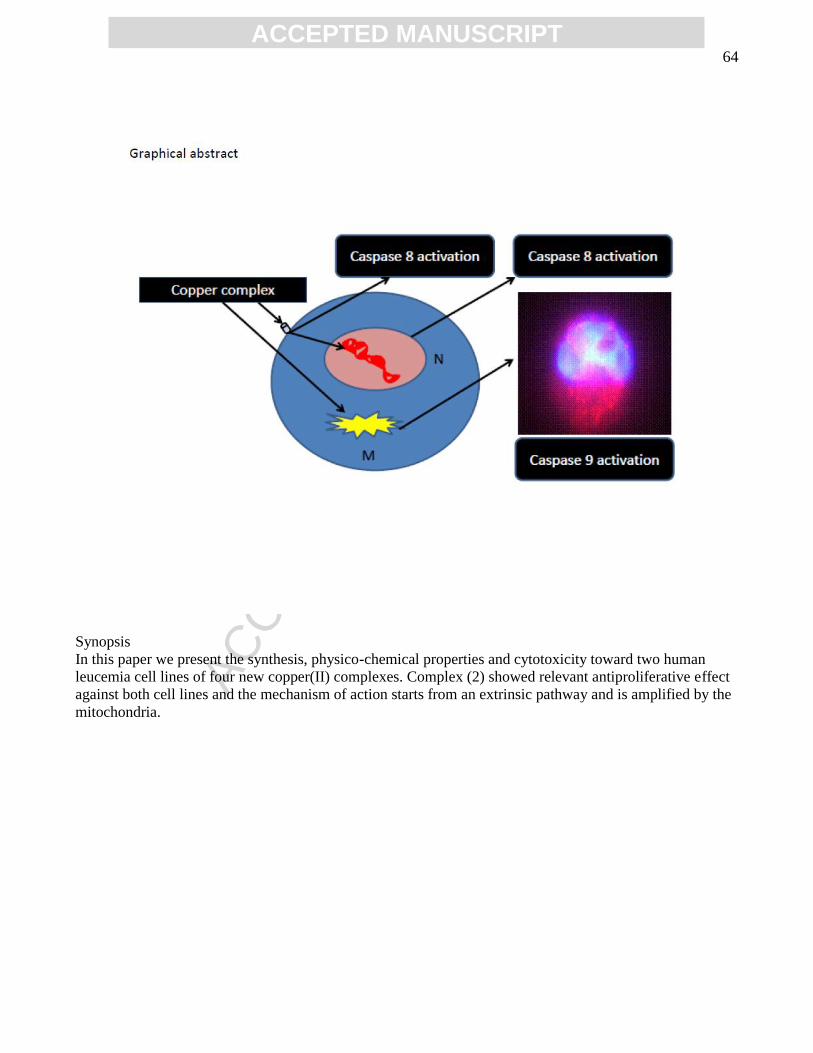
ACC
EPTE
D M
ANU
SCR
IPT
ACCEPTED MANUSCRIPT64
Synopsis
In this paper we present the synthesis, physico-chemical properties and cytotoxicity toward two human
leucemia cell lines of four new copper(II) complexes. Complex (2) showed relevant antiproliferative effect
against both cell lines and the mechanism of action starts from an extrinsic pathway and is amplified by the
mitochondria.

ACC
EPTE
D M
ANU
SCR
IPT
ACCEPTED MANUSCRIPT65
Highlights:
• New copper(II) complexes containing naphthalene rings (alpha and beta); • X-ray diffractions studies and several investigations in solution were performed; • Complex (2) is almost four times less toxic than cisplatin, based on LD50 values; • Complex (2) exhibits higher activitity than cisplatin against U937; • 5-The apoptosis signal triggered by complex (2) starts from an extrinsic pathway.