in patients with low-tension glaucoma and chronic simple glaucoma
TRANSCRIPT
British Journal ofOphthalmology, 1983, 67, 818-821
A comparative study of visual field defects seen
in patients with low-tension glaucoma and chronicsimple glaucomaR. A. HITCHINGS AND S. A. ANDERTON
From the Glaucoma Unit, Moorfields Eye Hospital, High Holborn, London WCJV 7AN
SUMMARY In a study comparing the visual field defects of patients with chronic simple andlow-tension glaucoma differences in the nature of the defect were found between the 2 groups.Patients with low-tension glaucoma had far 'steeper sided' defects than those seen in chronic simpleglaucoma (p <0-01).
The concept of a 'soft' or low-tension glaucoma hasbeen known for over 120 years.' However, a con-
troversy still exists concerning the exact nature of thiscondition. Ideas on pathogenesis range from low-tension glaucoma being a variety of chronic open-
angle glaucoma with the laminar cribrosa 'giving way'at normal intraocular tension2 to the idea that low-tension glaucoma is a variety of anterior ischaemicneuropathy.3 Comparison of the visual field defectsseen in low-tension and open-angle glaucoma mightbe expected to show differences between the 2conditions. However, even the type of the visual fielddefect seen in patients with low-tension glaucoma hasprovoked considerable discussion. In a recent criticalreview combined with examination of 32 of his owncases Levene4 concluded that a far higher proportion(92%) of patients with low-tension glaucoma hadvisual field defects to within 50 of fixation comparedwith 20% of 160 consecutive cases of chronic open-
angle glaucoma. However, Motolko and coworkers'could find no difference in the nature of the visualfield defectwhen comparing 160 eyes with low-tensionglaucoma with 154 eyes having chronic open-angleglaucoma matched according to cup-disc (C/D) ratio.
It seemed worthwhile to carry out a prospectivestudy of the visual field defects seen in these 2conditions.
Material and methods
This study compares the nature of the visual fielddefect seen in patients with low-tension glaucomamatched with patients having chronic open-angleCorrespondence to Mr R. A. Hitchings.
818
glaucoma. Each patient with low-tension glaucomafulfilled the following criteria: (a) 'glaucomatouscupping'; (b) nerve fibre bundle type of visual fielddefect; (c) intraocular pressures below 21 mmHgboth at presentation and at the time of 24-hourphasing when off all treatment: (d) open angles.These patients all had progressive low-tension
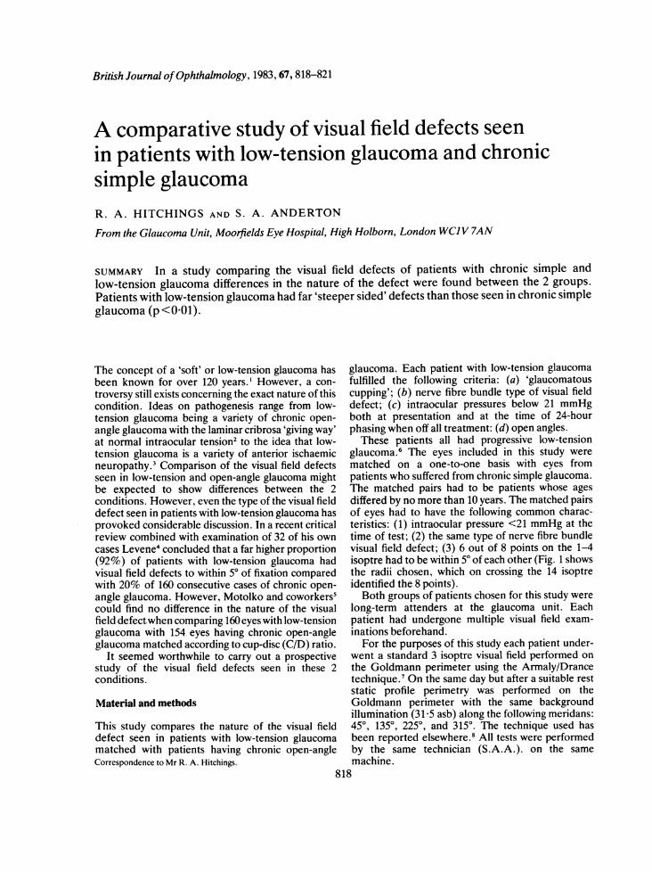
glaucoma.6 The eyes included in this study werematched on a one-to-one basis with eyes frompatients who suffered from chronic simple glaucoma.The matched pairs had to be patients whose agesdiffered by no more than 10 years. The matched pairsof eyes had to have the following common charac-teristics: (1) intraocular pressure <21 mmHg at thetime of test; (2) the same type of nerve fibre bundlevisual field defect; (3) 6 out of 8 points on the 1-4isoptre had to be within 50 of each other (Fig. 1 showsthe radii chosen, which on crossing the 14 isoptreidentified the 8 points).Both groups of patients chosen for this study were
long-term attenders at the glaucoma unit. Eachpatient had undergone multiple visual field exam-inations beforehand.
For the purposes of this study each patient under-went a standard 3 isoptre visual field performed onthe Goldmann perimeter using the Armaly/Drancetechnique.' On the same day but after a suitable reststatic profile perimetry was performed on theGoldmann perimeter with the same backgroundillumination (31-5 asb) along the following meridans:450, 1350, 2250, and 3150. The technique used hasbeen reported elsewhere.8 All tests were performedby the same technician (S.A.A.). on the samemachine.
Visualfield defects seen in patients with low-tension glaucoma and chronic simple glaucoma
{':(6 75 W
HARRGSI[R -AOmen
,'~ v X ';r+ ,. </ DIAGRAM INDICATING,, - ,i v \\ ' | - -< } X >\ '\ BTRE EIGHT AERIDIANS
-CUSED WHEN COMPARING
THE VISUAL FIELDSF BOTHGROUPS
\=s x t ,--'95 ,~~~~~~~~~~~~~~~~~~~~~~~~~345
210 \ 7 //t 030
0.315
4 1.00 2 2 2Y3752 300
o"~~~~~ ~l.I Ve
The 'steepness' of the slope of the visual field defectwas compared in those eyes that had a normalhemifield on profile perimetry. For this study the'steepness' was measured as the number of degreesalong the particular radius from the start of the defect(top of the slope) until the defect became absolute(the bottom of the slope). The top of the slope wasidentified by comparing the perimetry profile of theaffected radius with the diametrically opposite profilein the normal hemifield (i.e., 450 and 225°, 1350 and3150). A perimetry profile was considered 'normal' ifno depression exceeding 0 5 log unit intensityoccurred. In comparisons of the 2 opposite profilesthe start of a slope was identified when, at the samedistance from fixation, the sensitivity on one side was>0 5 log unit less than that on the other side.The figures for steepness on the 2 meridians
measured for each eye were averaged. The averagedfigures were used to compare the steepness of theslope of the visual field defect in the 2 groups.
Results
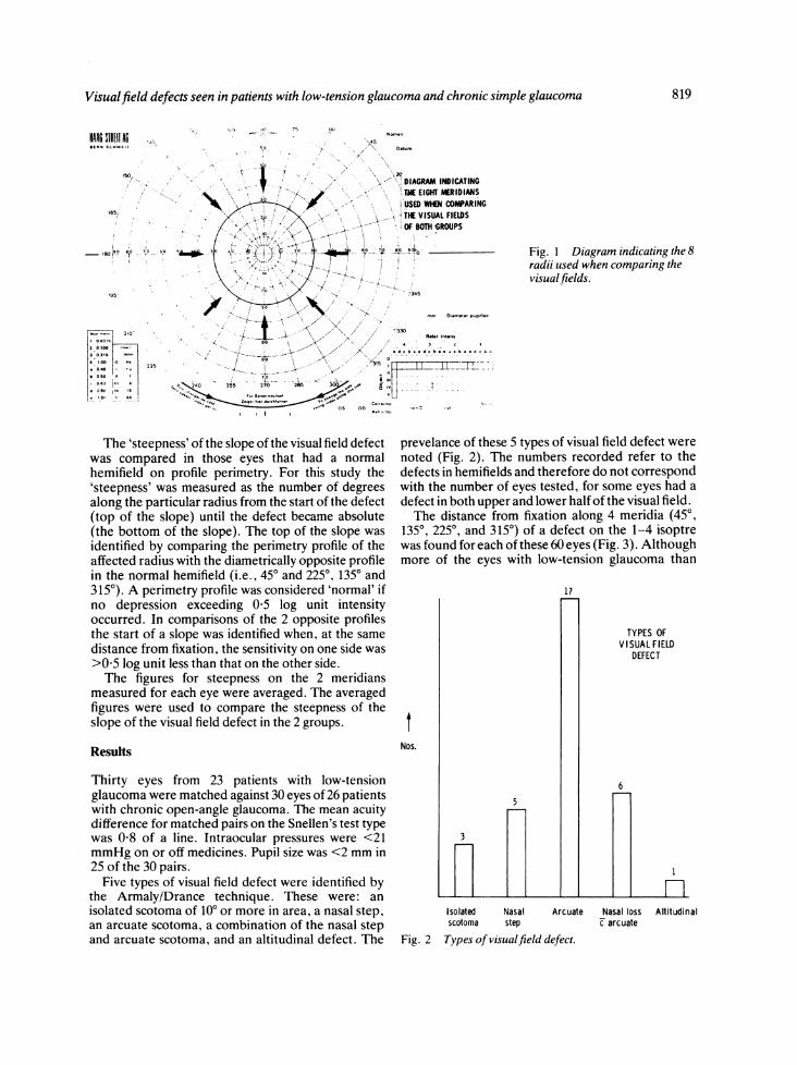
Thirty eyes from 23 patients with low-tensionglaucoma were matched against 30 eyes of 26 patientswith chronic open-angle glaucoma. The mean acuitydifference for matched pairs on the Snellen's test typewas 0-8 of a line. Intraocular pressures were <21mmHg on or off medicines. Pupil size was <2 mm in25 of the 30 pairs.Five types of visual field defect were identified by
the Armaly/Drance technique. These were: anisolated scotoma of 100 or more in area, a nasal step,an arcuate scotoma, a combination of the nasal stepand arcuate scotoma, and an altitudinal defect. The
Fig. 1 Diagram indicating the 8radii used when comparing thevisualfields.
prevelance of these 5 types of visual field defect werenoted (Fig. 2). The numbers recorded refer to thedefects in hemifields and therefore do not correspondwith the number of eyes tested, for some eyes had adefect in both upper and lower half of the visual field.The distance from fixation along 4 meridia (450,
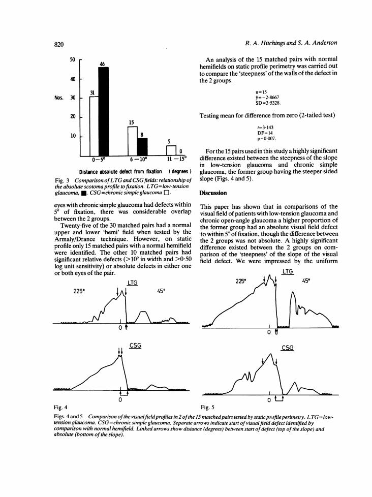
1350, 2250, and 3150) of a defect on the 1-4 isoptrewas found for each of these 60 eyes (Fig. 3). Althoughmore of the eyes with low-tension glaucoma than
tNos.
3
5
17
Isolated Nasal Arcuatescotoma step
Fig. 2 Types of visualfield defect.
TYPES OFVISUAL F IELD
DEFECT
nAltitudinal
6
Nasal lossc arcuate
819
R. A. Hitchings and S. A. Anderton
5046
40 -
Nos. 30 -
20-15
10 85
00-5o 6 -100 11 -150
Distance absolute defect from fixation ( degreesFig. 3 Comparison ofLTG and CSGfields: relationship ofthe absolute scotoma profile tofixation. LTG=low-tensionglaucoma, *. CSG=chronicsimpleglaucoma El.
eyes with chronic simple glaucoma had defects within50 of fixation, there was considerable overlapbetween the 2 groups.
Twenty-five of the 30 matched pairs had a normalupper and lower 'hemi' field when tested by theArmaly/Drance technique. However, on staticprofile only 15 matched pairs with a normal hemifieldwere identified. The other 10 matched pairs hadsignificant relative defects (>100 in width and >050log unit sensitivity) or absolute defects in either oneor both eyes of the pair.
LTG225° 1
o t
An analysis of the 15 matched pairs with normalhemifields on static profile perimetry was carried outto compare the 'steepness' of the walls of the defect inthe 2 groups.
n=15y=-2-8667SD=3-5328.
Testing mean for difference from zero (2-tailed test)t=3 143DF= 14p=0007.
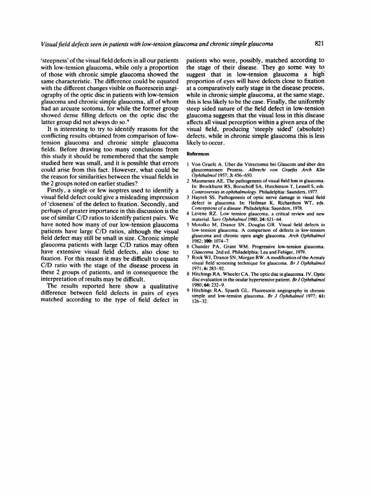
For the 15 pairs used in this study a highly significantdifference existed between the steepness of the slopein low-tension glaucoma and chronic simpleglaucoma, the former group having the steeper sidedslope (Figs. 4 and 5).
Discussion
This paper has shown that in comparisons of thevisual field of patients with low-tension glaucoma andchronic open-angle glaucoma a higher proportion ofthe former group had an absolute visual field defectto within 50 of fixation, though the difference betweenthe 2 groups was not absolute. A highly significantdifference existed between the 2 groups on com-parison of the 'steepness' of the slope of the visualfield defect. We were impressed by the uniform
LTG225 450
450
CSG
0 0Fig.4 Fig. 5Figs. 4 and 5 Comparison ofthe visualfieldprofiles in 2 ofthe 15 matchedpairs tested bystaticprofileperimetry. LTG=low-tension glaucoma. CSG=chronic simple glaucoma. Separate arrows indicate start of visualfield defect identified bycomparison with normal hemifield. Linked arrows show distance (degrees) between start ofdefect (top of the slope) andabsolute (bottom ofthe slope).
820
Visualfield defects seen in patients with low-tension glaucoma and chronic simple glaucoma
'steepness' of the visual field defects in all our patientswith low-tension glaucoma, while only a proportionof those with chronic simple glaucoma showed thesame characteristic. The difference could be equatedwith the different changes visible on fluorescein angi-ography of the optic disc in patients with low-tensionglaucoma and chronic simple glaucoma, all of whomhad an arcuate scotoma, for while the former groupshowed dense filling defects on the optic disc thelatter group did not always do so.9
It is interesting to try to identify reasons for theconflicting results obtained from comparison of low-tension glaucoma and chronic simple glaucomafields. Before drawing too many conclusions fromthis study it should be remembered that the samplestudied here was small, and it is possible that errorscould arise from this fact. However, what could bethe reason for similarities between the visual fields inthe 2 groups noted on earlier studies?
Firstly, a single or few isoptres used to identify avisual field defect could give a misleading impressionof 'closeness' of the defect to fixation. Secondly, andperhaps of greater importance in this discussion is theuse of similar C/D ratios to identify patient pairs. Wehave noted how many of our low-tension glaucomapatients have large C/D ratios, although the visualfield defect may still be small in size. Chronic simpleglaucoma patients with large C/D ratios may oftenhave extensive visual field defects, also close tofixation. For this reason it may be difficult to equateC/D ratio with the stage of the disease process inthese 2 groups of patients, and in consequence theinterpretation of results may be difficult.The results reported here show a qualitative
difference between field defects in pairs of eyesmatched according to the type of field defect in
patients who were, possibly, matched according tothe stage of their disease. They go some way tosuggest that in low-tension glaucoma a highproportion of eyes will have defects close to fixationat a comparatively early stage in the disease process,while in chronic simple glaucoma, at the same stage,this is less likely to be the case. Finally, the uniformlysteep sided nature of the field defect in low-tensionglaucoma suggests that the visual loss in this diseaseaffects all visual perception within a given area of thevisual field, producing 'steeply sided' (absolute)defects, while in chronic simple glaucoma this is lesslikely to occur.
References
I Von Graefe A. Uber die Vitrectomie bei Glaucom und uber denglaucomatosen Prozess. Albrecht von Graefes Arch KlinOphthalmol 1957; 3: 456-650.
2 Maumenee AE. The pathogenesis of visual field loss in glaucoma.In: Brockhurst RS, Boruchoff SA, Hutchinson T, Lessell S, eds.Controversies in ophthalmology. Philadelphia: Saunders, 1977.
3 Hayreh SS. Pathogenesis of optic nerve damage in visual fielddefect in glaucoma. In: Heilman K, Richardson WT, eds.Conceptions ofa disease. Philadelphia: Saunders, 1978.
4 Levene RZ. Low tension glaucoma, a critical review and newmaterial. Surv Ophthalmol 1980; 24: 621-64.
5 Motolko M, Drance SN, Douglas GR. Visual field defects inlow-tension glaucoma. A comparison of defects in low-tensionglaucoma and chronic open angle glaucoma. Arch Ophthalmol1982; 100:1074-7.
6 Chander PA, Grant WM. Progressive low-tension glaucoma.Glaucoma. 2nd ed. Philadelphia: Lea and Febiger, 1979.
7 Rock WJ, Drance SN, Morgan RW. A modification of the Armalyvisual field screening technique for glaucoma. Br J Ophthalmol1971; 6: 283-92.
8 Hitchings RA, Wheeler CA. The optic disc in glaucoma. IV. Opticdisc evaluation in the ocular hypertensive patient. BrJ Ophthalmol1980; 64:232-9.
9 Hitchings RA, Spaeth GL. Fluorescein angiography in chronicsimple and low-tension glaucoma. Br J Ophthalmol 1977; 61:126-32.
821