immunomodulatory effects of yersinia pestis ... · suppressive activity was not observed when it...
TRANSCRIPT
CLINICAL AND VACCINE IMMUNOLOGY, Jan. 2010, p. 49–55 Vol. 17, No. 11556-6811/10/$12.00 doi:10.1128/CVI.00336-09Copyright © 2010, American Society for Microbiology. All Rights Reserved.
Immunomodulatory Effects of Yersinia pestis Lipopolysaccharideson Human Macrophages�
Motohiro Matsuura,1* Hideyuki Takahashi,2 Haruo Watanabe,2Shinji Saito,1,3 and Kazuyoshi Kawahara4
Department of Infection and Immunity, School of Medicine, Jichi Medical University, Tochigi 329-0498, Japan1; Department ofBacteriology, National Institute of Infectious Disease, Tokyo 162-8640, Japan2; Graduate School of
Comprehensive Human Sciences, University of Tsukuba, Ibaraki 305-8575, Japan3; andCollege of Engineering, Kanto Gakuin University, Yokohama 236-8501, Japan4
Received 6 August 2009/Returned for modification 17 September 2009/Accepted 26 October 2009
In the current study, we investigated the activity of lipopolysaccharide (LPS) purified from Yersinia pestisgrown at either 27°C or 37°C (termed LPS-27 and LPS-37, respectively). LPS-27 containing hexa-acylated lipidA, similar to the LPS present in usual gram-negative bacteria, stimulated an inflammatory response in humanU937 cells through Toll-like receptor 4 (TLR4). LPS-37, which did not contain hexa-acylated lipid A, exhibitedstrong antagonistic activity to the TLR4-mediated inflammatory response. The phagocytic activity in the cellswas not affected by LPS-37. To estimate the activity of LPS in its bacterial binding form, formalin-killedbacteria (FKB) were prepared from Y. pestis cells grown at 27°C or 37°C (termed FKB-27 and FKB-37,respectively). FKB-27 strongly stimulated the inflammatory response. This activity was suppressed in thepresence of an anti-TLR4 antibody but not an anti-TLR2 antibody. In addition, this activity was almostcompletely suppressed by LPS-37, indicating that the activity of FKB-27 is predominantly derived from theLPS-27 bacterial binding form. In contrast, FKB-37 showed no antagonistic activity. The results arising fromthe current study indicate that Y. pestis causes infection in humans without stimulating the TLR4-based defensesystem via bacterial binding of LPS-37, even when bacterial free LPS-37 is not released to suppress the defensesystem. This is in contrast to the findings for bacteria that possess agonistic LPS types, which are easilyrecognized by the defense system via the bacterial binding forms.
Yersinia pestis is the causative agent of bubonic, septicemic,and pneumonic plague in humans and is primarily a rodentpathogen that is transmitted intradermally to humans throughthe bite of an infected flea (24). Y. pestis must survive in twodifferent temperature ranges. One temperature range repre-sents that of a flea residing in rodent burrows or mammalianhair (21°C to 28°C), while the other represents the body tem-perature of the infected rodent or human (37°C to 41°C) (2, 4,24). It has been demonstrated that the various cellular com-ponents of this bacterium are differentially expressed at thesetemperature ranges. The production of several virulent factors,such as the fraction 1 antigen (6), the pH 6 antigen (14), andYop proteins (33), are known to be upregulated during growthof the bacterium at 37°C. In contrast, the production of murinetoxin (12), which is required for the survival of Y. pestis in themidgut of fleas, is synthesized at approximately 27°C and isdownregulated at 37°C.
Y. pestis is a gram-negative bacterium that contains bacteriallipopolysaccharide (LPS) as its major cell wall component. Thelipid moiety in LPS that is responsible for immune systemactivation is lipid A and is composed of a glucosamine-disac-charide backbone carrying acyl chains and phosphate groups.The hexa-acylated type of lipid A was found to be the majorcomponent of the Escherichia coli LPS, which exhibits strong
immunological activities (8, 13, 17). This particular type oflipid A is widely found among usual gram-negative bacteria,but it is not the sole type of lipid A species. The specific typesof lipid A have been shown to differ among bacterial species,and heterogeneous types of lipid A often coexist in a singlespecies (28). We have previously reported that the lipid Apresent in Y. pestis LPS is heterogeneous (from hexa-acylatedto triacylated types) when the bacterium is grown at 27°C andshifts to the hypoacylated types (tetra- and triacylated types)when it is grown at 37°C (16). Such a temperature-dependentshift of lipid A types in the LPS of Y. pestis was further con-firmed by another group (29), who demonstrated a similar shiftin two additional pathogenic species of Yersinia. Hexa-acylatedlipid A is able to strongly activate Toll-like receptor 4 (TLR4),while the tetra-acylated type is a poor activator and servesinstead as an antagonist when the target cells are human inorigin (10, 22). The alteration of lipid A to hypoacylated typesis therefore favorable for the bacterium, as it allows the bac-terium to evade the human innate immune system and avoidelimination. It has been indicated that lipid A alterations thatresult in the evasion of the bacteria from host TLR4 recogni-tion play a more important pathogenic role than the additionalvirulence factors of Y. pestis (23).
In the current study, we purified LPS or prepared formalin-killed bacteria (FKB) from Y. pestis grown at either 27°C or37°C and examined their influence on the immune systemresponse in human U937 cells. Our findings suggested thatthe LPS isolated from Y. pestis grown at 37°C (LPS-37)strongly suppressed the human TLR4-dependent inflamma-tory response when it was free from the bacterial body. This
* Corresponding author. Mailing address: Department of Infectionand Immunity, School of Medicine, Jichi Medical University, 3311-1Yakushiji, Shimotsuke, Tochigi 329-0498, Japan. Phone: 81-285-58-7332. Fax: 81-285-44-1175. E-mail: [email protected].
� Published ahead of print on 4 November 2009.
49
on March 3, 2019 by guest
http://cvi.asm.org/
Dow
nloaded from
suppressive activity was not observed when it was bound to thebacterial body. In contrast, the LPS isolated from Y. pestisgrown at 27°C (LPS-27) was found to be agonistic towardTLR4 signaling in both the bacterium-free and -bound forms,as FKB grown at 27°C (FKB-27) showed agonistic activitysimilar to that of LPS-27.
MATERIALS AND METHODS
Bacterial culture, extraction of LPS, and preparation of FKB. Virulent Y.pestis strain Yreka (National Institute of Infectious Disease, Tokyo, Japan) wascultured on brain heart infusion agar (Difco Laboratories, Detroit, MI) at 27°Cor 37°C for 48 h. The bacterial cells were suspended in saline to obtain heat-killed bacteria for the extraction of LPS by the method reported previously (16)and were purified via extraction with 45% phenol containing triethylamine andsodium deoxycholate (21). The LPS forms obtained following growth at 27°C and37°C were termed LPS-27 and LPS-37, respectively. To prepare FKB, bacterialcells were suspended in 0.3% formalin solution at a dose of 30 mg (wetweight)/ml (9 � 109 bacteria/ml) and were incubated at 37°C with shaking for 5days to kill the bacteria completely. These preparations, termed FKB-27 andFKB-37, respectively, were then washed and suspended in phosphate-bufferedsaline.
Cell culture. The murine macrophage cell line RAW264.7 and the humanmacrophage cell line U937 (both from the American Type Culture Collection,Manassas, VA) were used in the current study. To obtain mouse peritonealexudate cells (PECs), 7- to 8-week-old C3H/HeN or C3H/HeJ mice (JapanCharles River, Tokyo, Japan) were injected intraperitoneally with 2 ml of thio-glycolate broth (Difco Laboratories). PECs were then obtained 4 days later byperitoneal lavage. All animal experiments were carried out according to theguidelines of the Laboratory Animal Center, Jichi Medical University. Thecomplete medium (CM) used for cell culture was composed of RPMI 1640medium (ICN Biomedicals, Inc., Aurora, OH) supplemented with 2 mM L-glutamine, 100 U/ml penicillin, 100 �g/ml streptomycin, 0.2% NaHCO3, and10% heat-inactivated fetal bovine serum (FBS; Flow Laboratories Inc., Rock-ville, MD). The cells were cultured in a humidified chamber at 37°C with 5%CO2 and 95% air. RAW264.7 cells and PECs suspended in CM at 1 � 106
cells/ml were cultured overnight, and the adherent cells were used in all exper-iments. U937 cells were suspended in CM supplemented with 100 nM phorbolmyristate acetate (PMA; Sigma-Aldrich Co., St. Louis, MO) at a cell density of5 � 105/ml and were cultured for 3 days to induce differentiation into macro-phage-like cells. Only the adherent cells were used in the current study.
Cytokine assay. RAW264.7 and U937 cells were cultured in 24-well cultureplates (500 �l/well; Corning Inc., Corning, NY) and were stimulated with variousLPSs, TLR ligands, and FKB. LPS purified from Salmonella enterica serovarAbortus-Equi (7) was a kind gift from C. Galanos (MIP fur Immunbiology,Freiburg, Germany) and was used as a reference for the TLR4 ligand. Pam3-Cys-Ser-(Lys)4 hydrochloride (Pam3-Cys), purchased from Calbiochem, EMDBioscience, Inc. (San Diego, CA), was used as a TLR2 ligand; and flagellin (S.enterica serovar Typhimurium), purchased from InvivoGen (San Diego, CA), wasused as a TLR5 ligand. The culture supernatant obtained 6 h after stimulationwas assayed for mouse and human cytokines by a specific sandwich enzyme-linked immunosorbent assay (ELISA). The ELISA was performed according tothe instructions supplied by Endogen (Woburn, MA) and by using matchedantibody pairs. Each cytokine was quantified (in ng/ml or pg/ml) on the basis ofthe standard curve obtained in each assay. In the suppression experiments,anti-human TLR4/CD284 (anti-TLR4 antibody; MBL Co., Ltd., Nagoya, Japan)(1), anti-human CD282 (TLR2) (anti-TLR2 antibody; BD Pharmingen, FranklinLakes, NJ) (19), and a human TLR4 antagonist, compound 406 (Daiichi KagakuCo., Tokyo, Japan) (10, 22), were used as suppressors. Cells were stimulated inthe presence of suppressors (added 1 h prior to stimulation), and the culturesupernatant was assayed 6 h after stimulation.
Detection of cytokine mRNA by RT-PCR. U937 cells were cultured in 60-mmculture dishes (Corning Inc.) containing 3 ml of medium and were stimulatedwith LPS or Pam3-Cys for 2 h in the presence or the absence of LPS-37. TotalRNA was then extracted by using Isogen (Nippon Gene Co., Ltd., Tokyo, Japan).One microgram of total RNA was reverse transcribed by using SuperScript IIreverse transcriptase (RT; Invitrogen Co., Carlsbad, CA). Aliquots of cDNAequivalent to 50 ng of RNA for the following genes were subsequently amplifiedby using the indicated primers (3, 5): for human tumor necrosis factor alpha(TNF-�), sense primer 5�-AGA GGG AAG AGT TCC CCA GGG AC-3� andantisense primer 5�-TGA GTC GGT CAC CCT TCT CCA G-3�; for humaninterleukin-1� (IL-1�), sense primer 5�-CCA GCT ACG AAT CTC GGA CCA
CC-3� and antisense primer 5�-TTA GGA AGA CAC AAA TTG CAT GGTGAA GTC AGT-3�; for human IL-6, sense primer 5�-ATG AAC TCC TTC TCCACA AGC GC-3� and antisense primer 5�-GAA GAG CCC TCA GGC TGGACT G-3�; for human IL-8, sense primer 5�-ATG ACT TCC AAG CTG GCCGTG-3� and antisense primer 5�-TTA TGA ATT CTC AGC CCT CTT CAAAAA CTT CTC-3�; for human IL-10, sense primer 5�-ATG CCC CAA GCTGAG AAC CAA GAC CCA-3� and antisense primer 5�-TCT CAA GGG GCTGGG TCA GCT ATC CCA-3�; for human IL-12p40, sense primer 5�-AGAGGC TCT TCT GAC CCC CAG-3� and antisense primer 5�-CTC TTG CTCTTG CCC TGG ACC TG-3�; and for human GAPDH, sense primer 5�-TGAAGG TCG GAG TCA ACG GAT TTG GT-3� and antisense primer 5�-CATGTG GGC CAT GAG GTC CAC CAC-3�. The PCR conditions included aninitial denaturation at 94°C for 1 min, followed by 20 to 32 cycles at 94°C for 1min, 60°C for 1 min, and 72°C for 2 min. Amplification was repeated for 32 cyclesfor IL-6, IL-10, and IL-12p40; 25 cycles for TNF-� and GAPDH; and 20 cyclesfor IL-1� and IL-8. The resulting PCR products were separated by electrophore-sis on 1.2% agarose gels, and the gels were stained with ethidium bromide.
Phagocytic assay. U937 cells were cultured in 96-well culture plates (100�l/well; Corning Inc.) and subjected to phagocytic assays, in accordance with themethod described previously (36). Briefly, the cells were cultured for 1 h withcytochalasin D (a phagocytosis inhibitor; Wako Pure Chemical Industries, Ltd.,Osaka, Japan), anti-TLR4 antibody, or LPS-37. After removal of the culturesupernatant, the cells were cultured for 2 h in the presence of fluorescein-labeledEscherichia coli K-12 BioParticles (Molecular Probes, Inc., Eugene, OR). Theculture supernatant containing the particles was then removed, and the remain-ing extracellular fluorescence was quenched by the addition of trypan bluesolution (250 �g/ml, pH 4.4). After incubation at room temperature for 3 min,the dye was discarded and the fluorescence intensity (in relative fluorescenceunits) associated with the intracellular fluorescent particles was measured at anexcitation wavelength of 480 nm and an emission wavelength of 525 nm with aSPECTRAmax M5 microplate fluorometer (Molecular Devices Corp., Sunny-vale, CA).
Estimation of LPS levels. The LPS levels present in the supernatants of theFKB were measured by using an Endospecy test chromogenic endotoxin-specificassay kit (Seikagaku Co., Tokyo, Japan). Briefly, the test sample solution wascombined with the kit reagent in a 96-well microplate and the plate was incu-bated for 30 min at 37°C. The reaction was then stopped by adding 0.6 M aceticacid, and the color reaction was measured at 405 nm. LPS-27 and LPS-37 wereused to construct the standard curves for the calculation of the LPS levels in thesupernatants of FKB-27 and FKB-37, respectively.
Statistical analysis. The significance of any difference between the means forthe experimental groups was determined by Student’s t test. The means wereconsidered significantly different if P was �0.05.
RESULTS
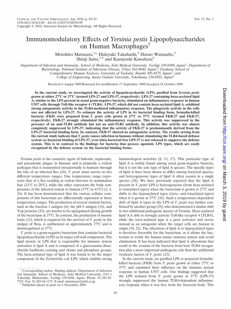
Cytokine production by mouse and human cells stimulatedwith Y. pestis LPS-27 and LPS-37. The mouse macrophage cellline RAW264.7 was stimulated with LPS-27, LPS-37, or a ref-erence Salmonella LPS; and the production of cytokines in thestimulated cells was assessed. The stimulation of cells withLPS-27 resulted in the production of both TNF-� and IL-6when it was administered at a low dose of only 1 ng/ml. Thestimulatory activity was similar to that achieved by the refer-ence Salmonella LPS. LPS-37 also had stimulatory activity;however, the stimulation was significantly weaker than that ofLPS-27. The activity of LPS-37 administered at 1,000 ng/mlwas comparable to that of LPS-27 administered at 1 ng/ml (Fig.1A). Similar results were also obtained when PECs isolatedfrom C3H/HeN mice were used as target cells. However, thetwo LPS forms failed to induce cytokine production whenPECs isolated from the TLR4 mutant mice (C3H/HeJ) wereused as the target cells (data not shown). These results indicatethat both LPS preparations function as specific mouse TLR4agonists and are sufficiently free from detectable contaminants(i.e., they are pure enough) to stimulate the other TLRs.
We next examined the effects of these LPSs in humancells. As shown in Fig. 1B, LPS-27 exhibited strong stimu-
50 MATSUURA ET AL. CLIN. VACCINE IMMUNOL.
on March 3, 2019 by guest
http://cvi.asm.org/
Dow
nloaded from
latory activity in human U937 cells comparable to that of thereference Salmonella LPS. In addition, the dose dependencyof LPS-27 for this activity was similar to that for the activa-tion of RAW264.7 cells. In contrast, the activity of LPS-37 in
the human cells was remarkably reduced and was undetectableat doses as high as 10 �g/ml. This moderate agonistic activityobserved in LPS-37-treated mouse cells and the antagonisticactivity observed in treated human cells were similar to the
FIG. 1. Effects of LPS-27 and LPS-37 isolated from Y. pestis on the stimulation of mouse macrophage RAW264.7 cells and human macrophageU937 cells. (A) Mouse RAW264.7 cells were stimulated with LPS-27, LPS-37, and reference Salmonella LPS at the indicated doses. (B) HumanU937 cells were stimulated as described for panel A. The cytokine levels in the culture supernatant at 6 h after stimulation were determined byELISA. The data are presented as the means � standard errors of triplicate samples, and a representative result from three independentexperiments is shown. h, human; m, mouse.
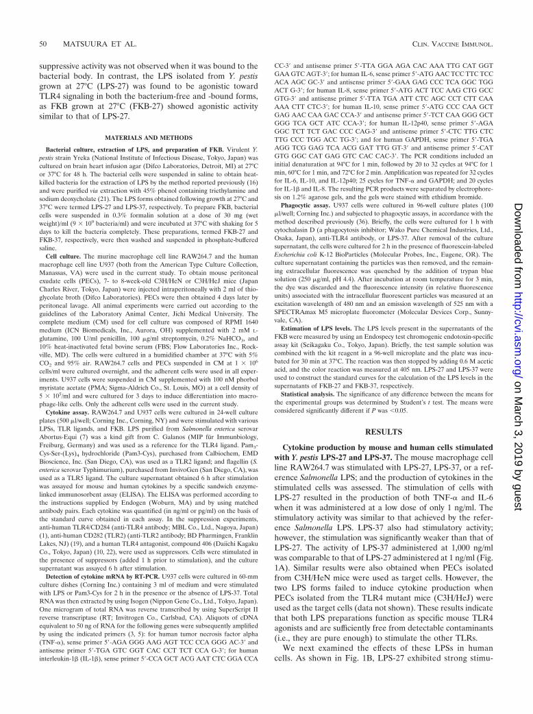
FIG. 2. Effects of Y. pestis LPS-37 on cytokine production in U937 cells stimulated with TLR4 and TLR2 agonists. (A) U937 cells cultured in thepresence of LPS-37 or compound 406 were stimulated with either Y. pestis LPS-27 (1 ng/ml) or Salmonella LPS (1 ng/ml). The cytokine levels in theculture supernatant were determined by ELISA. The data are presented as the means � standard errors for triplicate samples. (B) U937 cells culturedin the presence of 10 ng/ml LPS-37 were stimulated with 1 ng/ml of LPS-27 or Salmonella LPS. Total RNA was isolated 2 h after stimulation and subjectedto RT-PCR for determination of cytokine mRNA expression. PCR products were separated by electrophoresis on 1.2% agarose gels and stained withethidium bromide. (C) U937 cells cultured in the presence of LPS-37 were stimulated with Pam3-Cys (10 nM), and cytokine production was determinedas described for panel A. (D) U937 cells cultured with LPS-37 (10 ng/ml) were stimulated with Pam3-Cys (10 nM), and cytokine mRNA expression wasdetermined as described for panel B. Representative results of three (A and C) or two (B and D) experiments are presented.
VOL. 17, 2010 CHARACTERIZATION OF YERSINIA PESTIS LPS 51
on March 3, 2019 by guest
http://cvi.asm.org/
Dow
nloaded from
activities observed in cells treated with synthetic tetra-acylatedlipid A (compound 406).
Antagonistic activity of LPS-37 to stimulation of TLR4 inhuman cells. The suppressive effects of LPS-37 on the activa-tion of human cells with various ligands to extracellular TLRwere also examined. We used Y. pestis LPS-27 and the refer-ence Salmonella LPS as TLR4 ligands, Pam3-Cys as the TLR2ligand, and flagellin as the TLR5 ligand. Human U937 cellswere stimulated with these agonists in the presence of LPS-37or compound 406, and cytokine production was assessed. Wefound that the TLR4-dependent production of cytokines suchas TNF-� and IL-6 was strongly suppressed by LPS-37. Thisresult was similar to the response to compound 406 (Fig. 2A).The strong antagonistic activity of LPS-37 against humanTLR4 stimulation was confirmed by measuring the levels ofexpression of various cytokine mRNAs (Fig. 2B). LPS-37 wasnot observed to have suppressive effects against TLR2-stimu-lated cytokine production in terms of either protein productionor mRNA expression (Fig. 2C and D). Similarly, LPS-37 wasnot observed to have antagonistic activity against TLR5 stim-ulation by flagellin (data not shown). These results indicatethat LPS-37 acts as a strong antagonist against human TLR4but not the additional human extracellular TLRs, such asTLR2 and TLR5.
Effects of LPS-37 on the phagocytic activity of U937 cells.Antiphagocytic factors play an important role in the evasion ofbacteria from the host defense network by activating the innateimmune system. As LPS-37 strongly suppressed cytokine pro-duction on the basis of the host (human) innate immune re-sponse, we next investigated the effects of LPS-37 on thephagocytosis of bacteria by U937 cells. The phagocytic activityof U937 cells was assayed with fluorescein-labeled E. coli K-12BioParticles as the target bacteria, and the fluorescence of the
engulfed intracellular bacteria was measured by quenching thefluorescence of the extracellular bacteria with trypan blue.Treatment with LPS-37 or anti-human TLR4 antibody failed tosuppress the phagocytic activity in U937 cells, while the addi-tion of cytochalasin D showed a significant suppressive effect(Fig. 3). These results suggest that the suppression of TLR4signaling by LPS-37 does not affect the role of Y. pestis in theenhancement of resistance to bacterial phagocytes by U937cells.
Proinflammatory cytokines produce human cells stimulatedwith Y. pestis FKB. FKB-27 and FKB-37 were prepared by themethod used in the production of formalin vaccine, and theability of these FKB to stimulate cytokine production in U937cells was investigated. Similar to treatment with LPS, the ac-tivity of FKB-27 was found to be strong, while the activity ofFKB-37 was very weak, if it was present at all (Fig. 4A). Thecontribution of the free LPS released from the bacterial bodyto FKB-27 activity levels was found to be negligible. To esti-mate the amount of LPS released into the supernatant of the
FIG. 3. Effects of LPS-37 on the phagocytosis of bacteria by U937cells. A total of 5 � 104 U937 cells/well were cultured in 96-well platesin the presence of cytochalasin D, LPS-37, or anti-TLR4 antibody for1 h. The culture supernatant was then removed, and fluorescein iso-thiocyanate-labeled E. coli K-12 BioParticles were added at a dose of5 � 107/well. The cells were cultured for 2 h to phagocytose the E. coliparticles and were then treated with trypan blue to quench the extra-cellular fluorescence. The intensity of the fluorescence associated withintracellular fluorescent particles was then measured with a microplatefluorometer. Intensity is presented as relative fluorescence units. Thedata presented represent the means from triplicate assays; the errorbars indicate ranges and are representative of the results of threeexperiments. *, P � 0.05 compared to the results for the control, asdetermined by Student’s t test; **, P � 0.01 compared to the results forthe control, as determined by Student’s t test.
FIG. 4. Stimulation of cytokine production in U937 cells treated withformalin-killed Y. pestis. (A) U937 cells were stimulated with FKB-27 (Œ)and FKB-37 (F). The supernatants collected from FKB-27 (�) andFKB-37 (E) stock suspensions obtained by centrifugation were also usedto stimulate the cells. The cytokine levels in the culture supernatant weredetermined by ELISA. The data are presented as the means � standarderrors for triplicate samples. A representative result from three indepen-dent experiments is shown. (B) The amount of free LPS released fromeach FKB was estimated by use of an Endospecy test kit. The activity levelin each supernatant (Sup.), measured as the absorbance (A.) at 405 nm,was compared to that of the corresponding LPS. Data are presented asthe means � standard errors for duplicate samples. A representativeresult from two independent experiments is shown.
52 MATSUURA ET AL. CLIN. VACCINE IMMUNOL.
on March 3, 2019 by guest
http://cvi.asm.org/
Dow
nloaded from
FKB stock suspension, each of the supernatants isolated fromthe FKB-27- and FKB-37 suspensions was analyzed by theEndospecy test, and the levels were compared to the activityof the corresponding LPS (Fig. 4B). We found that 5 pg ofLPS-27 was released from a suspension of 5 � 104 FKB-27 andthat 2 pg of LPS-37 was released from a suspension of 1 � 105
FKB-37. These results indicate that the effect of the releasedLPS on FKB activity was negligible.
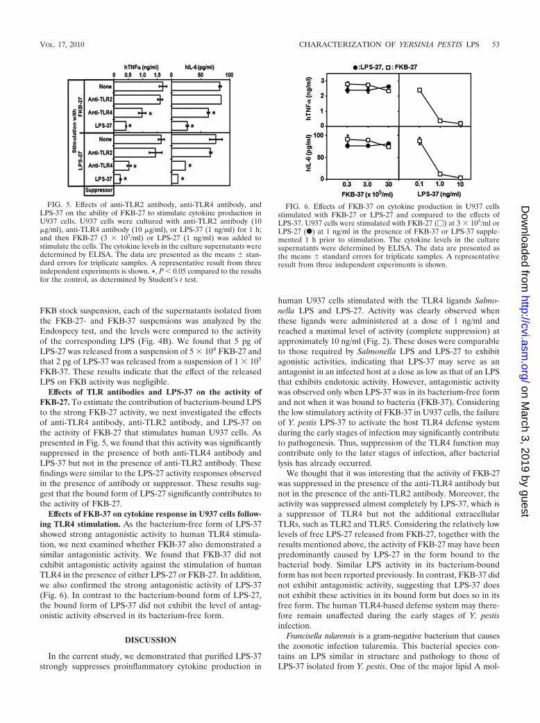
Effects of TLR antibodies and LPS-37 on the activity ofFKB-27. To estimate the contribution of bacterium-bound LPSto the strong FKB-27 activity, we next investigated the effectsof anti-TLR4 antibody, anti-TLR2 antibody, and LPS-37 onthe activity of FKB-27 that stimulates human U937 cells. Aspresented in Fig. 5, we found that this activity was significantlysuppressed in the presence of both anti-TLR4 antibody andLPS-37 but not in the presence of anti-TLR2 antibody. Thesefindings were similar to the LPS-27 activity responses observedin the presence of antibody or suppressor. These results sug-gest that the bound form of LPS-27 significantly contributes tothe activity of FKB-27.
Effects of FKB-37 on cytokine response in U937 cells follow-ing TLR4 stimulation. As the bacterium-free form of LPS-37showed strong antagonistic activity to human TLR4 stimula-tion, we next examined whether FKB-37 also demonstrated asimilar antagonistic activity. We found that FKB-37 did notexhibit antagonistic activity against the stimulation of humanTLR4 in the presence of either LPS-27 or FKB-27. In addition,we also confirmed the strong antagonistic activity of LPS-37(Fig. 6). In contrast to the bacterium-bound form of LPS-27,the bound form of LPS-37 did not exhibit the level of antag-onistic activity observed in its bacterium-free form.
DISCUSSION
In the current study, we demonstrated that purified LPS-37strongly suppresses proinflammatory cytokine production in
human U937 cells stimulated with the TLR4 ligands Salmo-nella LPS and LPS-27. Activity was clearly observed whenthese ligands were administered at a dose of 1 ng/ml andreached a maximal level of activity (complete suppression) atapproximately 10 ng/ml (Fig. 2). These doses were comparableto those required by Salmonella LPS and LPS-27 to exhibitagonistic activities, indicating that LPS-37 may serve as anantagonist in an infected host at a dose as low as that of an LPSthat exhibits endotoxic activity. However, antagonistic activitywas observed only when LPS-37 was in its bacterium-free formand not when it was bound to bacteria (FKB-37). Consideringthe low stimulatory activity of FKB-37 in U937 cells, the failureof Y. pestis LPS-37 to activate the host TLR4 defense systemduring the early stages of infection may significantly contributeto pathogenesis. Thus, suppression of the TLR4 function maycontribute only to the later stages of infection, after bacteriallysis has already occurred.
We thought that it was interesting that the activity of FKB-27was suppressed in the presence of the anti-TLR4 antibody butnot in the presence of the anti-TLR2 antibody. Moreover, theactivity was suppressed almost completely by LPS-37, which isa suppressor of TLR4 but not the additional extracellularTLRs, such as TLR2 and TLR5. Considering the relatively lowlevels of free LPS-27 released from FKB-27, together with theresults mentioned above, the activity of FKB-27 may have beenpredominantly caused by LPS-27 in the form bound to thebacterial body. Similar LPS activity in its bacterium-boundform has not been reported previously. In contrast, FKB-37 didnot exhibit antagonistic activity, suggesting that LPS-37 doesnot exhibit these activities in its bound form but does so in itsfree form. The human TLR4-based defense system may there-fore remain unaffected during the early stages of Y. pestisinfection.
Francisella tularensis is a gram-negative bacterium that causesthe zoonotic infection tularemia. This bacterial species con-tains an LPS similar in structure and pathology to those ofLPS-37 isolated from Y. pestis. One of the major lipid A mol-
FIG. 5. Effects of anti-TLR2 antibody, anti-TLR4 antibody, andLPS-37 on the ability of FKB-27 to stimulate cytokine production inU937 cells. U937 cells were cultured with anti-TLR2 antibody (10�g/ml), anti-TLR4 antibody (10 �g/ml), or LPS-37 (1 ng/ml) for 1 h;and then FKB-27 (3 � 105/ml) or LPS-27 (1 ng/ml) was added tostimulate the cells. The cytokine levels in the culture supernatants weredetermined by ELISA. The data are presented as the means � stan-dard errors for triplicate samples. A representative result from threeindependent experiments is shown. *, P � 0.05 compared to the resultsfor the control, as determined by Student’s t test.
FIG. 6. Effects of FKB-37 on cytokine production in U937 cellsstimulated with FKB-27 or LPS-27 and compared to the effects ofLPS-37. U937 cells were stimulated with FKB-27 (�) at 3 � 105/ml orLPS-27 (F) at 1 ng/ml in the presence of FKB-37 or LPS-37 supple-mented 1 h prior to stimulation. The cytokine levels in the culturesupernatants were determined by ELISA. The data are presented asthe means � standard errors for triplicate samples. A representativeresult from three independent experiments is shown.
VOL. 17, 2010 CHARACTERIZATION OF YERSINIA PESTIS LPS 53
on March 3, 2019 by guest
http://cvi.asm.org/
Dow
nloaded from
ecules present in the LPS isolated from F. tularensis was re-ported to exhibit a tetra-acylated structure that containedthree 3-OH C18 fatty acids, one C16 fatty acid, and one phos-phate group (25, 34). We have previously reported that LPS-37also exhibits a tetra-acylated structure but contains four 3-OHC14 fatty acids and two phosphate groups (16). In contrast toLPS-37, LPS isolated from F. tularensis was reported to beneither stimulatory nor antagonistic to human and murine cellsvia TLR4 (11). This difference was thought to be caused by thevariations in their compositions, as monophosphoryl lipid Ahas been reported to have weaker activity than diphosphoryllipid A (13) and longer acyl chains such as C16 have beenreported to have weaker activity than C14 (22). These so-calledsilent characteristics of F. tularensis LPS are thought to con-tribute to its capacity to evade mammalian immune defensemechanisms and to promote survival in an infected host (9, 30).More recently, the effects of the preventive administration of asynthetic TLR4 agonist on the protection of mice from exper-imental pneumonic tularemia have been demonstrated (18).Similar prophylactic activation of TLR4 may also prove usefulagainst Y. pestis infection.
The synthetic antagonist prototype termed compound 406 isa lipid IVA that is a biosynthetic precursor of E. coli lipid A(hexa-acylated) (26–27). This precursor structure, however, isnot normally found in the outer membrane of E. coli or otherusual members of the Enterobacteriaceae family. LPS is themajor structural component of the cell wall outer membrane ofgram-negative bacteria and plays an important role in thestabilization of the bacterial body. A mutant strain of E. colithat synthesizes only the lipid IVA precursor and that containsonly this type of lipid A species in its LPS has been reported togrow very slowly and to be unstable, as judged by its antibiotichypersensitivity and easy lysis following centrifugation (35).However, Y. pestis grown at 37°C does not appear to be sen-sitive to oscillatory shock or susceptible to antibacterial factorsin the serum of the infected host. It has been reported that theminimal LPS structure required for the viability of E. coli andSalmonella enterica serovar Typhimurium is lipid A glyco-sylated with 3-deoxy-D-manno-octulosonic acid (Kdo) residuesbut that Y. pestis can survive without Kdo residues in its LPS(32). In combination, these results suggest that Y. pestis differsfrom E. coli and other usual species of the Enterobacteriaceae,as it contains additional factors that may allow the constructionof a strong outer membrane and the formation of a stablebacterial body, thus compensating for its insufficient LPSstructure.
It has been reported that Y. pestis grown at a lower temper-ature (21°C) is readily phagocytosed by human neutrophils(15) but that Y. pestis grown at 37°C is resistant to phagocytosis(31). In the current study, we did not observe any antiphago-cytic effects of LPS-37 on bacterial phagocytes in U937 cells,even though LPS-37 strongly suppressed the TLR4-mediatedinflammatory responses of these cells. Several antiphagocyticfactors produced by Y. pestis are known to be upregulatedduring growth at 37°C. These factors include the type IIIsecretion system (TTSS) and several effecter Yop proteins ex-pressed on plasmid pCD1 (33) and the F1 capsule proteinexpressed on plasmid pMT1 (6, 20). These factors have beenshown to contribute to the extracellular survival of the bacte-rium following infection of humans (31). These results suggest
that LPS-37 does not function as an antiphagocytic factor orthat some additional factors expressed at 37°C are responsiblefor the antiphagocytic activity.
In conclusion, the current study reports on a possible rolefor the Y. pestis LPS during the infection of humans. LPS-37appears to preserve the bacterium from elimination from thehost via activation of the human TLR4 defense system duringthe early stages of infection when it is present in the bacte-rium-bound form and as a suppressor of the defense systemwhen it is present in the bacterium-free form. We also foundthat LPS-27, an agonistic form of LPS expressed by numer-ous gram-negative bacterial species, was easily recognizedby human TLR4 even in the bacterium-bound form.
REFERENCES
1. Akashi, S., H. Ogata, F. Kirikae, T. Kirikae, K. Kawasaki, M. Nishijima, R.Shimazu, Y. Nagai, K. Fukudome, M. Kimoto, and K. Miyake. 2000. Reg-ulatory roles for CD14 and phosphatidylinositol in the signaling via Toll-likereceptor 4-MD-2. Biochem. Biophys. Res. Commun. 268:172–177.
2. Anisimov, A. P., L. E. Lindler, and G. B. Pier. 2004. Intraspecific diversity ofYersinia pestis. Clin. Microbiol. Rev. 17:434–464.
3. Braun, M. C., J. He, C. Y. Wu, and B. L. Kelsall. 1999. Cholera toxinsuppresses interleukin (IL)-12 production and IL-12 receptor beta1 andbeta2 chain expression. J. Exp. Med. 189:541–552.
4. Brubaker, R. R. 1991. Factors promoting acute and chronic diseases causedby yersiniae. Clin. Microbiol. Rev. 4:309–324.
5. de Waal Malefyt, R., J. Abrams, B. Bennett, C. G. Figdor, and J. E. de Vries.1991. Interleukin 10 (IL-10) inhibits cytokine synthesis by human monocytes:an autoregulatory role of IL-10 produced by monocytes. J. Exp. Med. 174:1209–1220.
6. Du, Y., R. Rosqvist, and A. Forsberg. 2002. Role of fraction 1 antigen ofYersinia pestis in inhibition of phagocytosis. Infect. Immun. 70:1453–1460.
7. Galanos, C., O. Luderitz, and O. Westphal. 1979. Preparation and propertiesof standard lipopolysaccharide from Salmonella abortus equi. Zentralbl.Bacteriol. Microbiol. Hyg. Abt. 1 Orig. Reihe A 243:226–244.
8. Galanos, C., O. Luderitz, E. T. Rietschel, O. Westphal, H. Brade, L. Brade,M. Freudenberg, U. Schade, M. Imoto, H. Yoshimura, et al. 1985. Syntheticand natural Escherichia coli free lipid A express identical endotoxic activi-ties. Eur. J. Biochem. 148:1–5.
9. Gallagher, L. A., E. Ramage, M. A. Jacobs, R. Kaul, M. Brittnacher, and C.Manoil. 2007. A comprehensive transposon mutant library of Francisellanovicida, a bioweapon surrogate. Proc. Natl. Acad. Sci. U. S. A. 104:1009–1014.
10. Golenbock, D. T., R. Y. Hampton, N. Qureshi, K. Takayama, and C. R.Raetz. 1991. Lipid A-like molecules that antagonize the effects of endotoxinson human monocytes. J. Biol. Chem. 266:19490–19498.
11. Hajjar, A. M., M. D. Harvey, S. A. Shaffer, D. R. Goodlett, A. Sjostedt, H.Edebro, M. Forsman, M. Bystrom, M. Pelletier, C. B. Wilson, S. I. Miller,S. J. Skerrett, and R. K. Ernst. 2006. Lack of in vitro and in vivo recognitionof Francisella tularensis subspecies lipopolysaccharide by Toll-like receptors.Infect. Immun. 74:6730–6738.
12. Hinnebusch, B. J., A. E. Rudolph, P. Cherepanov, J. E. Dixon, T. G. Schwan,and A. Forsberg. 2002. Role of Yersinia murine toxin in survival of Yersiniapestis in the midgut of the flea vector. Science 296:733–735.
13. Homma, J. Y., M. Matsuura, S. Kanegasaki, Y. Kawakubo, Y. Kojima, N.Shibukawa, Y. Kumazawa, A. Yamamoto, K. Tanamoto, T. Yasuda, et al.1985. Structural requirements of lipid A responsible for the functions: astudy with chemically synthesized lipid A and its analogues. J. Biochem.98:395–406.
14. Huang, X. Z., and L. E. Lindler. 2004. The pH 6 antigen is an antiphagocyticfactor produced by Yersinia pestis independent of Yersinia outer proteinsand capsule antigen. Infect. Immun. 72:7212–7219.
15. Jarrett, C. O., E. Deak, K. E. Isherwood, P. C. Oyston, E. R. Fischer, A. R.Whitney, S. D. Kobayashi, F. R. DeLeo, and B. J. Hinnebusch. 2004. Trans-mission of Yersinia pestis from an infectious biofilm in the flea vector.J. Infect. Dis. 190:783–792.
16. Kawahara, K., H. Tsukano, H. Watanabe, B. Lindner, and M. Matsuura.2002. Modification of the structure and activity of lipid A in Yersinia pestislipopolysaccharide by growth temperature. Infect. Immun. 70:4092–4098.
17. Kotani, S., H. Takada, M. Tsujimoto, T. Ogawa, I. Takahashi, T. Ikeda, K.Otsuka, H. Shimauchi, N. Kasai, J. Mashimo, et al. 1985. Synthetic lipid Awith endotoxic and related biological activities comparable to those of anatural lipid A from an Escherichia coli re-mutant. Infect. Immun. 49:225–237.
18. Lembo, A., M. Pelletier, R. Iyer, M. Timko, J. C. Dudda, T. E. West, C. B.Wilson, A. M. Hajjar, and S. J. Skerrett. 2008. Administration of a synthetic
54 MATSUURA ET AL. CLIN. VACCINE IMMUNOL.
on March 3, 2019 by guest
http://cvi.asm.org/
Dow
nloaded from
TLR4 agonist protects mice from pneumonic tularemia. J. Immunol. 180:7574–7581.
19. Lien, E., T. J. Sellati, A. Yoshimura, T. H. Flo, G. Rawadi, R. W. Finberg,J. D. Carroll, T. Espevik, R. R. Ingalls, J. D. Radolf, and D. T. Golenbock.1999. Toll-like receptor 2 functions as a pattern recognition receptor fordiverse bacterial products. J. Biol. Chem. 274:33419–33425.
20. Liu, F., H. Chen, E. M. Galvan, M. A. Lasaro, and D. M. Schifferli. 2006.Effects of Psa and F1 on the adhesive and invasive interactions of Yersiniapestis with human respiratory tract epithelial cells. Infect. Immun. 74:5636–5644.
21. Manthey, C. L., and S. N. Vogel. 1994. Elimination of trace endotoxinprotein from rough chemotype LPS. J. Endotoxin Res. 1:84–91.
22. Matsuura, M., M. Kiso, and A. Hasegawa. 1999. Activity of monosaccharidelipid A analogues in human monocytic cells as agonists or antagonists ofbacterial lipopolysaccharide. Infect. Immun. 67:6286–6292.
23. Montminy, S. W., N. Khan, S. McGrath, M. J. Walkowicz, F. Sharp, J. E.Conlon, K. Fukase, S. Kusumoto, C. Sweet, K. Miyake, S. Akira, R. J. Cotter,J. D. Goguen, and E. Lien. 2006. Virulence factors of Yersinia pestis areovercome by a strong lipopolysaccharide response. Nat. Immunol. 7:1066–1073.
24. Perry, R. D., and J. D. Fetherston. 1997. Yersinia pestis—etiologic agent ofplague. Clin. Microbiol. Rev. 10:35–66.
25. Phillips, N. J., B. Schilling, M. K. McLendon, M. A. Apicella, and B. W.Gibson. 2004. Novel modification of lipid A of Francisella tularensis. Infect.Immun. 72:5340–5348.
26. Raetz, C. R. 1993. Bacterial endotoxins: extraordinary lipids that activateeucaryotic signal transduction. J. Bacteriol. 175:5745–5753.
27. Raetz, C. R., C. M. Reynolds, M. S. Trent, and R. E. Bishop. 2007. Lipid Amodification systems in gram-negative bacteria. Annu. Rev. Biochem. 76:295–329.
28. Raetz, C. R., and C. Whitfield. 2002. Lipopolysaccharide endotoxins. Annu.Rev. Biochem. 71:635–700.
29. Rebeil, R., R. K. Ernst, B. B. Gowen, S. I. Miller, and B. J. Hinnebusch. 2004.Variation in lipid A structure in the pathogenic yersiniae. Mol. Microbiol.52:1363–1373.
30. Sjostedt, A. 2006. Intracellular survival mechanisms of Francisella tularensis,a stealth pathogen. Microbes Infect. 8:561–567.
31. Spinner, J. L., J. A. Cundiff, and S. D. Kobayashi. 2008. Yersinia pestis typeIII secretion system-dependent inhibition of human polymorphonuclear leu-kocyte function. Infect. Immun. 76:3754–3760.
32. Tan, L., and C. Darby. 2005. Yersinia pestis is viable with endotoxin com-posed of only lipid A. J. Bacteriol. 187:6599–6600.
33. Viboud, G. I., and J. B. Bliska. 2005. Yersinia outer proteins: role in mod-ulation of host cell signaling responses and pathogenesis. Annu. Rev. Mi-crobiol. 59:69–89.
34. Vinogradov, E., M. B. Perry, and J. W. Conlan. 2002. Structural analysis ofFrancisella tularensis lipopolysaccharide. Eur. J. Biochem. 269:6112–6118.
35. Vorachek-Warren, M. K., S. Ramirez, R. J. Cotter, and C. R. Raetz. 2002. Atriple mutant of Escherichia coli lacking secondary acyl chains on lipid A.J. Biol. Chem. 277:14194–14205.
36. Wan, C. P., C. S. Park, and B. H. Lau. 1993. A rapid and simple micro-fluorometric phagocytosis assay. J. Immunol. Methods 162:1–7.
VOL. 17, 2010 CHARACTERIZATION OF YERSINIA PESTIS LPS 55
on March 3, 2019 by guest
http://cvi.asm.org/
Dow
nloaded from