imaging case of the month ... · a tumor of the cpa is an unusual cause of ansd in a child with...
TRANSCRIPT

Imaging Case of the Month
Pilocytic Astrocytoma of the Cerebellopontine Angle ina Child Presenting With Auditory Neuropathy
Spectrum Disorder
*Frederike Schneider, *Martin Kompis, †Christoph Ozdoba, ‡Jurgen Beck,*Marco Caversaccio, and *Pascal Senn
*University Department of Otorhinolaryngology, Head and Neck Surgery, ÞUniversity Institute of Diagnosticand Interventional Neuroradiology, and þUniversity Department of Neurosurgery, Inselspital,
Bern, Switzerland
Auditory neuropathy spectrum disorder (ANSD) is aclinical syndrome with hearing loss characterized bymeasurable otoacoustic emissions (OAEs) and absent orabnormal brain stem evoked response audiometry find-ings (BERA) (1,2). Routine magnetic resonance imaging(MRI) has been advocated in children with ANSD be-cause cochlear, neural, or central abnormalities are ob-served in up to 64% of affected cases (2). In the twolargest reported imaging series comprising a combinedtotal of 221 children, developmental malformations, suchas cochlear nerve deficiency or hindbrain malformations,were predominantly observed, suggesting a benign originof ASND in general. Bilateral ANSD cases are approxi-mately 4 times more frequently associated with intracra-nial abnormalities compared with unilateral cases (2).In contrast, unilateral ANSD should remind the readerof tumor growth in the cerebellopontine angle (CPA),as previously reported in a single case (3) and with thepresented case below.
CASE PRESENTATION
A 4-year-old male patient was referred to the speech andhearing therapy unit by his mother with the concerns ofmild speech delay and a family history of speech disorders.The thorough clinical examination revealed minimal fa-cial asymmetry with respect to eye and mouth musculartone, House-Brackmann 1 and 2. Audiometric evaluationdemonstrated left-sided, severe sensorineural hearing loss,
preserved transient evoked OAEs (TEOAEs), and patho-logic BERA findings on the left side indicating unilateralANSD (Fig. 1). On the right side, all tests were normal(Fig. 1).
The unenhanced, T2-weighted axial images showed alarge, partially cystic, expansive tumor in the cerebello-pontine angle (CPA) on the left side with displacement ofthe brain stem and the lower Cranial Nerves VII and VIII(Fig. 1). Postgadolinium axial and coronal sequencesshowed strong enhancement of the CPA lesion (Fig. 2).
A retromastoidal craniotomy with subtotal tumor re-moval was performed in the neurosurgery department.Total resection was not possible because of unclear bor-ders between tumor mass and vital brain stem structures.Histopathology revealed a pilocytic astrocytoma Grade I.This rare, benign tumor occurs at a rate of 2 in 100,000people and mostly in children or adolescents.
At 1 year after surgery, the child had stable hearing lossin the left ear, normal facial nerve function, and no otherneurologic sequelae. Because of the residual tumor mass inthe CPA, regular follow-up MRI scans will be adminis-tered. If tumor progression would be detected, revisionsurgery or radiosurgery could be evaluated as furthertherapeutic options.
DISCUSSION AND CONCLUSION
A tumor of the CPA is an unusual cause of ANSD ina child with unilateral hearing loss. Tumors of neuro-epithelial origin like pilocytic astrocytomas, brainstemgliomas, medulloblastomas, or ependymomas are themost frequently observed tumors in the posterior fossa inchildhood (4). These tumors may also grow or extend intothe CPA; however, the differential diagnosis includevestibular schwannomas as the most common tumor ofthe CPA as well as meningiomas, epidermoids, atypical
Address correspondence and reprint requests to Pascal Senn, M.D.,Head of Cochlear Implant Division, University Department of Otorhino-laryngology, Head and Neck Surgery, Inselspital, 3010 Bern, Switzerland;E-mail: [email protected] authors disclose no conflicts of interest.
Otology & Neurotology00:00Y00 � 2014, Otology & Neurotology, Inc.
1
Copyright © 2014 Otology & Neurotology, Inc. Unauthorized reproduction of this article is prohibited.

teratoid/rhabdoid tumors, and germ cell tumors, amongothers. In the presented case with a large pilocytic as-trocytoma in the left CPA, stretching or compression ofthe auditory nerve and mechanical stress on the nerveblood supply are the main mechanisms explaining thedecreased neural function and the ANSD picture on theaffected side. The partially preserved wave I in the BERAindicates some synchronous activity of spiral ganglion
neurons; however, this signal could also contain cochlearmicrophonics because of a high variability of these mea-surements across ANSD cases (5). Nevertheless, in allcases of suspected or confirmed ANSD and particularly inunilateral cases, MRI and neuropediatric evaluation arehighly recommended to rule out tumor growth in the CPA.The MRI examination protocol should include axial andsagittal unenhanced T1-weighted images, axial T2-
FIG. 1. Unilateral, left-sided auditory neuropathy spectrum disorder (ANSD) in a 4-year-old child with severe sensorineural hearing loss,preserved otoacoustic emissions (TEOAE), and abnormal brain stem evoked response audiometry (BERA). The unenhanced, T2-weightedMR image shows an expansive, partially cystic tumor in the left cerebellopontine angle with displacement of the brain stem and the lowercranial nerves VII and VIII.
2 F. SCHNEIDER ET AL.
Otology & Neurotology, Vol. 00, No. 00, 2014
Copyright © 2014 Otology & Neurotology, Inc. Unauthorized reproduction of this article is prohibited.
weighted images, axial fluid-attenuated inversion recoveryimages of the entire brain, and a high-resolution 3D con-structive interference in the steady state (CISS or SPACE
more recently) sequence of the temporal bones. In case oftumor detection, contrast enhanced axial, coronary, and sag-ittal T1-weighted images should be acquired additionally.
REFERENCES
1. Buchmann CA, Roush A, Teagle HFB, et al. Auditory neuropathycharacteristics in children with cochlear nerve deficiency. Ear Hear2006;27:399Y408.
2. Roche JP, Huang BY, Castillo M, et al. Imaging characteristics ofchildren with auditory neuropathy spectrum disorder. Otol Neurotol2010;31:780Y8.
3. Nachman AJ. Retrocochlear hearing loss in infants: a case study ofjuvenile astrozytoma. Int J Audiol 2012;51:640Y4.
4. Mirone G, Schiabello L, Chibbaro S, et al. Pediatric primary pilocyticastrocytoma of the cerebellopontine angle: a case report. Childs NervSyst 2009;25:247Y51.
5. Starr A, Sininger Y, Nguyen T, et al. Cochlear receptor (microphonicand summating potentials, otoacoustic emissions) and auditorypathway (auditory brain stem potentials) activity in auditory neu-ropathy. Ear Hear 2001;22:91Y9.
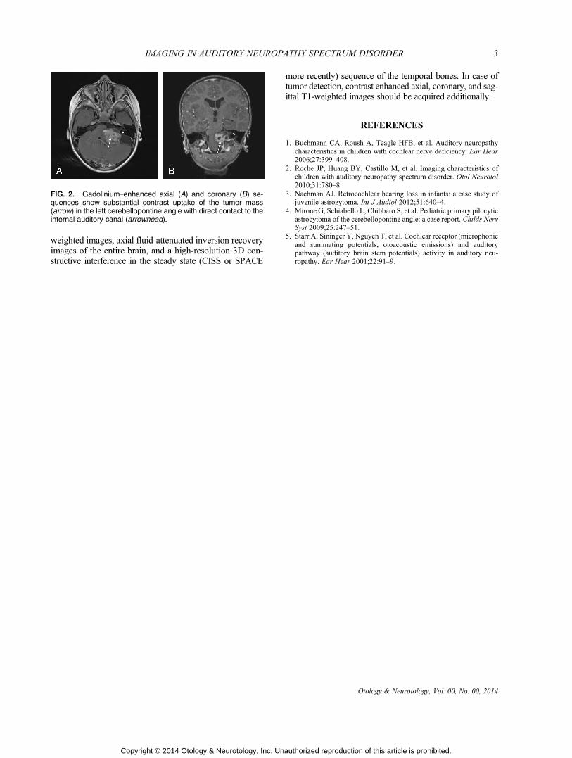
FIG. 2. GadoliniumYenhanced axial (A) and coronary (B) se-quences show substantial contrast uptake of the tumor mass(arrow) in the left cerebellopontine angle with direct contact to theinternal auditory canal (arrowhead).
3IMAGING IN AUDITORY NEUROPATHY SPECTRUM DISORDER
Otology & Neurotology, Vol. 00, No. 00, 2014
Copyright © 2014 Otology & Neurotology, Inc. Unauthorized reproduction of this article is prohibited.