image processing group profile 1994-2003...image processing group profile 1994-2003 image processing...
TRANSCRIPT

Faculty of Ele
Image Processing Group Profile
1994-2003
Image Processing Group ctrical Engineering and Computing, University of Zagreb

2
Published by: Image Processing Group Faculty of Electrical Engineering and Computing University of Zagreb, Croatia Publication date: May 2004 Copyright © by Image Processing Group
3
Contents
Contents _____________________________________________________ 3 About the Image Processing Group ________________________________ 5 A Short Biography of Prof. Sven Loncaric____________________________ 6 Image Processing Group Members ________________________________ 7
Faculty_____________________________________________________ 7 Graduate Assistants __________________________________________ 7 Graduate Students ___________________________________________ 7
Teaching Activities _____________________________________________ 8 Graduate Theses_____________________________________________ 9 Undergraduate Theses ________________________________________ 9
Research Activities ____________________________________________ 11 National Research Partners ___________________________________ 12 International Research Partners ________________________________ 12
Research and Development Projects ______________________________ 13 3-D Quantitative Analysis of Intracerebral Brain Hemorrhage__________ 14 Virtual Bronchoscopy_________________________________________ 15 WWW-based Virtual Endoscopy ________________________________ 16 Quantitative Analysis of Abdominal Aortic Aneurysm ________________ 17 Brain Perfusion Analysis ______________________________________ 18 Virtual Reality Markup Language Reference Center _________________ 19 Virtual Reality Reference Center________________________________ 20 Medical Image Segmentation Techniques ________________________ 21 Content-Based Image Retrieval ________________________________ 22 Cardiac Image Analysis_______________________________________ 23 A Web-Based Medical Teleconsultation System____________________ 24 Volume Visualization Software Modules __________________________ 25 Quantitative Analysis of SPECT Brain Images _____________________ 26 Image Management Techniques________________________________ 27 Intelligent Systems For Medical Image Analysis ____________________ 28 Deformable Models For Medical Image Analysis ___________________ 29 Neural Networks For Medical Image Analysis______________________ 30 Energy Minimization Techniques For Medical Image Analysis _________ 31
Image Processing Group Faculty of Electrical Engineering and Computing, University of Zagreb
Image Registration Methods ___________________________________ 32
4
Shape Description Methods ___________________________________ 33 Face Recognition Techniques__________________________________ 34 SVD-Based Techniques For Image Restoration ____________________ 35 Web-Based Electronic Journal Publishing Support System ___________ 36 Neural Networks For Financial Market Analysis ____________________ 37 CHAIRMAN Conference Management System_____________________ 38 COST B21 Project: Physiological Modelling of MR Image Formation____ 39 Biomedical Perfusion Image Sequence Analysis ___________________ 40 Serial Registration of Pre- and Intra-operative MR Brain Images _______ 41 Neural Networks for Ion Chromatography Optimization ______________ 42
Professional Activities __________________________________________ 43 Conference and Workshop Organization _________________________ 43 Invited Lectures _____________________________________________ 43 Journal Editorships __________________________________________ 44
Publications__________________________________________________ 45 Journal Papers _____________________________________________ 45 Books and Book Chapters_____________________________________ 45 Refereed Conference Papers, 2003 _____________________________ 46 Refereed Conference Papers, 2002 _____________________________ 46 Refereed Conference Papers, 2001 _____________________________ 46 Refereed Conference Papers, 2000 _____________________________ 47 Refereed Conference Papers, 1999 _____________________________ 48 Refereed Conference Papers, 1998 _____________________________ 49 Refereed Conference Papers, 1997 _____________________________ 50 Refereed Conference Papers, 1996 and older _____________________ 50
5
About the Image Processing Group
Image Processing Group (IPG) at the Department of Electronic Systems and Information Processing (ESIP) was founded in 1994 by Dr. Sven Loncaric. ESIP is one of the twelve departments which constitute the Faculty of Electrical Engineering and Computing at University of Zagreb. Faculty of Electrical Engineering and Computing is one of the most prestigious scientific institutions in Croatia. University of Zagreb has been founded in 1669 and has about 50,000 undergraduate and graduate students. Faculty of Electrical Engineering and Computing has about 100 permanent faculty members in all areas of electrical and computer engineering and computer science. Information about the IPG is available on the IPG web site:
http://ipg.zesoi.fer.hr For further information about the IPG activities please contact:
Professor Sven Loncaric Faculty of Electrical Engineering and Computing University of Zagreb Unska 3 10000 Zagreb Croatia Tel: +385-1-6129-891, +385-1-6129-911 (administrative assistant) Fax: +385-1-6129-652
Dr. Loncaric can also be reached at his e-mail address:
Image Processing Group Faculty of Electrical Engineering and Computing, University of Zagreb
6
A Short Biography of Prof. Sven Loncaric
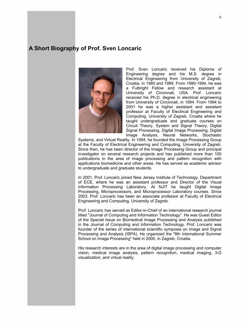
Prof. Sven Loncaric received his Diploma of Engineering degree and his M.S. degree in Electrical Engineering from University of Zagreb, Croatia, in 1985 and 1989. From 1990-1994, he was a Fulbright Fellow and research assistant at University of Cincinnati, USA. Prof. Loncaric received his Ph.D. degree in electrical engineering from University of Cincinnati, in 1994. From 1994 to 2001 he was a higher assistant and assistant professor at Faculty of Electrical Engineering and Computing, University of Zagreb, Croatia where he taught undergraduate and graduate courses on Circuit Theory, System and Signal Theory, Digital Signal Processing, Digital Image Processing, Digital Image Analysis, Neural Networks, Stochastic
Systems, and Virtual Reality. In 1994, he founded the Image Processing Group at the Faculty of Electrical Engineering and Computing, University of Zagreb. Since then, he has been director of the Image Processing Group and principal investigator on several research projects and has published more than 100 publications in the area of image processing and pattern recognition with applications biomedicine and other areas. He has served as academic advisor to undergraduate and graduate students. In 2001, Prof. Loncaric joined New Jersey Institute of Technology, Department of ECE, where he was an assistant professor and Director of the Visual Information Processing Laboratory. At NJIT he taught Digital Image Processing, Microprocessors, and Microprocessor Laboratory courses. Since 2003, Prof. Loncaric has been an associate professor at Faculty of Electrical Engineering and Computing, University of Zagreb. Prof. Loncaric has served as Editor-in-Chief of an international research journal titled "Journal of Computing and Information Technology". He was Guest Editor of the Special Issue on Biomedical Image Processing and Analysis published in the Journal of Computing and Information Technology. Prof. Loncaric was founder of the series of international scientific symposia on Image and Signal Processing and Analysis (ISPA). He organized the "8th International Summer School on Image Processing" held in 2000, in Zagreb, Croatia. His research interests are in the area of digital image processing and computer vision, medical image analysis, pattern recognition, medical imaging, 3-D visualization, and virtual reality.

7
Image Processing Group Members Research and development activities of the group have been carried out by a number of former and current graduate and undergraduate students. In his role as academic advisor, Dr. Loncaric has had opportunity to work with many excellent students, some of which are shown below. For a list of former undergraduate and graduate students please see IPG web site at: http://ipg.zesoi.fer.hr.
Faculty Sven Loncaric, director
Graduate Assistants Hrvoje Bogunovic, research and teaching assistant Tomislav Petkovic, research and teaching assistant Marko Subasic, research and teaching assistant
Graduate Students Hrvoje Regelja, graduate student Ivan Stajduhar, graduate student Adrijan Bozinovski, graduate student Sasa Galic, graduate student
Image Processing Group Faculty of Electrical Engineering and Computing, University of Zagreb

8
Teaching Activities
Prof. Sven Loncaric teaches undergraduate and graduate courses at Faculty of Electrical Engineering and Computing (FEEC), University of Zagreb. He is a member of the “Networks, Signals, and Systems” teaching group at FEEC. The group has seven teachers who teach courses in the area of signal and system theory. Prof. Loncaric has taught the following undergraduate courses:
• Systems and Signals • Linear Networks • Digital Signal Processing • Digital Image Processing • Neural Networks • Stochastic Processes in System Theory
The graduate courses currently taught by prof. Loncaric at FEEC are:
• Selected Topics in Image Processing • Digital Image Analysis • Neural Networks
At New Jersey Institute of Technology, USA, Dr. Loncaric taught "Digital Image Processing", "Microprocessors", and "Microprocessor Laboratory" courses. At Faculty of Mathematics, University of Zagreb, Prof. Loncaric offers a graduate course titled "Mathematical Methods in Image Processing". As part of international teaching activities Dr. Loncaric has been junior lecturer at "3rd IEEE EMBS Int'l Summer School on Biomedical Imaging", Berder, France, 1998. He has been invited lecturer for several years at "Int'l Summer School on Image Processing" held in Hungary and Croatia. Lecture notes and other information for all courses taught is available at the IPG web site: http://ipg.zesoi.fer.hr. Graduate assistants working on their graduate theses under prof. Loncaric's mentorship participate in teaching activities as teaching assistants for Digital Image Processing Laboratory and Stochastic Systems Laboratory. Teaching assistants also give tutoring sessions in several courses taught by prof. Loncaric. The list of graduate and undergraduate students mentored by Prof. Loncaric and titles of their theses are given in the following text.
9
Graduate Theses
Marko Subasic, "Geometric Deformable Models for Medical Image Analysis", 2003, MS Thesis
Srisheela Devabhaktuni, "Elastic Brain Image Registration using Mutual Information", 2003, MS Thesis
Domagoj Kovacevic, "Deformable Models for Medical Image Analysis", 2002, MS Thesis
Miroslav Koncar, "Distributed Information Systems for Electronic Health Record Management", 2002, MS Thesis
Ivan Vrancic, "Pattern Recognition for Financial Data Analysis", 2002, MS Thesis
Zoran Biuk, "Face Recognition from Multiple Views", 2001, MS Thesis
Zdravko Liposcak, "Face Recognition from Image Sequences", 2000, MS Thesis
Zeljko Devcic, "System identification methods for image degradation modeling", 1998, MS Thesis
Dubravko Cosic, "CT Head Image Analysis", 1997, MS Thesis
Zoran Dukic, "Shape Analysis using Morphological Signature Transform", 1996, MS Thesis
Undergraduate Theses
Tihomir Konosić, "Graphical User Interface for Volume Registration and Visualization", 2003 Marko Dvecko, "Volume Visualization Algorithms", 2003 Andrej Dolmac, "Volume Visualization Algorithms", 2003 Tomislav Petkovic, "Methods for Visual Quality Control of Plastic Products",
2002 Marijana Grgek, "Quantitative Analysis of 3-D MR Brain Images for Volume
Measurements", 2001 Ivan Stojic, "Spatio-temporal Segmentation using Simulated Annealing", 2001 Edgar Pek, "Telemedical System for Internet-based Consultations", 2001 Josip Krapac, "Computer Vision for Automated Quality Control", 2001 Hrvoje Bogunovic, "Segmentation from Image Sequences", 2001 Zeljko Tandaric, "Content-based Image Retrieval", 2001 Sinisa Skrinjar, "System for Automatic Generation of WML Pages", 2001 Ivan Ceskovic, "Quantitative Analysis of SPECT Images", 2001 Milan Matesin, "Intelligent System for Quantitative analysis of CT Images",
2001 Berislav Crkvenac, "Content-based Image Retrieval", 2000
Image Processing Group Faculty of Electrical Engineering and Computing, University of Zagreb
Dejan Marcetic, "Program for 3-D image visualization", 2000
10
Darije Ramljak, "3-D volume rendering visualization methods", 2000 Tomislav Markovinovic, "2-D and 3-D object thinning methods", 1999 Marko Subasic, "3-D stereo visualization using VTK", 1999 Tomislav Petrovic, "Neural networks for image analysis", 1999 Gordana Magdic, "ANSI Z39.50 standard for information servers", 1999 Zoran Majcenic, "Neural networks for medical image segmentation", 1999 Sasa Galic, "Optical flow techniques for cardiac imaging", 1999 Miroslav Koncar, "DICOM standard for radiological PACS", 1999 Boris Svilicic, "User interface for radiological PACS", 1999 Kresimir Prpa, "Image reconstruction from projections", 1999 Ivica Borscak, "Fractal image compression", 1999 Ivan Jakab, "Digital document processing and archiving", 1998 Damir Boraska, "Web-based electronic journal publishing system", 1998 Sasa Gavrilovic, "Web-based bibliographical information system", 1998 Jasmina Delic, "Databases for library information system", 1998 Kristina Peric, "A user interface for bibliographical information system", 1998 Ozren Rajkovic, "Marching cube algorithm for volume visualization", 1997 Ivan Vrancic, "Software system for network access to library information
databases", 1997 Domagoj Kovacevic, "Neural network segmentation of CT head images", 1996 Damir Cosic, "Volume vizualization methods", 1996 Damir Lisica, "Implementation of distance transform algorithms in Khoros
environment", 1995 Goran Dokmanovic, "Multimedia software for geographical information
systems", 1995 Kresimir Simunic, "Genetic algorithm-based image registration", 1995 Djordje Vukdragovic, "Energy minimization approaches for image
segmentation", 1995 Goran Polonji, "An implementation of Hough transform algorithm", 1995 Miro Zdilar, "Percepton neural network for pattern recognition", 1995
11
Research Activities
Image Processing Group brings together general expertise in the area of electronic systems and computer science and engineering. The primary emphasize in research is given to image processing, pattern recognition, computer vision, and medical imaging. Other areas of interest include 3-D visualization using virtual reality, and volume rendering methods. Computer vision and image processing have applications in many areas of human activity such as industry and medicine. The common task in all applications is a need for an image processing, e.g. to remove noise or restore image, followed by an image analysis in order to recognize certain parts of the scene and then perform image-based measurements or visualization of desired structures. Research areas in image processing and computer vision include techniques for image filtering, image enhancement, image restoration, image segmentation, image labeling, image understanding. A number of techniques is used in building the solutions for various computer vision problems including neural networks, genetic algorithms, expert systems, physics-based deformable models, clustering algorithms, optimization algorithms, and mathematical morphology. Biomedical image processing and analysis has been one of the main IPG research topics. Modern medical imaging modalities provide medical doctors with huge amount of information that requires quantitative analysis for diagnostic and interventional medical purposes. The image data can be two-dimensional (2-D) or three-dimensional (3-D) and can be a static image or an image sequence. 2-D or 3-D image data is typically obtained by means of a magnetic resonance (MR) or a computed tomography (CT) scanner, or by an ultrasound scanner. The structure of interest has to be extracted using pattern recognition methods. The obtained structure can be measured and visualized for use by a medical doctor. Modern medical imaging modalities use DICOM standard to enable digital exchange of image data. Image management techniques are used to store, retrieve, and display image data and integrate this information into radiological and other hospital information systems.
Image Processing Group Faculty of Electrical Engineering and Computing, University of Zagreb
12
National Research Partners
Croatian Ministry of Science and Technology
Croatian Academic and Research Network
Department of Radiology, Sestre Milosrdnice University Hospital, University of Zagreb, Croatia
Institute for Brain Research, Faculty of Medicine, University of Zagreb, Croatia
International Research Partners
Department of Neurosurgery, University of Medicine and Dentistry of New Jersey, USA
Computer Vision Laboratory, University of Ljubljana, Slovenia
Department of Electrical and Computer Engineering, New Jersey Institute of Technology, USA
Department of Electrical and Computer Engineering, University of Iowa, USA
Department of Applied Informatics, University of Szeged, Hungary
Department of Radiology, University Hospital Graz, Austria

13
Research and Development Projects
IPG topics of interest include all aspects of image processing and analysis. This includes theoretical basis and practical applications. Diverse areas such as neural networks, genetic algorithms, intelligent systems, mathematical morphology, and other are investigated and applied to problems in image processing and computer vision. A special research focus has been in medical image processing and analysis. A variety of contemporary medical imaging instruments provide physicians with a wealth of two-dimensional (2-D) and three-dimensional (3-D) image data. Interpretation of this data in everyday clinical practice is very important for medical diagnostics and intervention. Image interpretation represents a challenging task due to a large amount of acquired data. Intelligent medical image analysis is required for such quantitative image measurements. Volume visualization has played an important role in 3-D medical image processing and analysis. Volume rendering has gained a lot of popularity in recent years because of a realistic visualization of 3-D data. Virtual reality techniques and stereo visualization has enabled interactive fly-through visualizations of various body structures and organs and have been used both in education and diagnostics. In the following material, research activities of the IPG are presented with basic information about each research project. At the end of this report, the list of publications produced by IPG researchers has been provided.
Image Processing Group Faculty of Electrical Engineering and Computing, University of Zagreb
14
3-D Quantitative Analysis of Intracerebral Brain Hemorrhage
Supported by: Ministry of Science and Technology, Croatia National Institutes of Health, USA
Duration: May 1995-May 2001
People: Sven Loncaric, University of Zagreb, Croatia, principal co-investigator
Atam P. Dhawan, University of Cincinnati, USA, principal co-investigator
Dubravko Cosic, University of Zagreb, graduate student Domagoj Kovacevic, University of Zagreb, graduate student
Project description: Computed tomography (CT) allows three-dimensional (3-D) anatomical imaging of brain abnormalities such as human spontaneous intracerebral brain hemorrhage (ICH). With computerized image analysis, it is feasible to characterize the pathology of a selected volume of interest. The proposed research focuses on 3-D quantitative analysis to study the early evolution of the ICH. The underlying hypothesis is that the ICH volume and structure is related to the mortality and morbidity. Patients having ICH are scanned four times: within three hours after first symptoms, one hour later, eight hours later, and within twenty hours after first symptoms. During the course of the illness, 3-D changes in ICH volume and structure can be observed and analyzed. The important ICH features are volume, position in space, and shape of primary and edema region. Preliminary studies indicate that the ICH volume is significant for the survival of the patient. The position in space must be measured with respect to an invariant 3-D coordinate system so that the movement of the ICH across the scans can be determined. We have developed 3-D spatially weighted region growing algorithms with adaptive clustering for segmentation of ICH regions.
Publications:
D. Cosic and S. Loncaric. Computer System for Quantitative Analysis of ICH from CT Head Images. Proceedings of the 19th Annual International Conference of IEEE Engineering in Medicine and Biology Society, Chicago, USA, 1997.
S. Loncaric and D. Kovacevic. A Method for Segmentation of CT Head Images. Proceedings of the 9th International Conference on Image Analysis and Processing, pp. 388-395, Florence, Italy, 1997.
D. Cosic and S. Loncaric. A Rule-based Labeling of CT Head Image. Proceedings of the 6th Conference on Artificial Intelligence in Medicine Europe, Grenoble, France, Lecture Notes in Artificial Intelligence, pp. 453-456, Springer-Verlag, 1997.
S. Loncaric and D. Cosic and A. P. Dhawan. Hierarchical Segmentation of CT Head Images. Conference Proceedings of the 18th Annual International Conference of IEEE Engineering in Medicine and Biology Society, Amsterdam, Netherlands, 1996.
S. Loncaric and D. Cosic and A. P. Dhawan. Segmentation of CT Head Images. Computer Assisted Radiology '96 – Proc. of the International Symposium on Computer and Communication Systems for Image Guided Diagnosis and Therapy, pp. 1012-1012, Paris, France, 1996.
15
Virtual Bronchoscopy
Supported by: Central European Exchange Program for University Studies (CEEPUS)
Duration:
September 1997 – August 1998
People: Sven Loncaric, University of Zagreb, Croatia, principal co-investigator
Erich Sorantin, Karl-Franzens University of Graz, Austria, principal co-investigator
Attila Kuba, University of Szeged, Hungary, principal co-investigator
Tomislav Markovinovic, University of Zagreb, Croatia, undergraduate student
Tomislav Petrovic, University of Zagreb, Croatia, undergraduate student
Darije Ramljak, University of Zagreb, Croatia, undergraduate student
Project description: Virtual bronchoscopy is a visualization technique based on
computed tomography (CT) or magnetic resonance (MR) scanning of the organ of interest and extracting its 3-D model. The 3-D model can be interactively examined or fly-through visualizations along a path through the structure can be generated by volume or surface rendering. Such an approach is called virtual endoscopy and it has several advantages over conventional endoscopy where an instrument is inserted through a natural or minimally invasive opening on the human body. The advantages of virtual endoscopy are non-invasiveness of the technique and ability to move through structures where physical endoscope cannot enter. The main disadvantage of virtual endoscopy is that the color and the texture of organ surface cannot be restored from scanned images. The aim of the project is to realize the software application for virtual bronchoscopy i.e. for virtual endoscopy of the bronchial tubes. The input images are acquired by CT scanning. Image analysis must be conducted to determine the location of the bronchial tubes and to extract them. The goal of the procedure is to create a fly-through animation along the medial axis (skeleton) of the bronchial tubes. The 3-D skeleton is computed from segmented volume. The animation is created by volume rendering along the computed medial axis.
Publications:
S. Loncaric and T. Markovinovic and T. Petrovic and D. Ramljak and E. Sorantin. Construction of Virtual Environment for Endoscopy. Proceedings of the IEEE International Conference on Multimedia Computing and Systems, Florence, Italy, 1999.
S. Loncaric and D. Ramljak and T. Markovinovic and E. Sorantin. VRML-based Model Creation for Virtual Bronchoscopy. First Croatian Symposium on Computer Assisted Surgery, Zagreb, Croatia, 1999.
S. Loncaric. Virtual Reality in Medicine: An Overview. First Croatian Symposium on Computer Assisted Surgery, Zagreb, Croatia, 1999.
Image Processing Group Faculty of Electrical Engineering and Computing, University of Zagreb
16
WWW-based Virtual Endoscopy
Supported by: Central European Exchange Program for University Studies (CEEPUS)
Duration:
September 1998 – August 1999
People: Sven Loncaric, University of Zagreb, Croatia, principal co-investigator
Erich Sorantin, Karl-Franzens University of Graz, Austria, principal co-investigator
Attila Kuba, University of Szeged, Hungary, principal co-investigator
Tomislav Markovinovic, University of Zagreb, Croatia, undergraduate student
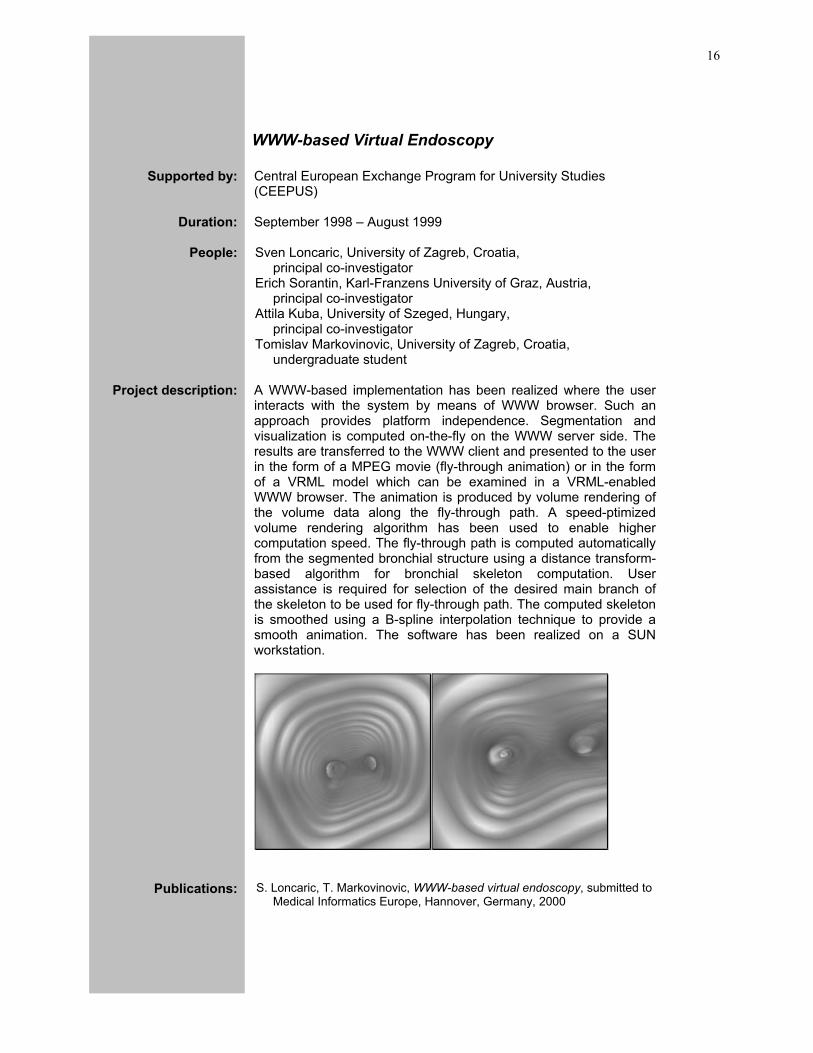
Project description: A WWW-based implementation has been realized where the user
interacts with the system by means of WWW browser. Such an approach provides platform independence. Segmentation and visualization is computed on-the-fly on the WWW server side. The results are transferred to the WWW client and presented to the user in the form of a MPEG movie (fly-through animation) or in the form of a VRML model which can be examined in a VRML-enabled WWW browser. The animation is produced by volume rendering of the volume data along the fly-through path. A speed-ptimized volume rendering algorithm has been used to enable higher computation speed. The fly-through path is computed automatically from the segmented bronchial structure using a distance transform-based algorithm for bronchial skeleton computation. User assistance is required for selection of the desired main branch of the skeleton to be used for fly-through path. The computed skeleton is smoothed using a B-spline interpolation technique to provide a smooth animation. The software has been realized on a SUN workstation.
Publications:
S. Loncaric, T. Markovinovic, WWW-based virtual endoscopy, submitted to Medical Informatics Europe, Hannover, Germany, 2000

17
Quantitative Analysis of Abdominal Aortic Aneurysm
Supported by: Central European Exchange Program for University Studies (CEEPUS)
Duration:
September 1999 – August 2000
People: Sven Loncaric, University of Zagreb, Croatia, principal co-investigator
Erich Sorantin, Karl-Franzens University of Graz, Austria, principal co-investigator
Attila Kuba, University of Szeged, Hungary, principal co-investigator
Domagoj Kovacevic, University of Zagreb, graduate student
Marko Subasic, University of Zagreb, graduate student
Project description: Abdominal aortic aneurysm (AAA) is a vascular disease which can
be treated by open surgical procedure or by a minimally invasive surgical procedure. In order to correctly select the size and shape of the stent graft device it is necessary to perform accurate measurements of the aneurysm. This can be done by CT or MR scanning. In this project a spiral CT scanner has been used for acquiring the chest images containing the aneurysm. Deformable models have been used for 3-D segmentation of AAA. In the first approach, a classical snake algorithm has been used in 2-D slices with additional forces acting between the slices. In the second approach, a level-set algorithm, according to Sethian, has been used for implementation of deformable model and its evaluation. The software has been implemented in MATLAB and C languages.
Publications:
S. Loncaric and D. Kovacevic and E. Sorantin. Semi-automatic Active Contour Approach to Segmentation of Computed Tomography Volumes. Proceedings of SPIE Medical Imaging, San Diego, USA, 2000
M. Subasic and S. Loncaric. 3-D deformable model for aneurysm segmentation, submitted to Medical Informatics Europe conference, Hannover, Germany, 2000
D. Kovacevic and S. Loncaric and E. Sorantin. Deformable Models for Medical Image Segmentation. First Croatian Symposium on Computer Assisted Surgery, Zagreb, Croatia, 1999.
Image Processing Group Faculty of Electrical Engineering and Computing, University of Zagreb
18
Brain Perfusion Analysis
Supported by: Central European Exchange Program for University Studies (CEEPUS)
Duration:
September 2000 – August 2001
People: Sven Loncaric, University of Zagreb, Croatia, principal co-investigator
Nada Besenski, University of Zagreb, Croatia, principal co-investigator
Erich Sorantin, Karl-Franzens University of Graz, Austria, principal co-investigator
Attila Kuba, University of Szeged, Hungary, principal co-investigator
Project description: The project is focused on quantitative image-based measurement of
diffusion and perfusion in brain ischemy. The brain images are acquired using magnetic resonance (MR) scanning. The analysis must be performed on single images as well as on image sequences to track time changes in patients with degenerative brain diseases. It is very important for diagnostic, therapeutic, and prospective reasons to determine the size and age of ischemia, and the size of penumbra in ischemic lesion. New MR imaging techniques such as diffusion weighted imaging and echoplanar perfusion enable such measurements. Image processing and analysis methods will be based on neural networks, expert systems, mathematical morphology, and other techniques. A clinical study will be conducted to determine the validity of the method.
19
Virtual Reality Markup Language Reference Center
Supported by: Croatian Academic and Research Network (CARNet)
Duration:
January 1996 – December 1997
People: Sven Loncaric, University of Zagreb, Croatia, principal investigator
Tomislav Markovinovic, University of Zagreb, Croatia, undergraduate student
Tomislav Petrovic, University of Zagreb, Croatia, undergraduate student
Darije Ramljak, University of Zagreb, Croatia, undergraduate student
Berislav Crkvenac, University of Zagreb, Croatia, undergraduate student
Project description: Recently, virtual reality has become an exciting research area with
many applications. Virtual Reality Modeling Language (VRML) is a quickly developing tool for description of 3-D objects which combines virtual reality technologies with network technologies. The aim of this project is to establish a referral center for VRML technology in Croatia. The project duration is one-year, starting in February 1997, and ending in February 1998. The project goals are:
• to establish a referent educational center for VRML technology
• to test hardware and software for virtual reality • to test VRML modelers and browsers on PC (Windows 95,
Windows NT, and Linux) and Sun Solaris platforms • to develop a model of Croatian territory in VRML format
Publications:
S. Loncaric and T. Markovinovic and T. Petrovic and D. Ramljak and E.
Sorantin. Construction of Virtual Environment for Endoscopy. Proceedings of the IEEE International Conference on Multimedia Computing and Systems, Florence, Italy, 1999.
S. Loncaric and D. Ramljak and T. Markovinovic and E. Sorantin. VRML-based Model Creation for Virtual Bronchoscopy. First Croatian Symposium on Computer Assisted Surgery, Zagreb, Croatia, 1999.
S. Loncaric. Virtual Reality in Medicine: An Overview. First Croatian Symposium on Computer Assisted Surgery, Zagreb, Croatia, 1999.
Image Processing Group Faculty of Electrical Engineering and Computing, University of Zagreb
20
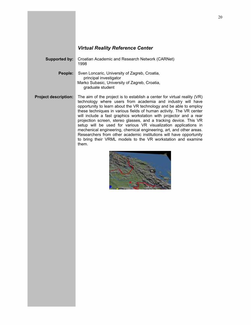
Virtual Reality Reference Center
Supported by: Croatian Academic and Research Network (CARNet) 1998
People: Sven Loncaric, University of Zagreb, Croatia, principal investigator
Marko Subasic, University of Zagreb, Croatia, graduate student
Project description: The aim of the project is to establish a center for virtual reality (VR)
technology where users from academia and industry will have opportunity to learn about the VR technology and be able to employ these techniques in various fields of human activity. The VR center will include a fast graphics workstation with projector and a rear projection screen, stereo glasses, and a tracking device. This VR setup will be used for various VR visualization applications in mechenical engineering, chemical engineering, art, and other areas. Researchers from other academic institutions will have opportunity to bring their VRML models to the VR workstation and examine them.
21
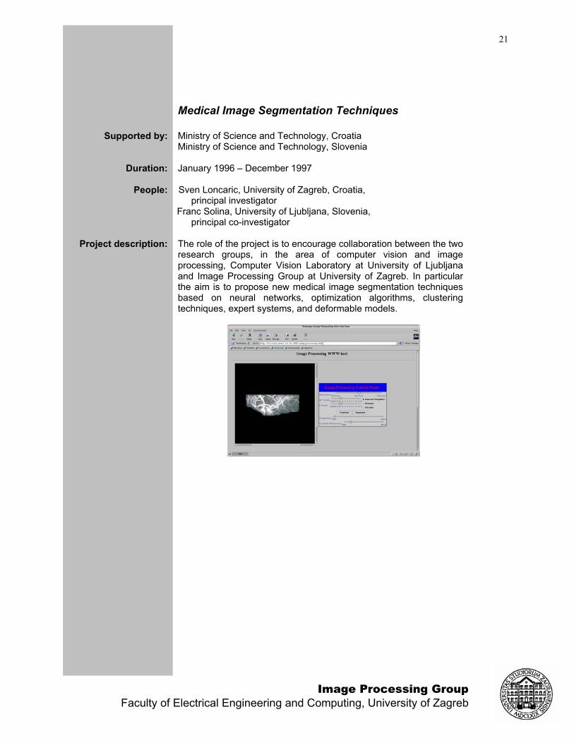
Medical Image Segmentation Techniques
Supported by: Ministry of Science and Technology, Croatia Ministry of Science and Technology, Slovenia
Duration:
January 1996 – December 1997
People: Sven Loncaric, University of Zagreb, Croatia, principal investigator
Franc Solina, University of Ljubljana, Slovenia, principal co-investigator
Project description: The role of the project is to encourage collaboration between the two
research groups, in the area of computer vision and image processing, Computer Vision Laboratory at University of Ljubljana and Image Processing Group at University of Zagreb. In particular the aim is to propose new medical image segmentation techniques based on neural networks, optimization algorithms, clustering techniques, expert systems, and deformable models.
Image Processing Group Faculty of Electrical Engineering and Computing, University of Zagreb
22
Content-Based Image Retrieval
Supported by: Pilot project
People: Sven Loncaric, University of Zagreb, Croatia, coordinator
Berislav Crkvenac, University of Zagreb, Croatia, undergraduate student
Zeljko Tandaric, University of Zagreb, Croatia, undergraduate student
Project description: Content-based image retrieval has become very important,
particularly in recent years with the growth of Internet and a huge number of images on the web sites. This fact demonstrates the need for tools for searching the Internet for images by content.Tools for indexing and retrieving the textual information from the Internet have been developed but development of content-based image retrieval have not been well researched. Tools for content-based image retrieval typically use color, shape, and texture features to classify images and define criteria of similarity. The difficulty is in defining a query image. In this project, we propose to develop a neural network based method for training the system to learn the similarity criteria for each particular application. Once the neural network is trained it is used for content-based search by comparing each image from the image database with query (example) image. The method will be tested on a class of medical images.
23
Cardiac Image Analysis
Supported by: Pilot project
People: Sven Loncaric, University of Zagreb, Croatia, principal co-investigator
James Duncan, Yale University, USA, principal co-investigator
Sasa Galic, University of Zagreb, Croatia, graduate student
Tvrtko Macan, University of Zagreb, Croatia, graduate student
Zoran Majcenic, University of Zagreb, Croatia, graduate student
Marko Subasic, University of Zagreb, Croatia, graduate student
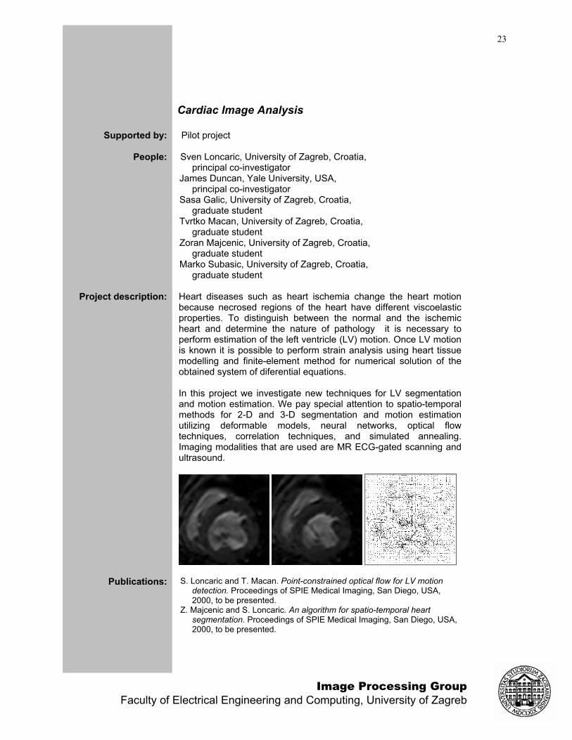
Project description: Heart diseases such as heart ischemia change the heart motion
because necrosed regions of the heart have different viscoelastic properties. To distinguish between the normal and the ischemic heart and determine the nature of pathology it is necessary to perform estimation of the left ventricle (LV) motion. Once LV motion is known it is possible to perform strain analysis using heart tissue modelling and finite-element method for numerical solution of the obtained system of diferential equations. In this project we investigate new techniques for LV segmentation and motion estimation. We pay special attention to spatio-temporal methods for 2-D and 3-D segmentation and motion estimation utilizing deformable models, neural networks, optical flow techniques, correlation techniques, and simulated annealing. Imaging modalities that are used are MR ECG-gated scanning and ultrasound.
Publications:
S. Loncaric and T. Macan. Point-constrained optical flow for LV motion detection. Proceedings of SPIE Medical Imaging, San Diego, USA, 2000, to be presented.
Z. Majcenic and S. Loncaric. An algorithm for spatio-temporal heart segmentation. Proceedings of SPIE Medical Imaging, San Diego, USA, 2000, to be presented.
Image Processing Group Faculty of Electrical Engineering and Computing, University of Zagreb
24
A Web-Based Medical Teleconsultation System
Supported by: Ministry of Science and Technology (An IT project)
People: Sven Loncaric, University of Zagreb, Croatia, principal co-investigator
Anamarija Margan, Cres, Croatia principal co-investigator
Miroslav Silovic, University of Zagreb graduate student
Edgar Pek, University of Zagreb undergraduate student
Project description: Medical consultations between ambulances in rural areas and
large medical centers are necessary for high quality health care. In particular, medical consultations between Croatian islands of Cres and Losinj and mainland medical centers in Rijeka and Zagreb are necessary for several reasons. Often, patient transport is not acceptable due to a high risk for the patient. In addition, transport generates additional expense and is often impossible in winter months due to weather and sea conditions, not to mention discomfort to the patient. In this paper, we describe a concept of a web-based system for medical teleconsultation. The system will be applied to communication between the islands of Cres, Losinj, and neighboring islands and university hospitals in Zagreb and Rijeka. The system consists of a teleconsultation module and a electronic Internet-based health record module. User interface is implemented in HTML and Phyton languages. WWW access to the system ensures platform independence, i.e. the system can be accessed using an ordinary Internet browser from any computer platform. The system is currently under development.
Publications: S. Loncaric and E. Pek and M. Silovic and A. Margan. Virtual Polyclinic: A Web-based system for medical teleconsultation. Proceedings of Emerging Technologies and Life Sciences: Medicine and Communication (MEDICOM), Remagen, Germany, 2000.
S. Loncaric and M. Silovic and E. Pek and A. Margan. A Web-based system for medical teleconsultation: First results. Proceedings of Telemedicine and Health Informatics, Milano, Italy, 2000.
25
Volume Visualization Software Modules
Supported by: Ministry of Science and Technology, Croatia VAMS
Duration:
October 1997-
People: Sven Loncaric, University of Zagreb, Croatia, coordinator
Josip Krapac, University of Zagreb, Croatia, undergraduate student
Dejan Marcetic, University of Zagreb, Croatia, undergraduate student
Darije Ramljak, University of Zagreb, Croatia, graduate student
Project description: The goal of this project is to develop software modules in the form
of dynamic libraries (DLLs) for Windows environment for use from a teleradiological application. The purpose of these modules is visualization of 3-D data such as computed tomography (CT) and magnetic resonance (MR) scans. The software modules must enable several visualization types. The first and the simpliest type is multi-planar visualization showing to the radiologist axial, coronal, and sagital views of the volume. The user must be able to navigate in all three planes and interpolation must be implemented to increase the resolution in Z direction. The second type of visualization is using a VRML surface model of the organ of interest. This type of visualization enables interactive examination of the volume of interest by means of a VRML viewer. The user can navigate through the volume in real time. The third type of visualization uses volume rendering for the best visualization of volume. The price to be paid for this are increased computational requirements and resulting lower speed. The user can specify a 3-D path for fly-through animation and the module will compute the rendered frames along the path and create a MPEG movie. The software modules will be integrated with the main program and tested by users. Graphical user interfaces for each module will be developed to enable effective communication with the software with minimal amount of interaction.
Publications: D. Cosic and S. Loncaric and O. Rajkovic. A Method for 3-D Image Representation Using a Surface Model. Proceedings of the IEEE 9th Mediterranean Electrotechnical Conference, pp. 82-85, Tel-Aviv, Israel, 1998.
Image Processing Group Faculty of Electrical Engineering and Computing, University of Zagreb
26
Quantitative Analysis of SPECT Brain Images
Supported by: pilot project
Duration:
October 1999-
People: Sven Loncaric, University of Zagreb, Croatia, coordinator
Srecko Loncaric, Faculty of Medicine, University Hospital Rebro, coordinator
Ivan Ceskovic, University of Zagreb, Croatia, undergraduate student
Project description: Single Photon Emission Computed Tomography (SPECT) medical
imaging modality has been used for functional imaging of various organs in the human body including brain. Functional imaging provides information about the function of an organ as opposed to modalities that provide anatomical information about the structures such as computed tomography (CT). In emision tomography a radioactive contrast medium is injected into the patient and radiation that is emitted as a result of radioactive decay is detected by a camera. A wide spectrum of different contrast media exists that are used for different organs and that reveal various aspects of the organ function. In SPECT, the 3-D organ is reconstructed from multiple projections to provide spatial information about the intensity of activity in different parts of the organ. SPECT imaging is used in studies of tumors, thyroid glands, and other organs. A disadvantage of SPECT with respect to CT is reduced resolution what makes image processing procedures more difficult. The goal of this project is to develop image processing and analysis algorithms and procedures for measurements of tumor size. The procedures must enable temporal studies to track tumor changes in time and evaluate the effect of therapy to the patient. The acquired images are obtained from the SPECT scanner in digital form by means of DICOM standard. The program will be developed for Windows PC environment and for UNIX environment.
27
Image Management Techniques
Supported by: pilot project
Duration:
October 1999-
People: Sven Loncaric, University of Zagreb, Croatia, coordinator
Miroslav Koncar, University of Zagreb, Croatia, graduate student
Dzumridin Ibraimi, University of Zagreb, undergraduate student
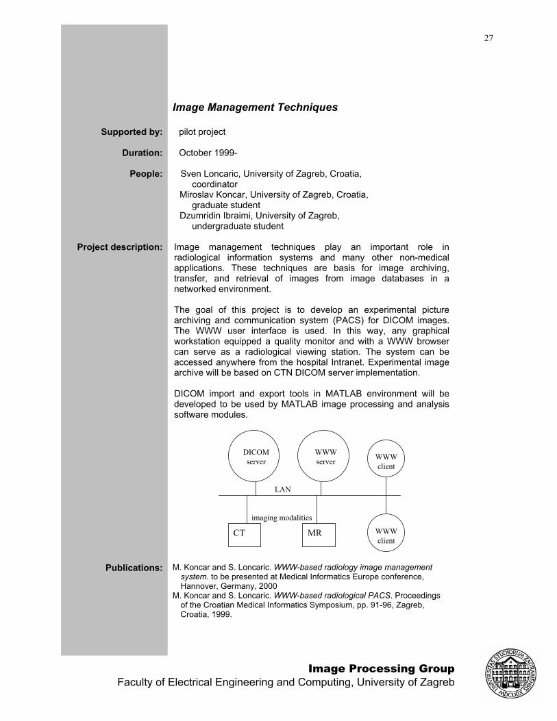
Project description: Image management techniques play an important role in
radiological information systems and many other non-medical applications. These techniques are basis for image archiving, transfer, and retrieval of images from image databases in a networked environment. The goal of this project is to develop an experimental picture archiving and communication system (PACS) for DICOM images. The WWW user interface is used. In this way, any graphical workstation equipped a quality monitor and with a WWW browser can serve as a radiological viewing station. The system can be accessed anywhere from the hospital Intranet. Experimental image archive will be based on CTN DICOM server implementation. DICOM import and export tools in MATLAB environment will be developed to be used by MATLAB image processing and analysis software modules.
DICOMserver
WWWserver
LAN
CT MR
imaging modalities
WWWclient
WWWclient
Publications:
M. Koncar and S. Loncaric. WWW-based radiology image management system. to be presented at Medical Informatics Europe conference, Hannover, Germany, 2000
M. Koncar and S. Loncaric. WWW-based radiological PACS. Proceedings of the Croatian Medical Informatics Symposium, pp. 91-96, Zagreb, Croatia, 1999.
Image Processing Group Faculty of Electrical Engineering and Computing, University of Zagreb
28
Intelligent Systems For Medical Image Analysis
Supported by: pilot project
Duration:
October 1999-
People: Sven Loncaric, University of Zagreb, Croatia, coordinator
Domagoj Kovacevic, University of Zagreb, Croatia, graduate student
Milan Matesin, University of Zagreb, Croatia, undergraduate student
Project description: Image interpretation is a challenging task. The reason for this is that
for accurate image interpretation it is necessary segment the scene into its parts and to perform recognition of objects in the scene. This task requires intelligence to be successfully performed. Implementation of intelligent procedures on a computer constitutes the field of artificial intelligence. In the area of artificial intelligence, a number of different methods have been studied. In particular expert systems have been used in many image understanding problems. A subgroup of expert systems are rule-based expert systems which are a useful tool for solution of specialized image understanding problems. In this project we will study intelligent systems that are useful for implementation of artificial image interpretation systems. After the initial image processing, the image is segmented into some uniform regions. A set of features is assigned to each region describing the properties of the region and its relation to the neighboring regions. Rules are formed to describe the knowledge about the structure of the image, i.e. what are the possible and meaningful configurations of regions. The task of rule-based expert system is to use input regions and rules to perform image labeling, i.e. to identify which region represents what part of the scene. Non-fuzzy and fuzzy rule-based expert systems will be investigated. The goal of the project is to develop a software for intelligent interpretation of medical images of particular class such as brain images, abdominal images, etc. The software will utilize the concepts of intelligent systems including rule-based expert systems and other techniques.
Publications: S. Loncaric and D. Kovacevic and D. Cosic. Fuzzy Expert System for Edema Segmentation. Proceedings of the IEEE 9th Mediterranean Electrotechnical Conference, pp. 1476-1479, Tel-Aviv, Israel, 1998.
D. Cosic and S. Loncaric. A Rule-based Labeling of CT Head Image. Proceedings of the 6th Conference on Artificial Intelligence in Medicine Europe, Grenoble, France, Lecture Notes in Artificial Intelligence, pp. 453-456, Springer-Verlag, 1997.

29
Deformable Models For Medical Image Analysis
Supported by: Central European Exchange Program for University Studies (CEEPUS)
Duration:
October 1998-October 2001
People: Sven Loncaric, University of Zagreb, Croatia, coordinator
Erich Sorantin, Karl-Franzens University Graz, Austria, coordinator
Marijana Grgek, University of Zagreb, Croatia, undergraduate student
Domagoj Kovacevic, University of Zagreb, Croatia, undergraduate student
Marko Subasic, University of Zagreb, Croatia, graduate student
Project description: Physics-based deformable models are a powerful tool for
segmentation of 2-D and 3-D structures in medical image analysis. In this project we investigate medical image analysis techniques based on deformable models. In deformable models, a model of shape border or surface is deformed under the influence of external and internal forces acting on the model. The deformation process is governed by the motion partial differential equations which have to be solved in orer to determine the dynamics of the model. In image analysis we are only interested in the final solution and not in the intermediate time steps. We investigate different implementations of the algorithm such as classical snake and baloon based approach and alternative level-set implementation of the algorithm. The goal of the project is to develop algorithms and software modules for specific problems in medical image analysis including segmentation of the brain ventricle, abdominal aortic aneurysm segmentation, and heart left ventricle segmentation.
Publications:
M. Subasic and S. Loncaric. 3-D deformable model for aneurysm segmentation. submitted to Medical Informatics Europe conference, Hannover, Germany, 2000
S. Loncaric and D. Kovacevic and E. Sorantin. Semi-automatic Active Contour Approach to Segmentation of Computed Tomography Volumes. Proceedings of SPIE Medical Imaging, San Diego, USA, 2000, to be presented.
D. Kovacevic and S. Loncaric. Deformable Contour-Based Method For Medical Image Segmentation. Proceedings of the 21st Int'l Conference Information Technology Interfaces, pp. 145-150, Pula, Croatia, 1999.
D. Kovacevic and S. Loncaric and E. Sorantin. Deformable Models for Medical Image Segmentation. First Croatian Symposium on Computer Assisted Surgery, Zagreb, Croatia, 1999.
Image Processing Group Faculty of Electrical Engineering and Computing, University of Zagreb
30
Neural Networks For Medical Image Analysis
Supported by: Ministry of Science and Technology, Croatia
Duration:
January 1996-
People: Sven Loncaric, University of Zagreb, Croatia, coordinator
Ivan Stojic, University of Zagreb, Croatia, undergraduate student
Domagoj Kovacevic, University of Zagreb, Croatia, undergraduate student
Project description: Segmentation of computed tomography (CT) images is an important
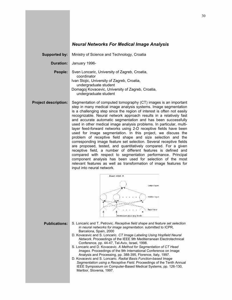
step in many medical image analysis systems. Image segmentation is a challenging step since the region of interest is often not easily recognizable. Neural network approach results in a relatively fast and accurate automatic segmentation and has been successfully used in other medical image analysis problems. In particular, multi-layer feed-forward networks using 2-D receptive fields have been used for image segmentation. In this project, we discuss the problem of receptive field shape and size selection and the corresponding image feature set selection. Several receptive fields are proposed, tested, and quantitatively compared. For a given receptive field, a number of different features is defined and compared with respect to segmentation performance. Principal component analysis has been used for selection of the most relevant features as well as transformation of image features for input into neural network.
Publications:
S. Loncaric and T. Petrovic. Receptive field shape and feature set selection in neural networks for image segmentation. submitted to ICPR, Barcelona, Spain, 2000
D. Kovacevic and S. Loncaric. CT Image Labeling Using Hopfield Neural Network. Proceedings of the IEEE 9th Mediterranean Electrotechnical Conference, pp. 44-47, Tel-Aviv, Israel, 1998.
S. Loncaric and D. Kovacevic. A Method for Segmentation of CT Head Images. Proceedings of the 9th International Conference on Image Analysis and Processing, pp. 388-395, Florence, Italy, 1997.
D. Kovacevic and S. Loncaric. Radial Basis Function-based Image Segmentation using a Receptive Field. Proceedings of the Tenth Annual IEEE Symposium on Computer-Based Medical Systems, pp. 126-130, Maribor, Slovenia, 1997.
31
Energy Minimization Techniques For Medical Image Analysis
Supported by: Ministry of Science and Technology, Croatia
Duration:
October 1997-
People: Sven Loncaric, University of Zagreb, Croatia, coordinator
Zoran Majcenic, University of Zagreb, Croatia, undergraduate student
Project description: Energy minimization techniques are a useful tool for many image
processing and analysis tasks. In this approach the problem is formulated in terms of an energy function which has to be minimized. The solution to this minimization problem represents the solution of the original image analysis problem. Using such an approach image analysis problems are reduced to energy function minimization problems. Function minimization problems can be solved using a number of numerical techniques. A major problem in such algorithms is an unwanted convergence to a one of the local minimas. To avoid this problem a number of stochastic techniques have been introduced which search the optimization space in stochastic manner to avoid the premature convergence to a local minimum. The goal of the project is to develop new and computationally efficient SA algorithms for 2-D and 3-D image analysis problems such as image segmentation and motion estimation. The algorithms have been applied to the problem of brain image segmentation, and heart motion estimation and segmentation. Multiresolution SA algorithms have been developed to reduce the complexity and speed-up computations.
Publications:
Z. Majcenic and S. Loncaric. An algorithm for spatio-temporal heart segmentation. Proceedings of SPIE Medical Imaging, San Diego, USA, 2000, to be presented.
S. Loncaric and Z. Majcenic. An efficient SA algorithm for brain hemorrhage image analysis. First Croatian Symposium on Computer Assisted Surgery, Zagreb, Croatia, 1999.
S. Loncaric and Z. Majcenic. Multiresolution simulated annealing for brain image analysis. Proceedings of SPIE Medical Imaging, Vol. 3661, pp. 1139-1146, San Diego, USA, 1999.
Z. Majcenic and S. Loncaric. CT image labeling using simulated annealing algorithm. Proceedings of the IX European Signal Processing Conference, Vol. 4, pp. 2513-2516, Island of Rhodos, Greece, 1998.
S. Loncaric and Z. Majcenic. Multiresolution CT Head Image Analysis using Simulated Annealing. Proceedings of the 20th Int’l Conference Information Technology Interfaces, pp. 257-262, Pula, Croatia, 1998.
S. Loncaric and Z. Majcenic. Multiresolution CT Head Image Analysis using Simulated Annealing. Proceedings of the 12th International Symposium Computer Assisted Radiology and Surgery, pp. 889-889, Tokyo, Japan, 1998.
Image Processing Group Faculty of Electrical Engineering and Computing, University of Zagreb
32
Image Registration Methods
Supported by: Ministry of Science and Technology, Croatia
Duration:
October 1997-
People: Sven Loncaric, University of Zagreb, Croatia, coordinator
Kresimir Simunic, University of Zagreb, Croatia, graduate student
Project description: Image registration or matching techniques are important in many
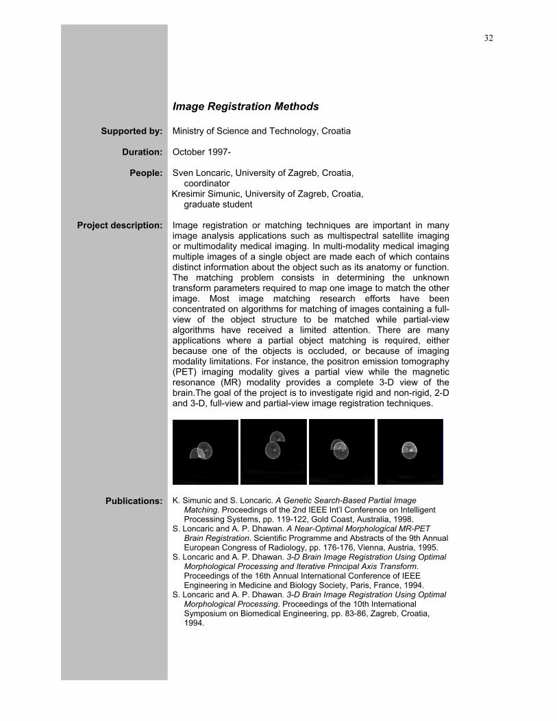
image analysis applications such as multispectral satellite imaging or multimodality medical imaging. In multi-modality medical imaging multiple images of a single object are made each of which contains distinct information about the object such as its anatomy or function. The matching problem consists in determining the unknown transform parameters required to map one image to match the other image. Most image matching research efforts have been concentrated on algorithms for matching of images containing a full-view of the object structure to be matched while partial-view algorithms have received a limited attention. There are many applications where a partial object matching is required, either because one of the objects is occluded, or because of imaging modality limitations. For instance, the positron emission tomography (PET) imaging modality gives a partial view while the magnetic resonance (MR) modality provides a complete 3-D view of the brain.The goal of the project is to investigate rigid and non-rigid, 2-D and 3-D, full-view and partial-view image registration techniques.
Publications:
K. Simunic and S. Loncaric. A Genetic Search-Based Partial Image Matching. Proceedings of the 2nd IEEE Int’l Conference on Intelligent Processing Systems, pp. 119-122, Gold Coast, Australia, 1998.
S. Loncaric and A. P. Dhawan. A Near-Optimal Morphological MR-PET Brain Registration. Scientific Programme and Abstracts of the 9th Annual European Congress of Radiology, pp. 176-176, Vienna, Austria, 1995.
S. Loncaric and A. P. Dhawan. 3-D Brain Image Registration Using Optimal Morphological Processing and Iterative Principal Axis Transform. Proceedings of the 16th Annual International Conference of IEEE Engineering in Medicine and Biology Society, Paris, France, 1994.
S. Loncaric and A. P. Dhawan. 3-D Brain Image Registration Using Optimal Morphological Processing. Proceedings of the 10th International Symposium on Biomedical Engineering, pp. 83-86, Zagreb, Croatia, 1994.
33
Shape Description Methods
Supported by: Ministry of Science and Technology, Croatia
Duration:
October 1997-
People: Sven Loncaric, University of Zagreb, Croatia, coordinator
Atam P.Dhawan, University of Cincinnati, USA coordinator
Zoran Djukic, University of Zagreb, Croatia, graduate student
Project description: The shape of the object is a silhouette of the object (e.g. obtained by illuminating the object by an infinitely distant light source). A shape description method generates a descriptor vector (also called a feature vector) from the given shape (binary image). The required properties of a shape description scheme are invariance to translation, scale, and rotation. This is required because these three transformation do not change the shape of the object. The problem of shape description is one of the most important problems in image analysis. It has been pursued by many authors and there is a huge amount of work related to it. The goal of the project is to investigate new techniques for description of shape using various concepts such as deformable models, mathematical morphology, image transforms, and other techniques. In particular, we are interested in techniques that have perceptual basis in a human visual perception system.
Publications:
S. Loncaric. A Survey of Shape Analysis Techniques. Pattern Recognition, Vol. 31, No. 8, pp. 983-1001, 1998.
S. Loncaric and A. P. Dhawan. Near-optimal MST-based Shape Description using Genetic Algorithm. Pattern Recognition, vol 28, pp. 571-579, 1995.
S. Loncaric. Morphological Signature Transform for Shape Representation and Matching. (Ph.D. Thesis), Univ. of Cincinnati, ECE Department, 1994.
Z. Dukic and S. Loncaric. An Object-oriented Implementation of Morphological Operations using Element Vector Representation. Journal of Computing and Information Technology, vol. 4, pp. 265-269, 1996.
S. Loncaric. Shape Description Methods: A Review. Proceedings of the 40th Anniversary Conference KoREMA, pp. 441-448, Zagreb, Croatia, 1995.
Z. Dukic and S. Loncaric. An Object-Oriented Implementation of Morphological Operations Using Element Vector Representation. Proceedings of the 18th International Conference Information Technology Interfaces, pp. 541-546, Pula, Croatia, 1996.
Z. Dukic and S. Loncaric. Erosion and Dilation based Morphological Signature Transform for Shape Description. SPIE Electronic Imaging '97, Nonlinear Image Processing VIII, San Jose, USA, 1997.
S. Loncaric and A. P. Dhawan. A morphological signature transform for shape description. Pattern Recognition, vol 26, pp. 1029-1037, 1993.
Image Processing Group Faculty of Electrical Engineering and Computing, University of Zagreb
34
Face Recognition Techniques
Supported by: pilot project
Duration:
January 1999-
People: Sven Loncaric, University of Zagreb, Croatia, coordinator
Zdravko Liposcak, University of Zagreb, Croatia, graduate student
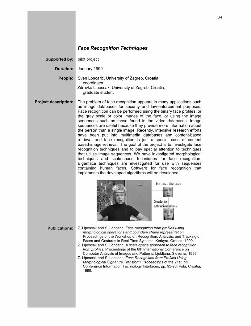
Project description: The problem of face recognition appears in many applications such
as image databases for security and law-enforcement purposes. Face recognition can be performed using the binary face profiles, or the gray scale or color images of the face, or using the image sequences such as those found in the video databases. Image sequences are useful because they provide more information about the person than a single image. Recently, intensive research efforts have been put into multimedia databases and content-based retrieval and face recognition is just a special case of content based-image retrieval. The goal of the project is to investigate face recognition techniques and to pay special attention to techniques that utilize image sequences. We have investigated morphological techniques and scale-space techniques for face recognition. Eigenface techniques are investigated for use with sequences containing human faces. Software for face recognition that implements the developed algorithms will be developed.
Publications:
Z. Liposcak and S. Loncaric. Face recognition from profiles using morphological operations and boundary shape representation. Proceedings of the Workshop on Recognition, Analysis, and Tracking of Faces and Gestures in Real-Time Systems, Kerkyra, Greece, 1999.
Z. Liposcak and S. Loncaric. A scale-space approach to face recognition from profiles. Proceedings of the 8th International Conference on Computer Analysis of Images and Patterns, Ljubljana, Slovenia, 1999.
Z. Liposcak and S. Loncaric. Face Recognition from Profiles Using Morphological Signature Transform. Proceedings of the 21st Int'l Conference Information Technology Interfaces, pp. 93-98, Pula, Croatia, 1999.
35
SVD-Based Techniques For Image Restoration
Supported by: Ministry of Science and Technology, Croatia
Duration:
October 199
People: Sven Loncaric, University of Zagreb, Croatia, coordinator
Zeljko Devcic, University of Zagreb, Croatia, graduate student
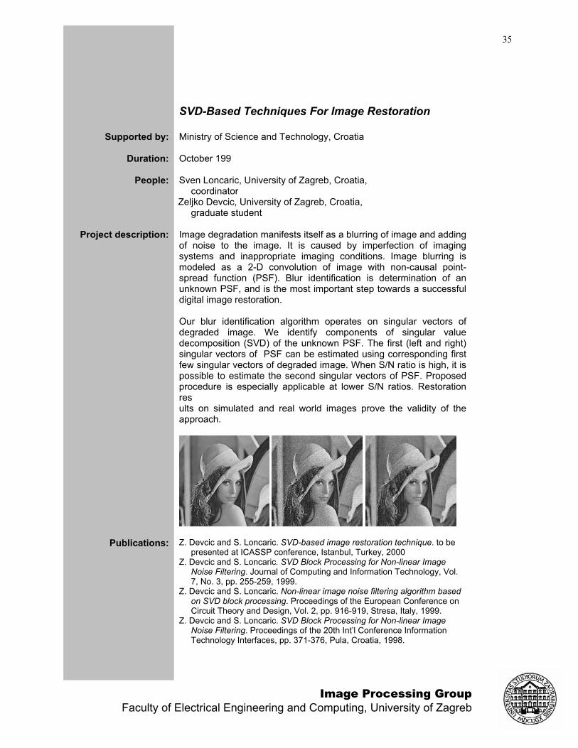
Project description: Image degradation manifests itself as a blurring of image and adding
of noise to the image. It is caused by imperfection of imaging systems and inappropriate imaging conditions. Image blurring is modeled as a 2-D convolution of image with non-causal point-spread function (PSF). Blur identification is determination of an unknown PSF, and is the most important step towards a successful digital image restoration. Our blur identification algorithm operates on singular vectors of degraded image. We identify components of singular value decomposition (SVD) of the unknown PSF. The first (left and right) singular vectors of PSF can be estimated using corresponding first few singular vectors of degraded image. When S/N ratio is high, it is possible to estimate the second singular vectors of PSF. Proposed procedure is especially applicable at lower S/N ratios. Restoration res ults on simulated and real world images prove the validity of the approach.
Publications:
Z. Devcic and S. Loncaric. SVD-based image restoration technique. to be presented at ICASSP conference, Istanbul, Turkey, 2000
Z. Devcic and S. Loncaric. SVD Block Processing for Non-linear Image Noise Filtering. Journal of Computing and Information Technology, Vol. 7, No. 3, pp. 255-259, 1999.
Z. Devcic and S. Loncaric. Non-linear image noise filtering algorithm based on SVD block processing. Proceedings of the European Conference on Circuit Theory and Design, Vol. 2, pp. 916-919, Stresa, Italy, 1999.
Z. Devcic and S. Loncaric. SVD Block Processing for Non-linear Image Noise Filtering. Proceedings of the 20th Int’l Conference Information Technology Interfaces, pp. 371-376, Pula, Croatia, 1998.
Image Processing Group Faculty of Electrical Engineering and Computing, University of Zagreb

36
Web-Based Electronic Journal Publishing Support System
Supported by: Ministry of Science and Technology, An IT project
Duration: October 2000 – October 2001
People: Sven Loncaric, University of Zagreb, Croatia, principal investigator
Marko Subasic, University of Zagreb, Croatia, graduate student
Hrvoje Bogunovic, University of Zagreb, Croatia, graduate student
Edgar Pek, University of Zagreb, Croatia, graduate student
Project description: The aim of the project was to develop a software system for
completely computerized preparation and publishing of paper version and electronic version of a technical journal. The main function of the software is to provide support for manuscript tracking and editorial actions such as review process and electronic publishing. The developed software is in routine use for support of a scientific journal titled "Journal of Computing and Information Technology" and can be accessed at http://cit.zesoi.fer.hr.
37
Neural Networks For Financial Market Analysis
Supported by: Pilot project
Duration:
Jan 2000-Jan 2002
People: Sven Loncaric, University of Zagreb, Croatia, coordinator
Ivan Vrancic, University of Zagreb, Croatia, graduate student
Project description: Analysis of bank customers represents an important task in banking
market planning. An example is planning of promotions for new banking services and credit card users. The analysis must be performed to select the potential customers who are most appropriate for offering certain services. In practise, it is difficult to determine exact rules for partitioning or segmentation of customers. Therefore, it is necessary to use learning to implicitly extract the required features that are important for market segmentation. Neural network is a powerful tool for problems where no explicit relation exists between the input and output data. Through a learning process, a neural network can learn the input-output relation in question. In this application a feed-forward neural network can be trained using the current users of various banking services and their profiles. After the network is trained profile data for new potential customers can be input to the network and the network will compute output indicating customers who are best potential candidates for certain services. A common learning algorithm used for feed-forward networks is back-propagation learning. The goal of the project is to investigate techniques used in banking market segmentation and develop a neural network-based procedure for market segmentation. The procedure will be tested on real data. The required software modules will be developed for the Windows platform.
Image Processing Group Faculty of Electrical Engineering and Computing, University of Zagreb

38
CHAIRMAN Conference Management System
Supported by: Ministry of Science and Technology, An IT project
Duration: October 2003 – October 2004
People: Sven Loncaric, University of Zagreb, Croatia, principal investigator
Hrvoje Bogunovic, University of Zagreb, Croatia, graduate student
Vjekoslav Levacic, University of Zagreb, Croatia, undergraduate student
Bojan Blazona, University of Zagreb, Croatia, undergraduate student
Boris Kuzmic, University of Zagreb, Croatia, undergraduate student
Project description: CHAIRMAN system is a conference management web-based
system for tracking many conference organization-related activities such as: author paper submission, reviewer assignment, collecting reviews, submission of revised and camera ready papers, program committee activities, preparation of conference proceedings, participant registration. The system also provides an on-site participant support during the conference. The software is based on Java Server Pages and MySQL database server. Further information about the project can be found at http://chairman.zesoi.fer.hr.
39
COST B21 Project: Physiological Modelling of MR Image Formation
Supported by: European COST Program
Duration: October 2003 –
People: Sven Loncaric, University of Zagreb, Croatia, Croatian national coordinator for COST B21
Project description: COST B21 project is an interdisciplinary networking project
connecting European research groups. The topics of interest include all aspects of magnetic resonance (MR) imaging and image processing and analysis of obtained MR images. Participation of the Image Processing Group is mostly concerned with image analysis aspects and development of techniques for segmentation of MR images.
Image Processing Group Faculty of Electrical Engineering and Computing, University of Zagreb
40
Biomedical Perfusion Image Sequence Analysis
Supported by: Philips Medical Systems, Netherlands
Duration: 2002 –
People: Sven Loncaric, University of Zagreb, Croatia, principal co-investigator
Drazenko Babic, Philips Medical Systems, Netherlands, principal co-investigator
Hrvoje Bogunovic, University of Zagreb, Croatia, graduate student
Project description: The goal of this project is to develop image analysis methods for
quantitative measurements of perfusion images of liver and brain obtained by X-ray imaging and contrast injection. The image analysis problem includes analysis of motion and extraction of curves showing propagation of contrast through various organs. Techniques for signal restoration from noise are used to fight the problem of noise. Specifically, Wiener filtering and wavelet-based denoising techniques are being researched. In analyis of contrast uptake curves, several different curve parameters are extracted to be used as features for medical diagnosis.
41
Serial Registration of Pre- and Intra-operative MR Brain Images
Supported by: Pilot project
Duration: 2002 –
People: Sven Loncaric, University of Zagreb, Croatia, principal investigator
Michael Schulder, University of Medicine and Dentistry of New Jersey, USA, principal co-investigator
Marko Subasic, University of Zagreb, Croatia, graduate student
Project description: The goal of serial image registration is to guide neurosurgeon during
resection of brain tumors, during which the surgeon takes intraoperative MR scans of the brain for the purpose of surgical navigation. Accurate registration requires biomechanical modeling of brain using finite element analysis. In particular, gravity-induced deformation and deformation caused by resection of tissue must be modeled.
Publications: S. Devabhaktuni, S. Loncaric, M. Schulder. Elastic Image Registration Of Diagnostic Brain Mr And Limited View Intra Operative Mr Using Mutual Information. 29th Annual Northeast Bioengineering Conference, Newark, NJ, USA, 2003
S. R. Subramaniam, S. Loncaric, M. Schulder. Simulation Of Patient Specific Brain Deformation Due To Brain Shift. 29th Annual Northeast Bioengineering Conference, Newark, NJ, USA, 2003
Image Processing Group Faculty of Electrical Engineering and Computing, University of Zagreb
42
Neural Networks for Ion Chromatography Optimization
Supported by: Pilot project
Duration: 2004 –
People: Sven Loncaric, University of Zagreb, Croatia, principal co-investigator
Stefica Cerjan–Stefanovic, University of Zagreb, Croatia, principal co-investigator
Tomislav Bolanca, University of Zagreb, Croatia, higher assistant
Melita Regelja, University of Zagreb, Croatia, graduate student
Hrvoje Regelja, University of Zagreb, Croatia, graduate student
Project description: The goal of this project is to investigate methods based on artificial
neural networks that will be used for optimization of ion chromatographic separation. Chemical expertise is brought to the project by Prof. Cerjan-Stefanovic and her team at Faculty of Chemical Engineering, University of Zagreb. Optimization in ion chromatography involves selection of experimental conditions for adequate separation and acceptable retention time for each individually samples. But, in chemical laboratories obtaining a balance between resolution and analysis time is not always easy. An efficient optimization method should be employed during the method development process in order to deal with these optimization problems and to reduce method development time. Computers assisted optimization procedures have to be used as an efficient aid for ion chromatography method development. Empirical modeling is a useful technique whereby a global optimum can be located. The use of chemometric protocols like experimental design in combination with artificial neural networks can be extremely beneficial in the systematic construction and optimization of response surfaces. For most simple optimization applications, traditional techniques, such as simplex method, are capable of locating a global optimum efficiently. However, for nonlinear behaviors, like those of ion chromatography, these methods are either time consuming or become trapped in local optima.
43
Professional Activities Prof. Loncaric has served as Program Committee member or reviewer for a number of international scientific meetings; the following is the list of meetings for which Prof. Loncaric played the main organizing roles. In particular, Prof. Loncaric has been the founder and organizer of a series of international symposia under title "Image and Signal Processing and Analysis (ISPA)". For more information please see ISPA web site at http://www.isispa.org.
Conference and Workshop Organization S. Loncaric, Program Chair. 3rd International Symposium on Image and Signal
Processing and Analysis. Rome, Italy, 2003
S. Loncaric and A. Santos and P. Clarysse. Biomedical Image Processing and Analysis. Tutorial workshop at MEDICON, Pula, Croatia, 2001
S. Loncaric, Co-Chair. 2nd International Symposium on Image and Signal Processing and Analysis. Pula, Croatia, 2001
S. Loncaric, Chair. First International Workshop on Image and Signal Processing and Analysis. Pula, Croatia, 2000
S. Loncaric, Chair. 8th International Summer School on Image Processing. Zagreb, Croatia, 2000
A. P. Dhawan and J. Udupa and S. Loncaric and A. Levy and M. Dole. Intelligent Biomedical Image Analysis. Tutorial Workshop at the 19th Annual International Conference of IEEE Engineering in Medicine and Biology Society, Chicago, USA, 1997.
A. P. Dhawan and J. Udupa and S. Loncaric and A. Levy and M. Dole. Intelligent Biomedical Image Analysis. Tutorial Workshop at 18th Annual International Conference of IEEE Engineering in Medicine and Biology Society, Amsterdam, Netherlands, 1996.
Invited Lectures
S. Loncaric. Prividna stvarnost u medicini (in Croatian language). Zbornik radova skupa "Telemedicina u Hrvatskoj", pp. 79-93, Akademija medicinskih znanosti Hrvatske, Zagreb, Croatia, 1998.
S. Loncaric. Virtual Reality in Medicine. Young lecturer at the Third IEEE EMBS International Summer School on Biomedical Imaging. Berder, France, 1998.
S. Loncaric. An Overview of Virtual Reality Techniques. Presented at the 6th Summer School on Image Processing, Szeged, Hungary, 1998.
S. Loncaric. Image Segmentation Techniques. Presented at the 5th Summer School on Image Processing, Szeged, Hungary, 1997.
S. Loncaric. Digital Image Processing and Analysis. Invited lecture, Croatian Society for Bio-Physics, Zagreb, Croatia, 1995.
Image Processing Group Faculty of Electrical Engineering and Computing, University of Zagreb

44
Journal Editorships
S. Loncaric, Editor-in-Chief, Journal of Computing and Information Technology, University Computing Center, Zagreb, 1999-present.
S. Loncaric, Guest Editor, Journal of Computing and Information Technology, Special Issue on Biomedical Image Processing and Analysis, Vol. 6, No. 2, 1998.
S. Loncaric, Editor, Journal of Computing and Information Technology, University Computing Center, Zagreb, 1997-1998.
45
Publications Publications describing research and development activities of the Image Processing Group are listed in the following sections. Most publications are available for full-text download in PDF format at the IPG web site at http://ipg.zesoi.fer.hr.
Journal Papers
M. Koncar and S. Loncaric. Concepts for Integrated Electronic Health Record Management System. Informatica Medica Slovenica, Vol. 8, No. 1, pp. 40-51, 2003
Z. Devcic and S. Loncaric. SVD Block Processing for Non-linear Image Noise Filtering. Journal of Computing and Information Technology, Vol. 7, No. 3, pp. 255-259, 1999.
S. Loncaric. A Survey of Shape Analysis Techniques. Pattern Recognition, Vol. 31, No. 8, pp. 983-1001, 1998.
D. Cosic and S. Loncaric. New Methods for Cluster Selection in Unsupervised Fuzzy Clustering. Automatika, vol. 37, pp. 133-137, 1996.
Z. Dukic and S. Loncaric. An Object-oriented Implementation of Morphological Operations using Element Vector Representation. Journal of Computing and Information Technology, vol. 4, pp. 265-269, 1996.
S. Loncaric and A. P. Dhawan. Morphological Signature Transform and Applications. Automatika, vol. 37, pp. 25-30, 1996.
S. Loncaric and A. P. Dhawan. Near-optimal MST-based Shape Description using Genetic Algorithm. Pattern Recognition, vol 28, pp. 571-579, 1995.
S. Loncaric and A. P. Dhawan. 3-D Image Analysis of Intra-cerebral Brain Hemorrhage from Digitized CT films. Computer Methods and Programs in Biomedicine, vol 46, pp. 207-216, 1995.
S. Loncaric and A. P. Dhawan. A morphological signature transform for shape description. Pattern Recognition, vol 26, pp. 1029-1037, 1993.
Books and Book Chapters
S. Loncaric, A. Neri, H. Babic, Eds., Proceedings of the 3rd Int'l Symposium on Image and Signal Processing and Analysis, Rome, Italy, 2003
E. Sorantin, E. Balogh, A. Vilanova Bartol, K. Palgyi, L.G. Nyl, S. Loncaric, M. Subasic, D. Kovacevic. Virtual Dissection of the Colon. In D. Caramella, C. Bartolozzi, Eds., 3D Image Processing, Springer-Verlag, 2002
S. Loncaric, H. Babic, Eds., Proceedings of the 2nd Int'l Symposium on Image and Signal Processing and Analysis, Pula, Croatia, 2001
S. Loncaric, Ed., Proceedings of the First Int'l Workshop on Image and Signal Processing and Analysis, Pula, Croatia, 2000
S. Loncaric, Ed., Proceedings of the 8th Int'l Summer School on Image Processing, Zagreb, Croatia, 2000
Image Processing Group Faculty of Electrical Engineering and Computing, University of Zagreb
46
Refereed Conference Papers, 2003
H. Bogunovic, E. Pek, S. Loncaric, V. Mornar. An Electronic Journal Management System. 25th Int'l Conf. on Information Technology Interfaces, Cavtat, Croatia, 2003
P. Schedlbauer, E. Sorantin, H.A. Deutschmann, D. Kovacevic, M. Subasic, S. Loncaric, B. Georgieva, H. Schöllnast, K.A. Hausegger, Segmentation of infrarenal aortic aneurysm based on spiral CT data: A new approach using active contour models, European Congress of Radiology, Vienna, Austria, 2003
S. Devabhaktuni, S. Loncaric, M. Schulder. Elastic Image Registration Of Diagnostic Brain Mr And Limited View Intra Operative Mr Using Mutual Information. 29th Annual Northeast Bioengineering Conference, Newark, NJ, USA, 2003
S. R. Subramaniam, S. Loncaric, M. Schulder. Simulation Of Patient Specific Brain Deformation Due To Brain Shift. 29th Annual Northeast Bioengineering Conference, Newark, NJ, USA, 2003
Refereed Conference Papers, 2002
T. Petkovic, J. Krapac, S. Loncaric, and M. Sercer. Automated visual inspection of plastic products. Procedings of the 11th International Electrotechnical and Computer Science Conference ERK 2002, pp. 283-286, Portoroz, Slovenia, 2002
M. Subasic, D. Kovacevic, and S. Loncaric. Segmentation of Abdominal Aortic Aneurysm using Deformable Models. Proceedings of East-West-Vision 2002, pp. 61-66, Graz, Austria, 2002
M. Koncar and S. Loncaric. Concepts for Integrated Electronic Health Record Management System. Presented at 15th Int'l Conf. on Computer-Based Medical Systems, Maribor, Slovenia, 2002
P.Schedlbauer, E.Sorantin, K.Hausegger, D.Kovacevic, S.Loncaric. Segmentation of Infrarenal Aortic aneurysms based on Spiral CT data - A new Approach using Active Contour Models, 3rd Workshop on Multislice CT, 3D Imaging, and Virtual Endoscopy, Rome, Italy, 2002
M. Subasic and S. Loncaric and E. Sorantin. 3-D image analysis of abdominal aortic aneurysm. Proceedings of SPIE Medical Imaging, San Diego, USA, 2002.
Refereed Conference Papers, 2001
M. Matesin and S. Loncaric and D. Petravic. A Rule-based Approach to Stroke Lesion Analysis from CT Brain Images. Proceedings of 2nd Int'l Symposium on Image and Signal Processing and Analysis, pp. 219-223, Pula, Croatia, 2001.
T. Macan and S. Loncaric. 3-D Cardiac Motion Estimation by Point-Constrained Optical Flow Algorithm. Proceedings of 2nd Int'l Symposium on Image and Signal Processing and Analysis, pp. 255-259, Pula, Croatia, 2001.
47
Z. Biuk and S. Loncaric. Face Recognition from Multi-Pose Image Sequence. Proceedings of 2nd Int'l Symposium on Image and Signal Processing and Analysis, pp. 319-324, Pula, Croatia, 2001.
E. Pek and S. Loncaric and A. Margan. Internet-based Medical Teleconsultation System. Proceedings of 2nd Int'l Symposium on Image and Signal Processing and Analysis, pp. 657-661, Pula, Croatia, 2001.
D. Kovacevic and S. Loncaric. Energy minimization-based interactive image segmentation. Proceedings of SPIE Medical Imaging, San Diego, USA, 2001, pp. 1542-1549
M. Subasic and S. Loncaric and E. Sorantin. 3-D deformable model segmentation of abdominal aortic aneurysm. Proceedings of SPIE Medical Imaging, San Diego, USA, 2001, pp. 388-394
S. Loncaric and I. Ceskovic and S. Loncaric and R. Petrovic. 3-D quantitative analysis of brain SPECT images. Proceedings of SPIE Medical Imaging, San Diego, USA, 2001, pp. 1689-1695
S. Galic and S. Loncaric. Cardiac image segmentation using spatio-temporal clustering. Proceedings of SPIE Medical Imaging, San Diego, USA, 2001, pp. 1199-1206
T. Macan and S. Loncaric. Hybrid Optical Flow and Segmentation Technique for LV Motion Detection. Proceedings of SPIE Medical Imaging, San Diego, USA, 2001, pp. 475-482
Marko Subasic, Sven Loncaric, Erich Sorantin. Shape-Specific Adaptations for Level-Set Deformable Model-Based Segmentation. Proceedings of Computer Vision Winter Workshop, pp. 104-113, Bled, Slovenia, 2001.
Refereed Conference Papers, 2000
Z. Devcic and S. Loncaric. Simultaneous estimation of PSF and global frame shifts at lower S/N ratios. Proceedings of the First Int'l Workshop on Image and Signal Processing and Analysis, pp. 41-46, Pula, Croatia, 2000.
Z. Liposcak and S. Loncaric. Prediction and verification for face detection. Proceedings of the First Int'l Workshop on Image and Signal Processing and Analysis, pp. 107-111, Pula, Croatia, 2000.
S. Loncaric and E. Pek and M. Silovic and A. Margan. Virtual Polyclinic: A Web-based system for medical teleconsultation. Proceedings of Emerging Technologies and Life Sciences: Medicine and Communication (MEDICOM), Remagen, Germany, 2000.
S. Loncaric and M. Silovic and E. Pek and A. Margan. A Web-based system for medical teleconsultation: First results. Proceedings of Telemedicine and Health Informatics, Milano, Italy, 2000.
Z. Devcic and S. Loncaric. Blur Identification Using Averaged Spectra of Degraded Image Singular Vectors. IEEE Int'l Conference on Acoustics, Speech, and Signal Processing, ICASSP, Istanbul, Turkey, 2000.
S. Loncaric and Z. Majcenic. Optical flow algorithm for cardiac motion estimation. World Congress on Medical Physics and Biomedical Engineering, Chicago, USA, 2000.
Image Processing Group Faculty of Electrical Engineering and Computing, University of Zagreb

48
S. Loncaric and M. Subasic and Erich Sorantin. 3-D deformable model for aortic aneurysm segmentation from CT images. World Congress on Medical Physics and Biomedical Engineering, Chicago, USA, 2000.
C. Halmai, D. Cosic, A. Kuba, E. Sorantin, S. Loncaric, K.H Hausegger. 3-D extension of infrarenal aortic aneurysms extracted from spiral CT. European Congress of Radiology, Vienna, 2000
S. Loncaric and D. Kovacevic and E. Sorantin. Semi-automatic Active Contour Approach to Segmentation of Computed Tomography Volumes. Proceedings of SPIE Medical Imaging, San Diego, USA, 2000.
S. Loncaric and T. Macan. Point-constrained optical flow for LV motion detection. Proceedings of SPIE Medical Imaging, San Diego, USA, 2000.
Z. Majcenic and S. Loncaric. An algorithm for spatio-temporal heart segmentation. Proceedings of SPIE Medical Imaging, San Diego, USA, 2000.
M. Koncar and S. Loncaric. WWW-based radiology image management system, submitted to Medical Informatics Europe conference, Hannover, Germany, 2000
S. Loncaric, T. Markovinovic. Web-based virtual endoscopy, submitted to Medical Informatics Europe conference, Hannover, Germany, 2000
M. Subasic and S. Loncaric. 3-D image analysis of abdominal aortic aneurysm, submitted to Medical Informatics Europe conference, Hannover, Germany, 2000
Refereed Conference Papers, 1999 M. Koncar and S. Loncaric. WWW-based radiological PACS. Proceedings of
the Croatian Medical Informatics Symposium, pp. 91-96, Zagreb, Croatia, 1999.
Z. Liposcak and S. Loncaric. Face recognition from profiles using morphological operations and boundary shape representation. Proceedings of the Workshop on Recognition, Analysis, and Tracking of Faces and Gestures in Real-Time Systems, Kerkyra, Greece, 1999.
Z. Devcic and S. Loncaric. Non-linear image noise filtering algorithm based on SVD block processing. Proceedings of the European Conference on Circuit Theory and Design, Vol. 2, pp. 916-919, Stresa, Italy, 1999.
Z. Liposcak and S. Loncaric. A scale-space approach to face recognition from profiles. Proceedings of the 8th International Conference on Computer Analysis of Images and Patterns, Ljubljana, Slovenia, 1999.
Z. Liposcak and S. Loncaric. Face Recognition from Profiles Using Morphological Signature Transform. Proceedings of the 21st Int'l Conference Information Technology Interfaces, pp. 93-98, Pula, Croatia, 1999.
D. Kovacevic and S. Loncaric. Deformable Contour-Based Method For Medical Image Segmentation. Proceedings of the 21st Int'l Conference Information Technology Interfaces, pp. 145-150, Pula, Croatia, 1999.
S. Loncaric and T. Markovinovic and T. Petrovic and D. Ramljak and E. Sorantin. Construction of Virtual Environment for Endoscopy. Proceedings
49
of the IEEE International Conference on Multimedia Computing and Systems, Florence, Italy, 1999.
S. Loncaric, A. P. Dhawan, D. Kovacevic, D. Kovacevic, D. Cosic, J. Broderick, T. Brott. Image-based Measurements of Intracerebral Brain Hemorrhage. First Croatian Symposium on Computer Assisted Surgery, Zagreb, Croatia, 1999.
S. Loncaric and Z. Majcenic. An efficient SA algorithm for brain hemorrhage image analysis. First Croatian Symposium on Computer Assisted Surgery, Zagreb, Croatia, 1999.
S. Loncaric and D. Ramljak and T. Markovinovic and E. Sorantin. VRML-based Model Creation for Virtual Bronchoscopy. First Croatian Symposium on Computer Assisted Surgery, Zagreb, Croatia, 1999.
S. Loncaric. Virtual Reality in Medicine: An Overview. First Croatian Symposium on Computer Assisted Surgery, Zagreb, Croatia, 1999.
S. Loncaric and Z. Majcenic. Multiresolution simulated annealing for brain image analysis. Proceedings of SPIE Medical Imaging, Vol. 3661, pp. 1139-1146, San Diego, USA, 1999.
S. Loncaric and A. P. Dhawan and D. Kovacevic and D. Cosic and J. Broderick and T. Brott. Quantitative Intracerebral Brain Hemorrhage Analysis. Proceedings of SPIE Medical Imaging, Vol 3661, pp. 886-894, San Diego, USA, 1999.
Refereed Conference Papers, 1998
Z. Majcenic and S. Loncaric. CT image labeling using simulated annealing algorithm. Proceedings of the IX European Signal Processing Conference, Vol. 4, pp. 2513-2516, Island of Rhodos, Greece, 1998.
D. Sersic and S. Loncaric. Enhancement of Mammographic Images for detection of Microcalcifications. Proceedings of the IX European Signal Processing Conference, Vol. 2, pp. 693-696, Island of Rhodos, Greece, 1998.
K. Simunic and S. Loncaric. A Genetic Search-Based Partial Image Matching. Proceedings of the 2nd IEEE Int’l Conference on Intelligent Processing Systems, pp. 119-122, Gold Coast, Australia, 1998.
S. Loncaric and Z. Majcenic. Multiresolution CT Head Image Analysis using Simulated Annealing. Proceedings of the 20th Int’l Conference Information Technology Interfaces, pp. 257-262, Pula, Croatia, 1998.
Z. Devcic and S. Loncaric. SVD Block Processing for Non-linear Image Noise Filtering. Proceedings of the 20th Int’l Conference Information Technology Interfaces, pp. 371-376, Pula, Croatia, 1998.
S. Loncaric and Z. Majcenic. Multiresolution CT Head Image Analysis using Simulated Annealing. Proceedings of the 12th International Symposium Computer Assisted Radiology and Surgery, pp. 889-889, Tokyo, Japan, 1998.
D. Kovacevic and S. Loncaric. CT Image Labeling Using Hopfield Neural Network. Proceedings of the IEEE 9th Mediterranean Electrotechnical Conference, pp. 44-47, Tel-Aviv, Israel, 1998.
Image Processing Group Faculty of Electrical Engineering and Computing, University of Zagreb

50
S. Loncaric and D. Kovacevic and D. Cosic. Fuzzy Expert System for Edema Segmentation. Proceedings of the IEEE 9th Mediterranean Electrotechnical Conference, pp. 1476-1479, Tel-Aviv, Israel, 1998.
D. Cosic and S. Loncaric and O. Rajkovic. A Method for 3-D Image Representation Using a Surface Model. Proceedings of the IEEE 9th Mediterranean Electrotechnical Conference, pp. 82-85, Tel-Aviv, Israel, 1998.
Refereed Conference Papers, 1997
D. Cosic and S. Loncaric. Computer System for Quantitative Analysis of ICH from CT Head Images. Proceedings of the 19th Annual International Conference of IEEE Engineering in Medicine and Biology Society, Chicago, USA, 1997.
S. Loncaric and D. Kovacevic. A Method for Segmentation of CT Head Images. Proceedings of the 9th International Conference on Image Analysis and Processing, pp. 388-395, Florence, Italy, 1997.
D. Kovacevic and S. Loncaric. Radial Basis Function-based Image Segmentation using a Receptive Field. Proceedings of the Tenth Annual IEEE Symposium on Computer-Based Medical Systems, pp. 126-130, Maribor, Slovenia, 1997.
D. Cosic and S. Loncaric. A Rule-based Labeling of CT Head Image. Proceedings of the 6th Conference on Artificial Intelligence in Medicine Europe, Grenoble, France, Lecture Notes in Artificial Intelligence, pp. 453-456, Springer-Verlag, 1997.
Z. Dukic and S. Loncaric. Erosion and Dilation based Morphological Signature Transform for Shape Description. SPIE Electronic Imaging '97, Nonlinear Image Processing VIII, San Jose, USA, 1997.
Refereed Conference Papers, 1996 and older
S. Loncaric and D. Cosic and A. P. Dhawan. Hierarchical Segmentation of CT Head Images. Conference Proceedings of the 18th Annual International Conference of IEEE Engineering in Medicine and Biology Society, Amsterdam, Netherlands, 1996.
D. Cosic and S. Loncaric. Two Methods for ICH Segmentation. Proceedings of the 11th International Symposium on Biomedical Engineering, pp. 63-66, Zagreb, Croatia, 1996.
D. Cosic and S. Loncaric. New Methods for Cluster Selection in Unsupervised Fuzzy Clustering. Proceedings of the 41th Conference KoREMA'96, vol. 4, pp. 1-3, Opatija, Croatia, 1996.
Z. Dukic and S. Loncaric. An Object-Oriented Implementation of Morphological Operations Using Element Vector Representation. Proceedings of the 18th International Conference Information Technology Interfaces, pp. 541-546, Pula, Croatia, 1996.
S. Loncaric and D. Cosic and A. P. Dhawan. Segmentation of CT Head Images. Computer Assisted Radiology '96 - Proceedings of the International

51
Image Processing Group Faculty of Electrical Engineering and Computing, University of Zagreb
Symposium on Computer and Communication Systems for Image Guided Diagnosis and Therapy, pp. 1012-1012, Paris, France, 1996.
S. Loncaric and D. Cosic and A. P. Dhawan. 3-D Quantification of Intracerebral Brain Hemorrhage. IEEE Engineering in Medicine and Biology Society 2nd International Summer School on Biomedical Imaging, Berder, France, 1996.
S. Loncaric and A. P. Dhawan. A Near-Optimal Morphological MR-PET Brain Registration. Scientific Programme and Abstracts of the 9th Annual European Congress of Radiology, pp. 176-176, Vienna, Austria, 1995.
S. Loncaric. Shape Description Methods: A Review. Proceedings of the 40th Anniversary Conference KoREMA, pp. 441-448, Zagreb, Croatia, 1995.
S. Loncaric and A. P. Dhawan. Morphological Signature Transform and Applications. Proceedings of the 40th Anniversary Conference KoREMA, pp. 449-453, Zagreb, Croatia, 1995.
S. Loncaric and A. P. Dhawan. 3-D Brain Image Registration Using Optimal Morphological Processing and Iterative Principal Axis Transform. Proceedings of the 16th Annual International Conference of IEEE Engineering in Medicine and Biology Society, Paris, France, 1994.
S. Loncaric and A. P. Dhawan. 3-D Brain Image Registration Using Optimal Morphological Processing. Proceedings of the 10th International Symposium on Biomedical Engineering, pp. 83-86, Zagreb, Croatia, 1994.