identification of glucose-derived cross-linking sites in ribonuclease a
TRANSCRIPT

Identification of Glucose-Derived Cross-Linking Sites in Ribonuclease A
Zhenyu Dai,† Benlian Wang,‡ Gang Sun,§ Xingjun Fan,| Vernon E. Anderson,† andVincent M. Monnier*,†,|
Department of Biochemistry, Case Western Reserve University, Cleveland, Ohio 44106, Center for Proteomicsand Mass Spectrometry, Case Western Reserve University, Cleveland, Ohio 44106, Department of InternalMedicine, Washington University, St. Louis, Missouri 63110, and Department of Pathology, Case Western
Reserve University, Cleveland, Ohio 44106
Received December 21, 2007
The accumulation of glycation derived cross-links has been widely implicated in extracellular matrixdamage in aging and diabetes, yet little information is available on the cross-linking sites in proteinsand the intra- versus intermolecular character of cross-linking. Recently, glucosepane, a 7-memberedheterocycle formed between lysine and arginine residues, has been found to be the single major cross-link known so far to accumulate during aging. As an approach toward identification of glucose derivedcross-linking sites, we have preglycated ribonuclease A first for for 14 days with 500 mM glucose,followed by a 4-week incubation in absence of glucose. MALDI-TOF analysis of tryptic digests revealedthe presence of Amadori products (∆m/z ) 162) at K1, K7, K37 and K41, in accordance with previousstudies. In addition, K66, K98 and K104 were also modified by Amadori products. Intramolecularglucosepane cross-links were observed at K41-R39 and K98-R85. Surprisingly, the only intermolecularcross-link observed was the 3-deoxyglucosone-derived DODIC at K1-R39. The identity of cross-linkedpeptides was confirmed by sequencing with tandem mass spectrometry. Recombinant ribonuclease Amutants R39A, R85A, and K91A were produced, purified, and glycated to further confirm the importanceof these sites on protein cross-linking. These data provide the first documentation that bothintramolecular and intermolecular cross-links form in glucose-incubated proteins.
Keywords: glycation • ribonuclease A • cross-linking • glycation sites • glucosepane • DODIC • AGEs
Introduction
The Maillard Reaction is initiated by reaction of a reducingsugar with a primary amine to form an unstable Schiff base,followed by its rearrangement to relatively stable Amadoriproducts. These can undergo fragmentation, oxidation andrearrangements resulting in a plethora of compounds namedadvanced glycation end products (AGEs). In the past 30 years,the quantitative relationship between accumulation of AGEsin tissues and the extent of tissue pathology has been exten-sively studied in animals as well as in humans. AGE-likefluorescence (excitation at 370 nm, emission at 440 nm) hasbeen shown to increase in collagen from aged and diabeticpatients.1 In retinal vessels of diabetic rats, AGE-specificfluorescence increased 2.6-fold after 26 weeks of diabetes.2
Similar increases in AGE fluorescence have been found inproteins from diabetic lens3 and renal cortex.4 AGE accumula-tion was accompanied by histological evidence of diabetictissue damage. The senescent extracellular matrix exhibits
characteristics including decreased solubility,5 decreased pro-teolytic digestibility,6,7 and accumulation of yellow and fluo-rescent material,8 all of which are commonly associated withdiabetes. These age-related and diabetes-enhanced changes arethought to result in part from AGE-derived cross-links.
The accumulation of AGEs on proteins depends on reducingsugar concentration and the protein turnover rate. The lowturnover rate of collagen, lens protein and neural proteinspredisposes them to AGE accumulation. Changes in collagen-rich tissues, such as arteries, lungs, and joints, have beencorrelated with hypertension, emphysema, and decreased jointmobility.9
In collagen, cross-links derived from lysyl oxidase activityhave been unequivocally proven to be involved in determiningthe mechanical character of immature collagen.10 In contrast,the glycation derived cross-links have been hypothesized, butnot proven, to be responsible for the collagen stiffening duringaging and in diabetes. The type of cross-links formed, that is,inter- versus intrafibrillar cross-links, could have a very differentmechanical impact on the tissue properties.
Few studies have been done on the glycation-derived cross-linking sites of proteins in spite of their potential importanceon the mechanical character. One of the reasons for the slowprogress in this field may be linked to the low level of individualAGEs found in vivo. Biemel et al. provided convincing evidence
* To whom correspondence should be addressed: Vincent M. Monnier,Department of Pathology, Case Western Reserve University, Cleveland, OH44120. Tel: (216) 368-6238. E-mail: [email protected].
† Department of Biochemistry, Case Western Reserve University.‡ Center for Proteomics and Mass Spectrometry, Case Western Reserve
University.§ Department of Internal Medicine, Washington University.| Department of Pathology, Case Western Reserve University.
2756 Journal of Proteome Research 2008, 7, 2756–2768 10.1021/pr700874a CCC: $40.75 2008 American Chemical SocietyPublished on Web 05/24/2008

that glucosepane is the dominant cross-link in both humanserum albumin and lens protein11 (Figure 1). Our laboratoryprovided additional data supporting the premise that glu-cosepane is the single major glycation derived cross-link knownto date in aging human skin and glomerular basement col-lagen.12 In addition, its levels are dramatically elevated indiabetes,13 a condition that is associated with increased stiffnessof all collagen-rich tissues. Glucosepane levels increased up to∼2 nmol/mg collagen in old nondiabetic controls, and furtherincreased to ∼4.3 nmol/mg in diabetic patients.12 Thesenumbers can be translated into one cross-link for every 2-5triple helical collagen molecules in diabetic and nondiabeticaged controls, respectively. This level of modification mightcontribute to the accumulation of collagen matrix due toimpaired proteolytic digestion in diabetes and aging, andperhaps even increased matrix stiffening, particularly if thecross-links are intermolecular or interfibrillar.
Because of the highly repetitive structure of the helicaldomain of the type I collagen molecule, and the anticipateddifficulty in making unequivocal structural assignments, weopted to first approach the problem of site specificity usingribonuclease A, a protein that has been widely used in glycationstudies.
Experimental Procedures
Reagents. RNase A from bovine pancreas (Type XII-A, g90%SDS-PAGE purity) and D-(13C6)glucose were from Sigma (St.Louis, MO). Chelex 100 Resin was from Bio-Rad (Hercules, CA).Deionized water (18.2 MΩ cm-1) was used for all experiments.Sequencing grade trypsin was purchased from Promega (Madi-son, WI). Chymotrypsin and endoproteinase Asp-N were fromRoche Applied Science (Indianapolis, IN). All other reagentswere obtained in the highest quality available from Sigma (StLouis, MO), unless indicated otherwise.
Preparation of Glycated RNase A. Bovine RNase A at aconcentration of 50 mg/mL was incubated with a 1:1 mixtureof 250 mM D-glucose and 250 mM D-(13C6)glucose in 100 mMdeaerated, Chelex-treated sodium phosphate buffer containing1 mM diethylenetriamine pentaacetic acid (DTPA) at 37 °C for2 weeks.14 All in vitro incubations were in these “anaerobic”conditions. Glucose was removed by dialysis against 4 L of thesame buffer for 2 days with one buffer change. Spectra/Por 7dialysis tubing (Spectrumlabs, Rancho Dominguez, CA, 2 kDaMWCO) was used. Upon determination of protein concentra-
tion, this “preglycated” preparation15 was further incubatedwith an equal amount of freshly added native RNase A for 4weeks at 37 °C. Chloroform and toluene (0.15% each, v/v) wereadded to prevent bacterial growth during the incubation.
Enzymatic Digestion for Peptide Mapping. Prior to enzy-matic digestion, protein samples (20 µL) were diluted to 500µL with water and concentrated to 20 µL using an Ultrafree-0.5centrifugal filter unit (Millipore, Billerica, MA, 5 kDa MWCO).Per manufacturer’s information, three cycles of concentrationshould remove ∼99% of the initial salt content. The resultingprotein sample was subjected to in-solution digestion bytrypsin in the presence of RapiGest SF (Waters, Milford, MA).The ratio of enzyme to protein substrates was 1:100 and thedigestion was carried out in a 50 mM NH4HCO3 buffer (pH8.0) at 37 °C for 3 h. The ultracentrifuged protein was alsosubjected to digestion by chymotrypsin and endoproteinaseAsp-N. Chymotryptic digestion was carried out in 100 mM Tris-HCl, 10 mM CaCl2, pH 7.8, whereas the Asp-N digestion bufferwas 50 mM Tris-HCl, pH 8.0. In both digestions, the ratio ofenzyme to protein substrates was around 1:100 and thedigestion was carried out at 37 °C (endoproteinase Asp-N) orat 25 °C (chymotrypsin) overnight. Before digestion, proteinsamples were reduced by 5 mM dithiothreitol (60 °C, 30 min)and alkylated with 15 mM iodoacetamide (30 min in the dark).
Matrix-Assisted Laser Desorption and Ionization MassSpectrometry (MALDI). The digested peptides were analyzedby using a prOTOF 2000 MALDI O-TOF mass spectrometer(PerkinElmer, Inc.). The prOTOF mass spectrometer typicallyhas a mass accuracy of better than 10 ppm. Samples weremixed with an equal volume of R-cyano-4-hydroxycinnamicacid (Fluka, St. Louis, MO) matrix solution (10 mg/mL in 50%acetonitrile, 0.1% TFA). Typically, 1 µL of the mixture wasapplied onto the laser target probe and was air-dried beforebeing introduced into the mass spectrometer. For identificationof possible glycation modified peptides, a database includingall possible m/z values of Amadori product, glucosepane, andDODIC modified peptides was generated and used to comparewith the isotopic doublet or triplet peaks found in the MALDIexperiment. Laser-desorbed positive ions were analyzed afteracceleration by 19 kV in the reflectron mode for the peptidedigest. Other instrument settings were the following: laserenergy at 65%; laser rate, 100.0 Hz; declustering potential, 25.0
Figure 1. Structure of Amadori product, glucosepane, and DODIC with corresponding ∆m/z values.
Identification of Glucose-Derived Cross-Linking Sites in RNase A research articles
Journal of Proteome Research • Vol. 7, No. 7, 2008 2757

V; cooling flow, 200 mL/min; mass range, 600.0-5000.0 Da;focus flow, 200.0 mL/min.
Liquid Chromatography-Tandem Mass Spectrometry (LC-MS/MS) Analysis. LC-MS/MS analyses of the proteolytic digestswere performed using a quadrupole ion trap mass spectrometer(model LTQ) from Thermo-Finnigan (San Jose, CA) coupledwith an Ettan MDLC system (GE Healthcare, Piscataway, NJ),chromatographed with a gradient of 0-60% acetonitrile/0.1%formic acid for 30 min. The spectra were acquired by data-dependent methods, consisting of a full scan and then MS/MS on the six most abundant precursor ions at a collisionenergy of 35%. The previously selected precursor ions wererepeated twice during 45 s and then were excluded for 180 s.The data were submitted to a BioWorks Rev. 3.3 databasesearch. Further interpretation of the tandem mass spectra ofthe modified peptides was assisted by an Excel spreadsheet thatgenerated predicted fragment ions from glycated peptidesavailable from V. Anderson.
Mutagenesis of RNase A. A synthetic gene for bovine RNaseA was a gift from the Genex Corp. This gene was placed inM13mp18 (United States Biochemical).16 RNase A in M13mp18was subcloned into pET22b(+) (Novagen) between the MscIand HindIII restriction enzyme sites. Mutant plasmids for theexpression of mutant RNase A, R39A, R85A, and K91A wereconstructed with a Quickchange Site-Directed Mutagenesis Kit(Stratagene Cloning Systems). The sequence of the primersdesignated to replace the Arg39 codon with Ala codon was 5′-CT AGA AAC TTG ACC AAG GAC GCA (for Ala) TGT AAG CCAGTT AAC ACA T-3′. The sequence of the primers designatedto replace the Arg85 codon with Ala codon was 5′-G TCC ATCACT GAC TGT GCT (for Ala) GAG ACA GGC TCG AGT-3′. Thesequence of the primers designated to replace the Lys91 codonwith Ala codon was 5′- CGT GAG ACA GGC TCG AGT GCG (forAla) TAT CCT AAT TGT GCT TAC-3′. Mutagenesis was con-firmed by DNA sequencing at Biotic Solutions, Inc. Themutagenized plasmids were transformed into XL1-Blue super-competent cells. The plasmids containing RNase A mutantsR39A, R85A, K91A were transformed into the Escherichia colicell line BL21-Gold(DE3)pLysS (Stratagene) which was storedat -70 °C.
Production and Purification of Wild-Type and MutantRNase A. The frozen cells were plated on a 50 µg/mL ampicillinand 34 µg/mL chloramphenicol agar plate, and the cells weregrown overnight at 37 °C. A single colony of BL21-Gold(DE3)pLysS containing an alanine mutant in pET22b(+)from the plate was transferred to 50 mL of 2× YT LB mediumwith 50 µg/mL ampicillin and 34 µg/mL chloramphenicol at37 °C and agitated with a gyratory shaker overnight at 225 rpm.This culture was then diluted to 1 L of the same medium andshaken under the same conditions until the optical density at600 nm reached 0.8. At this time, protein expression wasinduced by addition of 2 mL of 0.5 M IPTG to 1 L of cells. Afterinduction, the cells were shaken at 37 °C for another 3 h. Thecells were then centrifuged at 6000 rpm and the pellet wascollected and stored at -80 °C. The pellet from 1 L cell culturewas suspended in 140 mL of 100 mM NaCl and incubated onice for 15 min. The cells were sonicated for 5 min with 2 s pulsesto lyse the cells and break up the genomic DNA. The lysed cellswere centrifuged and resuspended in 140 mL of 100 mM Tris-HCl (pH 8.0). The suspension was centrifuged and the pelletwas suspended in 10 mL of solubilization buffer (20 mM Tris-HCl, pH 8.0; 7 M guanidine-HCl; 10 mM DTT; 10 mM EDTA; 1mM PMSF). This solution was stirred under nitrogen for 2 h to
solubilize the RNase A mutants. The soluble portion was dilutedto 100 mL of 20 mM acetic acid solution and dialyzed against4 L of 20 mM acetic acid solution. The resulting solution wasfurther diluted to 600 mL of refolding buffer (0.1 M Tris-aceticacid buffer, pH 8.0; 0.1 M NaCl; 3.0 mM reduced glutathione;0.6 mM oxidized glutathione). This solution was kept at roomtemperature overnight for refolding. After refolding, the volumeof this solution was reduced using an ultrafiltration cell(Amicon) with an ultrafiltration membrane (MWCO 3000).
R39A, R85A, and K91A were purified separately on a SPSepharose fast flow cation exchange column (GE Healthcare).The protein solution was applied to this cation exchangecolumn, washed with a buffer of 25 mM Hepes and 1 mM EDTAat pH 7, and eluted with a linear gradient from 5 to 500 mMNaCl. The flow was monitored by UV absorption at 280 nm.Major UV active peaks were collected and characterized bySDS-PAGE. The fractions containing mutant RNase A werecombined and dialyzed against the equilibration buffer of 5mM potassium phosphate and 40 mg/L CaCl2 at pH 6.0. Thepreparation was concentrated and applied to a Macro-PrepCeramic Hydroxyapatite column (Bio-Rad Laboratories). Pro-teins were washed with the equilibration buffer and eluted withan 80 min linear gradient (5-500 mM) of potassium phosphatebuffer. The fractions with highest purity RNase A (based onSDS-PAGE) were collected.
Results
Throughout the studies described below, we have usedincubation of D-glucose and 13C6 D-glucose in an equimolarratio in order to obtain a characteristic isotopic signature ofmodified peptides by mass spectrometric analysis of proteolyticdigests. Doublet peaks separated by 6 Da were obtained ifmodification was on a single residue, and triplet peaks (1:2:1)were obtained if the peptide incorporated two modifications.This strategy helped us unequivocally identify the glucose-modified peptide signals. Furthermore, by using preglycationunder strict anaerobic conditions for 2 weeks to preventglycoxidation, followed by dialysis to remove free glucose andoxoaldehydes, we hoped to minimize modifications by reactiveintermediates in free form such as glyoxal, methylglyoxal, and3-deoxyglucosone.
Sequence Coverage of RNase A from MALDI Analysis. Thesequence coverage of RNase A following tryptic digestion fromMALDI experiments was at least 85%. The high sequencecoverage of the enzymatic digestions decreased the chance ofmissing modified peptides whose corresponding native pep-tides were not sensitive in mass spectrometry. In comparison,chymotrypsin digestion only covered 50% of the sequence,while Asp-N digestion covered almost 100% of the sequence.The sequence coverage is apparently not directly related to thenumber of cutting sites by different enzymes because chymot-rypsin yields the smallest average peptide size while having thelowest sequence coverage. The coverage difference betweendifferent enzymes may be linked to the charge distribution onthe individual peptides after enzymatic cutting. For example,chymotryptic cutting may lead to more peptides which haveno positive charged residues. These noncharged peptidesappear with significantly reduced intensities in MALDI peptidemaps.
Amadori- and Glucosepane-Modified Peptides Are MajorModifications in Ribonuclease A Incubated with Glucose. Itis well-established that oxidative conditions during incubationof proteins with glucose favor the formation of certain AGEs
research articles Dai et al.
2758 Journal of Proteome Research • Vol. 7, No. 7, 2008
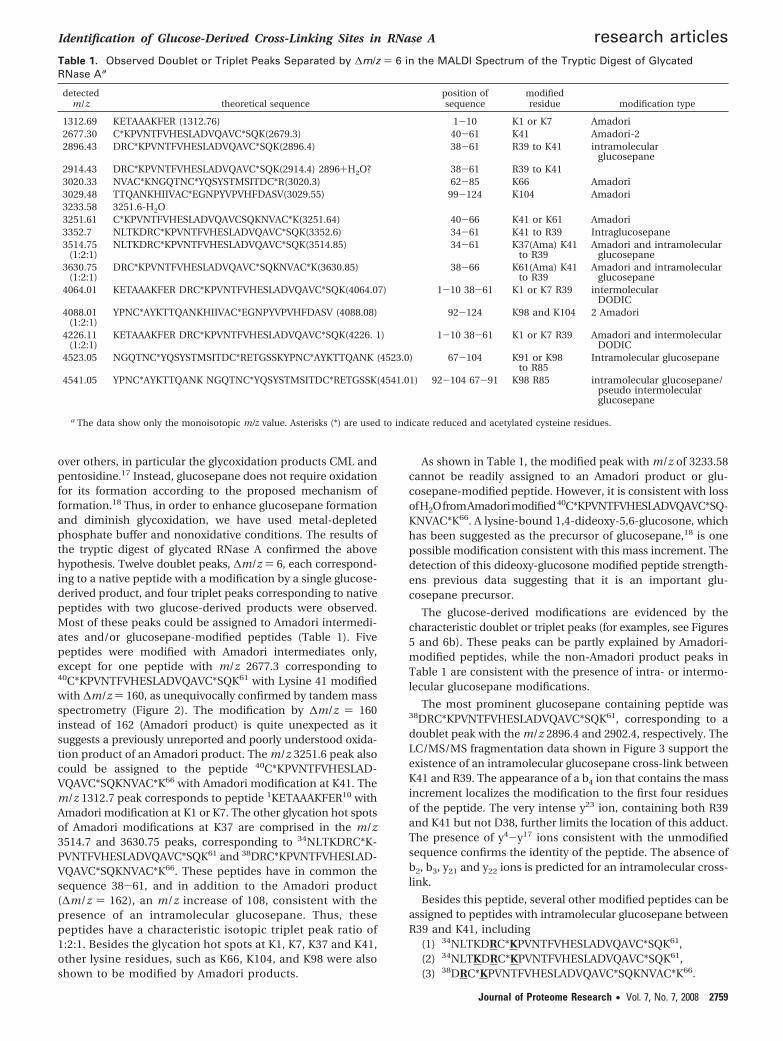
over others, in particular the glycoxidation products CML andpentosidine.17 Instead, glucosepane does not require oxidationfor its formation according to the proposed mechanism offormation.18 Thus, in order to enhance glucosepane formationand diminish glycoxidation, we have used metal-depletedphosphate buffer and nonoxidative conditions. The results ofthe tryptic digest of glycated RNase A confirmed the abovehypothesis. Twelve doublet peaks, ∆m/z ) 6, each correspond-ing to a native peptide with a modification by a single glucose-derived product, and four triplet peaks corresponding to nativepeptides with two glucose-derived products were observed.Most of these peaks could be assigned to Amadori intermedi-ates and/or glucosepane-modified peptides (Table 1). Fivepeptides were modified with Amadori intermediates only,except for one peptide with m/z 2677.3 corresponding to40C*KPVNTFVHESLADVQAVC*SQK61 with Lysine 41 modifiedwith ∆m/z ) 160, as unequivocally confirmed by tandem massspectrometry (Figure 2). The modification by ∆m/z ) 160instead of 162 (Amadori product) is quite unexpected as itsuggests a previously unreported and poorly understood oxida-tion product of an Amadori product. The m/z 3251.6 peak alsocould be assigned to the peptide 40C*KPVNTFVHESLAD-VQAVC*SQKNVAC*K66 with Amadori modification at K41. Them/z 1312.7 peak corresponds to peptide 1KETAAAKFER10 withAmadori modification at K1 or K7. The other glycation hot spotsof Amadori modifications at K37 are comprised in the m/z3514.7 and 3630.75 peaks, corresponding to 34NLTKDRC*K-PVNTFVHESLADVQAVC*SQK61 and 38DRC*KPVNTFVHESLAD-VQAVC*SQKNVAC*K66. These peptides have in common thesequence 38-61, and in addition to the Amadori product(∆m/z ) 162), an m/z increase of 108, consistent with thepresence of an intramolecular glucosepane. Thus, thesepeptides have a characteristic isotopic triplet peak ratio of1:2:1. Besides the glycation hot spots at K1, K7, K37 and K41,other lysine residues, such as K66, K104, and K98 were alsoshown to be modified by Amadori products.
As shown in Table 1, the modified peak with m/z of 3233.58cannot be readily assigned to an Amadori product or glu-cosepane-modified peptide. However, it is consistent with lossofH2OfromAmadorimodified40C*KPVNTFVHESLADVQAVC*SQ-KNVAC*K66. A lysine-bound 1,4-dideoxy-5,6-glucosone, whichhas been suggested as the precursor of glucosepane,18 is onepossible modification consistent with this mass increment. Thedetection of this dideoxy-glucosone modified peptide strength-ens previous data suggesting that it is an important glu-cosepane precursor.
The glucose-derived modifications are evidenced by thecharacteristic doublet or triplet peaks (for examples, see Figures5 and 6b). These peaks can be partly explained by Amadori-modified peptides, while the non-Amadori product peaks inTable 1 are consistent with the presence of intra- or intermo-lecular glucosepane modifications.
The most prominent glucosepane containing peptide was38DRC*KPVNTFVHESLADVQAVC*SQK61, corresponding to adoublet peak with the m/z 2896.4 and 2902.4, respectively. TheLC/MS/MS fragmentation data shown in Figure 3 support theexistence of an intramolecular glucosepane cross-link betweenK41 and R39. The appearance of a b4 ion that contains the massincrement localizes the modification to the first four residuesof the peptide. The very intense y23 ion, containing both R39and K41 but not D38, further limits the location of this adduct.The presence of y4-y17 ions consistent with the unmodifiedsequence confirms the identity of the peptide. The absence ofb2, b3, y21 and y22 ions is predicted for an intramolecular cross-link.
Besides this peptide, several other modified peptides can beassigned to peptides with intramolecular glucosepane betweenR39 and K41, including
(1) 34NLTKDRC*KPVNTFVHESLADVQAVC*SQK61,(2) 34NLTKDRC*KPVNTFVHESLADVQAVC*SQK61,(3) 38DRC*KPVNTFVHESLADVQAVC*SQKNVAC*K66.
Table 1. Observed Doublet or Triplet Peaks Separated by ∆m/z ) 6 in the MALDI Spectrum of the Tryptic Digest of GlycatedRNase Aa
detectedm/z theoretical sequence
position ofsequence
modifiedresidue modification type
1312.69 KETAAAKFER (1312.76) 1-10 K1 or K7 Amadori2677.30 C*KPVNTFVHESLADVQAVC*SQK(2679.3) 40-61 K41 Amadori-22896.43 DRC*KPVNTFVHESLADVQAVC*SQK(2896.4) 38-61 R39 to K41 intramolecular
glucosepane2914.43 DRC*KPVNTFVHESLADVQAVC*SQK(2914.4) 2896+H2O? 38-61 R39 to K413020.33 NVAC*KNGQTNC*YQSYSTMSITDC*R(3020.3) 62-85 K66 Amadori3029.48 TTQANKHIIVAC*EGNPYVPVHFDASV(3029.55) 99-124 K104 Amadori3233.58 3251.6-H2O3251.61 C*KPVNTFVHESLADVQAVCSQKNVAC*K(3251.64) 40-66 K41 or K61 Amadori3352.7 NLTKDRC*KPVNTFVHESLADVQAVC*SQK(3352.6) 34-61 K41 to R39 Intraglucosepane3514.75
(1:2:1)NLTKDRC*KPVNTFVHESLADVQAVC*SQK(3514.85) 34-61 K37(Ama) K41
to R39Amadori and intramolecular
glucosepane3630.75
(1:2:1)DRC*KPVNTFVHESLADVQAVC*SQKNVAC*K(3630.85) 38-66 K61(Ama) K41
to R39Amadori and intramolecular
glucosepane4064.01 KETAAAKFER DRC*KPVNTFVHESLADVQAVC*SQK(4064.07) 1-10 38-61 K1 or K7 R39 intermolecular
DODIC4088.01
(1:2:1)YPNC*AYKTTQANKHIIVAC*EGNPYVPVHFDASV (4088.08) 92-124 K98 and K104 2 Amadori
4226.11(1:2:1)
KETAAAKFER DRC*KPVNTFVHESLADVQAVC*SQK(4226. 1) 1-10 38-61 K1 or K7 R39 Amadori and intermolecularDODIC
4523.05 NGQTNC*YQSYSTMSITDC*RETGSSKYPNC*AYKTTQANK (4523.0) 67-104 K91 or K98to R85
Intramolecular glucosepane
4541.05 YPNC*AYKTTQANK NGQTNC*YQSYSTMSITDC*RETGSSK(4541.01) 92-104 67-91 K98 R85 intramolecular glucosepane/pseudo intermolecularglucosepane
a The data show only the monoisotopic m/z value. Asterisks (*) are used to indicate reduced and acetylated cysteine residues.
Identification of Glucose-Derived Cross-Linking Sites in RNase A research articles
Journal of Proteome Research • Vol. 7, No. 7, 2008 2759

Figure 2. Tandem mass spectrum of glycated RNase A tryptic peptide with molecular doubly charged ion at m/z 1339. This signal wasassigned to peptide 40C*KPVNTFVHESLADVQAVC*SQK61 with modification of K41 by ∆m/z ) 160 (oxidized Amadori). Every b iongreater than b2 contained this increment, while every y ion up to y20 was consistent with the unmodified peptide sequence, stronglyimplicating the modification of K41 by an oxidized Amadori product.
Figure 3. Tandem mass spectrum of the glycated RNase A tryptic peptide corresponding 36DRC*KPVNTFVHESLADVQAVC*SQK61 withprecursor m/z 1449.4 (double charged). The presence of ions b4 and y23, both containing the glucosepane mass increment, suggestthe presence of intermolecular glucosepane cross-linking K41 and R39. b18-b22 and y23 ions are doubly charged. The inset showsthe corresponding singly charged peptide with m/z 2896.4 and its m + 6 signal at 2902.4.
research articles Dai et al.
2760 Journal of Proteome Research • Vol. 7, No. 7, 2008

Figure 4. (a) Tandem mass spectrum of the glycated RNase A tryptic peptide, triply protonated precursor m/z 972.3, corresponding to36DRC*KPVNTFVHESLADVQAV C*SQK61, can be interpreted as an intramolecular modification by DODIC cross-linking residues K41and R39. The b11-b21, b23, and y17 ions are doubly charged. (b) Selected ion chromatograms demonstrating the coelution of peptideswith m/z 2896.4 (presumably glucosepane) and m/z 2914.4 (presumably DODIC or glucosepane plus 1 mol H2O) suggest that the peptideswith m/z 2914.4 may have different origins.
Identification of Glucose-Derived Cross-Linking Sites in RNase A research articles
Journal of Proteome Research • Vol. 7, No. 7, 2008 2761

Peptide 1 and 2 actually are same peptides with differentmodifications: peptide 1 only has an intramolecular glu-cosepane modification, whereas peptide 2 has both an in-tramolecular glucosepane and an Amadori product, presumablyat K37, resulting in the missed tryptic cleavage. Of relevancein the above analyses and throughout these studies is that thepeptide containing the glucosepane modification at K41 andR39 was a major signal, strongly suggesting that this site is ahighly favored target for glucosepane formation.
The m/z 2914.4 cannot be easily explained by either Amadorior glucosepane modifications. Instead, it is consistent with thepeptide 34DRC*KPVNTFVH ESLADVQAVC*SQK61 with, how-ever, a ∆m/z of 126. This mass increment is 18 Da greater thanthat of an intramolecular glucosepane modification, that is,potentially due to addition of H2O. Another possible AGE cross-link, DODIC (Figure 1), also has a six carbon backbonederivable from glucose that would yield M and M + 6 doubletswith ∆m/z ) 126 from the unmodified peptide. The LC/MS/MS spectrum of this peptide strengthened this tentative as-signment. As shown in Figure 4a, the existence of the predicted∆m/z ) 126 b4 ion again limits the modification to the fragment34DRC*K37, suggesting the modification could be DODIC.Interestingly, when we tried to find the triply charged peptidewith m/z 972.1 (Mr ) 2914.4 Da) in the LC/MS spectrum, wefound that there are two major species with different elutiontimes at 22.47 and 24.38 min (Figure 4b). The second 2914.4peak coeluted with the peak with m/z 2896.4, leading us tospeculate that the first 2914.4 peptide may be modified by, forexample, DODIC, whereas the second 2914.4 peptide might bemodified by glucosepane (2896.4) plus a water molecule addedin the mass spectrometer. Coelution may suggest in-sourcefragmentation, that is, loss of H2O.
A major question this work hoped to address is the extentto which glucose participates in intermolecular cross-linking.The peptide with m/z of 4541.0 matches the theoretical massof the peptide 67NGQTNC*YQSYSTMSITDC*RETGSSK91 cross-linked to 92YPNC*AYKTTQANK104, by glucosepane. Glucosep-ane would necessarily cross-link R85 and K98, as K104 and K91must be unmodified to allow for tryptic digestion. But with theexistence of the nearby doublet peak with m/z of 4523.0, weneed to reconsider the origin of the peak at 4541.0 (Figure 5).This hypothesized cross-linked peptide, 67NGQTNC*YQSYSTM-SITDC*RETGSSK91s92YPNC*AYKTTQANK104, is comprised ofsequential tryptic peptides. As indicated by the 3-D crystalstructure of RNase A (Figure 7), residues K98 and R85 are veryclose to each other, making formation of an intramolecularglucosepane probable and it would be indistinguishable bymass spectrometry from an intermolecular cross-link. With allthese factors considered, the peak at m/z 4541.0 may actuallyoriginate by hydrolysis of the peak with m/z 4523.0. In aglycated protein containing an intramolecular cross-link, theappearance of an M + 18 peak may be due to addition of waterupon enzymatic hydrolysis of an internal peptide bond. In thiscase of RNase A with glucosepane cross-linking K98 and R85,either partial tryptic digestion at K91 or chymotryptic digestionat Y97 will result in two mass spectral doublets separated by18 Da. The ratio of original peptide and the M + 18 peptidederived from enzymatic hydrolysis will depend on the efficiencyof enzymatic digestion (on K91-Y92 in tryptic digestion andon Y97-K98 in chymotryptic digestion).
The search for intermolecular cross-linking sites revealedunexpected results. The anticipated intermolecular glucosepanecross-link has proven elusive. However, after searching for all
glucosepane-based theoretical m/z values compatible withcross-linked peptides, we could not identify candidate glu-cosepane cross-linked tryptic peptides consistent either withm/z of 4064.01 (doublet peak) or 4226.1 (1:2:1 triplet peak).Interestingly, both of these m/z values matched theoreticalvalues of intermolecular cross-links assuming the cross-link hada molecular mass 18 Da greater than glucosepane. These dataprompted a search for other intermolecular cross-links whichcontain all 6 carbons of a single glucose. Again, the DODICcross-link is consistent with these mass spectral data. Thetandem mass spectrum of the 4064.01 peptide supported thepresence of an intermolecular cross-link with ∆m/z of 126between K1 and R39 (Figure 6a). The b ion, y ion, b′ ion and y′ion series establish the presence of both peptides, 1KETAAAK-FER10 and 38DRC*KPVNTFVHESLADVQAVC*S QK61. The ap-pearance of b′4 and b′5 also suggests that the cross-linking sitecontains K1 instead of K7. In the same manner, the peptidewith m/z value of 4226.1 also fits the cross-linking of peptide1KETAAAKFER10 and 38DRC*KPVNTFVHESLADVQAVC*S QK61
by DODIC, with additional Amadori modification at K7 or K41(Figure 6b).
In order to strengthen the above observations, digestionswith the enzymes Asp-N and chymotrypsin were also carriedout to verify the presence of the modification by Amadori,glucosepane and DODIC (Table 2). These two enzymes yieldrelatively low amounts of modified peptides and some of themcannot be assigned readily so that less information wasobtained than with tryptic digestion.
In the Asp-N digestion map, peptides with m/z of 1480.7 and1880.9 confirmed the existence of a major intramolecular
Figure 5. Both tryptic and chymotryptic peptides indicate thepresence of an intramolecular glucosepane cross-link betweenK98 and R85. Incomplete enzymatic hydrolysis between thecross-linked residues would yield a pseudointermolecular-glucosepane, resulting in an additional m/z of 18 after diges-tion. Thus, m/z 4523.0 corresponds to peptide 67NGQTNC-*YQSYSTMSITDC*RETGSSKYPNC*AYKTTQANK104 with anintramolecular glucosepane at K98 and R85. The m/z of 4259.8corresponds to peptide 80SITDC*RETGSSKYPNC*AYKTTQAN-KHIIVAC*EGNPY115 with an intramolecular glucosepane at R85and K98/K104.
research articles Dai et al.
2762 Journal of Proteome Research • Vol. 7, No. 7, 2008

Figure 6. (a) Tandem mass spectrum of the quadruply charged precursor ion, m/z 1016.8. y22 ion suggests the cross-linking at R39,while b′4, b′5, and y′4 ion suggests that K1 instead of K7 is the cross-linking site. The b11-b14, b16-b20, b′4, b′5, y21 and y22 ions aretriply charged, and the b′8 ion is quadruply charged. The data are compatible with an intermolecular DODIC cross-link. (b) MALDImass spectrum of the tryptic digestion of glycated RNase A. Two sets of peaks point to intermolecular cross-linking by DODIC betweenK1 and R39. The two peptides cross-linked are 1KETAAAKFER10 and 38DRC*KPVNTFVHESLADVQAVC*SQK61.
Identification of Glucose-Derived Cross-Linking Sites in RNase A research articles
Journal of Proteome Research • Vol. 7, No. 7, 2008 2763

glucosepane between K41 and R39. A relative weak doubletpeak (∆m/z ) 6), with m/z 1898.9 shows that an intramolecularDODIC cross-link could be present between K41 and R39. Thedoublet peak with m/z 2830.3 and 2836.3 corresponds to thepeptide 14DSSTSAASSSNYC*NQMMKSRNLTK37, with an Ama-dori modification at K31 or K37. On the basis of the data withtrypsin and the known glycation hot spots, modification at K37is more likely than at K31. In the same manner, the peptidewith m/z 3604.6 is assigned to 53DVQAVC*SQKNVAC*KN-
GQTNC*YQSYS TMSIT82, with Amadori modification at K61 orK66. Combined with the data from tryptic digestion, K66 wouldbe the more likely site. Both a weak doublet peak at 4142.8and a strong double peak at 4574.2 suggest an Amadorimodification at K91, K98 or K104. The two sets of doublet peakswith m/z 2444.1 and 3734.7 did not correspond to modificationof predicted Asp-N peptides. But it is possible that peak 2444.1comes from an intramolecular glucosepane-modified peptide,whereas peak 3734.7 comes from an Amadori-modified peptide
Table 2. Modified Peptides Mapped by Enzymatic Digestion with (a) Asp-N and (b) Chymotrypsin
detected m/z theoretical sequenceposition ofsequence
modifiedresidue
modificationtype
(a) Asp-N1394.61898.9 DRC*KPVNTFVHESLA (1898.9) 38-52 R39-K41 Intra-DODIC1480.7 DRC*KPVNTFVH(1480.7) 38-48 R39-K41 Intraglucosepane1880.9 DRC*KPVNTFVHESLA (1880.9) 38-52 R39-K41 Intraglucosepane2444.1 Originated from 2336.0 Intraglucosepane2830.3? DSSTSAASSSNYC*NQMMKSRNLTK (2830.3) 14-37 K31 or K37 Amadori3604.6 DVQAVC*SQKNVAC*KNGQTNC*YQSYSTMSIT (3604.6) 53-82 K61 or K66 Amadori3734.7 Originated from 3572.5 Amadori4142.8 ETGSSKYPNC*AYKTTQANKHIIVAC*EGNPYVPVHF (4143.0) 86-120 K91 or K98
or K104Amadori
4574.2 DC*RETGSSKYPNC*AYKTTQANKHIIVAC*EGNPYVPVHF (4574.2) 83-120 K91 or K98or K104
Amadori
(b) Chymotrypsin811.1903.41076.01343.61381.61473.7 TKDRC*KPVNTF(1473.7) 36-46 R39 to K41/K37 Intraglucosepane2124.82206.1 KTTQANKHIIVAC*EGNPY(2206.1) 98-115 K98 or K104 Amadori2271.0 SITDC*RETGSSKYPNC*AY (2271.0) 80-97 K91 Amadori4259.8 SITDC*RETGSSKYPNC*AY 80-97 R85 Interglucosepane
KTTQANKHIIVAC*EGNPY(4260.0) 98-115 K98 or K104 Or intramolecular glucosepane4277.8 SITDC*RETGSSKYPNC*AYKTTQANKHIIVAC*EGNPY(4278.0) 80-115 R85 to K98? intramolecular glucosepane
Table 3. The Modifications Affected by Mutations of the Predicted Intra- or Intermolecular Cross-Linking Sitesa
observed peptides WT R39A R85A K91A
2677.2 (K41 Amadori-2) + 3321.6 (K41 Amadori)(36-61) + +2896.4 (K41-R39 Intraglucosepane) + 3321.6(K41 Amadori)(36-61) + +2914.4 (K41-R39 Intra-DODIC) + 3321.6(K41 Amadori)(36-61) + +2953.4 + + +3020.3 (K66 Amadori)(62-85) + + 4421.8(62-98) +3029.4 (K104 Amadori) + + + +3233.5 (K41 or K61 Amadori-H2O) + 3419.6 (K41 Amadori-H2O)(38-66) + +3251.5 (K41 or K61 Amadori) + 3437.5 (K41 Amadori)(38-66) + +3351.6 (K41-R39 Intraglucosepane) + 3321.6(K41 Amadori)(36-61) + +3380.7 + + + +3514.7(T) (K41-R39 Intraglucosepane
and K37 Amadori)+ 3481.6(K37 and K41 Amadori)(34-61) + +
3630.7(T) (K41-R39 Intraglucosepaneand K61 Amadori)
+ 3481.6(K37 and K41 Amadori)(34-61) +
4046.0 (K1-R39 Interglucosepane) + 1312.7 (K1 Amadori) (1-10) + +4064.0 (K1-R39 Inter-DODIC) + 1312.7 (K1 Amadori) (1-10) + +4082.0 (K1-R39 Inter-DODIC+H2O) + 1312.7 (K1 Amadori) (1-10) + +4226.0(T) (K1-R39 Interglucosepane
and K7 Amadori)+ + + +
4523.0 (K98-R85Intraglucosepane)
+ + 4491.9 (K98 Amadori)(67-104)
4465.9 (K98-R85 intraglucosepane)(67-104)
4541.0 (K98-R85 Intraglucosepane/pseudo interglucosepane)
+ + 4491.9 (K98 Amadori)(67-104)
4465.9 (K98-R85intraglucosepane) (67-104)
4558.9 (K98-R85 Intra-DODIC/pseudointer-DODIC)
+ + 4491.9 (K98 Amadori)(67-104)
4483.9 (K98-R85 intra-DODIC)(67-104)
4581.0 (K1-R85inter glucosepane-H2O)
+ +
4599.0 (K1-R85 inter glucosepane) + +
a The plus sign (+) means that the m/z value (stand for modified peptides) is found in the tryptic digest of glycated RNase A wild-type or mutants.
research articles Dai et al.
2764 Journal of Proteome Research • Vol. 7, No. 7, 2008

due to the appearance of their prominent corresponding nativepeptides (m/z ) 2336.0 and 3572.5, respectively).
Finally, in the chymotryptic digestion map, the peptide withm/z 1473.7 confirms the major intramolecular glucosepanebetween K41 and R39. Furthermore, the peptide with m/z2271.0 indicates that the peptide 80SITDC*RETGSSKYPNC*AY97
is modified by an Amadori product at K91, a site whosemodification was not identified in the tryptic digest. Thepeptides with m/z 4259.8 and 4277.8 have been discussedbefore and could represent the same intramolecular glu-cosepane-modified peptide (vide supra) and after enzymatichydrolysis between the cross-linked residues.
The tryptic mapping of wild-type and RNase A mutants R39A,R85A, and K91A incubated with glucose is summarized in Table3. As predicted, when R39 is changed to A39, the assigned R39-K41 intramolecular cross-links and K1-R39 intermolecularcross-links disappeared. Instead, new peptides with K1 or K41modified with Amadori product were shown as glucose-modified peaks, such as peptide doublet peaks with m/z of1312.7, 3321.6, 3419.6, 3437.5, 3481.6 (triplet). In contrast,peptides with cross-linking sites different from R39, such asK98-R85 and K1-R85, were not affected by the R39A mutation.Similarly, the mutation R85A only affects R85 containing cross-linking, such as K98-R85 and K1-R85. The newly identifiedpeptide with m/z of 4491.5 is assigned to peptide 67NGQTNC*YQ-SYSTMSITDC*RETGSSKYPNC*AYKTTQANK104, with possibleAmadori modification at K98, due to lack of R85. All thepeptides using R39 as the proposed cross-linking site remainunaffected.
Discussion
As Biemel et al.19 proposed, both glucosepane and DODICare derived from Amadori products. Consequently, clarifyingthe glycation hot spots for the Amadori product was a necessarystep prior to investigating the sites of glucosepane or DODICformation. Two studies by Watkins et al. and Brock et al. agreedon the glycation hot spots of RNase A with the order of K41,K7, K37 and K120,21 with slight differences. Watkins groupshowed that K1 is more glycated than K37 based on radioactiv-ity measurement of [14C]glucose, which should be more reliablethan the semiquantitative methods of mass spectrometry. Intheir study, 38% of K41, 29% of K7, 24% of K1 and 9% of K37were glycated by Amadori products. One can assume thatbecause the Amadori product is the precursor of glucosepane
and DODIC, the sites that are preferentially modified byAmadori product accumulation will have a greater potentialfor glucosepane formation, particularly if there is a proximalarginine residue.
Our observation confirmed the four glycation hot spots listedabove. Additional glycation sites also were indicated by enzy-matic peptide maps as K66, K91, K98, and K104. The identifica-tion of additional sites of modification may be due to thedifferent incubation conditions and digestion procedures. Forexample, we have used 500 mM glucose, while previous studiesby Watkins et al. and Brock et al. have used 400 mM glucose.We used nonoxidative incubation conditions, while previousstudies did not exclude O2. As expected, the major cross-linkidentified under our nonoxidative conditions is glucosepaneas evidenced by the dominant tryptic peptide with m/z 2896.4(residues 38-61) in the digestion of glycated RNase. In thisaspect, our in vitro nonoxidative incubation results conformwell with the predominant level of glucosepane observedduring analysis of in vivo human tissue modifications.11,12
Most established protocols and routine methods used toidentify and confirm cross-links in vivo utilize complete acidhydrolysis or exhaustive enzymatic digestion of proteins to theconstituent amino acids, and amino acids with modifications.11,22
The cross-links identified by these protocols will include bothintra- and intermolecular cross-links. Thus, discrimination ofthe cross-links as being primarily intra- or intermolecular isimportant for understanding of the in vivo implication of cross-linking.
Whether intra- or intermolecular glucosepane cross-links areformed should depend on the availability of nearby arginineresidue(s) at the major sites of Amadori product accumulation.
Table 4. The Relative Surface Accessibility (RSA) Values ofLysines and Arginines Are Predicted by SARIG Server
lysines and arginineson RNase A RSA
K1 118.0K7 31.5K31 46.9K37 45.1K41 21.7K61 44.3K66 69.2K91 75.0K98 39.6K104 26.8R10 31.7R33 23.4R39 58.2R85 42.5
Figure 7. Distance of all nearby lysine and arginine pairs in RNaseA. Calculated lysine and arginine distance: K41 to R39, 7.4 Å; K37to R39, 13.6 Å; K98 to R85, 4.0 Å; K7 to R10, 8.1 Å.
Identification of Glucose-Derived Cross-Linking Sites in RNase A research articles
Journal of Proteome Research • Vol. 7, No. 7, 2008 2765

Thus, we calculated the distance between several pairs ofnearby lysine and arginine residues from the crystal structure1RBX deposited in the protein data bank (Dunbar, J.; Yennawar,H. P.; Banerjee, S.; Farber, G. K., unpublished results) usingPyMOL (DeLano, W.L., The PyMOL Molecular Graphics System(2002) on World Wide Web http://www.pymol.org). Thesedistances are highlighted in Figure 7.
From the values of the paired lysine/arginine distances, themost likely intramolecular cross-links are K98-R85 andK41-R39. In the MALDI tryptic peptide map, the intensity ofthe peptide with intramolecular glucosepane between K41 andR39 (m/z 2896.4) is much higher than the peptide withintramolecular glucosepane between K98 and R85 (m/z 4523.0).K41 has been unequivocally established as the most prevalentAmadori accumulation site, whereas K98 is not an establishedglycation hot spot, although our data proved the presence ofthe Amadori product on K98. Other factors may have minoreffects on the level of intramolecular glucosepane formationat different sites, such as the relative surface accessibility oflysine and arginine. As shown in Table 4, the relative surfaceaccessibility (RSA) values of lysines and arginines are predictedby the Structural Analysis of Residue Interaction Graphs(SARIG) server provided by Weizmann Institute of Science(bioinfo2.weizmann.ac.il/pietro/SARIG/). The more the argi-nine is exposed, the greater propensity for intramolecularglucosepane formation. The lowest accessibility of K41 will notinhibit the intramolecular glucosepane formation as long as ithas a local arginine “mate”. The highest accessibility of R39makes it not only a suitable local “mate” for K41, but also agood candidate for intermolecular cross-linking.
The existence of both modifications with ∆m/z 108 and 126is very interesting. These paired modifications suggest that bothglucosepane and DODIC modifications are occurring at thesame sites. Initially, DODIC modifications were not anticipatedbecause of low levels observed in vivo as compared to glu-cosepane. Glucosepane represents the dominant cross-link incollagen12 and in lens protein (132.3-241.7 pmol glucosepane/mg of protein compared to 1.3-8.0 pmol DODIC/mg of
protein).11 However, the appearance of DODIC at the samesites as intramolecular glucosepane is quite reasonable becauseglucosepane and DODIC share the same precursor, the ami-noketose. An interesting part of this work is the relative peakintensity of intramolecular glucosepane and intramolecularDODIC modifications at different intracross-linking sites. At theK41 and R39 cross-linking site, the intramolecular glucosepanecontaining peptide ion (m/z 2896.4) is much more intense thanthe intramolecular DODIC containing peptide ion (m/z 2914.4).While at the K98 and R85 cross-linking site, instead, theintramolecular DODIC containing peptide ion (4541.0) is moreintense than the intramolecular glucosepane containing pep-tide ion (4523.0). The peptide ion at m/z 4541.0 may be derivedfrom two sources, that is, the peptide 67NGQTNC*YQSYSTMSITDC*RETGSSKYPNC*AYKTTQANK104 with an intramo-lecular DODIC modification, or the peptide 67NGQTNC*YQ-SYSTMSITDC*RETGSSKYPNC*AYKTTQANK104 with intramo-lecular glucosepane modification and an additional trypticcleavage at K91.
An important aspect of this study was to identify the sites ofglucose-derived intermolecular cross-links. As noted above, thepeaks at m/z 4064.0 and 4226.1 fit the theoretical masses forintermolecular glucosepane containing peptides. Furthermore,the LC/MS/MS fragmentation data of the peptide with m/z4064.0 not only confirmed the presence of both peptides, butalso suggested that K1 and R39 were the cross-linked residues.As shown in Table 2, the m/z value of 4226.1 was consistentwith the same peptides being cross-linked with an additionalAmadori modification at K7. As shown in Table 4, the relativesurface accessibilities of K1 and R39 are the highest amonglysines and arginines of RNase A, respectively. Although theaccumulation of Amadori product at K1 is not the dominantsite in RNase A, K1 is one of the glycation hot sites. With allthese factors taken into consideration, the major intercross-link found between K1 and R39 is not so surprising.
However, the observation of DODIC as the only intercross-link observed is quite surprising. In in vitro incubations ofbovine serum albumin with glucose,19 Biemel et al. reported
Figure 8. Schematic representation of glucose mediated cross-link formation in RNase A.
research articles Dai et al.
2766 Journal of Proteome Research • Vol. 7, No. 7, 2008

about 3-fold higher yields of glucosepane than DODIC. Instead,glucosepane is present at 10-40 times more than DODIC inboth human serum albumin and human lens proteinsamples.11 The authors attribute the dominance of glucosepanein vivo to that of its precursor, N6-(2,3-dihydroxy-5,6-dioxo-hexyl)-L-lysinate, which is irreversibly linked to the protein andinaccessible to the detoxifying reductases and thus becomes apersistent glycation agent. This character is quite different fromother important intermediates including 3-DG, MGO, and GO,which exist in equilibrium between free and loosely protein-associated forms.
Although Lederer et al. initially proposed a mechanism ofglucosepane formation through DODIC as a precursor,23 thepresence of DODIC and the absence of glucosepane in theincubation of BSA with a 3-deoxyosone mixture exclude DODICas a precursor of glucosepane.19 Using o-phenylenediamine asa trapping agent for protein bound dicarbonyl groups, Biemelet al. suggest that N6-(2,3-dihydroxy-5,6-dioxohexyl)-L-lysinateis the only prominent lysine linked R-diketo compound formedfrom the incubation of glucose with lysozyme.24 Later theyproposed a mechanism for the formation of glucosepane fromAmadori products via N6-(2,3-dihydroxy-5,6-dioxohexyl)-L-ly-sinate and the seven member ring intermediate, azepanone.Thus, the formation of glucosepane and DODIC only sharesthe step from lysine to Amadori product. The formation ofglucosepane through N6-(2,3-dihydroxy-5,6-dioxohexyl)-L-ly-sinate requires several enolization steps along the sugarbackbone to effect the intramolecular oxidation-reductionreactions, whereas these steps are not required in DODICformation. We observed that intramolecular glucosepane ishighly favored in the K41-R39 cross-link over DODIC. Interest-ingly, Biemel et al. observed that the three lysine residues K33,K96, and K116 of lysozyme, which all have partner arginine(s)within 5 Å, have the highest rate of transformation fromAmadori to N6-(2,3-dihydroxy-5,6-dioxohexyl)-L-lysinate.24 Thiscorrelation suggested that the guanidino group of argininecatalyzes this transformation.
By analogy, the lysine sites with nearby arginine residues inRNase A could also favor the formation of glucosepane, whereasthose lysine residues without nearby arginine(s) will be morelikely to form DODIC. In our case, the highly glycated lysineK41 is more prone to form intramolecular glucosepane due toclose proximity of R39. Because of extremely low surfaceaccessibility, K41 can form neither intermolecular glucosepanenor intermolecular DODIC cross-links. Among all other glyca-tion hot spots, K1 has high surface accessibility and relativelyhigh Amadori accumulation, which makes it a good candidatesite to form an intermolecular cross-link. Without nearbyarginine-catalyzed enolization, DODIC formation is favored atthis site. This process is presented in Figure 8.
In summary, this work demonstrates that the Amadoriproducts, glucosepane, and DODIC are major glucose-derivedmodifications accumulating in RNase A under nonoxidativeglycation conditions. Two intramolecular glucosepane cross-links were found at sites R39-K41 and R85-K98. At the siteR39-K41, an intramolecular DODIC cross-link was also ob-served. The only intermolecular cross-link was also observedat K1-R39, but surprisingly found to be a DODIC cross-link.
Abbreviations: 3-DG, 3-deoxyglucosone; AGE(s), advancedglycation end product(s); DODIC, 3-deoxyglucosone-derivedimidazolium cross-link; DTPA, diethylenetriamine pentaaceticacid; LC/MS, liquid chromatography-mass spectrometry; MAL-DI, matrix-assisted laser desorption and ionization; RNase A,
ribonuclease A; ROS, reactive oxygen species; RP-HPLC, re-versed phase-high performance liquid chromatography; TFA,trifluoroacetic acid.
Acknowledgment. This work was supported by agrant from the National Institute on Aging AG-18426 and inpart by NEI grant EY-07099.
References(1) Monnier, V. M.; Vishwanath, V.; Frank, K. E.; Elmets, C. A.;
Dauchot, P.; Kohn, R. R. Relation between complications of typeI diabetes mellitus and collagen-linked fluorescence. N. Engl.J. Med. 1986, 314 (7), 403–8.
(2) Hammes, H. P.; Martin, S.; Federlin, K.; Geisen, K.; Brownlee, M.Aminoguanidine treatment inhibits the development of experi-mental diabetic retinopathy. Proc. Natl. Acad. Sci. U.S.A. 1991, 88(24), 11555–8.
(3) Nakayama, H.; Mitsuhashi, T.; Kuwajima, S.; Aoki, S.; Kuroda, Y.;Itoh, T.; Nakagawa, S. Immunochemical detection of advancedglycation end products in lens crystallins from streptozocin-induced diabetic rat. Diabetes 1993, 42 (2), 345–50.
(4) Mitsuhashi, T.; Nakayama, H.; Itoh, T.; Kuwajima, S.; Aoki, S.;Atsumi, T.; Koike, T. Immunochemical detection of advancedglycation end products in renal cortex from STZ-induced diabeticrat. Diabetes 1993, 42 (6), 826–32.
(5) Schnider, S. L.; Kohn, R. R. Effects of age and diabetes mellituson the solubility and nonenzymatic glucosylation of human skincollagen. J. Clin. Invest. 1981, 67 (6), 1630–5.
(6) Hamlin, C. R.; Luschin, J. H.; Kohn, R. R. Partial characterizationof the age-related stabilizing factor of post-mature human col-lagen--II. By the use of trypsin. Exp. Gerontol. 1978, 13 (6), 415–23.
(7) Hamlin, C. R.; Luschin, J. H.; Kohn, R. R. Partial characterizationof the age-related stabilizing factor of post-mature human col-lagen--I. By the use of bacterial collagenase. Exp. Gerontol. 1978,13 (6), 403–14.
(8) LaBella, F. S.; Paul, G. Structure of collagen from human tendonas influenced by age and sex. J. Gerontol. 1965, 20, 54–9.
(9) Paul, R. G.; Bailey, A. J. Glycation of collagen: the basis of its centralrole in the late complications of ageing and diabetes. Int. J. Bio-chem. Cell Biol. 1996, 28 (12), 1297–310.
(10) Kivirikko, K. I.; Peltonen, L. Abnormalities in copper metabolismand disturbances in the synthesis of collagen and elastin. Med.Biol. 1982, 60 (2), 45–8.
(11) Biemel, K. M.; Friedl, D. A.; Lederer, M. O. Identification andquantification of major maillard cross-links in human serumalbumin and lens protein. Evidence for glucosepane as thedominant compound. J. Biol. Chem. 2002, 277 (28), 24907–15.
(12) Sell, D. R.; Biemel, K. M.; Reihl, O.; Lederer, M. O.; Strauch, C. M.;Monnier, V. M. Glucosepane is a major protein cross-link of thesenescent human extracellular matrix. Relationship with diabetes.J. Biol. Chem. 2005, 280 (13), 12310–5.
(13) Monnier, V. M.; Mustata, G. T.; Biemel, K. L.; Reihl, O.; Lederer,M. O.; Zhenyu, D.; Sell, D. R. Cross-linking of the extracellularmatrix by the maillard reaction in aging and diabetes: an updateon ”a puzzle nearing resolution. Ann. N.Y. Acad. Sci. 2005, 1043,533–44.
(14) Price, D. L.; Rhett, P. M.; Thorpe, S. R.; Baynes, J. W. Chelatingactivity of advanced glycation end-product inhibitors. J. Biol.Chem. 2001, 276 (52), 48967–72.
(15) Khalifah, R. G.; Todd, P.; Booth, A. A.; Yang, S. X.; Mott, J. D.;Hudson, B. G. Kinetics of nonenzymatic glycation of ribonucleaseA leading to advanced glycation end products. Paradoxical inhibi-tion by ribose leads to facile isolation of protein intermediate forrapid post-Amadori studies. Biochemistry 1996, 35 (15), 4645–54.
(16) Laity, J. H.; Shimotakahara, S.; Scheraga, H. A. Expression of wild-type and mutant bovine pancreatic ribonuclease A in Escherichiacoli. Proc. Natl. Acad. Sci. U.S.A. 1993, 90 (2), 615–9.
(17) Miyata, T.; Wada, Y.; Cai, Z.; Iida, Y.; Horie, K.; Yasuda, Y.; Maeda,K.; Kurokawa, K.; van Ypersele de Strihou, C. Implication of anincreased oxidative stress in the formation of advanced glycationend products in patients with end-stage renal failure. Kidney Int.1997, 51 (4), 1170–81.
(18) Biemel, K. M.; Conrad, J.; Lederer, M. O. Unexpected carbonylmobility in aminoketoses: the key to major Maillard crosslinks.Angew. Chem., Int. Ed. Engl. 2002, 41 (5), 801–4.
(19) Biemel, K. M.; Reihl, O.; Conrad, J.; Lederer, M. O. Formationpathways for lysine-arginine cross-links derived from hexoses and
Identification of Glucose-Derived Cross-Linking Sites in RNase A research articles
Journal of Proteome Research • Vol. 7, No. 7, 2008 2767

pentoses by Maillard processes: unraveling the structure of apentosidine precursor. J. Biol. Chem. 2001, 276 (26), 23405–12.
(20) Watkins, N. G.; Thorpe, S. R.; Baynes, J. W. Glycation of aminogroups in protein. Studies on the specificity of modification ofRNase by glucose. J. Biol. Chem. 1985, 260 (19), 10629–36.
(21) Brock, J. W.; Hinton, D. J.; Cotham, W. E.; Metz, T. O.; Thorpe,S. R.; Baynes, J. W.; Ames, J. M. Proteomic analysis of the sitespecificity of glycation and carboxymethylation of ribonuclease.J. Proteome Res. 2003, 2 (5), 506–13.
(22) Sell, D. R.; Monnier, V. M. Structure elucidation of a senescencecross-link from human extracellular matrix. Implication of pen-toses in the aging process. J. Biol. Chem. 1989, 264 (36), 21597–602.
(23) Lederer, M. O.; Klaiber, R. G. Cross-linking of proteins by Maillardprocesses: characterization and detection of lysine-arginine cross-links derived from glyoxal and methylglyoxal. Bioorg. Med. Chem.1999, 7 (11), 2499–507.
(24) Biemel, K. M.; Lederer, M. O. Site-specific quantitative evaluationof the protein glycation product N6-(2,3-dihydroxy-5,6-dioxo-hexyl)-L-lysinate by LC-(ESI)MS peptide mapping: evidence forits key role in AGE formation. Bioconjugate Chem. 2003, 14 (3),619–28.
PR700874A
research articles Dai et al.
2768 Journal of Proteome Research • Vol. 7, No. 7, 2008