iastm: taking the sore thumb ucsf assistant clinical ... · 9/5/2015 2 epidermis: thick skin dermis...
TRANSCRIPT

9/5/2015
1
Vital Importance of the
Connective Tissue System
DaPrato, DPT, SCS 2015
IASTM: Taking the sore thumb out of manual therapy
Disclosures
• UCSF Assistant Clinical Professor
• Outpatient Sports-Ortho perspective
• Created the approach Myofascial Decompression Techniques
Objectives:
• Describe the layers and subunits of the connective tissue system. (CT)
• Summarize the role collagen, ground substance, and other CT elements may be affecting movement patterns
• Define the terms tensegrity, thixotropy, as they relate to stress-strain curves
• Distinguish the effect of stretching on length-tension vs. CT elements
• Describe common IASTM interventions to improve mobility of the CT system
Integument
Skin layers Epidermis: thin skin

9/5/2015
2
Epidermis: thick skin Dermis
Connective tissue development• Ectoderm and Mesoderm gives rise to the
CT of skin and fascia above and around
muscle tissue.
Fascial Layers• Superficial
• Deep = Aponerotic & Epimysial
TLF, TFL, rectus sheath…
• Intermuscular
• Visceral
Living tissue is hydrated and dynamic
analogous…

9/5/2015
3
Dynamic nature of living tissue
• The way we think about movement in the human body is clouded by the way we learned anatomy
▫ Cadaver tissue, hard muscles, web like fascia
▫ Surgery observation; as soon as you cut into it you have changed it.
▫ AND what we observe in surgery = tourniquets used; even though living tissue, not the same
Functions of muscles
• We learn action but as we move in real world multiple
▫ ERs become IRs past 90 degrees
Jean-Claude GUIMBERTEAU, MD: strolling under the skin
Skin: more complex that we learned
Retinacula Cutis 4th International Fascia Congress
• Benchmark sciences filling in the gaps of traditional anatomy
9/5/2015
4
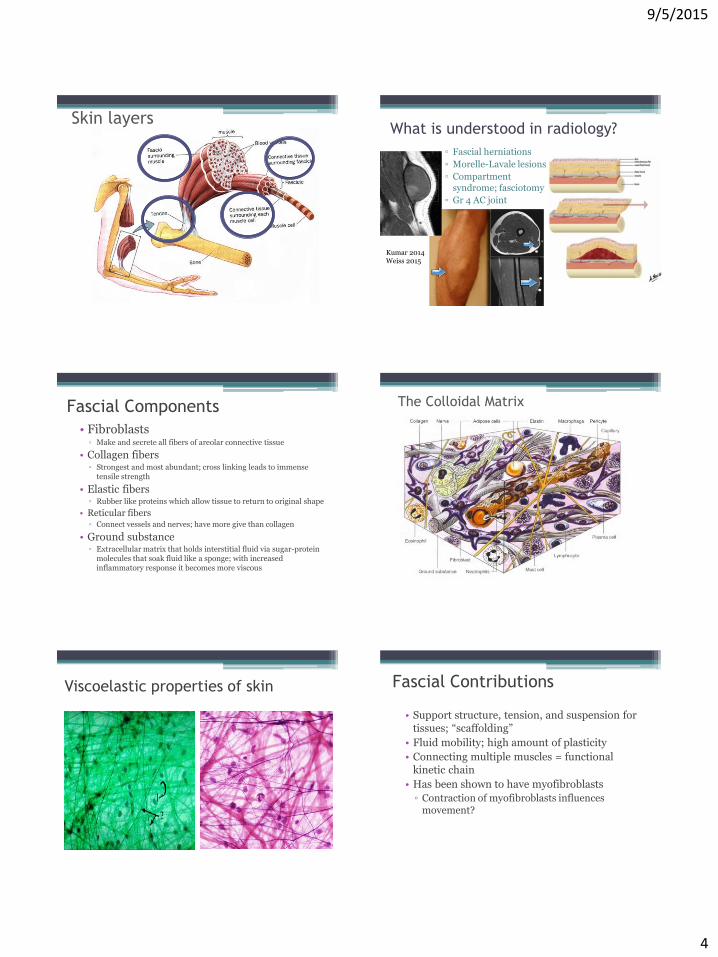
Skin layersWhat is understood in radiology?
▫ Fascial herniations
▫ Morelle-Lavale lesions
▫ Compartment syndrome; fasciotomy
▫ Gr 4 AC joint
Kumar 2014Weiss 2015
Fascial Components
• Fibroblasts▫ Make and secrete all fibers of areolar connective tissue
• Collagen fibers▫ Strongest and most abundant; cross linking leads to immense
tensile strength
• Elastic fibers▫ Rubber like proteins which allow tissue to return to original shape
• Reticular fibers▫ Connect vessels and nerves; have more give than collagen
• Ground substance▫ Extracellular matrix that holds interstitial fluid via sugar-protein
molecules that soak fluid like a sponge; with increased inflammatory response it becomes more viscous
The Colloidal Matrix
Viscoelastic properties of skin Fascial Contributions
• Support structure, tension, and suspension for tissues; “scaffolding”
• Fluid mobility; high amount of plasticity
• Connecting multiple muscles = functional kinetic chain
• Has been shown to have myofibroblasts
▫ Contraction of myofibroblasts influences movement?

9/5/2015
5
Muscles within the Fascial layers
• Twitch of skin – horse, cow, pig…
• Hair stand up - Arrector Pili
• Myofibroblasts
Viscoelastic properties: Collagen
• Dermis is made up of 80% collagen, dry weight, and of that collagen, 85% is type I
• Type 3 collagen is ~15% of dermal collagen, but is higher in immature tissue
• With age, ratio of type 1:3 collagen increases
• Increased collagen fiber density with age= decreased ground substance space
Viscoelastic properties Viscoelastic properties:
Ground substance – with GAGs
• Glycosaminoglycans
▫ Proteoglycans and repeating disaccharide units
▫ Commonly hyaluronan and chontratin sulfate; including dermatan sulfate
▫ Bind water in normal healthy tissue and proteins
▫ In aged skin, less binding to water and bind more to elastoic material= thickened
Viscoelastic properties: Thixotropic
Effect• Thixotropy is the property of certain gels or
fluids that are thick (viscous) under normal
conditions, but flow (become thin, less viscous) over time when shaken, agitated, or otherwise stressed.
Viscoelastic properties:
Creep and Hysteresis
• In relation to manual therapy, creep is the distortion of tissues as a function of pressure
over time
• Hysteresis is the exchange of heat and energy as tissues are distorted; permanent deformation. Microtrauma.
• With MFR 90-120 seconds is the time for generally the first barrier (R1) to release and push into new range of extensibility.
• Tendon Hysteresis in 5-10 minutes(Kubo 2001)

9/5/2015
6
Stress-strain curves; human tissueWhat really happens when we
stretch?• Sensory endpoint theory (Weppler & Magnusson 2010)
▫ Very little evidence that Torque/angle curves shift; even w/ 8 weeks
▫ More likely that the perception of the stretch sensation occurs later in the application of similar force
▫ PF stretch doesn’t change relfex pathway (Hayes 2012)
Stretching Soft tissue mobility: Folding
• CT ability to compress upon itself
• Shoulder Elevation= inferior capsule and axillary fold stretched, but also superior and anterior structures need to fold
• End Range hip flexion in supine = hams and
glute flexibility, but also anterior hip folding
Fascia encapsulates and supports Fascia encapsulates and supports

9/5/2015
7
TensegrityTensegrity
• “Tensional integrity”
• Fuller 1950’s first visualized by the sculptor Kenneth Snelson (Snelson, 1996).Fuller defines tensegrity systems as structures that stabilize their shape by continuous tension rather than by continuous compression
• Micro: studies of both cultured cells and whole tissues indicate that cell shape stability depends on a balance between microtubules and opposing contractile microfilaments
Tensegrity Mechanical CT Changes
Inflammation or Trauma
ECM response
Collagen cross-linkingCollagen
cross-linking
Ground substance viscosity
Tensegrity affected
Secondary movement
dysfunctions
Cascade of events:• Ge et al. 2008 studied involvement of central
sensitization mechanisms in local pain syndromes
• pain perception may result from a deregulation in peripheral afferent and central nervous system pathways- “chronic excitability”
Trigger Points
• Lower levels of oxygen, nutrients, blood perfusion
• Increased calcium levels, leading to excessive chronic muscle contracture, spasms
• Stress can lead to abnormal excess afferent stimulation
• Can have shortening of sarcomeres

9/5/2015
8
Fascial mechanics• Translating forces =“Slings”
• Lats to TLF to contra glute max and down lateral thigh =ITB Tx
Myofascial Lines
• Work of Thomas Myers
• Myofascial Tracks= muscles, tendons, ligaments and fascia
• Bony Stations= joints or insertional sites at bony landmark
• Can be static or motion driven▫ Picture: Pec minor, biceps,
coracobrachialis, rectus abdominis
▫ Or ab scar restricting shoulder or lumbar motion
Superficial Back Line
Includes:
Plantar fascia
Achilles tendon and Gastrocnemius
Hamstings
Sacrotuberous ligament
Thoracolumbar fascia
Erector spinae
Scalp fascia
Superficial Back Line
Lateral Line
• Often involved with leg length differences and pelvic obliquities
Includes:
Peroneals
Anterior ligament of the head of fibula
ITB and TFL
Superior fibers of glute max, medius
External and internal obliques
Splenius capitis and SCM
Lateral Line

9/5/2015
9
The Spiral Line
The Serape=double spiralIncludes:
Splenius capitis and cervicis
Rhomboids
Serratus anterior
Ext/Int obl. & ab aponerosis
TFL and ITB
Tib anterior
Peroneals
Bicep femoris to sacrotuberousligament
Lumbar fascia and erector spinae
Myofascial Lines
• Fascial planes and CT restrictions affect dynamic movement – scar adhesions
• Body takes the path of least resistance
• Interventions with traditional manual therapy
• Interventions with negative pressure applications: www.MyofascialDecompression.com
• Interventions with IASTM
• Manual therapy for lymphatic flow
Both picture thoracic extension; but may be restricted for different reasons
Compensations and Adaptations
“A best-practice model for managing patients with musculoskeletal complaints has yet to be identified.”-Tim Flynn (JOSPT 2007)
Consider hip strength for runners with foot pain, or
Use of foot othosis as a method of treating PFPS
Why does IASTM work?
• Bridge to the IASTM picture
• Mechanical connective tissue change
• Fascial plane restriction and scar adhesions
• Trigger points and metabolic exchange
• Fluid mobility
• Counterirritant; C fiber instead of A fiber; Gated
Pain generating mechanisms
• Receptors in TL Fascia (Schilder 2014 Pain)
▫ Muscle pain could be more akin to projection hyperalglesia, and not an “issue in the tissue.”
• IE: PF pain – likely cartilage or other soft tissue restrictions?
▫ Grooves in PF joint in cadavers
• OR IE: THR patients that have the same hip pain come back 6 months later

9/5/2015
10
IASTM = Instrument Assisted STM
• Mostly a Pro-inflammatory approach
▫ Some techniques are for flushing in acute phases
• Effect at the superficial fascia level, deep fascial membrane, epimyseum
▫ mechanoreceptors
Where are the Pacinian corpuscles?
=
Where are the Meisner corpuscles?
=
Where are the Ruffini corpuscles?
=
IASTM• ASTYM
• Graston
• SASTM
• FAKTR
• Iamtools
• GuaSha Orthopedic
• Target Point
• Fuzion Tool
• BioEdge
= GuaSha
IASTM• Self trigger point tools – theracane, TrP tools…
• Snowman with a hat, PAs
• Belt techniques for hip and shoulder
▫ Are you just moving the joint? Or are you also with an inferior hip glide pushing through RF trigger point region, and other contractile tissue that may reset sensitivity thresholds
• Negative pressure devices
Negative Pressure
Ultrasound With Myofascial Decompression

9/5/2015
11
MFD Techniques Negative Pressure
Negative Pressure Negative Pressure
Proof of Concept Study Proof of Concept Study

9/5/2015
12
GuaSha and IASTM
Basic Steps:• Watch the area with movement
patterns
• Sweep area with hands/fingers
• Trace and isolate with Vectoring
▫ 4 directions, find most limited.
▫ Compare contralateral
▫ Add in rotational vector as well
IASTM techniques
• Superficial scrape
▫ Sense percussive info from tool
• Scrape parallel fibers
▫ Into the motion barrier = direct = subacute and chronic
▫ Away from motion barrier =indirect = acute or irritable
• Diagonal and perpendicular
• Add movement
Contraindications
• Eyes; genitalia?• Unhealed wounds• Hemophilia, leukemia, active TB• Thrombocytopenia• Later stages of pregnancy• Influenza of fever• Moderate/severe anemia• Moderate/severe cardiac
conditions, high BP• Vasculitis• Cancer active• Skin elasticity disorders-EDS??
Precautions-Patients that are over eager
- addictive personalities, chronic pain?
-Blood thinners-Healing or thin skin
▫ Elderly, Psoriasis
-Pregnancy-Areas of ecchymosis
▫ Previous STM-Venous stasis and varicose veins-DM; tissue healing and neuropathy-Swollen tissue
▫ especially pitting edema
Results? Outcomes?

9/5/2015
13
Future Studies
• Perfusion and Diffusion MRI with MFD vs Grastonvs ASTYM vs other tools/techniques/modalities
• Pre, during, and 8-48 hrs after for different Tx
• Fascial changes on magnified MRI
• Local metabolic change; on a nano level
▫ IE: why does US work in some patients?
ReferencesBooks: • Functional Atlas of the Human Fascial System, Carla Stecco MD, 2015, 1st
Edition• The Endless Web. Fascial Anatomy and Physical Reality. L. Schultz, R.
Feitis, 1996. North Atlantic Books. • Anatomy Trains. Thomas Myers, 2008, 3rd editionJournals: • Abbott RD, et. Al. Stress and matrix-responsive cytoskeletal remodeling in fibroblasts.
J Cell Physiol. 2013 Jan; 228(1):50-7• Ates, F, et al. Muscle lengthening surgery causes differential acute mechanical effects
in both targeted and non-targeted synergistic muscles. Journal of Electromyography and Kinesiology, 23, 1198-1205, 2013
• Ballyns, Jet al. , Objective ultrasonic measures for characterizing myofascial trigger points associated with cervical pain. J. Ultrasound Med., vol. 30, pp. 1331-1340, 2011
• Bhattacharya V, et. Al. Detail microscopic analysis of deep fascia of lower limb and its surgical implication. Indian J Plast Surg. 2010 Jul;43(2):135-40
• Bonilla-Yoon, et al. The Morel-Lavallée lesion: pathophysiology, clinical presentation, imaging features, and treatment options. Emerg Radiol. 2014 Feb;21(1):35-43.
• Borgini E, Stecco A, Day J. A., Stecco, C. How much time is required to modify a fascial fibrosis? J Bodyw MovTher, 2010 Vol 14(4): 318 – 325
• Cao TV, Hicks MR, Standley PR. In vitro biomechanical strain regulation of fibroblast wound healing. The Journal of the American Osteopathic Association. Nov 2013;113(11):806-818
References• Ge HY,et al. Topographical mapping and mechanical pain sensitivity of myofascial trigger
points in the infraspinatus muscle. Eur J Pain. 2008 Oct;12(7):859-65 • Findley, T., Chaudhry, H., & Dhar, S. (2014). Transmission of muscle force to fascia during
exercise. Journal of Bodywork and Movement Therapies. In press 2014• Holm L, et al. Contraction intensity and feeding affect collagen and myofibrillar synthesis
rates differently in human skeletal muscle.Am J Physiol (Endo), 298: E257-E269, 2010• Kim KT, Kim YJ, Lee JW, et al.. Can necrotizing infectious fasciitis be differentiated from
nonnecrotizing infectious fasciitis with MR imaging? Radiology 2011;259(3):816–824• Kubo K, et. al. Is passive stiffness in human muscles related to the elasticity of tendon
structures? Eur J Appl Physiol. 2001 Aug;85(3-4):226-32 • Kumar S, Kumar S. Morel-Lavallee lesion in distal thigh: A case report. J Clin Orthop Trauma.
2014 Sep;5(3):161-6• Langevin HM, Nedergaard M, Howe AK. Cellular control of connective tissue matrix tension.
J Cell Biochem. 2013 Aug; 114(8):1714-9• May DA, et. al. Abnormal signal intensity in skeletal muscle at MR imaging: patterns, pearls,
and pitfalls. Radiographics. 2000 Oct;20 Spec No:S295-315• Schilder A, et al. Sensory findings after stimulation of the thoracolumbar fascia with
hypertonic saline suggest its contribution to low back pain. Pain 155:222-231, 2014• Schleip R. et al. Passive muscle stiffness may be influenced by active contractility of
intramuscular connective tissue. Medical Hypotheses 2006; 66(1): 66-71• Weppler CH, Magnusson SP. Increasing muscle extensibility: a matter of increasing length or
modifying sensation? Phys Ther. 2010 Mar;90(3):438-49 • Willard FH,et al. The thoracolumbar fascia: anatomy, function and clinical considerations. . J
Anat 2012; 221(6):507-36
Other recommended CE work
• www.ptrehab.ucsf.edu/education/continuing-education
• Janda courses or Movement Links• MyofascialDecompression.com• Kaiser Residency and fellowship• IPA PNF and FM• Great Lakes MFR course• Kinetacore.com• Spinalmanipulation.org• Myopain seminars• Systemicdryneedling.com
Thank You!!