hydrolysis of a novel lysosomotropic enzyme substrate for ... · homogenate (7.5-45 pg protein),...
TRANSCRIPT
Hydrolysis of a novel lysosomotropic enzyme substrate for ,&galactosidase within intact cells
Christine R. Kaneski,' Stephanie A. French, Marc-Raleigh Brescia, Michael J. Harbour, and Stephen P. E Miller
Developmental and Metabolic Neurology Branch, National Institute of Neurological Disorders and Stroke, National Institutes of Health, 9000 Rockville Pike, Bethesda, MD 20892
Abstract With the goal of improving the detection of lysosomal sphingolipid hydrolases within intact cells, we have re- cently synthesized a new fluorophor, 0-[4-(l-imidazolyl)butyl]- 2,3-dicyano-1,4-hydroquinonyl 0-D-galactopyranoside (Im-DCH-0- Gal). In the present study, we evaluated the interaction of Im- DCH-P-Gal and its tetraacetate derivative, Im-DCH-P- Gal(OAc)4, with living human fibroblasts. Im-DCH-@-Gal was shown to be a specific substrate for human lysosomal p- galactosidase in cell homogenates. Im-DCH-@-GaI(OAc), was taken up and hydrolyzed by normal fibroblasts under physiologi- cal culture conditions. Very little hydrolysis of Im-DCH-p- Gal(OAc), was observed in fibroblasts genetically deficient in lysosomal acid 0-galactosidase or in normal cells pretreated with the lysosomal inhibitors chloroquine and ammonium chloride. Analysis of substrate processing by cells indicated that normal and acid p-galactosidase-deficient cells showed similar rates of uptake and deacetylation of Im-DCH-P-Gal(OAc),, with an 80% decrease in the rate of deglycosylation of substrate by p- galactosidase-deficient fibroblasts. However, under our condi- tions, the fluorescent product was not well retained by cells. Our results indicate that this novel class of compounds may be useful in measuring lysosomal enzyme function in intact cells and may have application as a fluorescent marker for genet- ically altered cells.-Kaneski, C. R., s. A. French, M-R. Bres- cia, M. J. Harbour, and S. P. F. Miller. Hydrolysis of a novel lysosomotropic enzyme substrate for 0-galactosidase within in- tact cells. J Lipid Res. 1994. 35: 1441-1451.
Supplementary key words lysosomal storage diseases GM, ganglio- sidosis sphingolipid hydrolases glycosphingolipid metabolism fluorescent substrates
Glycosphingolipids are catabolized within lysosomes by a series of specific hydrolases that sequentially remove hexose residues from the polysaccharide moiety of these complex molecules. There are a number of genetic dis- eases in which an inheritable deficiency in one or more of these enzymes blocks the degradative pathway, resulting in the accumulation of lipid within the lysosomes. This accumulation disrupts cellular function and causes a vari- ety of systemic and neurologic disorders (1).
Although lysosomal glycosphingolipid hydrolases have been well characterized through studies of cell homogenates
and purified enzyme preparations, the understanding of the functions of these enzymes in intact cells has been hin- dered by the extreme hydrophobicity of their natural sub- strates. In addition, the lack of a useful chromophore in these substrates made it difficult to perform real-time measurement of intracellular enzymatic activity.
The importance of the cellular microenvironment in the modulation of in situ enzyme activity has been demonstrated in several studies showing that lysosomal hydrolases may have a much lower activity in vivo than is found in biochemical assays (2) and are affected by changes in intralysosomal p H (3, 4). In addition, sphin- golipid hydrolases are known to be associated with activa- tor proteins in vivo (5-7), but these enzyme-activator as- sociations may be disrupted by procedures used for biochemical assays.
O u r laboratory has developed a series of unique fluorescent enzyme substrates based on 2,3-dicyanohydro- quinone (DCH) (Fig. 1). D C H is a well-characterized fluorescent compound (8) that has been used in cultured cells to measure intracellular p H (9-14) and to test for cell viability (15). O u r enzymatic substrate consists of a D C H ring modified with a carbohydrate group to serve as a sub- strate for the enzyme, and a basic amine substituent, N- alkylimidazole, to promote concentration of the substrate within lysosomes. This design is based upon the lysosomo- tropic properties of amines that have pK, values near 7 (16, 17). These moderately basic amines have been shown to concentrate in subcellular compartments with low pH, such as lysosomes (18, 19). Another important property of
Abbreviations: Im-DCH-&Gal, 0-[4-(l-imidazolyl)butyl]-2,3-dicyano- 1,Chydroquinonyl P-D-galactopyranoside; Im-DCH-P-Gal(OAc)+, 0-[4-(1- imidazolyl)butyl]-2,3-dicyano-1,4-hydr~uinonyl~-~-~alactopyranoside tetraacetate; Im-DCH, O-[4-(l-imidazolyl)butyl]-2,3-dicyano-l,4-hydro- quinone; DCH, 2,3-dicyanohydroquinone; FBS, fetal bovine serum; TLC, thin-layer chromatography.
'To whom correspondence should be addressed.
Journal of Lipid Research Volume 35, 1994 1441
by guest, on July 17, 2018w
ww
.jlr.orgD
ownloaded from
tured in McCoy’s 5A medium supplemented with insulin (10 pg/ml), transferrin (10 pg/ml), sodium selenite (10 nM), bovine serum albumin (5 mg/ml), nonessential
OR amino acids, L-glutamine (2 mM), penicillin (100 unitdml), and streptomycin (100 pg/ml). This medium formulation was designated McCoy-SF.
Preparation of substrate
0- [ 4-( 1-imidazolyl)butyl] -2,3-dicyano-1,4-hydroquinonyl 0-D-galactopyranoside (Im-DCH-@-Gal) and its tetraace- tate derivative (Im-DCH-@-Gal(OAc),) were synthesized as previously described (20). For cell culture experiments, the substrates were dissolved in hot DMSO and added rapidly to pre-warmed (37OC) McCoy-SF medium to give a final concentration of 0.5% DMSO in medium. The resulting suspension was sonicated for 15 sec using a
CN
&Galactosidase
v* IAI
I Isl qoH CN
CN
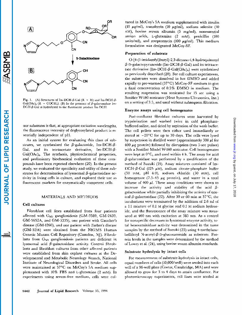
Fig. 1. (A) Structures of Im-DCH-0-Gal (R = H) and Im-DCH-(3- Gal(OAc), (R = COCH,). (B) In the presence of 0-galactosidase Im- DCH-0-Gal is hydrolyzed to the fluorescent product Im-DCH.
our substrates is that, at appropriate excitation wavelengths, the fluorescence intensity of deglycosylated product is es- sentially independent of pH.
As an initial system for evaluating this class of sub- strates, we synthesized the @-galactoside, Im-DCH-0- Gal, and its tetraacetate derivative, Im-DCH-P- Gal(OAc),. The synthesis, physicochemical properties, and preliminary biochemical evaluation of these com- pounds have been reported elsewhere (20). In the present study, we evaluated the specificity and utility of these sub- strates for determination of lysosomal 0-galactosidase ac- tivity in living cells in culture, and explored their use as fluorescent markers for enzymatically competent cells.
MATERIALS AND METHODS
Cell cultures
Fibroblast cell lines established from four patients affected with G M ~ gangliosidosis (GM-3589, GM-2439, GM-5652A, and GM-5335), one patient with Gaucher’s disease (GM-1260), and one patient with Farber’s disease (GM-3214) were obtained from the NIGMS Human Genetic Mutant Cell Repository (Camden, NJ). Fibrob- lasts from G M ~ gangliosidosis patients are deficient in lysosomal acid 0-galactosidase activity. Control fibrob- lasts and fibroblast cultures from other affected patients were established from skin explant cultures at the De- velopmental and Metabolic Neurology Branch, National Institute of Neurological Disorders and Stroke. All cells were maintained at 37OC on McCoy’s 5A medium sup- plemented with 10% FBS and L-glutamine (2 mM). In experiments using serum-free medium, cells were cul-
Sonifier W-140 sonicator (Heat Systems-Ultrasonics, Inc.) on a setting of 3.5, and used without subsequent filtration.
Enzyme assays using cell homogenates
Post-confluent fibroblast cultures were harvested by trypsinization and washed twice in cold phosphate- buffered saline, and dried by aspiration of the wash buffer. The cell pellets were then either used immediately or stored at -2OOC for up to 30 days. The cells were lysed by suspension in distilled water (approximately 300 pl for 400 p g protein) followed by disruption (two 5-sec pulses) with a Sonifier Model W-140 sonicator. Cell homogenates were kept at O O C , and used within 4 h. The assay for acid @-galactosidase was performed by a modification of the method of Suzuki (21). Assay mixtures consisted of Im- DCH-@-Gal (125 p M ) , sodium citrate-phosphate buffer (50 mM, pH 4.0), sodium chloride (50 mM), cell homogenate (7.5-45 pg protein), and water in a total volume of 400 p1. These assay conditions were shown to increase the activity and stability of the acid 0- galactosidase while partially inhibiting the activity of neu- tral 0-galactosidase (22). After 30 or 60 min at 37OC, the incubations were terminated by the addition of 2.6 ml of a 1:l mixture of 0.1 M glycine and 0.1 M sodium hydrox- ide, and the fluorescence of the assay mixture was meas- ured at 460 nm with excitation at 385 nm. As a control for nonspecific decreases in lysosomal enzyme activity, to- tal hexosaminidase activity was determined in the same samples by the method of Suzuki (23) using 4-methylum- belliferyl N-acetyl-@-D-glucosaminide as substrate. Pro- tein levels in the samples were determined by the method of Lowry et al. (24), using bovine serum albumin standards.
Substrate hydrolysis by intact cells
For measurement of substrate hydrolysis in intact cells, equal numbers of cells (10,00O/well) were seeded into each well of a 96-well plate (Costar, Cambridge, MA) and were allowed to grow for 3 to 4 days to attain confluence. For photomicroscopy experiments, cell lines were seeded at
1442 Journal of Lipid Research Volume 3 5 , 1994
by guest, on July 17, 2018w
ww
.jlr.orgD
ownloaded from
3,000 cells/well into 8-chambered glass microscope slides (Nunc, Inc., Naperville, IL) and, after 3-4 days, were used while still in log-phase growth. At the beginning of each experiment, cells were rinsed with Hank's balanced salt solution (HBSS) and pre-incubated with McCoy-SF to eliminate exogenous 0-galactosidases present in FBS. After overnight incubation, the medium was removed and the cells were refed with 200 $/well McCoy-SF contain- ing the appropriate concentration of substrate. Plates were maintained at 37OC in a humidified CO2 incubator (5% COP in air) and were allowed to equilibrate for 2 h before the initial fluorescence reading.
Slides were loaded overnight at 37OC with 200 pl/well substrate in McCoy-SF, rinsed 3 times with HBSS, refed with 200 pllwell McCoy-SF, and then incubated at 37OC for an additional 0-24 h. At intervals, the culture medium was removed and the cells were covered with a small amount of HBSS under a glass coverslip to maintain via- bility during the observation period.
Uptake, metabolism, and release of I ~ - D C H - ~ - G ~ ~ ( O A C ) ~
Normal and affected cells were seeded at equal num- bers of cells in 25-cm2 tissue culture flasks with growth medium and were allowed to incubate for 4 days to reach confluence. Cells were rinsed with HBSS and incubated for 24 h with McCoy-SF. The medium was then removed and the cells were refed with 5 ml McCoy-SF containing 100 pM Im-DCH-0-Gal(OAc)4. After 18 h of incubation with substrate, the culture supernatants were harvested and the cultures were refed with 5 ml McCoy-SF medium without substrate. At 0, 2, 4, 7.5, and 24 h after refeeding, culture supernatants from duplicate flasks were collected and processed separately. The cells from each flask were har- .vested by scraping into 1.0 ml of water. The cell and medium samples were stored at -7OOC until the final collection.
The concentration of Im-DCH was determined by fluorescence at 465 nm (excitation = 392 nm) in a 1:3 mixture of 0.05 M aqueous sodium hydroxide and methanol. The total concentration of Im-DCH-P-Gal plus Im-DCH-P-Gal(OAc), was measured by fluores- cence at 390 nm (excitation = 350 nm) in water-methanol 1:3. The ratio of the galactoside to the tetraacetate was de- termined by thin-layer chromatography (silica gel) using a chloroform-methanol-water 40:lO:l solvent system, fol- lowed by fluorescence scanning using a 355 nm short- wavelength cutoff filter (Shimadzu CS-9000 Scanning Densitometer, xenon lamp, excitation = 320 nm). The fluorescence intensity data from the thin-layer plates were standardized against adjacent lanes containing known amounts of each compound. Protein levels in the cell homogenates were determined by the Bio-Rad protein as- say (Bio-Rad Chemical Division, Richmond, CA) using BSA as standard.
Fluorescence measurements
Fluorescence of assay mixtures and cell homogenates was determined using a Perkin-Elmer 204-A Fluorescence Spectrophotometer with 10 nm slit widths and 1 cm x 1 cm quartz cuvettes. Solvent blank values were subtracted from the sample readings, and the results were reported as uncorrected fluorescence. The conversion of fluores- cence data to concentrations of the fluorescent product was performed using standard curves made from Im- DCH. Excitation and emission maxima for Im-DCH at pH 7 are 370 and 460 nm, respectively. Excitation and emission maxima for both Im-DCH-P-Gal and Im-DCH- 0-Gal(OAc)4 are 350 and 390 nm, respectively (20).
For tissue culture plates, total fluorescence of individual wells was measured using a MicroFLUOR fluorescence microplate scanner (Dynatech Laboratories, Inc., Chan- tilly, VA) equipped with a low-pressure mercury vapor lamp. Fluorescence was measured using a 365 nm broad- band excitation filter and 450 nm narrowband emission filter combined with a 400 nm UV blocking nonfluores- cent filter. In each experiment, background fluorescence of cells was determined using control wells containing cells in McCoy-SF medium without substrate. Wells con- taining substrate in medium, but no cells, were included in each plate to confirm that no spontaneous hydrolysis of substrate had occurred. In addition, standard wells con- taining a 2 pM concentration of the product, Im-DCH, were included for each plate to monitor variations in in- strument sensitivity.
Fluorescence photographs were taken with a Zeiss Ax- iovert 405M microscope using a 365 nm broadband exci- tation filter, a 395 nm interference filter, and a 420 nm longpass emission filter. Photographs were made using Kodak Ektachrome 200 film.
RESULTS
Specificity of Im-DCH-0-Gal in cell homogenates
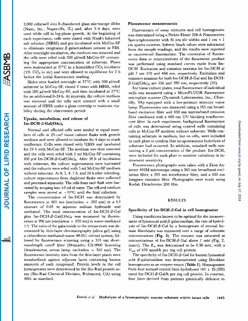
Using conditions known to be optimal for the measure- ment of lysosomal acid @-galactosidase, the rate of hydrol- ysis of Im-DCH-0-Gal by a homogenate of normal hu- man fibroblasts was measured over a range of substrate concentrations (Fig. 2). The enzyme was saturated at concentrations of Im-DCH-6-Gal above 1 mM (Fig. 2, insert). The K,,, was determined to be 0.30 mM, with a VmM of 470 nmol/h per mg cell protein.
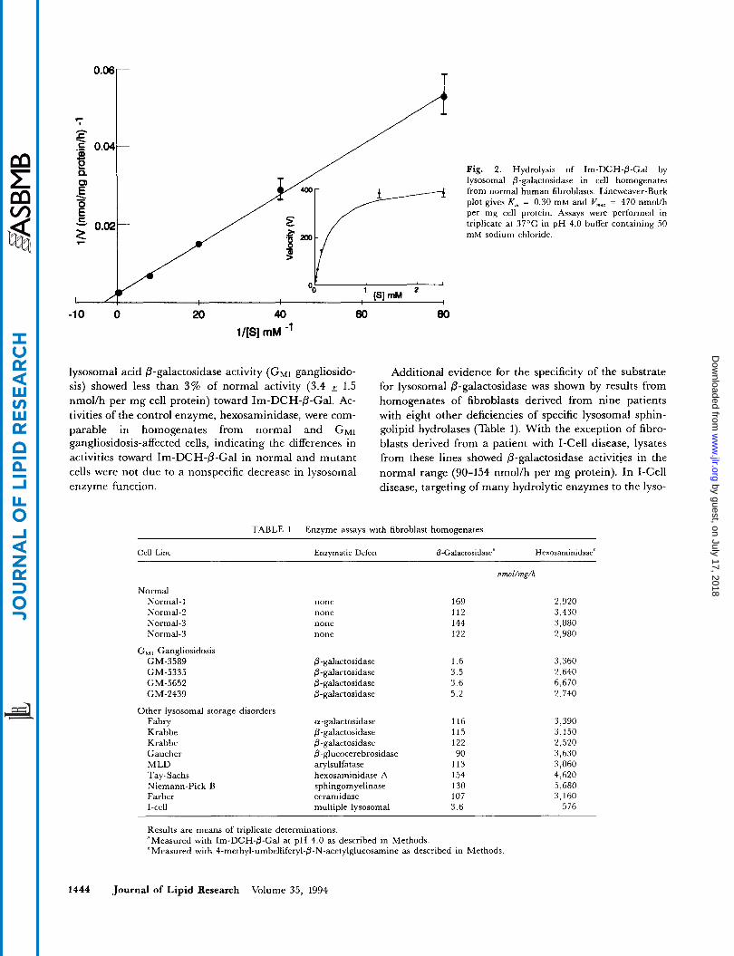
The specificity of Im-DCH-0-Gal for human lysosomal acid 0-galactosidase was demonstrated using fibroblast homogenates as an enzyme source (Table 1). Homogenates from four normal control lines hydrolyzed 140 * 26 (SD) nmol Im-DCH-0-Gallh per mg cell protein. In contrast, four lines derived from patients genetically deficient in
Kaneski et al. Hydrolysis of a lysosomotropic enzyme substrate within intact cells 1443
by guest, on July 17, 2018w
ww
.jlr.orgD
ownloaded from
O T T
Fig. 2. Hydrolysis of Im-DCH-@-Gal by lysosomal 0-galactosidase in cell homogenates from normal human fibroblasts. Lineweaver-Burk plot gives K , = 0.30 m M and Vmar = 470 nmolih per mg cell protein. Assays wem performed in triplicate at 37'C in pH 4.0 buffer containing 50 mM sodium chloride.
-10 0 20 40 60 80 1/[S] mM -'
lysosomal acid @-galactosidase activity ( G M ~ gangliosido- sis) showed less than 3% of normal activity (3.4 * 1.5 nmol/h per mg cell protein) toward Im-DCH-P-Gal. Ac- tivities of the control enzyme, hexosaminidase, were com- parable in homogenates from normal and G M ~ gangliosidosis-affected cells, indicating the differences in activities toward Im-DCH-@-Gal in normal and mutant cells were not due to a nonspecific decrease in lysosomal enzyme function.
Additional evidence for the specificity of the substrate for lysosomal 0-galactosidase was shown by results from homogenates of fibroblasts derived from nine patients with eight other deficiencies of specific lysosomal sphin- golipid hydrolases (Table 1). With the exception of fibro- blasts derived from a patient with I-Cell disease, lysates from these lines showed 0-galactosidase activities in the normal range (90-154 nmol/h per mg protein). In I-Cell disease, targeting of many hydrolytic enzymes to the lyso-
TABLE 1. Enzyme assays with fibroblast homogenates
Cell Line Enzymatic Defect &Galactosidase" Hexosamintdase'
Normal Normal-1 Normal-2 Normal-3 Normal-3
GM, Gangliosidosis GM-3589 GM-5335 GM-5652 GM-2439
Other lysosomal storage disorders Fabry Krabbe Krabbe Gaucher MLD Tay-Sachs Niemann-Pick B Farber I-cell
none none none none
@-galactosidase @-galactosidase @-galactosidase @-galactosidase
a-galactosidase @-galactosidase @-galactosidase @-glucocerebrosidase arylsulfatase hexosaminidase A sphingom yelinase ceramidase multiDle lysosomal
nmol/mg/h
169 112 144 122
1.6 3.5 3.6 5.2
116 115 122 90
113 154 130 107 3.6
2,920 3,430 3,880 2.980
3,360 2,640 6,670 2,740
3,390 3,150 2,520 3,630 3,060 4,620 5,680 3,160
576
Results are means of triplicate determinations. "Measured with Im-DCH-P-Gal at pH 4.0 as described in Methods. 'Measured with 4-methyl-umbelliferyl-@-N-acetylglucosamine as described in Methods
1444 Journal of Lipid Research Volume 35, 1994
by guest, on July 17, 2018w
ww
.jlr.orgD
ownloaded from
somes by the mannose-6-phosphate pathway is disrupted (25), leading to a marked intracellular deficiency in acid 0-galactosidase and hexosaminidase, as well as other sphingolipid hydrolases.
Specificity of Im-DCH-P-Gd for acid lysosomal P -galac tos idase
Several different P-galactosidases occur in human tis- sues besides the lysosomal acid 0-galactosidase isoen- zymes that are deficient in G M ~ gangliosidosis (EC 3.2.1.23): a “neutral” P-galactosidase with a pH optimum of 5.8, and a second lysosomal enzyme, galactosylcera- mide 0-galactosidase (EC 3.2.1.46) (26-28). When assay conditions were modified to optimize activity of neutral 0- galactosidase, the rate of hydrolysis of Im-DCH-0-Gal by normal fibroblast homogenates was reduced to 6.1 +_ 1.0 nmol/h per mg protein. This rate represents a 20-fold reduction in activity compared to hydrolysis of Im-DCH- P-Gal by acid 0-galactosidase. This decrease is consistent with previous findings that, in liver, neutral 0-galactosidase accounts for approximately 5% of the activity toward other artificial substrates (29).
Cell lysates from two cell lines derived from patients ge- netically deficient in galactosylceramide 0-galactosidase (Krabbe’s disease) showed hydrolytic activity toward Im- DCH-@-Gal that was within the normal range (Table 1). Galactosylceramide 0-galactosidase hydrolyzes the P- glycosidic linkage of galactose in galactosylceramide and lactosylceramide, but is distinct from the acid 6- galactosidase that is deficient in G M ~ gangliosidosis (30). Because the 0-galactosidase that is deficient in G M ~ gan- gliosidosis is approximately 5 - to 10-times more active than galactosylceramide @-galactosidase in cultured fibroblasts (21), a decrease in hydrolytic activity toward Im-DCH-0-Gal due to a defect in the latter enzyme would not be detectable in our assay. Therefore, it cannot be determined from these data whether or not Im-DCH- @-Gal is also a substrate for the galactocerebroside- specific enzyme.
Comparison of the hydrolysis of Im-DCH-&Gal and Im-DCH-P-Gal(OAc), in intact cells
When normal fibroblasts in microplate culture were ex- posed to Im-DCH-0-Gal (100 p~ in McCoy’s SF) using a variety of culture and loading conditions, including the presence and absence of FBS in the medium and lyophili- zation of the substrate with low density lipoprotein, no significant increase in fluorescence was detected during incubation periods of up to 24 h.
When the more hydrophobic tetraacetate, Im-DCH-P- G a l ( 0 A ~ ) ~ (100 p M in McCoy’s SF), was substituted as substrate in the culture medium, a significant increase in fluorescence over time was observed. After 21 h of incuba- tion, fluorescence of wells treated with Im-DCH-P- G a l ( 0 A ~ ) ~ increased from 329 ~f: 9 units to 743 +_ 45
units, an average increase of 414 f 46 units in 21 h. Therefore, I ~ - D C H - P - G ~ ~ ( O A C ) ~ was selected as sub- strate for further studies in intact cells.
A second galactoside substrate was synthesized, in which the lysosomotropic substituent (imidazolyl-butyl) was replaced by a strongly hydrophobic moiety, CH3 (CH2)17. When cells were treated with C 18-DCH-/3-Gal or its tetraacetate derivative using a variety of conditions, no significant increase in fluorescence of cells or medium was observed during incubation periods as long as 48 h. As a result, these extremely insoluble octadecyl derivatives were not studied further and all subsequent experiments were performed using Im-DCH-@-Gal(OAc),.
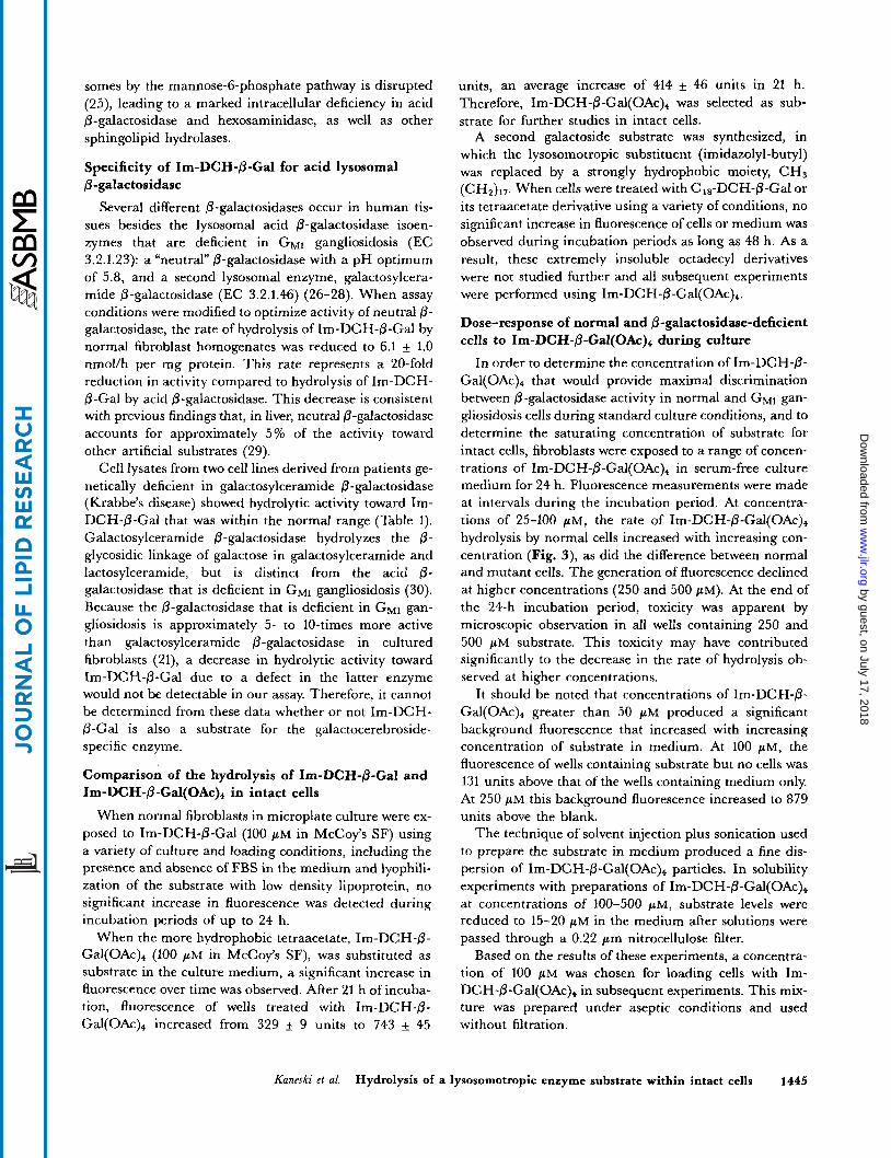
Dose-response of normal and 0-galactosidase-deficient cells to I ~ - D C H - ~ - G ~ ~ ( O A C ) ~ during culture
In order to determine the concentration of Im-DCH-P- Gal(OAc), that would provide maximal discrimination between 0-galactosidase activity in normal and G M ~ gan- gliosidosis cells during standard culture conditions, and to determine the saturating concentration of substrate for intact cells, fibroblasts were exposed to a range of concen- trations of I ~ - D C H - P - G ~ ~ ( O A C ) ~ in serum-free culture medium for 24 h. Fluorescence measurements were made at intervals during the incubation period. At concentra- tions of 25-100 pM, the rate of I ~ - D C H - P - G ~ ~ ( O A C ) ~ hydrolysis by normal cells increased with increasing con- centration (Fig. 3), as did the difference between normal and mutant cells. The generation of fluorescence declined at higher concentrations (250 and 500 pM). At the end of the 24-h incubation period, toxicity was apparent by microscopic observation in all wells containing 250 and 500 pM substrate. This toxicity may have contributed significantly to the decrease in the rate of hydrolysis ob- served at higher concentrations.
It should be noted that concentrations of Im-DCH-P- Gal(OAc), greater than 50 pM produced a significant background fluorescence that increased with increasing concentration of substrate in medium. At 100 p ~ , the fluorescence of wells containing substrate but no cells was 131 units above that of the wells containing medium only. At 250 pM this background fluorescence increased to 879 units above the blank.
The technique of solvent injection plus sonication used to prepare the substrate in medium produced a fine dis- persion of I ~ - D C I - I - ~ - G ~ ~ ( O A C ) ~ particles. In solubility experiments with preparations of Im-DCH-P-Gal(OAc), at concentrations of 100-500 pM, substrate levels were reduced to 15-20 in the medium after solutions were passed through a 0.22 pm nitrocellulose filter.
Based on the results of these experiments, a concentra- tion of 100 pM was chosen for loading cells with Im- DCH-P-Gal(OAc), in subsequent experiments. This mix- ture was prepared under aseptic conditions and used without filtration.
Kaneski et al. Hydrolysis of a lysosomotropic enzyme substrate within intact cells 1445
by guest, on July 17, 2018w
ww
.jlr.orgD
ownloaded from
I I S
I , 0 100 200 300 400 500
-200 ! Im-DCH-BGal(OAc)4 (uM)
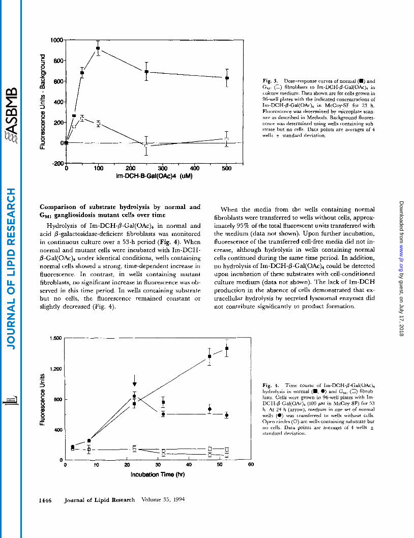
Comparison of substrate hydrolysis by normal and GMl gangliosidosis mutant cells over time
Hydrolysis of I ~ - D C H - P - G ~ ~ ( O A C ) ~ in normal and acid /3-galactosidase-deficient fibroblasts was monitored in continuous culture over a 53-h period (Fig. 4). When normal and mutant cells were incubated with Im-DCH- fi-Gal(OA~)~ under identical conditions, wells containing normal cells showed a strong, time-dependent increase in fluorescence. In contrast, in wells containing mutant fibroblasts, no significant increase in fluorescence was ob- served in this time period. In wells containing substrate but no cells, the fluorescence remained constant or slightly decreased (Fig. 4).
Fig. 3. Dose-response curves of normal (m) and G , , (0) fibroblasts to Im-DCH-@-Gal(OAc), in culture medium. Data shown are for cells grown in 96-well plates with the indicated concentrations of Im-DCH-@-Gal(OAc)+ in McCoy-SF for 23 h. Fluorescence was determined by microplate scan- ner as described in Methods. Background fluores- cence was determined using wells containing sub- strate but no cells. Data points are averages of 4 wells + standard deviation.
When the media from the wells containing normal fibroblasts were transferred to wells without cells, approx- imately 95% of the total fluorescent units transferred with the medium (data not shown). Upon further incubation, fluorescence of the transferred cell-free media did not in- crease, although hydrolysis in wells containing normal cells continued during the same time period. In addition, no hydrolysis of I ~ - D C H - @ - G ~ ~ ( O A C ) ~ could be detected upon incubation of these substrates with cell-conditioned culture medium (data not shown). The lack of Im-DCH production in the absence of cells demonstrated that ex- tracellular hydrolysis by secreted lysosomal enzymes did not contribute significantly to product formation.
I I I
0 10 20 30 40 50 60
Incubation Time (hr)
Fig. 4. Time course of Im-DCH-fi-Gal(OAc), hydrolysis in normal (m, 0) and G,, (0) fibrob- lasts. Cells were grown in 96-well plates with Im- DCH-@-Gal(OAc)+ (100 p~ in McCoy-SF) for 53 h. At 24 h (arrow), medium in one set of normal wells (0) was transferred to wells without cells. Open circles (0) are wells containing substrate but no cells. Data points are averages of 4 wells + standard deviation.
1446 Journal of Lipid Research Volume 3 5 , 1994
by guest, on July 17, 2018w
ww
.jlr.orgD
ownloaded from
Effect of lysosomal inhibitors
In order to provide further evidence that hydrolysis of Im-DCH-P-Gal(OAc)+ occurred in the lysosomes of the cells, normal fibroblasts were pretreated for 3 days with the lysosomal inhibitors ammonium chloride (10 mM) and chloroquine (100 pM). At these concentrations, the inhibitors have been found to reduce activity of lysosomal enzymes in treated fibroblasts by 85-95% by raising the intralysosomal pH (19, 31). The hydrolysis of Im-DCH-P-
Gal(OAc)+, measured in terms of fluorescent units above background per 24 h, was reduced in normal fibroblasts by 91% 11% in cells pretreated with chloroquine and by 79% 8% in cells pretreated with ammonium chloride.
Use of I ~ - D C H - P - G ~ ~ ( O A C ) ~ for fluorescence microscopy
Because the emission wavelengths of Im-DCH-P- G a l ( 0 A ~ ) ~ and its products lie within the visible spec-
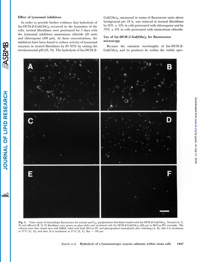
Fig. 5 . Time course of intracellular fluorescence for normal and G,,, gangliosidosis fibroblasts loaded with Im-DCH-P-Gal(OAc)+. Normal (A, C, E) and affected (B, D, F) fibroblasts were grown on glass slides and incubated with Im-DCH-P-Cal(OAc)+ (100 PM in McCoy-SF) overnight. The cultures were then rinsed once with HBSS, refed with fresh McCoy-SF, and photographed immediately after refeeding (A, B), after 6 h incubation at 37OC (C, D), and after 24 h incubation at 37OC (E, F). Bar = 100 pm.
Kuneski et al. Hydrolysis of a lysosomotropic enzyme substrate within intact cells 1447
by guest, on July 17, 2018w
ww
.jlr.orgD
ownloaded from
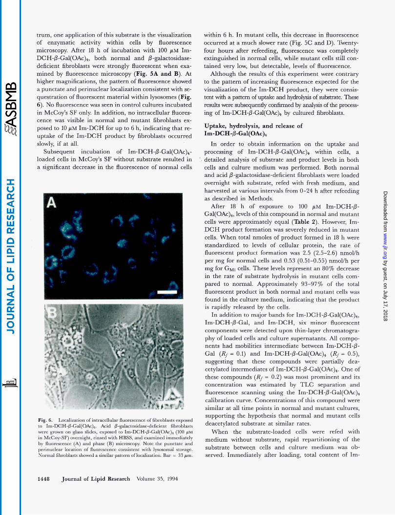
t m , one application of this substrate is the visualization of enzymatic activity within cells by fluorescence microscopy After 18 h of incubation with 100 pM Im- DCH-P-Gal(OAc)4, both normal and &galactosidase- deficient fibroblasts were strongly fluorescent when exa- mined by fluorescence microscopy (Fig. 5A and B). At higher magnifications, the pattern of fluorescence showed a punctate and perinuclear localization consistent with se- questration of fluorescent material withii lysosomes (Fig. 6). No fluorescence was seen in control cultures incubated in McCoy’s SF only In addition, no intracellular fluores- cence was visible in normal and mutant fibroblasts ex- posed to 10 pM Im-DCH for up to 6 h, indicating that re- uptake of the Im-DCH product by fibroblasts occurred slowly, if at all.
Subsequent incubation of I~-DCH-&G~~(OAC)~- loaded cells in McCoy’s SF without substrate resulted in a significant decrease in the fluorescence of normal cells
:.
Fig. 6. Localization of intracellular fluorescence of fibroblasts exposed to Im-DCH-fl-Gal(OAc)*. Acid fl-galactosidase-deficient fibroblasts were grown on glass slides, exposed to Im-DCH-&Gal(OAc)+ (100 p~ in McCoy-SF) overnight, rinsed with HBSS, and examined immediately by fluorescence (A) and phase (B) microscopy. Note the punctate and perinuclear location of fluorescence consistent with lysosomal storage. Normal fibroblasts showed a similar pattem of localization. Bar = 33 Fm.
1448 Joumal of Lipid Research Volume 35, 1994
within 6 h. In mutant cells, this decrease in fluorescence occurred at a much slower rate (Fig. 5C and D). Twenty- four hours after refeedig, fluorescence was completely extinguished in normal cells, while mutant cells still con- tained very low, but detectable, levels of fluorescence.
Although the results of this experiment were contrary to the pattern of increasing fluorescence expected for the visualization of the Im-DCH product, they were consis- tent with a pattern of uptake and hydrolysis of substxate. These d t s were subsequently confirmed by analysis of the process- ing of Im-DCH-@-Gal(OAc), by cultured fibroblasts.
Uptake, hydrolysis, and release of I ~ - D C H - & G ~ ~ ( O A C ) ~
In order to obtain information on the uptake and processing of I~-DCH-P-G~~(OAC)~ within cells, a detailed analysis of substrate and product levels in both cells and culture medium was performed. Both normal and acid 8-galactosidase-deficient fibroblasts were loaded overnight with substrate, refed with fresh medium, and harvested at various intervals from 0-24 h after refeeding as described in Methods.
After 18 h of exposure to 100 pM Im-DCH-P- Gal(OAc)4, levels of this compound in normal and mutant cells were approximately equal (Table 2). However, Im- DCH product formation was severely reduced in mutant cells. When total nmoles of product formed in 18 h were standardized to levels of cellular protein, the rate of fluorescent product formation w a s 2.5 (2.5-2.6) nmol/h per mg for normal cells and 0.53 (0.51-0.55) nmol/h per mg for G M ~ cells. These levels represent an 80% decrease in the rate of substrate hydrolysis in mutant cells com- pared to normal. Approximately 93-9776 of the total fluorescent product in both normal and mutant cells was found in the culture medium, indicating that the product is rapidly released by the cells.
In addition to major bands for I~-DCH-@-G~~(OAC)~, Im-DCH-&Gal, and Im-DCH, six minor fluorescent components were detected upon thin-layer chromatogra- phy of loaded cells and culture supernatants. All compo- nents had mobilities intermediate between Im-DCH-6- Gal (Rj = 0.1) and I ~ - D C H - ~ ~ - G ~ ( O A C ) ~ (Rj = 0.5), suggesting that these compounds were partially dea- cetylated intermediates of Im-DCH-fl-Gal(OAc)4. One of these compounds (Rj = 0.2) was most prominent and its concentration was estimated by TLC separation and fluorescence scanning using the Im-DCH-P-Gal(OAc)+ calibration curve. Concentrations of this compound were similar at all time points in normal and mutant cultures, supporting the hypothesis that normal and mutant cells deacetylated substrate at similar rates.
When the substrate-loaded cells were refed with medium without substrate, rapid repartitioning of the substrate betweep cells and culture medium was ob- served. Immediately after loading, total content of Im-
by guest, on July 17, 2018w
ww
.jlr.orgD
ownloaded from
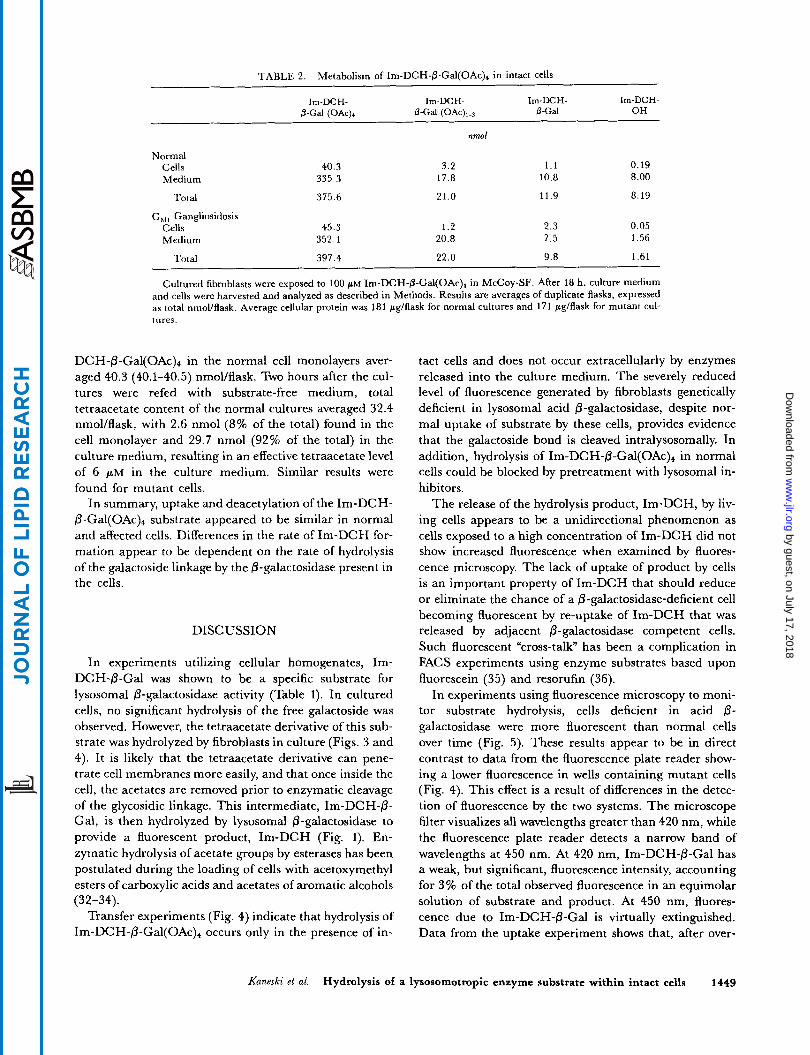
TABLE 2. Metabolism of Im-DCH-fi-Gd(OAc)+ in intact cells
Im-DCH- Im-DCH- Im-DCH- Im-DCH- &Gal (OAc), 0-Gal (OAC),.~ 0-Gal OH
nmol
Normal Cells 40.3 3.2 1.1 0.19 Medium 335.3 17.8 10.8 8.00
Total 375.6 21.0 11.9 8.19
GM, Gangliosidosis Cells 45.3 1.2 2.3 0.05 Medium 352.1 20.8 7.5 1.56
Total 397.4 22.0 9.8 1.61
Cultured fibroblasts were exposed to 100 p~ Im-DCH-fi-Gal(OAc)4 in McCoy-SF. After 18 h, culture medium and cells were harvested and analyzed as described in Methods. Results are averages of duplicate flasks, expressed as total nmollflask. Average cellular protein was 181 pg/flask for normal cultures and 171 pg/flask for mutant cul- tures
D C H - / ~ - G ~ ~ ( O A C ) ~ in the normal cell monolayers aver- aged 40.3 (40.1-40.5) nmol/flask. Two hours after the cul- tures were refed with substrate-free medium, total tetraacetate content of the normal cultures averaged 32.4 nmol/flask, with 2.6 nmol (8% of the total) found in the cell monolayer and 29.7 nmol (92% of the total) in the culture medium, resulting in an effective tetraacetate level of 6 p M in the culture medium. Similar results were found for mutant cells.
In summary, uptake and deacetylation of the Im-DCH- P-Gal(OAc), substrate appeared to be similar in normal and affected cells. Differences in the rate of Im-DCH for- mation appear to be dependent on the rate of hydrolysis of the galactoside linkage by the @-galactosidase present in the cells.
DISCUSSION
In experiments utilizing cellular homogenates, Im- DCH-0-Gal was shown to be a specific substrate for lysosomal @-galactosidase activity (Table 1). In cultured cells, no significant hydrolysis of the free galactoside was observed. However, the tetraacetate derivative of this sub- strate was hydrolyzed by fibroblasts in culture (Figs. 3 and 4). It is likely that the tetraacetate derivative can pene- trate cell membranes more easily, and that once inside the cell, the acetates are removed prior to enzymatic cleavage of the glycosidic linkage. This intermediate, Im-DCH-0- Gal, is then hydrolyzed by lysosomal &galactosidase to provide a fluorescent product, Im-DCH (Fig. 1). En- zymatic hydrolysis of acetate groups by esterases has been postulated during the loading of cells with acetoxymethyl esters of carboxylic acids and acetates of aromatic alcohols
Transfer experiments (Fig. 4) indicate that hydrolysis of Im-DCH-@-Gal(OAc), occurs only in the presence of in-
(32-34).
tact cells and does not occur extracellularly by enzymes released into the culture medium. The severely reduced level of fluorescence generated by fibroblasts genetically deficient in lysosomal acid 0-galactosidase, despite nor- mal uptake of substrate by these cells, provides evidence that the galactoside bond is cleaved intralysosomally. In addition, hydrolysis of Im-DCH-/.?-Gal(OAc), in normal cells could be blocked by pretreatment with lysosomal in- hibitors.
The release of the hydrolysis product, Im-DCH, by liv- ing cells appears to be a unidirectional phenomenon as cells exposed to a high concentration of Im-DCH did not show increased fluorescence when examined by fluores- cence microscopy. The lack of uptake of product by cells is an important property of Im-DCH that should reduce or eliminate the chance of a 0-galactosidase-deficient cell becoming fluorescent by re-uptake of Im-DCH that was released by adjacent @-galactosidase competent cells. Such fluorescent “cross-talk” has been a complication in FACS experiments using enzyme substrates based upon fluorescein (35) and resorufin (36).
In experiments using fluorescence microscopy to moni- tor substrate hydrolysis, cells deficient in acid 0- galactosidase were more fluorescent than normal cells over time (Fig. 5). These results appear to be in direct contrast to data from the fluorescence plate reader show- ing a lower fluorescence in wells containing mutant cells (Fig. 4). This effect is a result of differences in the detec- tion of fluorescence by the two systems. The microscope filter visualizes all wavelengths greater than 420 nm, while the fluorescence plate reader detects a narrow band of wavelengths at 450 nm. At 420 nm, Im-DCH-@-Gal has a weak, but significant, fluorescence intensity, accounting for 3% of the total observed fluorescence in an equimolar solution of substrate and product. At 450 nm, fluores- cence due to Im-DCH-P-Gal is virtually extinguished. Data from the uptake experiment shows that, after over-
Kaneski et ai. Hydrolysis of a lysosomotropic enzyme substrate within intact cells 1449
by guest, on July 17, 2018w
ww
.jlr.orgD
ownloaded from
night incubation with Im-DCH-@-Gal(OAc),, the in- tracellular ratio of all forms of substrate to product is ap- proximately 200:l in normal cells and 900:l in acid @-galactosidase-deficient cells (Table 2). These results suggest that much of the fluorescence observed in the pho- tomicrographs may be due to the presence of Im-DCH-@- Gal(OAc)4 and its deacylated forms in the cells. While the demonstration of slower degradation of substrate by mu- tant cells may be viewed as a model of the storage process, substrate fluorescence interferes with visualization of in- tracellular enzymatic activity. Preliminary studies indi- cate that, with the use of a microscope emission filter set that more strongly discriminates against substrate (wavelengths > 450 nm), normal cells may be distin- guished from mutant by an increased fluorescence of product over time.
This novel class of DCH-based substrates has several applications for the study of lysosomal sphingolipid hydrolases in living cells. The fact that these substrates are taken up and processed by viable cells under physiological conditions, and that the Im-DCH product fluoresces at physiological temperature and pH, allows for the study of environmental factors influencing enzymatic activity. The low toxicity of these compounds permits the study of en- zyme function over time. In addition, with the 96-well plate system, a number of different cell lines or treatments may be compared simultaneously under identical condi- tions. Further information on the influence of the microenvironment in lysosomal enzyme function could ultimately lead to the development of new therapeutic strategies in the treatment of sphingolipid storage diseases.
Another potential application of these Im-DCH-based substrates would be sorting of genetically corrected cells according to their intracellular enzyme content using FACS technology. The utility of fluorescent enzymatic substrates for this procedure has been demonstrated by Nolan and co-workers (35) for murine fibroblast and lym- phoid cells genetically transduced with the @-galactosidase reporter gene, lacZ. In our system, normal cells may be selected on the basis of decreased levels of galactoside fluorescence or of increased levels of deglycosylated product over time. Further studies are in progress on the use of these DCH-based fluorescent substrates to detect and separate normal and mutant cells in mixed popula- tions on the basis of intracellular activity of specific glycolipid hydrolases. I Manuscript recemed 17 Nouembo 1993 and in revisedfom I 8 February 1994.
REFERENCES
1. Scriver, C. R., A. L. Beaudet, W. S. Sly, D. Valle, J. B. Stanbury, J. B. Wyngaarden, and D. S. Fredrickson, edi- tors. 1989. The Metabolic Basis of Inherited Disease. Vol
2.
3.
4.
5.
6.
7.
8.
9.
10.
11.
12.
13.
14.
15.
16.
17.
18.
19.
20.
11. McGraw-Hill, Inc., New York. 1565-1839. Bieberich, E., and G. Legler. 1989. Intracellular activity of lysosomal glucosylceramidase measured with 4-nonylum- belliferyl /3-glucoside. Biol. Chem. Hoppe-Seyler. 370: 809-817. Ivleva, T. S., T. A. Oglobilina, L. L. Litinskaya, and G. Y. Wiederschain. 1991. Estimation and comparison of lysosomal and cytoplasmic pH of human fibroblasts from healthy donors and patients with lysosomal storage dis- eases. Biomed. Sci. 2: 398-402. van Weely, S., M. van den Berg, J. A. Barranger, M. C. Miranda, J. M. Tager, and J. M. F. G. Aerts. 1993. Role of pH in determining the cell-type-specific residual activity of glucocerebrosidase in Type I Gaucher disease. J. Clin. Zn- vest. 91: 1167-1175. Kishimoto, Y., M. Hiraiwa, and J. S. OBrien. 1992. Sapo- sins: structure, function, distribution, and molecular genetics. J. Lipid Res. 33: 1255-1267. OBrien, J. S., and Y. Kishimoto. 1991. Saposin proteins: structure, function, and role in human lysosomal storage disorders. FASEB J. 5: 301-308. Sandhoff, K., G. van Echten, M. Schroder, D. Schnabel, and K. Suzuki. 1992. Metabolism of glycolipids: the role of glycolipid-binding proteins in the function and pathobi- ochemistry of lysosomes. Biochem. Sac. Tram. 20: 695-699. Brown, R. G., and G. Porter. 1977. Effect of pH on the emission and absorption characteristics of 2,3-dicyano-p- hydroquinone. J. Chem. Sac. Faraday Zans. I 73: 1281-1285. Alabaster, O., K. C. Carr, and L. Leondaridis. 1984. Tumor cell heterogeneity: its determination by flow cyto- metric analysis of intracellular pH. Methods Achieu. Exp. Pathol. 11: 96-110. Gillies, R. J., J. Cook, M. H. Fox, and K. A. Giuliano. 1987. Flow cytometric analysis of intracellular pH in 3T3 cells. Am. J. Physiol. 253: C121-Cl25. Hirsch-Kauffmann, M., G. Valet, J. Wieser, and M. Schweiger. 1985. Progressive muscular dystrophy (Duchenne): biochemical studies by flow-cytometry. Hum. Genet. 69: 332-336. Kurtz, I., and R. S. Balaban. 1985. Fluorescence emission spectroscopy of 1,4-dihydroxyphthalonitrile. Biophys. J. 48:
Musgrove, E., C. Rugg, and D. Hedley. 1986. Flow cyto- metric measurement of cytoplasmic pH: a critical evalua- tion of available fluorochromes. Cytometry. 7: 347-355. Valet, G., A. Raffael, L. Moroder, E. Wunsch, and G. Ruhenstroth-Bauer. 1981. Fast intracellular pH determina- tion in single cells by flow-cytometry. Naturwissenschaften. 68:
Valet, G., H. H. Warnecke, and H. Kahle. 1984. New possi- es of cytostatic drug testing on patient tumor cells by
flow cytometry. Blut. 49: 37-43. De Duve, C., T. De Barsy, B. Poole, A. Trouet, P. Tulkens, and F. Van Hoof. 1974. Lysosomotropic agents. Biochem. Pharmacal. 23: 2495-2531. Firestone, R. A,, J. M. Pisano, and R. J. Bonney. 1979. Lysosomotropic agents. 1. Synthesis and cytotoxic action of lysosomotropic detergents. J. Mea'. Chem. 22: 1130-1133. Miller, D. K., E. Griffiths, J. Lenard, and R. A. Firestone. 1983. Cell killing by lysosomotropic detergents. J Cell Bioi.
Wilson, P. D., R. A. Firestone, and J. Lenard. 1987. The role of lysosomal enzymes in killing of mammalian cells by the lysosomotropic detergent N-dodecylimidazole. ,I. Cell Biol. 104: 1223-1229. Miller, S. P. F., S. A. French, and C. R. Kaneski. 1991. Syn-
499-508.
265-266.
97: 1841-1851.
1450 Journal of Lipid Research Volume 35, 1994
by guest, on July 17, 2018w
ww
.jlr.orgD
ownloaded from
thesis and characterization of a novel lysosomotropic en- zyme substrate that fluoresces at intracellular pH. J Or&
Suzuki, K. 1977. Globoid cell leukodystrophy (Krabbe dis- ease) and G M , gangliosidosis. In Practical Enzymology of the Sphingolipidoses. R. H. Glew and S. P. Peters, editors. Alan R. Liss, New York. 101-136.
22. Ho, M. W., and J. S. OBrien. 1971. Differential effect of chloride ions on 0-galactosidase isoenzymes: a method for separate assay. Clin. Chim. Acta. 32: 443-450.
23. Suzuki, K. 1978. Enzymic diagnosis of sphingolipidoses. Methods Enzymol. Volume L. Complex carbohydrates. Part C. V. Ginsburg, editor. Academic Press, New York, San Francisco, London. 456-488.
24. Lowry, 0. H., N. J. Rosebrough, A. L. Farr, and R. J. Ran- dall. 1951. Protein measurement with Folin phenol reagent. J. Biol. Chem. 193: 265-275.
25. Nolan, C. M., and W. S. Sly. 1989. I-cell disease and pseudo-Hurler polydystrophy: disorders of lysosomal en- zyme phosphorylation and localization. In The Metabolic Basis of Inherited Disease. C. R. Scriver, A. L. Beaudet, W. S. Sly, D. Valle, J. B. Stanbury, J. B. Wyngaarden, and D. S. Fredrickson, editors. McGraw-Hill, Inc., New York. 1589-1601. Kobayashi, T., and K. Suzuki. 1981. A taurodeoxycholate- activated galactosylceramidase in the murine intestine. J. Biol. Chem. 256: 1133-1137.
27. Tanaka, H., M. Meisler, and K. Suzuki. 1975. Activity of human hepatic 0-galactosidase toward natural glycosphin- golipid substrates. Biochim. Biophys. Acta. 398: 452-463.
28. Wenger, D. A., M. Sattler, and C. Clark. 1975. Effect of bile salts on lactosylceramide 0-glactosidase activities in human
C h n . 56: 30-34. 21.
26.
29.
30.
31.
32.
33.
34.
35.
36.
brain, liver, and cultured skin fibroblasts. Biochim. Biophys. Acta. 409: 297-303. OBrien, J. S. 1983. The gangliosidoses. In The Metabolic Basis of Inherited Disease. J. B. Stanbury, J. B. Wyn- gaarden, D. S. Fredrickson, J. L. Goldstein, and M. S. Brown, editors. McGraw-Hill, Inc., New York. 945-969. Tanaka, H., and K. Suzuki. 1975. Lactosylceramide 0- galactosidase in human sphingolipidoses. J. Biol. Chem.
Lie, S. O., and B. Shofield. 1973. Inactivation of lysosomal function in normal cultured human fibroblasts by chloro- quine. Biochem. Phannacol. 22: 3109-3114. Borle, A. B., and K. W. Snowdowne. 1987. Methods for the measurement of intracellular ionized calcium in mam- malian cells: Comparison of four classes of Ca2+ indicators. In Calcium and Cell Function, Vol VII. W. Y. Cheung, edi- tor. Academic Press, Orlando. 159-200. Thomas, J. A., R. N. Buchsbaum, A. Zimniak, and E. Racker. 1979. Intracellular pH measurements in Ehrlich ascites tumor cells utilizing spectroscopic probes generated in situ. Biochemistry. 18: 2210-2218. Tsien, R. Y. 1981. A non-disruptive technique for loading calcium buffers and indicators into cells. Nature. 290: 527-528. Nolan, G. P., S. Fiering, J-F. Nicolas, and L. A. Herzen- berg. 1988. Fluorescence-activated cell analysis and sorting of viable mammalian cells based on 0-D-galactosidase ac- tivity after transduction of Escherichia coli 1ac.Z Pmc. Nutl. Acad. Sci. USA. 85: 2603-2607. Wittrup, K. D., and J. E. Bailey. 1988. A single-cell assay of @-galactosidase activity in Sacchummyces cerevisiae. Cytome-
250: 2324-2332.
tv. 9: 394-404.
Kuneski et al. Hydrolysis of a lysosomotropic enzyme substrate within intact cells 1451
by guest, on July 17, 2018w
ww
.jlr.orgD
ownloaded from