host-dependent trigger of caspases and apoptosis by ...iai.asm.org/content/75/6/2903.full.pdf ·...
TRANSCRIPT
INFECTION AND IMMUNITY, June 2007, p. 2903–2913 Vol. 75, No. 60019-9567/07/$08.00�0 doi:10.1128/IAI.00147-07Copyright © 2007, American Society for Microbiology. All Rights Reserved.
Host-Dependent Trigger of Caspases and Apoptosis byLegionella pneumophila�†
Marina Santic,1,2 Rexford Asare,2 Miljenko Doric,1 and Yousef Abu Kwaik2*Department of Microbiology and Parasitology, University of Rijeka, Rijeka, Croatia,1 and Department of
Microbiology and Immunology, Room 406, University of Louisville College of Medicine,319 Abraham Flexner Way 55A, Louisville, Kentucky 402022
Received 29 January 2007/Returned for modification 23 March 2007/Accepted 26 March 2007
The Dot/Icm system of Legionella pneumophila triggers activation of caspase-3 during early stages of infectionof human macrophages, but apoptosis is delayed until late stages of infection. During early stages of infectionof mouse macrophages, the organism triggers rapid caspase-1-mediated cytotoxicity, which is mediated bybacterial flagellin. However, it is not known whether caspase-1 is triggered by L. pneumophila in humanmacrophages or whether caspase-3 is activated in permissive or nonpermissive mouse macrophages. Usingsingle-cell analyses, we show that the wild-type strain of L. pneumophila does not trigger caspase-1 activationthroughout the intracellular infection of human monocyte-derived macrophages (hMDMs), even when theflagellated bacteria escape into the cytoplasm during late stages. Using single-cell analyses, we show that theDot/Icm system of L. pneumophila triggers caspase-3 but not caspase-1 within permissive A/J mouse bonemarrow-derived primary macrophages by 2 to 8 h, but apoptosis is delayed until late stages of infection. WhileL. pneumophila triggers a Dot/Icm-dependent activation of caspase-1 in nonpermissive BALB/c mouse-derivedmacrophages, caspase-3 is not activated at any stage of infection. We show that robust intrapulmonaryreplication of the wild-type strain of L. pneumophila in susceptible A/J mice is associated with late-stageDot/Icm-dependent pulmonary apoptosis and alveolar inflammation. In the lungs of nonpermissive BALB/cmice, L. pneumophila does not replicate and does not trigger pulmonary apoptosis or alveolar inflammation.Thus, similar to hMDMs, L. pneumophila does not trigger caspase-1 but triggers caspase-3 activation duringearly and exponential replication in permissive A/J mouse-derived macrophages, and apoptosis is delayed untillate stages of infection. The Dot/Icm type IV secretion system is essential for pulmonary apoptosis in thegenetically susceptible A/J mice.
The ability of Legionella pneumophila to cause pneumonia isdependent on its capacity to invade and replicate within alveolarmacrophages, monocytes, and potentially alveolar epithelial cells(1, 38, 43). Upon entry into the host cell, L. pneumophila modu-lates the biogenesis of the phagosome into a replicative niche thatis halted from maturation through the “default” endosomal-lyso-somal degradation pathway (17, 32, 42, 44). The L. pneumophila-containing phagosome (LCP) intercepts early secretory vesiclesfrom the endoplasmic reticulum exit sites, which allows the or-ganism to remodel the LCP membrane to become rough endo-plasmic reticulum (RER) derived within minutes of its biogenesisfrom the plasma membrane (16, 18, 39, 41). Upon activation ofhuman macrophages by gamma interferon, the LCP fuses to thelysosomes and fails to be remodeled by the RER (33). In contrastto what was found for L. pneumophila, recent studies have shownthat Legionella longbeachae is trafficked to, and replicates within,a nonacidified, late-endosome-like phagosome that is remodeledby the RER (5). The Dot/Icm type IV secretion system of L.pneumophila is essential for evasion of endocytic fusion and forremodeling of the LCP into an RER-derived compartment (16,18, 32, 35, 39, 41, 42, 44). These manipulations of host cell pro-
cesses during early stages are thought to be mediated by theinjection of effectors by the Dot/Icm transporter directly from thebacterium into the host cell (8, 28). During late stages of infectionof human macrophages and Acanthamoeba polyphaga, the bac-teria escape into the host cell cytosol, where they reside for 2to 6 h prior to lysis of the host cell plasma membrane (24).
In addition to evasion of vesicle traffic by L. pneumophiladuring early stages of infection, the bacterium also inducesDot/Icm-dependent activation of caspase-3 in human macro-phages (12–14, 25, 27, 49). There are at least 14 caspases(cysteine proteases) that trigger the activation of two distinctapoptosis signaling pathways, designated the extrinsic and in-trinsic pathways, that converge on the activation of caspase-3,which is the executioner of rapid apoptosis (30, 36). Interest-ingly, the Dot/Icm-mediated activation of caspase-3 by L. pneu-mophila in human macrophages during early stages of infec-tion seems to be novel, since it is independent of the extrinsicand intrinsic pathways of apoptosis (25). Interestingly, despitethe robust activation of caspase-3 during early and exponentialreplication of L. pneumophila within human macrophages,apoptosis is not triggered until termination of intracellularreplication (3, 25), which is a novel modulation of caspase-3activity that halts it from the rapid dismantling of the cell.Recent data have shown that the delay in apoptosis of L.pneumophila-infected human macrophages is associated withinduction of strong Dot/Icm-dependent antiapoptotic signalsthat are mediated by NF-�B and non-NF-�B signaling mech-anisms (2, 20).
* Corresponding author. Mailing address: Department of Microbi-ology and Immunology, Room 406, University of Louisville College ofMedicine, 319 Abraham Flexner Way 55A, Louisville, KY 40202. Phone:(502) 852-4117. Fax: (502) 852-7531. E-mail: [email protected].
† Supplemental material for this article may be found at http://iai.asm.org/.
� Published ahead of print on 9 April 2007.
2903
on October 21, 2018 by guest
http://iai.asm.org/
Dow
nloaded from
Among inbred mouse strains, the A/J strain is the only onesusceptible to L. pneumophila infection, while all the otherstrains are relatively resistant (46–48). This genetic suscepti-bility is attributed to a polymorphism in the gene encoding theneuronal apoptosis inhibitory protein (naip5) (11, 45). Thenaip family of genes are evolutionary conserved from viruses tohumans, and some encode proteins that possess antiapoptoticactivity, due to inhibition of caspase-3 and caspase-7 (10, 21).However, caspase-3 is not required for the infection of mousemacrophages by L. pneumophila (26, 31), which is distinct fromthat of human macrophages (25). In mouse macrophages thatare nonpermissive for intracellular proliferation of L. pneumo-phila, the bacterial flagellin (FlaA) triggers caspase-1-mediatedproinflammatory rapid cell death/pyropoptosis (26, 31). Themechanism and the role of Naip5 in activation of caspase-1 byL. pneumophila are not known.
It is not known whether caspase-1 is triggered by L. pneu-mophila in human macrophages or whether caspase-3 is acti-vated in permissive or nonpermissive mouse macrophages. It isalso not known whether similar kinetics of apoptosis in tissueculture systems is also exhibited in the lungs of animal models.Here, we show that within human monocyte-derived macro-phages (hMDMs) and A/J mouse macrophages, L. pneumo-phila does not trigger caspase-1 activation throughout the in-tracellular infection, despite the escape of highly flagellated L.pneumophila bacteria into the cytosol of hMDMs during latestages of infection. L. pneumophila triggers differential and
temporal early activation of caspase-3 in A/J mouse-derivedmacrophages, similar to that in hMDMs, but caspase-3 is nottriggered in the resistant BALB/c mouse-derived macro-phages. Our data show that Dot/Icm-mediated pulmonaryapoptosis is triggered during late stages of intrapulmonary repli-cation in susceptible A/J mice. In contrast, L. pneumophila failsto induce pulmonary apoptosis in BALB/c mice, despite rapidcaspase-1-mediated cell death in primary macrophages in vitro.
MATERIALS AND METHODS
Animals, bacteria, and macrophages. Female pathogen-free A/J and BALB/cmice, 8 to 9 weeks of age, were used in all experiments. The mice were housedin specific-pathogen-free conditions within the animal care facility. The virulentclinical isolate of L. pneumophila strain AA100 and its isogenic dotA mutant(GL10) have been described previously (49). The wild-type Francisella novicidastrain U112 has also been described previously (19). The bacteria were main-tained frozen at �80°C and, prior to use, were grown on buffered charcoal yeastextract agar for 72 h. The plates for gfp-transformed AA100 or its isogenic dotAmutant were supplemented with 5 �g/ml chloramphenicol. After cultivation, thebacteria were washed by centrifugation and resuspended in sterile saline.
To prepare mouse bone marrow-derived macrophages (mBMDM), bone mar-row samples were isolated from healthy A/J or BALB/c mice and were preparedas described previously (26). To prepare hMDMs, peripheral blood monocyteswere isolated from healthy volunteers with no history of tularemia or Legion-naires’ disease and hMDMs were prepared as we described previously (33). Thevolunteers were 25 to 45 years old, with no history of pneumonia or any under-lying chronic disease.
Inoculation of animals. Mice were inoculated intratracheally, as we describedpreviously (4, 23, 29). Briefly, the mice were anesthetized by intraperitonealinjection of ketamine (2.5 mg/mouse). A total of 50 �l of the L. pneumophila
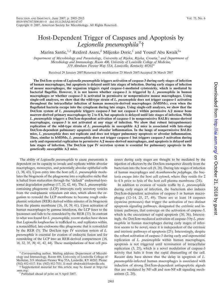
FIG. 1. Temporal activation of caspase-1 in hMDMs is triggered by L. pneumophila during late but not early stages of infection. Humanmacrophages were infected with L. pneumophila AA100 or the dotA mutant. F. tularensis (Ft) was used as a positive control. Representativeconfocal microscopy images of hMDMs are shown. The cells were stained for active caspase-1 (C-1) and for apoptosis by TUNEL (T) assays.Quantification of the percentages of cells with active caspase-1 (C1) in addition to the double positives (C1�T) is shown in panel B and is basedon examination of 100 infected cells from three different coverslips. The data are representative of three experiments, and error bars representstandard deviations.
2904 SANTIC ET AL. INFECT. IMMUN.
on October 21, 2018 by guest
http://iai.asm.org/
Dow
nloaded from
suspension (106 CFU) in sterile water was inoculated directly into the trachea byusing a 26-gauge needle, followed by 10 to 20 �l of air. Control animals wereinoculated with saline only and were sacrificed at different time points.
Quantitation of L. pneumophila bacteria in pulmonary tissues of mice. Atdifferent time points after inoculation of bacteria, the mice were humanelysacrificed. The lungs were aseptically excised, finely minced, and homogenized ina tissue homogenizer with 5 ml of sterile distilled water. The numbers of CFU ofL. pneumophila AA100 or the dotA mutant strain in the lungs were determinedby a plate dilution method using buffered charcoal yeast extract agar. After 3days of incubation at 37°C, the colonies were enumerated and the results wereexpressed as numbers of CFU per lung.
Pulmonary histopathology. The histological changes and apoptosis in thelungs of A/J and BALB/c mice in response to L. pneumophila were assessed bylight and confocal microscopy. At 2, 24, and 48 h after inoculation, the mice werehumanely sacrificed using CO2 asphyxiation. Before lung removal, the pulmo-nary vasculature was perfused with 10 ml of saline containing 5 mM EDTA viathe right ventricle. The excised lungs were inflated and fixed in 10% neutral
formalin for 24 h, dehydrated, and embedded in paraffin. Sections were cut andstained with eosin and hematoxylin for analyses of the infiltration process in thelungs of infected mice. In addition, sections (5 �m) were cut and labeling ofapoptotic cells was carried out using terminal deoxynucleotidyltransferase-me-diated dUTP-biotin nick end labeling (TUNEL) with an in situ cell death de-tection kit as recommended by the manufacturer (Roche, Indianapolis, IN). Thehistopathology of the lung tissue stained with eosin and hematoxylin was ana-lyzed by light microscopy. An analysis of the histology of the lung tissue in thepresence of intracellular green fluorescent protein (GFP)-expressing bacteriaand apoptotic cells (TUNEL positive) was carried out using laser scanningconfocal microscopy. On average, 10 0.2-�m-thick serial sections of each imagewere captured and stored for further analyses, using Adobe Photoshop CSversion 8.0 (Adobe Photoshop, Inc.).
TEM. For examination of apoptosis in the lungs of A/J mice by transmissionelectron microscopy (TEM), mouse lungs were removed and placed in 2.5%glutaraldehyde as described previously (24). Briefly, the lungs were postfixed byimmersion in 2% osmium tetroxide in 0.1 M sodium Sorenson’s buffer for 1 h,
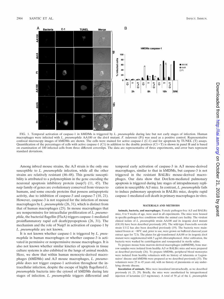
FIG. 2. Dot/Icm-mediated early activation of caspase-3 but not caspase-1 in A/J mouse-derived mBMDM upon infection by L. pneumophila.A/J mouse mBMDM were infected with L. pneumophila AA100 or the dotA mutant or with F. tularensis (Ft) as a positive control. Representativeconfocal microscopy images at 8 and 18 h after infection are shown in panels A and C. The cells were stained for active caspase-1 (C-1) or C-3and apoptosis by TUNEL (T) assays. Quantification of the percentages of cells with active caspase-1 (C1) and C3 in addition to the double positives(C1�T or C3�T) is shown in panels B and D and is based on examination of 100 infected cells from three different coverslips. The data arerepresentative of three experiments, and error bars represent standard deviations.
VOL. 75, 2007 LEGIONELLA HOST-SPECIFIC CASPASE ACTIVATION 2905
on October 21, 2018 by guest
http://iai.asm.org/
Dow
nloaded from
followed by dehydration in acetone and infiltration and embedment in Epon 12epoxy resin (24). Ultrathin sections (0.1 �m) were then cut, stained with uranylacetate and lead citrate, and examined with a Philips TEM (Morgagni 268D;Philips, The Netherlands) at 80 kV.
Caspase activation and TUNEL assays. To assess activation of caspase-1 andcaspase-3 by confocal microscopy, 2.5 � 105 mBMDM or hMDMs on glasscoverslips were infected with L. pneumophila AA100, the dotA mutant, or F.novicida at a multiplicity of infection of 10 for 1 h, followed by incubation for 2,8, and 18 h. For caspase-1 activation, macrophages were stained for 1 h withFAM-YVAD-FMK (Immunochemistry Technologies, Bloomington, IN) as rec-ommended by the manufacturer. As a positive control for caspase-1 activation inhMDMs, macrophages were treated with 10 mM Simvastatin (Calbiochem, SanDiego, CA) (9). For caspase-3 activation, after infection and fixation, the cellswere incubated with anti-active caspase-3 rabbit polyclonal antiserum for 1 h,followed by a goat anti-rabbit immunoglobulin G secondary antibody conjugatedto Alexa red (Molecular Probes, Inc., Eugene, OR).
Apoptotic nuclei were labeled with TUNEL according to the manufacturer’sinstructions (Boehringer Mannheim Corporation, Indianapolis, IN). Cells wereexamined with a Zeiss Axiophot Photomicroscope Leica TCS NT confocal laserscanning microscope. A minimum of 100 cells per sample were examined, and
apoptosis was quantified as the percentage of apoptotic cells (TUNEL-positivenuclei).
Statistical analyses. All experiments were performed at least three times, andthe data shown are representative of one experiment. Statistical analyses wereperformed using the two-tailed Student t test.
RESULTS
L. pneumophila does not trigger activation of caspase-1throughout the infection of primary human macrophages. Al-though caspase-1 has been shown to be triggered by L. pneu-mophila within mouse-derived macrophages, it is not knownwhether caspase-1 is triggered by L. pneumophila during anystage of the infection of human macrophages. We utilizedprimary hMDMs to examine potential activation of caspase-1by L. pneumophila AA100 or the dotA mutant throughout theintracellular infection. As a positive control for caspase-1 ac-
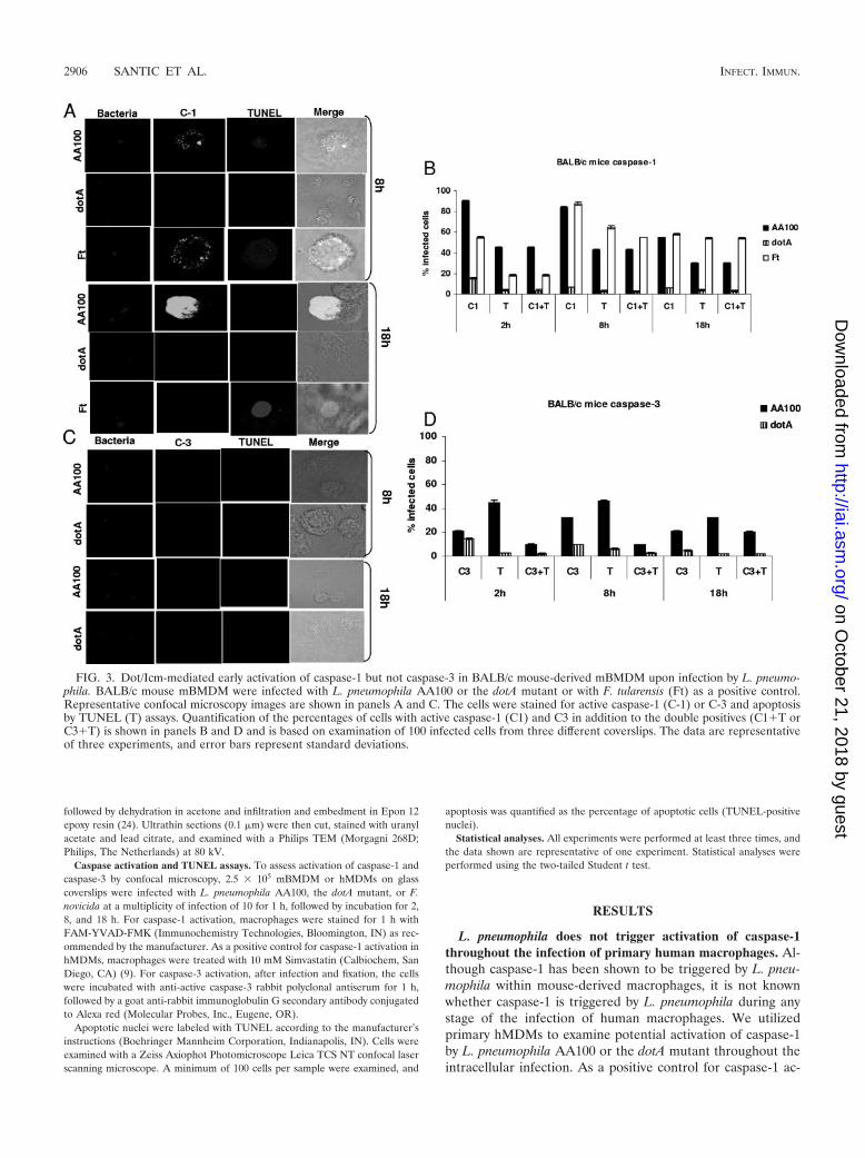
FIG. 3. Dot/Icm-mediated early activation of caspase-1 but not caspase-3 in BALB/c mouse-derived mBMDM upon infection by L. pneumo-phila. BALB/c mouse mBMDM were infected with L. pneumophila AA100 or the dotA mutant or with F. tularensis (Ft) as a positive control.Representative confocal microscopy images are shown in panels A and C. The cells were stained for active caspase-1 (C-1) or C-3 and apoptosisby TUNEL (T) assays. Quantification of the percentages of cells with active caspase-1 (C1) and C3 in addition to the double positives (C1�T orC3�T) is shown in panels B and D and is based on examination of 100 infected cells from three different coverslips. The data are representativeof three experiments, and error bars represent standard deviations.
2906 SANTIC ET AL. INFECT. IMMUN.
on October 21, 2018 by guest
http://iai.asm.org/
Dow
nloaded from
tivation, cells were pretreated with Simvastatin or infected byFrancisella tularensis. By 2 h after infection of hMDMs with allthe strains, caspase-1 activation was minimal, which was notsignificantly different from that for uninfected cells (Student’st test, P � 0.2) (Fig. 1). By 8 h after infection of hMDMs withL. pneumophila AA100, only �10% of the infected cells werepositive for caspase-1 and also apoptotic (Student’s t test, P �0.1) (Fig. 1). While �80% of L. pneumophila-infected hMDMswere apoptotic by 18 h postinfection, only �10% of themexhibited caspase-1 activation (Fig. 1). Similar results wereobtained for hMDMs (data not shown). Therefore, the largenumber of infected hMDMs that became apoptotic at 18 hwere not associated with activation of caspase-1. When thecells were labeled for active caspase-3, L. pneumophila trig-gered time-dependent early activation of caspase-3, and late-stage apoptosis was inhibited by the caspase-3 inhibitor (datanot shown), consistent with many previously published datafrom independent laboratories (12–14, 25, 27, 49). Caspase-1activation was exhibited in control hMDMs treated with Sim-vastatin or infected by F. tularensis as positive controls (Fig. 1).The caspase-1 activity and apoptosis in hMDMs infected withthe dotA mutant were not significantly different from those foruninfected cells at all time points after infection (Student’s ttest, P � 0.3) (Fig. 1). We conclude that L. pneumophila doesnot trigger caspase-1 activation throughout the infection ofhuman macrophages, while caspase-3 is highly activatedthroughout the intracellular infection. Importantly, late-stageapoptosis in hMDMs is not associated with activation ofcaspase-1.
Differential activation of caspases and apoptosis by L. pneu-mophila in permissive mouse macrophages. Although caspase-3has been shown to be triggered by L. pneumophila within
human macrophages, it is not known whether L. pneumophilatriggers caspase-3 in mouse-derived macrophages. We utilizedsingle-cell analyses to examine the kinetics of potential activa-tion of caspase-3 and apoptosis in primary mBMDM obtainedform A/J permissive and BALB/c nonpermissive mice. We alsoexamined the kinetics of caspase-1 activation throughout theinfection to decipher whether apoptosis triggered during latestages of infection was mediated by caspase-1 or caspase-3. Weused Francisella tularensis as a positive control for caspase-1activation (22).
A/J mBMDM infected by L. pneumophila AA100 or its dotAmutant were examined for the kinetics of caspase-3 activationand apoptosis using single-cell analyses by confocal micros-copy. Approximately 20% of the cells infected by L. pneumo-phila AA100 exhibited activation of caspase-3 at 2 h afterinfection, which was significantly different from what wasfound for cells infected by the dotA mutant or uninfected cells(Student’s t test, P � 0.01), but only a few cells were positivefor TUNEL (Fig. 2). At 8 h postinfection, �70% of L. pneu-mophila-infected A/J mBMDM exhibited caspase-3 activation,which was significantly different from what was found for cellsinfected by the dotA mutant or uninfected cells (Student’s ttest, P � 0.003), but only a few infected cells underwent apop-tosis (Fig. 2). By 18 h after infection with L. pneumophilaAA100, a large number of the cells were lysed, and �60% ofthe remaining infected cells exhibited caspase-3 activationand were also apoptotic (Student’s t test, P � 0.001) (Fig. 2).Apoptosis was inhibited when the infected cells were pre-treated with the caspase-3 inhibitor but not when the in-fected cells were pretreated with the caspase-1 inhibitor(data not shown). The dotA mutant control neither activatedcaspase-3 nor triggered apoptosis at any time point after
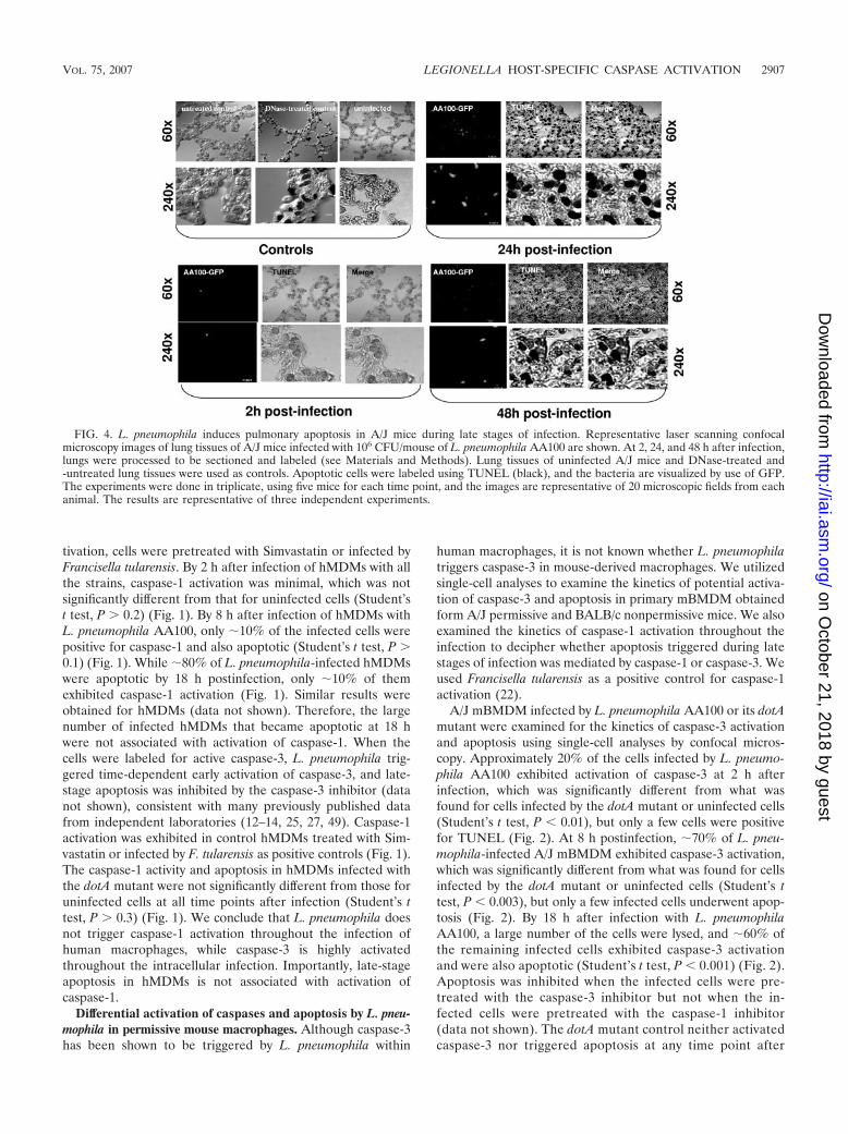
FIG. 4. L. pneumophila induces pulmonary apoptosis in A/J mice during late stages of infection. Representative laser scanning confocalmicroscopy images of lung tissues of A/J mice infected with 106 CFU/mouse of L. pneumophila AA100 are shown. At 2, 24, and 48 h after infection,lungs were processed to be sectioned and labeled (see Materials and Methods). Lung tissues of uninfected A/J mice and DNase-treated and-untreated lung tissues were used as controls. Apoptotic cells were labeled using TUNEL (black), and the bacteria are visualized by use of GFP.The experiments were done in triplicate, using five mice for each time point, and the images are representative of 20 microscopic fields from eachanimal. The results are representative of three independent experiments.
VOL. 75, 2007 LEGIONELLA HOST-SPECIFIC CASPASE ACTIVATION 2907
on October 21, 2018 by guest
http://iai.asm.org/
Dow
nloaded from
infection of A/J mBMDM, which was not significantly dif-ferent from what was found for uninfected cells (Student’s ttest, P � 0.3) (Fig. 2).
At 2 to 8 h after infection of A/J mBMDM with L. pneumo-phila AA100, caspase-1 activation and apoptosis were not sig-nificantly different from what was found for cells infected bythe dotA mutant or uninfected cells (Student’s t test, P � 0.2)(Fig. 2). By 18 h postinfection, �80% of the L. pneumophilaAA100-infected cells were TUNEL positive but only 10% ofthem exhibited caspase-1 activation. The dotA mutant did notactivate caspase-1 in A/J mBMDM, and only a few cells un-derwent apoptosis at all time points after infection (Fig. 2),which was not significantly different from what was found foruninfected cells (Student’s t test, P � 0.2). Infection of A/JmBMDM by the F. tularensis control triggered time-dependentactivation of caspase-1.
L. pneumophila triggers temporal and differential rapid activa-tion of caspase-1 and apoptosis in resistant BALB/c mouse-de-rived macrophages. Although caspase-1 has been shown to betriggered by L. pneumophila within nonpermissive mouse-
derived macrophages, it is not known whether activation ofcaspase-3 is also triggered by L. pneumophila at any stage ofinfection. Therefore, we used single-cell analysis to examinethe temporal kinetics of activation of caspase-1 and caspase-3in BALB/c mBMDM. The data showed that L. pneumophilaAA100 triggered robust activation of caspase-1 at 2 to 8 h afterinfection of BALB/c mBMDM, when most infected cells werepositive for caspase-1 activity and were also apoptotic (Stu-dent’s t test, P � 0.001) (Fig. 3). In contrast, infection ofBALB/c mBMDM with the dotA mutant triggered minimalcaspase-1 activation and apoptosis, a result not significantlydifferent from what was found for uninfected cells (Student’s ttest, P � 0.1) (Fig. 3). The cells infected by F. tularensis exhib-ited time-dependent activation of caspase-1 (Fig. 3).
In contrast, caspase-3 activation was minimal at 2 h afterinfection of BALB/c mBMDM by L. pneumophila AA100,which was not significantly different from what was found fordotA-infected and uninfected cells (Student’s t test, P � 0.3).At 8 h after infection with the wild-type strain of L. pneumo-phila �30% of infected cells exhibited activation of caspase-3
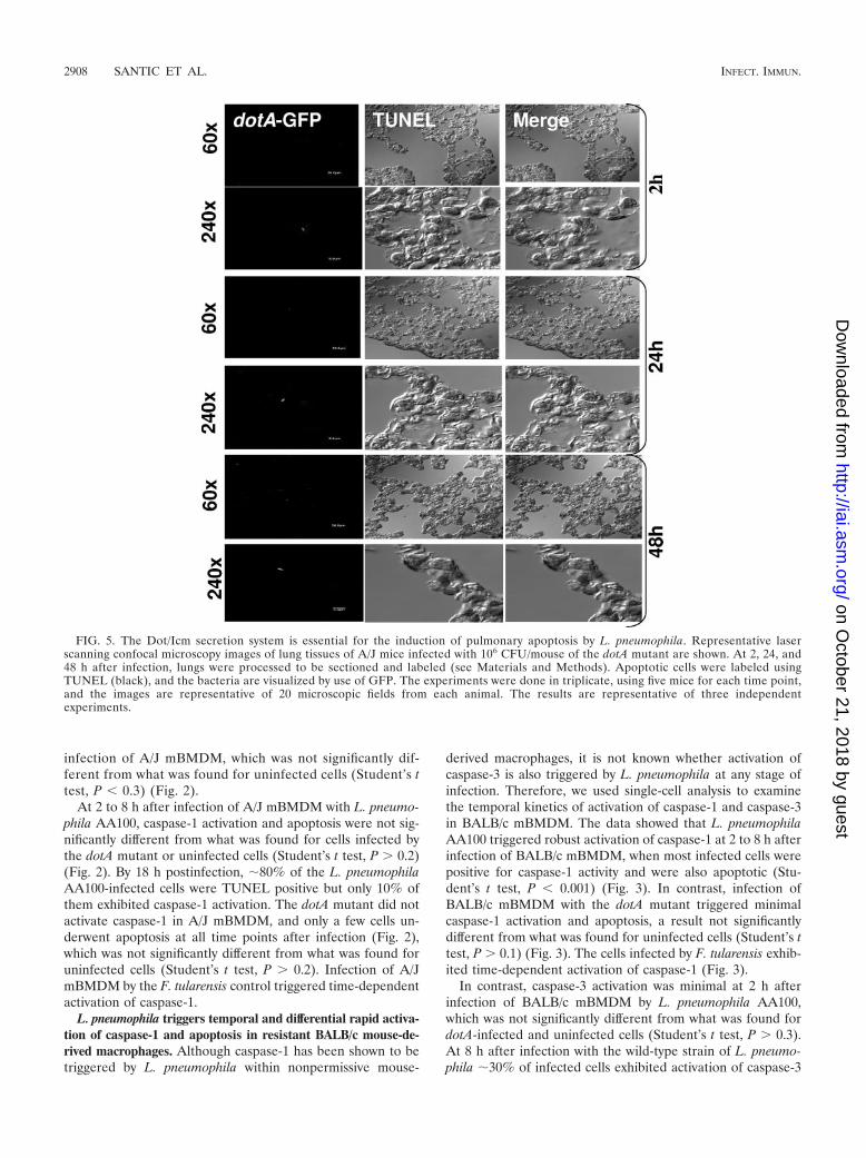
FIG. 5. The Dot/Icm secretion system is essential for the induction of pulmonary apoptosis by L. pneumophila. Representative laserscanning confocal microscopy images of lung tissues of A/J mice infected with 106 CFU/mouse of the dotA mutant are shown. At 2, 24, and48 h after infection, lungs were processed to be sectioned and labeled (see Materials and Methods). Apoptotic cells were labeled usingTUNEL (black), and the bacteria are visualized by use of GFP. The experiments were done in triplicate, using five mice for each time point,and the images are representative of 20 microscopic fields from each animal. The results are representative of three independentexperiments.
2908 SANTIC ET AL. INFECT. IMMUN.
on October 21, 2018 by guest
http://iai.asm.org/
Dow
nloaded from
(Student’s t test, P � 0.05) and �10% of these were alsopositive for TUNEL. Similar results were obtained at 18 h afterinfection by L. pneumophila AA100 (Fig. 3). The dotA mutanttriggered very low levels of activation of caspase-3 at all timepoints after infection, which was similar to what was found foruninfected macrophages (Student’s t test, P � 0.2). Takentogether, our results show that L. pneumophila does not triggercaspase-3 in nonpermissive BALB/c mouse-derived primarymacrophages.
L. pneumophila induces Dot/Icm-dependent pulmonary in-flammation and apoptosis in A/J mice. L. pneumophila AA100replicated in the lungs of A/J mice, where the number ofbacteria peaked at 48 h after infection but did not replicatewithin the lungs of BALB/c mice, and the dotA mutant did notreplicate in any mice, consistent with previous published ob-servations (see Fig. S1 in the supplemental material). Inflam-matory infiltration was first evident in lung tissue from L.pneumophila AA100-infected A/J mice at 24 h after infectionand became more severe by 48 h postinfection (see Fig. S2 inthe supplemental material). However, there was no detectableinflammatory infiltration in the lungs of A/J mice infected withthe dotA mutant or BALB/c mice infected with L. pneumophilaAA100 at any time points after infection (see Fig. S3 in thesupplemental material).
We examined by in situ cell analyses the kinetics of apoptosisin the lung tissues of A/J and BALB/c mice infected with L.pneumophila AA100 and the dotA mutant by laser scanningconfocal microscopy. As a positive control, we used DNase-treated sections of lung tissue. As negative controls, we usedDNase-untreated sections and lung tissue sections of unin-
fected mice inoculated with saline (Fig. 4). Any nuclei stainedblack by TUNEL were considered apoptotic, regardless of theintensity of the staining.
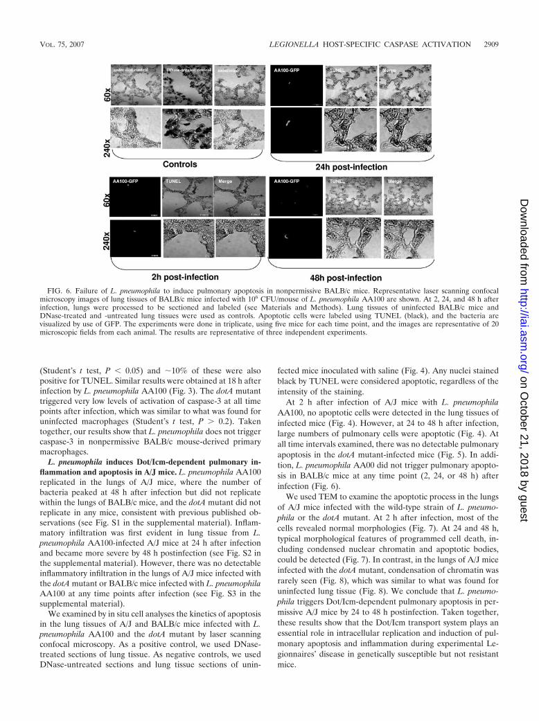
At 2 h after infection of A/J mice with L. pneumophilaAA100, no apoptotic cells were detected in the lung tissues ofinfected mice (Fig. 4). However, at 24 to 48 h after infection,large numbers of pulmonary cells were apoptotic (Fig. 4). Atall time intervals examined, there was no detectable pulmonaryapoptosis in the dotA mutant-infected mice (Fig. 5). In addi-tion, L. pneumophila AA00 did not trigger pulmonary apopto-sis in BALB/c mice at any time point (2, 24, or 48 h) afterinfection (Fig. 6).
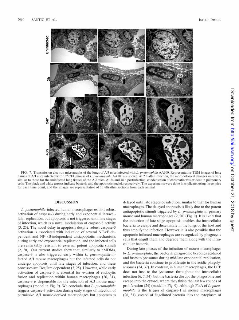
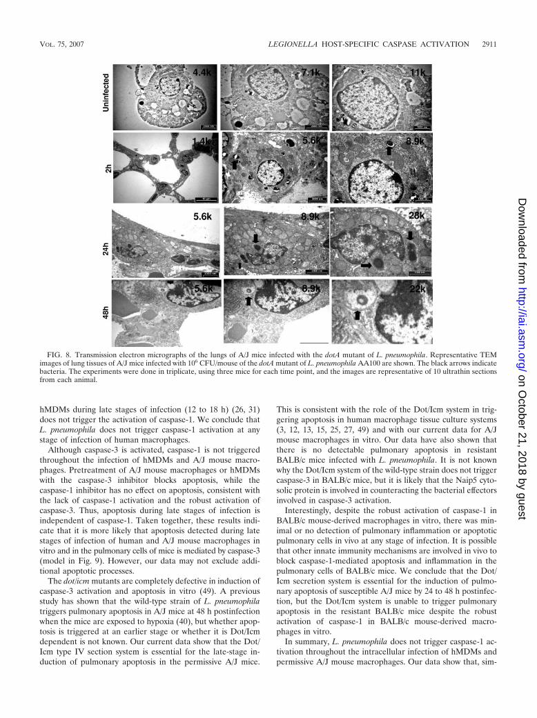
We used TEM to examine the apoptotic process in the lungsof A/J mice infected with the wild-type strain of L. pneumo-phila or the dotA mutant. At 2 h after infection, most of thecells revealed normal morphologies (Fig. 7). At 24 and 48 h,typical morphological features of programmed cell death, in-cluding condensed nuclear chromatin and apoptotic bodies,could be detected (Fig. 7). In contrast, in the lungs of A/J miceinfected with the dotA mutant, condensation of chromatin wasrarely seen (Fig. 8), which was similar to what was found foruninfected lung tissue (Fig. 8). We conclude that L. pneumo-phila triggers Dot/Icm-dependent pulmonary apoptosis in per-missive A/J mice by 24 to 48 h postinfection. Taken together,these results show that the Dot/Icm transport system plays anessential role in intracellular replication and induction of pul-monary apoptosis and inflammation during experimental Le-gionnaires’ disease in genetically susceptible but not resistantmice.
FIG. 6. Failure of L. pneumophila to induce pulmonary apoptosis in nonpermissive BALB/c mice. Representative laser scanning confocalmicroscopy images of lung tissues of BALB/c mice infected with 106 CFU/mouse of L. pneumophila AA100 are shown. At 2, 24, and 48 h afterinfection, lungs were processed to be sectioned and labeled (see Materials and Methods). Lung tissues of uninfected BALB/c mice andDNase-treated and -untreated lung tissues were used as controls. Apoptotic cells were labeled using TUNEL (black), and the bacteria arevisualized by use of GFP. The experiments were done in triplicate, using five mice for each time point, and the images are representative of 20microscopic fields from each animal. The results are representative of three independent experiments.
VOL. 75, 2007 LEGIONELLA HOST-SPECIFIC CASPASE ACTIVATION 2909
on October 21, 2018 by guest
http://iai.asm.org/
Dow
nloaded from
DISCUSSION
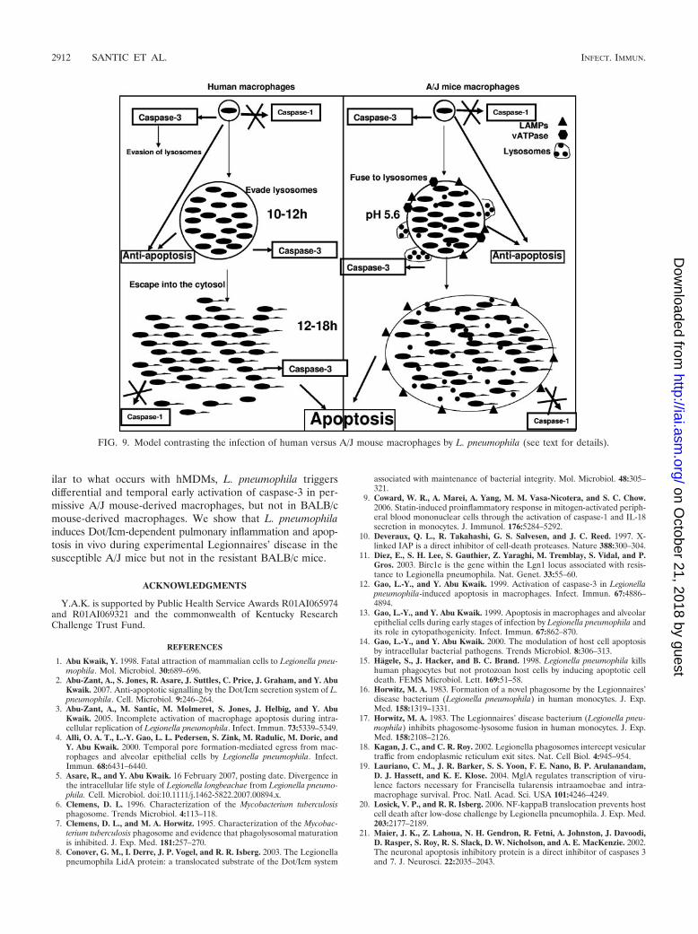
L. pneumophila-infected human macrophages exhibit robustactivation of caspase-3 during early and exponential intracel-lular replication, but apoptosis is not triggered until late stagesof infection, which is a novel modulation of caspase-3 activity(3, 25). The novel delay in apoptosis despite robust caspase-3activation is associated with induction of several NF-�B-de-pendent and NF-�B-independent antiapoptotic mechanismsduring early and exponential replication, and the infected cellsare remarkably resistant to external potent apoptotic stimuli(2, 20). Our current studies show that, similarly to hMDMs,caspase-3 is also triggered early within L. pneumophila-in-fected A/J mouse macrophages but the infected cells do notundergo apoptosis until late stages of infection, and theseprocesses are Dot/Icm dependent (3, 25). However, while earlyactivation of caspase-3 is essential for evasion of endocyticfusion and replication within human macrophages (26, 31),caspase-3 is dispensable for the infection of A/J mouse mac-rophages (model in Fig. 9). We conclude that L. pneumophilatriggers caspase-3 activation during early stages of infection ofpermissive A/J mouse-derived macrophages but apoptosis is
delayed until late stages of infection, similar to that for humanmacrophages. The delayed apoptosis is likely due to the potentantiapoptotic stimuli triggered by L. pneumophila in primarymouse and human macrophages (2, 20) (Fig. 9). It is likely thatthe induction of late-stage apoptosis enables the intracellularbacteria to escape and disseminate in the lungs of the host andthus amplify the infection. However, it is also possible that theapoptotic infected macrophages are recognized by phagocyticcells that engulf them and degrade them along with the intra-cellular bacteria.
During late phases of the infection of mouse macrophagesby L. pneumophila, the bacterial phagosome becomes acidifiedand fuses to lysosomes during mid-late exponential replication,and the bacteria continue to proliferate in the acidic phagoly-sosomes (34, 37). In contrast, in human macrophages, the LCPdoes not fuse to the lysosomes throughout the intracellularinfection (6, 7, 34), but the bacteria disrupt the phagosome andescape into the cytosol, where they finish the last few rounds ofproliferation (24) (model in Fig. 9). Although FlaA of L. pneu-mophila is the trigger of caspase-1 in mouse macrophages(26, 31), escape of flagellated bacteria into the cytoplasm of
FIG. 7. Transmission electron micrographs of the lungs of A/J mice infected with L. pneumophila AA100. Representative TEM images of lungtissues of A/J mice infected with 106 CFU/mouse of L. pneumophila AA100 are shown. At 2 h after infection, the morphological changes were verysimilar to those for the uninfected lung tissues of the A/J mice. At 24 and 48 h postinfection, condensation of chromatin was evident in pulmonarycells. The black and white arrows indicate bacteria and the apoptotic nuclei, respectively. The experiments were done in triplicate, using three micefor each time point, and the images are representative of 10 ultrathin sections from each animal.
2910 SANTIC ET AL. INFECT. IMMUN.
on October 21, 2018 by guest
http://iai.asm.org/
Dow
nloaded from
hMDMs during late stages of infection (12 to 18 h) (26, 31)does not trigger the activation of caspase-1. We conclude thatL. pneumophila does not trigger caspase-1 activation at anystage of infection of human macrophages.
Although caspase-3 is activated, caspase-1 is not triggeredthroughout the infection of hMDMs and A/J mouse macro-phages. Pretreatment of A/J mouse macrophages or hMDMswith the caspase-3 inhibitor blocks apoptosis, while thecaspase-1 inhibitor has no effect on apoptosis, consistent withthe lack of caspase-1 activation and the robust activation ofcaspase-3. Thus, apoptosis during late stages of infection isindependent of caspase-1. Taken together, these results indi-cate that it is more likely that apoptosis detected during latestages of infection of human and A/J mouse macrophages invitro and in the pulmonary cells of mice is mediated by caspase-3(model in Fig. 9). However, our data may not exclude addi-tional apoptotic processes.
The dot/icm mutants are completely defective in induction ofcaspase-3 activation and apoptosis in vitro (49). A previousstudy has shown that the wild-type strain of L. pneumophilatriggers pulmonary apoptosis in A/J mice at 48 h postinfectionwhen the mice are exposed to hypoxia (40), but whether apop-tosis is triggered at an earlier stage or whether it is Dot/Icmdependent is not known. Our current data show that the Dot/Icm type IV section system is essential for the late-stage in-duction of pulmonary apoptosis in the permissive A/J mice.
This is consistent with the role of the Dot/Icm system in trig-gering apoptosis in human macrophage tissue culture systems(3, 12, 13, 15, 25, 27, 49) and with our current data for A/Jmouse macrophages in vitro. Our data have also shown thatthere is no detectable pulmonary apoptosis in resistantBALB/c mice infected with L. pneumophila. It is not knownwhy the Dot/Icm system of the wild-type strain does not triggercaspase-3 in BALB/c mice, but it is likely that the Naip5 cyto-solic protein is involved in counteracting the bacterial effectorsinvolved in caspase-3 activation.
Interestingly, despite the robust activation of caspase-1 inBALB/c mouse-derived macrophages in vitro, there was min-imal or no detection of pulmonary inflammation or apoptoticpulmonary cells in vivo at any stage of infection. It is possiblethat other innate immunity mechanisms are involved in vivo toblock caspase-1-mediated apoptosis and inflammation in thepulmonary cells of BALB/c mice. We conclude that the Dot/Icm secretion system is essential for the induction of pulmo-nary apoptosis of susceptible A/J mice by 24 to 48 h postinfec-tion, but the Dot/Icm system is unable to trigger pulmonaryapoptosis in the resistant BALB/c mice despite the robustactivation of caspase-1 in BALB/c mouse-derived macro-phages in vitro.
In summary, L. pneumophila does not trigger caspase-1 ac-tivation throughout the intracellular infection of hMDMs andpermissive A/J mouse macrophages. Our data show that, sim-
FIG. 8. Transmission electron micrographs of the lungs of A/J mice infected with the dotA mutant of L. pneumophila. Representative TEMimages of lung tissues of A/J mice infected with 106 CFU/mouse of the dotA mutant of L. pneumophila AA100 are shown. The black arrows indicatebacteria. The experiments were done in triplicate, using three mice for each time point, and the images are representative of 10 ultrathin sectionsfrom each animal.
VOL. 75, 2007 LEGIONELLA HOST-SPECIFIC CASPASE ACTIVATION 2911
on October 21, 2018 by guest
http://iai.asm.org/
Dow
nloaded from
ilar to what occurs with hMDMs, L. pneumophila triggersdifferential and temporal early activation of caspase-3 in per-missive A/J mouse-derived macrophages, but not in BALB/cmouse-derived macrophages. We show that L. pneumophilainduces Dot/Icm-dependent pulmonary inflammation and apop-tosis in vivo during experimental Legionnaires’ disease in thesusceptible A/J mice but not in the resistant BALB/c mice.
ACKNOWLEDGMENTS
Y.A.K. is supported by Public Health Service Awards R01AI065974and R01AI069321 and the commonwealth of Kentucky ResearchChallenge Trust Fund.
REFERENCES
1. Abu Kwaik, Y. 1998. Fatal attraction of mammalian cells to Legionella pneu-mophila. Mol. Microbiol. 30:689–696.
2. Abu-Zant, A., S. Jones, R. Asare, J. Suttles, C. Price, J. Graham, and Y. AbuKwaik. 2007. Anti-apoptotic signalling by the Dot/Icm secretion system of L.pneumophila. Cell. Microbiol. 9:246–264.
3. Abu-Zant, A., M. Santic, M. Molmeret, S. Jones, J. Helbig, and Y. AbuKwaik. 2005. Incomplete activation of macrophage apoptosis during intra-cellular replication of Legionella pneumophila. Infect. Immun. 73:5339–5349.
4. Alli, O. A. T., L.-Y. Gao, L. L. Pedersen, S. Zink, M. Radulic, M. Doric, andY. Abu Kwaik. 2000. Temporal pore formation-mediated egress from mac-rophages and alveolar epithelial cells by Legionella pneumophila. Infect.Immun. 68:6431–6440.
5. Asare, R., and Y. Abu Kwaik. 16 February 2007, posting date. Divergence inthe intracellular life style of Legionella longbeachae from Legionella pneumo-phila. Cell. Microbiol. doi:10.1111/j.1462-5822.2007.00894.x.
6. Clemens, D. L. 1996. Characterization of the Mycobacterium tuberculosisphagosome. Trends Microbiol. 4:113–118.
7. Clemens, D. L., and M. A. Horwitz. 1995. Characterization of the Mycobac-terium tuberculosis phagosome and evidence that phagolysosomal maturationis inhibited. J. Exp. Med. 181:257–270.
8. Conover, G. M., I. Derre, J. P. Vogel, and R. R. Isberg. 2003. The Legionellapneumophila LidA protein: a translocated substrate of the Dot/Icm system
associated with maintenance of bacterial integrity. Mol. Microbiol. 48:305–321.
9. Coward, W. R., A. Marei, A. Yang, M. M. Vasa-Nicotera, and S. C. Chow.2006. Statin-induced proinflammatory response in mitogen-activated periph-eral blood mononuclear cells through the activation of caspase-1 and IL-18secretion in monocytes. J. Immunol. 176:5284–5292.
10. Deveraux, Q. L., R. Takahashi, G. S. Salvesen, and J. C. Reed. 1997. X-linked IAP is a direct inhibitor of cell-death proteases. Nature 388:300–304.
11. Diez, E., S. H. Lee, S. Gauthier, Z. Yaraghi, M. Tremblay, S. Vidal, and P.Gros. 2003. Birc1e is the gene within the Lgn1 locus associated with resis-tance to Legionella pneumophila. Nat. Genet. 33:55–60.
12. Gao, L.-Y., and Y. Abu Kwaik. 1999. Activation of caspase-3 in Legionellapneumophila-induced apoptosis in macrophages. Infect. Immun. 67:4886–4894.
13. Gao, L.-Y., and Y. Abu Kwaik. 1999. Apoptosis in macrophages and alveolarepithelial cells during early stages of infection by Legionella pneumophila andits role in cytopathogenicity. Infect. Immun. 67:862–870.
14. Gao, L.-Y., and Y. Abu Kwaik. 2000. The modulation of host cell apoptosisby intracellular bacterial pathogens. Trends Microbiol. 8:306–313.
15. Hagele, S., J. Hacker, and B. C. Brand. 1998. Legionella pneumophila killshuman phagocytes but not protozoan host cells by inducing apoptotic celldeath. FEMS Microbiol. Lett. 169:51–58.
16. Horwitz, M. A. 1983. Formation of a novel phagosome by the Legionnaires’disease bacterium (Legionella pneumophila) in human monocytes. J. Exp.Med. 158:1319–1331.
17. Horwitz, M. A. 1983. The Legionnaires’ disease bacterium (Legionella pneu-mophila) inhibits phagosome-lysosome fusion in human monocytes. J. Exp.Med. 158:2108–2126.
18. Kagan, J. C., and C. R. Roy. 2002. Legionella phagosomes intercept vesiculartraffic from endoplasmic reticulum exit sites. Nat. Cell Biol. 4:945–954.
19. Lauriano, C. M., J. R. Barker, S. S. Yoon, F. E. Nano, B. P. Arulanandam,D. J. Hassett, and K. E. Klose. 2004. MglA regulates transcription of viru-lence factors necessary for Francisella tularensis intraamoebae and intra-macrophage survival. Proc. Natl. Acad. Sci. USA 101:4246–4249.
20. Losick, V. P., and R. R. Isberg. 2006. NF-kappaB translocation prevents hostcell death after low-dose challenge by Legionella pneumophila. J. Exp. Med.203:2177–2189.
21. Maier, J. K., Z. Lahoua, N. H. Gendron, R. Fetni, A. Johnston, J. Davoodi,D. Rasper, S. Roy, R. S. Slack, D. W. Nicholson, and A. E. MacKenzie. 2002.The neuronal apoptosis inhibitory protein is a direct inhibitor of caspases 3and 7. J. Neurosci. 22:2035–2043.
FIG. 9. Model contrasting the infection of human versus A/J mouse macrophages by L. pneumophila (see text for details).
2912 SANTIC ET AL. INFECT. IMMUN.
on October 21, 2018 by guest
http://iai.asm.org/
Dow
nloaded from
22. Mariathasan, S., D. S. Weiss, V. M. Dixit, and D. M. Monack. 2005. Innateimmunity against Francisella tularensis is dependent on the ASC/caspase-1axis. J. Exp. Med. 202:1043–1049.
23. Molmeret, M., O. A. T. All, M. Radulic, M. Susa, M. Doric, and Y. AbuKwaik. 2002. The C-terminus of IcmT is essential for pore formation and forintracellular trafficking of Legionella pneumophila within Acanthamoebapolyphaga. Mol. Microbiol. 43:1139–1150.
24. Molmeret, M., D. Bitar, L. Han, and Y. Abu Kwaik. 2004. Disruption of thephagosomal membrane and egress of Legionella pneumophila into the cyto-plasm during late stages of the intracellular infection of macrophages andAcanthamoeba polyphaga. Infect. Immun. 72:4040–4051.
25. Molmeret, M., S. D. Zink, L. Han, A. Abu-Zant, R. Asari, D. M. Bitar, andY. Abu Kwaik. 2004. Activation of caspase-3 by the Dot/Icm virulence systemis essential for arrested biogenesis of the Legionella-containing phagosome.Cell. Microbiol. 6:33–48.
26. Molofsky, A. B., B. G. Byrne, N. N. Whitfield, C. A. Madigan, E. T. Fuse, K.Tateda, and M. S. Swanson. 2006. Cytosolic recognition of flagellin by mousemacrophages restricts Legionella pneumophila infection. J. Exp. Med. 203:1093–1104.
27. Muller, A., J. Hacker, and B. Brand. 1996. Evidence for apoptosis of humanmacrophage-like HL-60 cells by Legionella pneumophila infection. Infect.Immun. 64:4900–4906.
28. Nagai, H., J. C. Kagan, X. Zhu, R. A. Kahn, and C. R. Roy. 2002. A bacterialguanine nucleotide exchange factor activates ARF on Legionella phago-somes. Science 295:679–682.
29. Pedersen, L. L., M. Radulic, M. Doric, and Y. Abu Kwaik. 2001. HtrAhomologue of Legionella pneumophila: an indispensable element for intra-cellular infection of mammalian but not protozoan cells. Infect. Immun.69:2569–2579.
30. Porter, A. G., and R. U. Janicke. 1999. Emerging roles of caspase-3 inapoptosis. Cell Death Differ. 6:99–104.
31. Ren, T., D. S. Zamboni, C. R. Roy, W. F. Dietrich, and R. E. Vance. 2006.Flagellin-deficient Legionella mutants evade caspase-1- and Naip5-mediatedmacrophage immunity. PLoS Pathog. 2:e18.
32. Roy, C. R., K. H. Berger, and R. R. Isberg. 1998. Legionella pneumophilaDotA protein is required for early phagosome trafficking decisions that occurwithin minutes of bacterial uptake. Mol. Microbiol. 28:663–674.
33. Santic, M., M. Molmeret, and Y. Abu Kwaik. 2005. Maturation of theLegionella pneumophila-containing phagosome into a phagolysosome withingamma interferon-activated macrophages. Infect. Immun. 73:3166–3171.
34. Sauer, J. D., J. G. Shannon, D. Howe, S. F. Hayes, M. S. Swanson, and R. A.Heinzen. 2005. Specificity of Legionella pneumophila and Coxiella burnetiivacuoles and versatility of Legionella pneumophila revealed by coinfection.Infect. Immun. 73:4494–4504.
35. Segal, G., M. Purcell, and H. A. Shuman. 1998. Host cell killing and bacterialconjugation require overlapping sets of genes within a 22-kb region of the
Legionella pneumophila chromosome. Proc. Natl. Acad. Sci. USA 95:1669–1674.
36. Shi, Y. 2002. Apoptosome: the cellular engine for the activation of caspase-9.Structure 10:285–288.
37. Sturgill-Koszycki, S., and M. S. Swanson. 2000. Legionella pneumophilareplication vacuoles mature into acidic, endocytic organelles. J. Exp. Med.192:1261–1272.
38. Swanson, M. S., and B. K. Hammer. 2000. Legionella pneumophila pathogen-esis: a fateful journey from amoebae to macrophages. Annu. Rev. Microbiol.54:567–613.
39. Swanson, M. S., and R. R. Isberg. 1995. Association of Legionella pneumo-phila with the macrophage endoplasmic reticulum. Infect. Immun. 63:3609–3620.
40. Tateda, K., J. C. Deng, T. A. Moore, M. W. Newstead, R. Paine III, N.Kobayashi, K. Yamaguchi, and T. J. Standiford. 2003. Hyperoxia mediatesacute lung injury and increased lethality in murine Legionella pneumonia:the role of apoptosis. J. Immunol. 170:4209–4216.
41. Tilney, L. G., O. S. Harb, P. S. Connelly, C. G. Robinson, and C. R. Roy.2001. How the parasitic bacterium Legionella pneumophila modifies itsphagosome and transforms it into rough ER: implications for conversion ofplasma membrane to the ER membrane. J. Cell Sci. 114:4637–4650.
42. Vogel, J. P., H. L. Andrews, S. K. Wong, and R. R. Isberg. 1998. Conjugativetransfer by the virulence system of Legionella pneumophila. Science 279:873–876.
43. Vogel, J. P., and R. R. Isberg. 1999. Cell biology of Legionella pneumophila.Curr. Opin. Microbiol. 2:30–34.
44. Wiater, L. A., K. Dunn, F. R. Maxfield, and H. A. Shuman. 1998. Early eventsin phagosome establishment are required for intracellular survival of Legion-ella pneumophila. Infect. Immun. 66:4450–4460.
45. Wright, E. K., S. A. Goodart, J. D. Growney, V. Hadinoto, M. G. Endrizzi,E. M. Long, K. Sadigh, A. L. Abney, I. Bernstein-Hanley, and W. F. Dietrich.2003. Naip5 affects host susceptibility to the intracellular pathogen Legion-ella pneumophila. Curr. Biol. 13:27–36.
46. Yamamoto, Y., T. W. Klein, and H. Friedman. 1992. Genetic control ofmacrophage susceptibility to infection by Legionella pneumophila. FEMSMicrobiol. Immunol. 89:137–146.
47. Yamamoto, Y., T. W. Klein, and H. Friedman. 1991. Legionella pneumophilagrowth in macrophages from susceptible mice is genetically controlled. Proc.Soc. Exp. Biol. Med. 196:405–409.
48. Yoshida, S., and Y. Mizuguchi. 1986. Multiplication of Legionella pneumo-phila Philadelphia-1 in cultured peritoneal macrophages and its correlationto susceptibility of animals. Can. J. Microbiol. 32:438–442.
49. Zink, S. D., L. Pedersen, N. P. Cianciotto, and Y. Abu Kwaik. 2002. TheDot/Icm type IV secretion system of Legionella pneumophila is essential forthe induction of apoptosis in human macrophages. Infect. Immun. 70:1657–1663.
Editor: W. A. Petri, Jr.
VOL. 75, 2007 LEGIONELLA HOST-SPECIFIC CASPASE ACTIVATION 2913
on October 21, 2018 by guest
http://iai.asm.org/
Dow
nloaded from