honeycomb monolith-structured silica with highly ordered, three-dimensionally interconnected...
TRANSCRIPT
pubs.acs.org/cm Published on Web 07/09/2009 r 2009 American Chemical Society
3476 Chem. Mater. 2009, 21, 3476–3478DOI:10.1021/cm901265y
Honeycomb Monolith-Structured Silica with Highly
Ordered, Three-Dimensionally Interconnected
Macroporous Walls
Jin-Woong Kim, Kohei Tazumi, Rika Okaji, andMasahiro Ohshima*
Department of Chemical Engineering, Kyoto University,Kyoto 615-8510, Japan
Received May 7, 2009Revised Manuscript Received July 4, 2009
Velev et al.1 were the first to report a uniquemethod forpreparation of highly ordered, three-dimensionally inter-connected silica structures using a colloid crystal as atemplate. Since then, materials with ordered structureshave been investigated intensively by many researchersover the past decade.1-10 The unique structure reportedby Velev’s group was determined by Zakhidov et al. to bean inverse opal.2 The resulting 3D structures have greatpotential as photonic crystals, catalysts, electrodes, andbiomaterials.11,12 Several methods, such as filtration,1,3,4
centrifugation,13 and drying,5 have been proposed forfabricating the regularly aligned colloid crystals. Infiltra-tion of an inorganic precursor into a prearranged tem-plate has often been used to prepare the inverse opalstructure.1-4 Recently, a new method was developed,involving direct introduction of colloid particles intoprecursor solutions.8,9 Iskandar et al.8 prepared a reverseopal structure from silica by dip coating a colloidalsuspension of SiO2 and polystyrene latex (PSL) mixturewithout preordered arrangement of PSL. Oh et al.9 con-ducted a sol-gel synthesis of the mesoporous silicatein the presence of the polymer latex. However, thesemethods do not provide materials of large size and uni-form structure. More development is necessary to dis-cover a simple method for preparation of relatively largestructures for commercial applications, such as catalystsand adsorbents.
Herein, we show that unidirectional freezing of a colloi-dal suspension composed of two kinds of monodispersednanoparticles is a simple method for producing a honey-comb monolith structure with highly ordered, three-di-mensionally interconnected macroporous walls. Unidirec-tional freezing is a relatively simple and cost-effectivemethod for the creation of microhoneycomb monolithstructures from various kinds of materials, such as cera-mics,14 polymers,15,16 and carbon.17 This method results inan aligned, porous structurewith smooth-walled or ladder-like-walled microscale tubes aligned along the freezingdirection, as well as a honeycomb structure in the cross-section. This structure has great potential as a catalystsupport,18 catalyst,19 substrate for drug delivery,20 orbioscaffold21 due to its inherently high contact efficiencyand the controllability of its unique morphology. Theunidirectional freezing method has been applied to severalsystems, including sol-gel22 and polymer-solvent sys-tems,15,16 where a solid-liquid phase separation can beinduced by lowering the temperature. The resulting solidphase can be utilized as a template. The liquid phase is latersolidified in the template by reaction, solvent evaporation,or binder materials. However, there have been no applica-tions of this method to prepare a highly ordered and three-dimensionally interconnected structure by adopting theparticle templating technique.Silica sol and monodispersed poly[styrene-(co-2-hydro-
xyethyl methacrylate)] (PSHEMA) latex were used for thisstudy. Silica sol (Snowtex OS) was kindly supplied byNissan Chemical Industries Co., Ltd. The diameter of thesilica nanoparticles was in the range of 8-11 nm. Mono-dispersed PSHEMA latex was prepared by extending theReese and Asher’s procedure.23 The details of the prepara-tion method are described in the Supporting Information.As shown in Figure S1 (Supporting Information), sphericalPSHEMA particles with uniform diameter were obtained.The average particle diameter of PSHEMA decreased withan increase of the content of sodium dodecyl sulfate.PSHEMA particles with an average diameter of 385 nmand aCVvalue of 2.86%were used for this study. The dried
(1) Velev, O. D.; Jede, T. A.; Lobo, R. F.; Lenhoff, A.M.Nature 1997,389, 447.
(2) Zakhidov, A. A.; Baughman, R. H.; Iqbal, Z.; Cui, C. X.;Khayrullin, I.; Dantas, S. O.; Marti, I.; Ralchenko, V. G. Science1998, 282, 897.
(3) Park, S. H.; Xia, Y. N. Adv. Mater. 1998, 10, 1045.(4) Holland, B. T.; Blanford, C. F.; Stein, A. Science 1998, 281, 538.(5) Yang, P.D.;Deng,T.; Zhao,D.Y.; Feng, P.Y.; Pine,D.;Chmelka,
B. F.; Whitesides, G. M.; Stucky, G. D. Science 1998, 282, 2244.(6) Iskandar, F.; Mikrajuddin; Okuyama, K. Nano Lett. 2001, 1, 231.(7) Iskandar, F.; Mikrajuddin; Okuyama, K. Nano Lett. 2002, 2, 389.(8) Iskandar, F.; Abdullah, M.; Yoden, H.; Okuyama, K. J. Appl.
Phys. 2003, 93, 9237.(9) Oh, C. G.; Baek, Y. Y.; Ihm, S. K. Adv. Mater. 2005, 17, 270.(10) Woo, S. W.; Dokko, K.; Sasajima, K.; Takei, T.; Kanamura, K.
Chem. Commun. 2006, 4099.(11) Stein, A.; Schroden, R. C.Curr. Opin. Solid StateMater. Sci. 2001,
5, 553.(12) Yuan, Z. Y.; Su, B. L. J. Mater. Chem. 2006, 16, 663.(13) Wijnhoven, J.; Vos, W. L. Science 1998, 281, 802.
(14) Fukasawa, T.; Ando,M.; Ohji, T.; Kanzaki, S. J. Am. Ceram. Soc.2001, 84, 230.
(15) Zhang, H. F.; Hussain, I.; Brust,M.; Butler,M. F.; Rannard, S. P.;Cooper, A. I. Nat. Mater. 2005, 4, 787.
(16) Kim, J.-W.; Taki, K.; Nagamine, S.; Ohshima, M.Chem. Eng. Sci.2008, 63, 3858.
(17) Mukai, S. R.; Nishihara, H.; Yoshida, T.; Taniguchi, K.; Tamon,H. Carbon 2005, 43, 1563.
(18) Nishihara, H.; Mukai, S. R.; Tamon, H. Carbon 2004, 42, 899.(19) Mukai, S. R.; Nishihara, H.; Shichi, S.; Tamon, H. Chem. Mater.
2004, 16, 4987.(20) Gutierrez, M. C.; Garcia-Carvajal, Z. Y.; Jobbagy, M.; Rubio, T.;
Yuste, L.; Rojo, F.; Ferrer,M. L.; delMonte, F.Adv. Funct.Mater.2007, 17, 3505.
(21) Ma, P. X.; Zhang, R. Y. J. Biomed. Mater. Res. 2001, 56, 469.(22) Nishihara, H.; Mukai, S. R.; Yamashita, D.; Tamon, H. Chem.
Mater. 2005, 17, 683.(23) Reese, C. E.; Asher, S. A. J. Colloid Interface Sci. 2002, 248, 41.
Communication Chem. Mater., Vol. 21, No. 15, 2009 3477
PSHEMA particles were added to the silica sol with a 74.2/25.8 PSHEMA/silica volume ratio. The PSHEMAparticleswere dispersed by stirring and ultrasonic treatment. Afterdispersion treatment, the colloidal solution was poured intoa polypropylene (PP) test tube, 100 mm in length and10 mm in diameter. The test tube was then frozen unidir-ectionally by immersion into a liquid nitrogen bath at aconstant rate. After freezing the solution completely, thesolidified sample was freeze-dried at 268 K for 4 days. Thefreeze-dried sample was then calcined at 723 K to removePSHEMA particles. The morphologies of the resultingporous samples were observed by scanning electron micro-scopy (SEM; Tiny-SEM 1540, Technex Lab Co. Ltd.). Inthe unidirectional freezing process, the freezing rate changesuntil a pseudo-steady state is established. Tamon and co-researchers18,22 suggested that the pseudo-steady state of icegrowth is established at least 5 cm above the bottom of thetube. Because of this, we harvested a sample from 5 cmabove the bottom of the tube for SEM observation.Figure 1a shows a photographic image of calcined
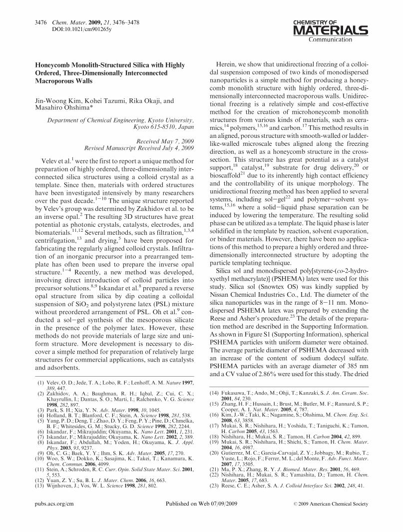
honeycomb monolith-structured silica, several centi-meters in length and about 8 mm in diameter. The testresults for laser transmission perpendicular and verticalto the freezing direction are illustrated in Figure 1b andFigure S2c (Supporting Information), respectively.Whenthe laser is perpendicular to the freezing direction, thestrength of transmitted light is strongest in the centerarea, and some light is transmitted through the sidewallbecause the porous structure has highly ordered, three-dimensionally interconnected macroporous walls. Thestrength of the light transmitted vertical to the freezingdirection was weaker than the strength of the lighttransmitted perpendicular to the freezing direction.Figure 2 shows typical SEM micrographs of prepared
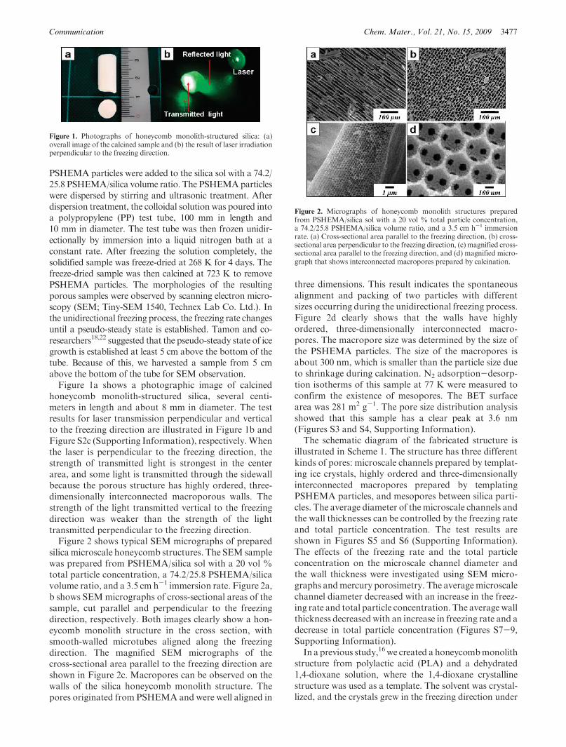
silica microscale honeycomb structures. The SEM samplewas prepared from PSHEMA/silica sol with a 20 vol %total particle concentration, a 74.2/25.8 PSHEMA/silicavolume ratio, and a 3.5 cm h-1 immersion rate. Figure 2a,b shows SEMmicrographs of cross-sectional areas of thesample, cut parallel and perpendicular to the freezingdirection, respectively. Both images clearly show a hon-eycomb monolith structure in the cross section, withsmooth-walled microtubes aligned along the freezingdirection. The magnified SEM micrographs of thecross-sectional area parallel to the freezing direction areshown in Figure 2c. Macropores can be observed on thewalls of the silica honeycomb monolith structure. Thepores originated from PSHEMA and were well aligned in
three dimensions. This result indicates the spontaneousalignment and packing of two particles with differentsizes occurring during the unidirectional freezing process.Figure 2d clearly shows that the walls have highlyordered, three-dimensionally interconnected macro-pores. The macropore size was determined by the size ofthe PSHEMA particles. The size of the macropores isabout 300 nm, which is smaller than the particle size dueto shrinkage during calcination. N2 adsorption-desorp-tion isotherms of this sample at 77 K were measured toconfirm the existence of mesopores. The BET surfacearea was 281 m2 g-1. The pore size distribution analysisshowed that this sample has a clear peak at 3.6 nm(Figures S3 and S4, Supporting Information).The schematic diagram of the fabricated structure is
illustrated in Scheme 1. The structure has three differentkinds of pores: microscale channels prepared by templat-ing ice crystals, highly ordered and three-dimensionallyinterconnected macropores prepared by templatingPSHEMA particles, and mesopores between silica parti-cles. The average diameter of the microscale channels andthe wall thicknesses can be controlled by the freezing rateand total particle concentration. The test results areshown in Figures S5 and S6 (Supporting Information).The effects of the freezing rate and the total particleconcentration on the microscale channel diameter andthe wall thickness were investigated using SEM micro-graphs andmercury porosimetry. The averagemicroscalechannel diameter decreased with an increase in the freez-ing rate and total particle concentration. The average wallthickness decreased with an increase in freezing rate and adecrease in total particle concentration (Figures S7-9,Supporting Information).In a previous study,16we created a honeycombmonolith
structure from polylactic acid (PLA) and a dehydrated1,4-dioxane solution, where the 1,4-dioxane crystallinestructure was used as a template. The solvent was crystal-lized, and the crystals grew in the freezing direction under
Figure 1. Photographs of honeycomb monolith-structured silica: (a)overall image of the calcined sample and (b) the result of laser irradiationperpendicular to the freezing direction.
Figure 2. Micrographs of honeycomb monolith structures preparedfrom PSHEMA/silica sol with a 20 vol % total particle concentration,a 74.2/25.8 PSHEMA/silica volume ratio, and a 3.5 cm h-1 immersionrate. (a) Cross-sectional area parallel to the freezing direction, (b) cross-sectional area perpendicular to the freezing direction, (c) magnified cross-sectional area parallel to the freezing direction, and (d) magnified micro-graph that shows interconnected macropores prepared by calcination.
3478 Chem. Mater., Vol. 21, No. 15, 2009 Kim et al.
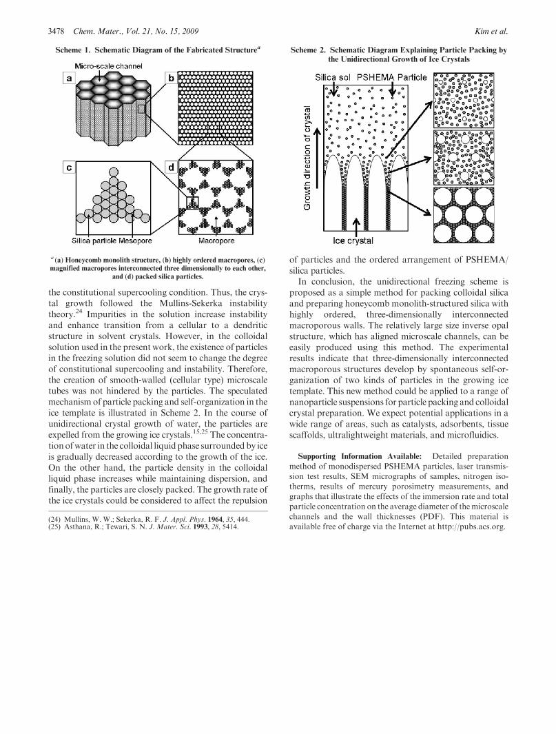
the constitutional supercooling condition. Thus, the crys-tal growth followed the Mullins-Sekerka instabilitytheory.24 Impurities in the solution increase instabilityand enhance transition from a cellular to a dendriticstructure in solvent crystals. However, in the colloidalsolution used in the present work, the existence of particlesin the freezing solution did not seem to change the degreeof constitutional supercooling and instability. Therefore,the creation of smooth-walled (cellular type) microscaletubes was not hindered by the particles. The speculatedmechanism of particle packing and self-organization in theice template is illustrated in Scheme 2. In the course ofunidirectional crystal growth of water, the particles areexpelled from the growing ice crystals.15,25 The concentra-tionofwater in the colloidal liquid phase surroundedby iceis gradually decreased according to the growth of the ice.On the other hand, the particle density in the colloidalliquid phase increases while maintaining dispersion, andfinally, the particles are closely packed. The growth rate ofthe ice crystals could be considered to affect the repulsion
of particles and the ordered arrangement of PSHEMA/silica particles.In conclusion, the unidirectional freezing scheme is
proposed as a simple method for packing colloidal silicaand preparing honeycomb monolith-structured silica withhighly ordered, three-dimensionally interconnectedmacroporous walls. The relatively large size inverse opalstructure, which has aligned microscale channels, can beeasily produced using this method. The experimentalresults indicate that three-dimensionally interconnectedmacroporous structures develop by spontaneous self-or-ganization of two kinds of particles in the growing icetemplate. This new method could be applied to a range ofnanoparticle suspensions for particle packing and colloidalcrystal preparation. We expect potential applications in awide range of areas, such as catalysts, adsorbents, tissuescaffolds, ultralightweight materials, and microfluidics.
Supporting Information Available: Detailed preparation
method of monodispersed PSHEMA particles, laser transmis-
sion test results, SEM micrographs of samples, nitrogen iso-
therms, results of mercury porosimetry measurements, and
graphs that illustrate the effects of the immersion rate and total
particle concentration on the average diameter of themicroscale
channels and the wall thicknesses (PDF). This material is
available free of charge via the Internet at http://pubs.acs.org.
Scheme 1. Schematic Diagram of the Fabricated Structurea
a (a) Honeycomb monolith structure, (b) highly ordered macropores, (c)magnified macropores interconnected three dimensionally to each other,
and (d) packed silica particles.
Scheme 2. Schematic Diagram Explaining Particle Packing by
the Unidirectional Growth of Ice Crystals
(24) Mullins, W. W.; Sekerka, R. F. J. Appl. Phys. 1964, 35, 444.(25) Asthana, R.; Tewari, S. N. J. Mater. Sci. 1993, 28, 5414.