histology …pass the tissues please…. i. intro to histology tissue definition: a group of similar...
TRANSCRIPT

Histology
…pass the tissues please…
I. Intro to Histology
Tissue definition: A group of similar cells working towards one unifying goal
Tissues components1. Similar cells2. Extracellular Matrix
- water, NaCl, ions, calcium, fibers, nutrients, etc..
The 4 types of tissues1. Epithelial
* covers body surfaces * lines hollow organs * lines body cavities and ducts * forms glands and secretions
2. Connective * protection and support * binding together (like glue) * energy storage
3. Muscle * movement and force (including peristalsis)
4. Nervous * coordinates body activities
I. Intro to Histology

I. Critical Thinking Question?
What tissue type does blood belong to?A. Epithelial
B. Connective
C. Muscle
D. Nerve
E. Blood isn’t a tissue

Location Covers external
surfaces Lines internal surfaces
such as cheeks, blood vessels, organs etc…
Glands
II. Epithelial Anatomy
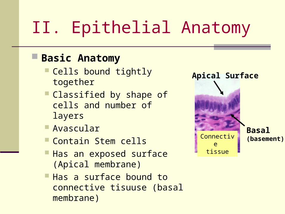
Apical Surface
Basal(basement)
Basic Anatomy Cells bound tightly together Classified by shape of cells
and number of layers Avascular Contain Stem cells Has an exposed surface
(Apical membrane) Has a surface bound to
connective tisuuse (basal membrane)
Connective tissue
II. Epithelial Anatomy
III. Epithelia Physiology
Function Provides protection internally and externally Controls permeability Provides for touch/stimuli Produces secretions:
exocrine – released onto surfaces digestive enzymes, sweat
endocrine – releases into tissue fluid and blood Hormones (chemical messengers) from pancreas,
thyroid, pituitary, etc
II. Epithelial Anatomy
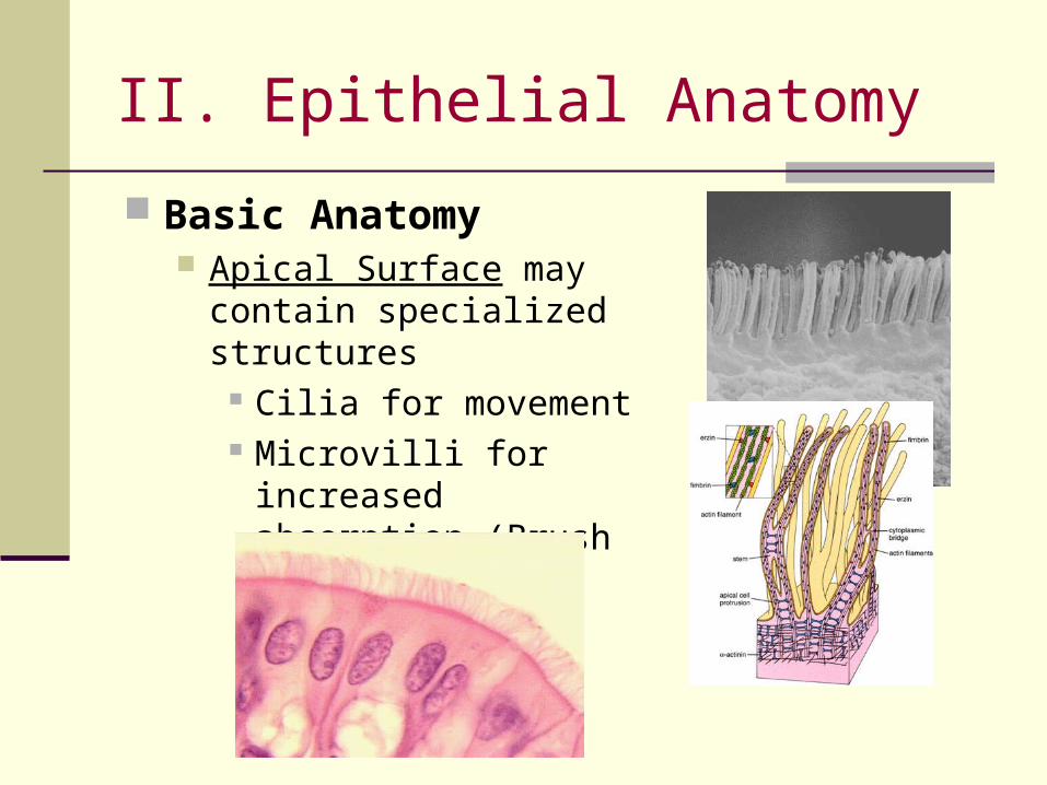
Basic Anatomy Apical Surface may contain
specialized structures Cilia for movement Microvilli for increased
absorption (Brush Borders)

II. Epithelial Anatomy
The Brush Border
II. Epithelial Anatomy
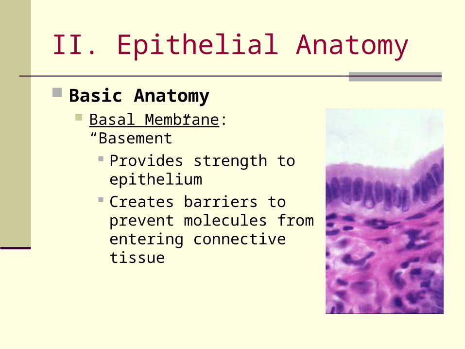
Basic Anatomy Basal Membrane: “Basement”
Provides strength to epithelium Creates barriers to prevent
molecules from entering connective tissue

II. Epithelial Anatomy
Shape Squamous: Flat Cubodial: cube-like Columnar: column-like Transitional: changes
By Layers Simple: single Stratified: multiple Pseudostratified:
appears multiple, but really simple
Classification Systems
Example. Simple cubodial = 1 cell thick of cube shaped cells

Simple Squamous Anatomy Lining of body cavities,
organs, blood vessels, alveoli lung surface
Serous Membranes
Physiology Diffusion Secretions
IV. Epithelial Tissues: Simple Squamous
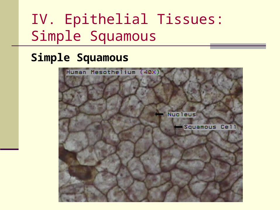
Simple Squamous
IV. Epithelial Tissues: Simple Squamous
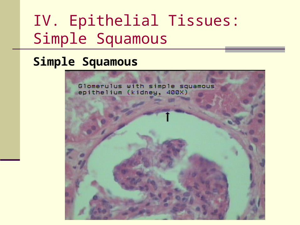
Simple Squamous
IV. Epithelial Tissues: Simple Squamous

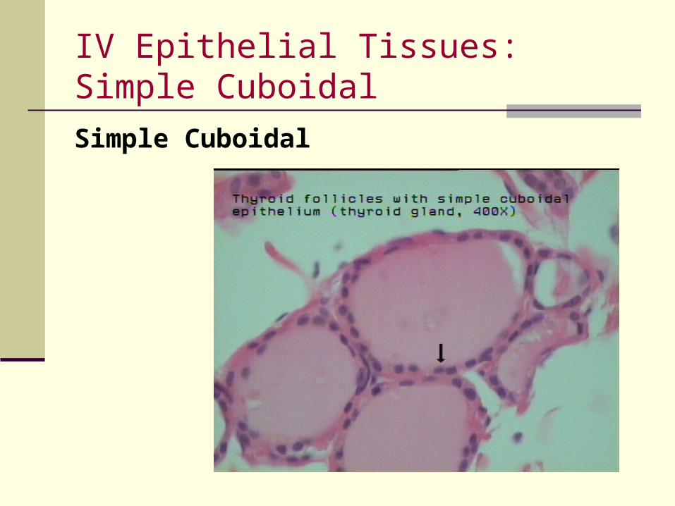
Simple CuboidalAnatomy Digestive tract, Kidney
tubules, glands
Physiology Absorption and
Secretions
IV Epithelial Tissues: Simple Cuboidal
IV Epithelial Tissues: Simple Cuboidal
Simple Cuboidal

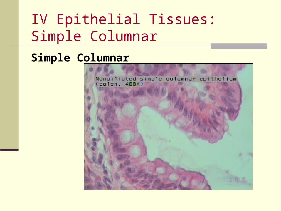
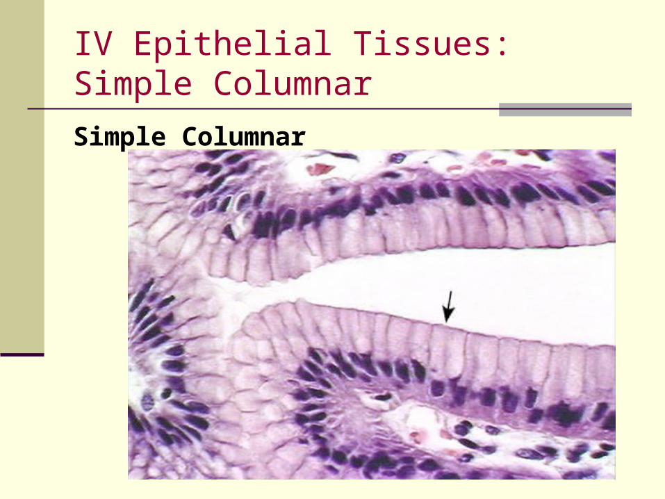
Simple ColumnarAnatomy Lining of digestive tract Modified by presence of
cilia Contains “Goblet cells”
Physiology Help move surface
material Absorption
IV Epithelial Tissues: Simple Columnar
IV Epithelial Tissues: Simple Columnar
Simple Columnar

Simple Columnar
IV Epithelial Tissues: Simple Cuboidal
IV Epithelial Tissues: Simple Columnar
Simple Columnar

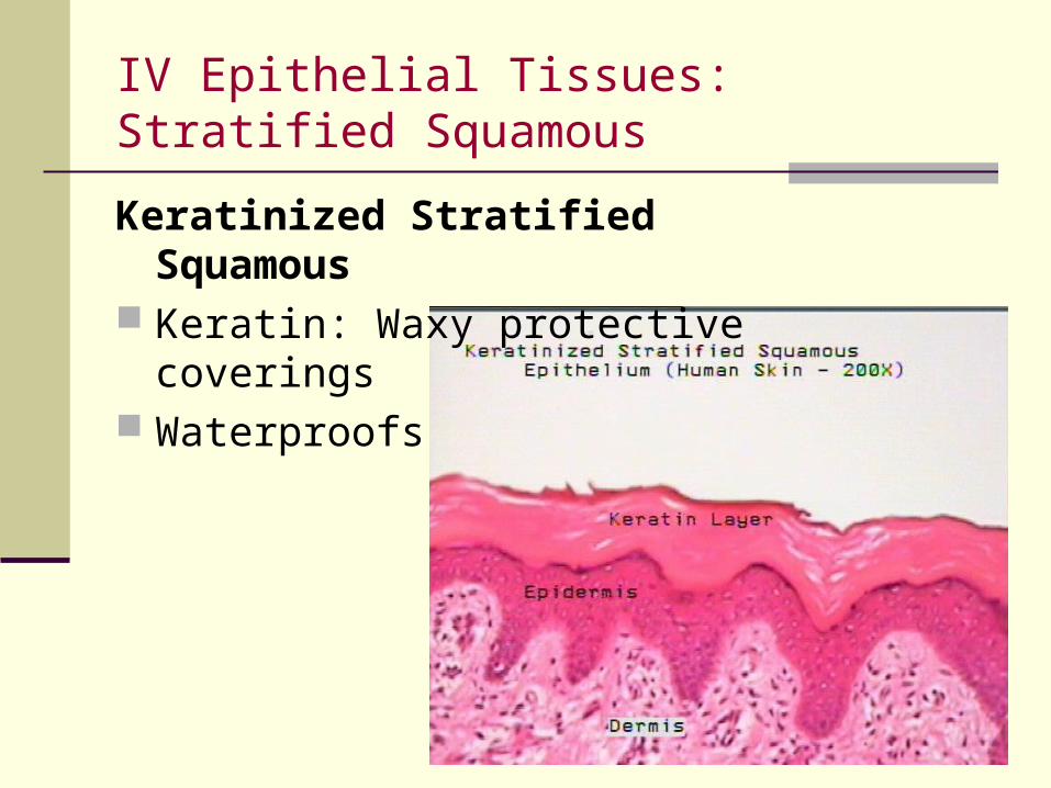
Stratified SquamousAnatomy Outer most layer-
squamous cells Inner- cuboidal or
columnar Lining of mouth,
esophagus, skin Can be keratinized
Physiology Protection Secretion Moistens membranes
IV Epithelial Tissues: Stratified Squamous

Stratified Squamous – non keratinized
IV Epithelial Tissues: Stratified Squamous
IV Epithelial Tissues: Stratified Squamous
Keratinized Stratified Squamous Keratin: Waxy protective coverings Waterproofs

IV Epithelial Tissues: Pseudostratified Columnar
Pseudostratified ColumnarAnatomy One layer All attach to the basal
membrane Appears stratified Upper respiratory tract
Physiology Move material

Pseudostratified Columnar
IV Epithelial Tissues: Pseudostratified Columnar

V. Connective Tissue Abundant extracellular material
Matrix (dominant part) or ground substance Fiber, cells in liquid, gel, or solid matrix
Never exposed to “outside environment” If exposed, causes a response for “damage
control” (ie. Bleeding)
Functions Bind and/or support other tissue Energy storage Defense of the body

Classification is based on the composition of matrix…
1. Connective Tissue Proper – loose and dense. subcutaneous, fat, tendons and ligaments
2. Fluid connective tissue
3. Supporting connective tissue
V. Connective Tissue

Connective Tissue
Tissue Proper Fluid Supporting
Blood Lymph
Cartilage Bone
Dense Regular
DenseLoose
Dense Irregular
V. Connective Tissue
VI. Connective Tissue ProperTissue Proper:
- Either loose or dense- Examples: subcutaneous, fat, tendons and ligaments
Tissue Proper is composed of … Fibroblasts – homeostasis of tissue Macrophages – engulf waste Fat cells – permanent residents Mast cells – near blood vessels, release chemicals to
elicit injury response

3 Fibers Collagen - long and straight, most
common fibers, strong but flexible
Elastic – branched and wavy, contains elastin, are elastic
Reticular – less common, thin, branching, interwoven framework of fibers
VI. Connective Tissue Proper

Loose or areolar Fewer fibers but all kinds Cushioning and support Deep to skin, between muscles, around
vessels Dense Fibrous
abundant, organized collagen fibers Tendons and ligaments Dense Regular or Dense irregular
VI. Connective Tissue Proper

Dense Regular collagen runs parallel, packed
tightly, aligned with forces Tendons and ligaments Provides attachements
Dense Irregular with collagen to provide
support and strength Gives skin its strength
VI. Connective Tissue Proper

VII. Adipose Adipose Connective
Tissue Loose connective Store large droplets of
fat Large “marshmellow”
looking cells Nuclei squished to
one side
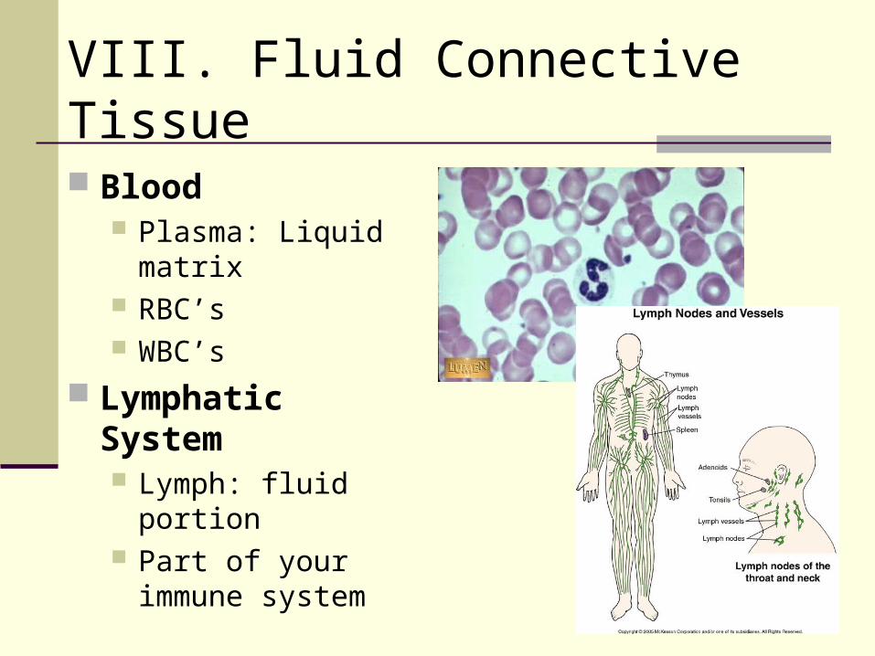
Blood Plasma: Liquid matrix RBC’s WBC’s
Lymphatic System Lymph: fluid portion Part of your immune
system
VIII. Fluid Connective Tissue

Cartilage Connective Tissue Rubbery consistency (matrix) Avascular
IX. Supporting Connective

Types of Cartilage Hyaline – most common
Joints, rib cage, respiratory tract Elastic
Mostly elastic fibers, Very flexible Outer ear, nose, epiglottis
Fibrocartilage Mostly collagen fibers, durable, strong, tough Backbone (resist compression, absorb shock…)
IX. Supporting Connective


Bone Osteocytes: bone cells Hardest connective
tissue Spongy bone
Ends of long bones Compact
Shafts of long bones Tightly organized
IX. Supporting Connective

Bone Connective Tissue

Three types
1. Skeletal
2. Smooth
3. Cardiac
Functions: Cells have ability to contract Locomotion Support Other body movement
X: Muscle
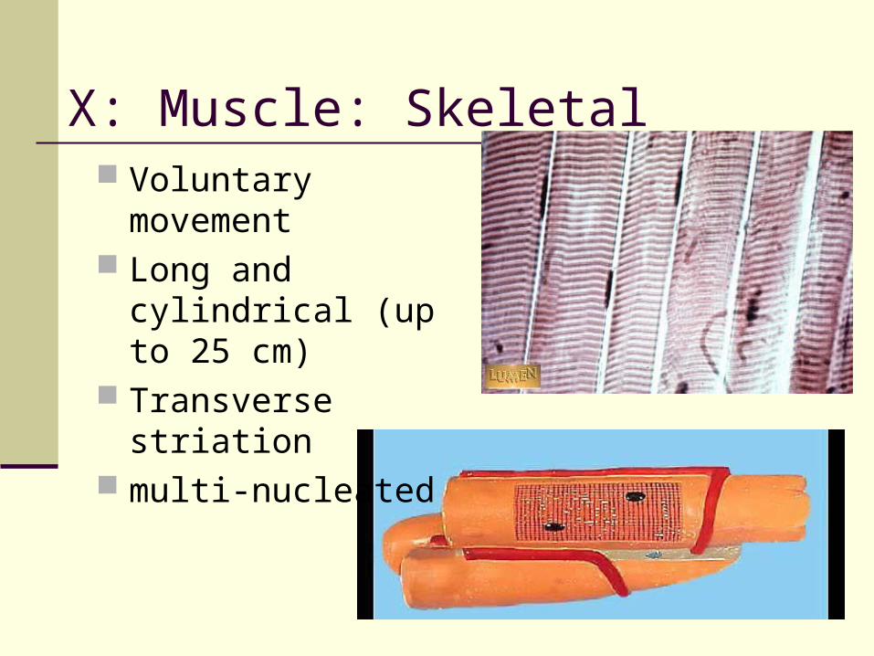
X: Muscle: Skeletal Voluntary movement Long and cylindrical
(up to 25 cm) Transverse striation multi-nucleated
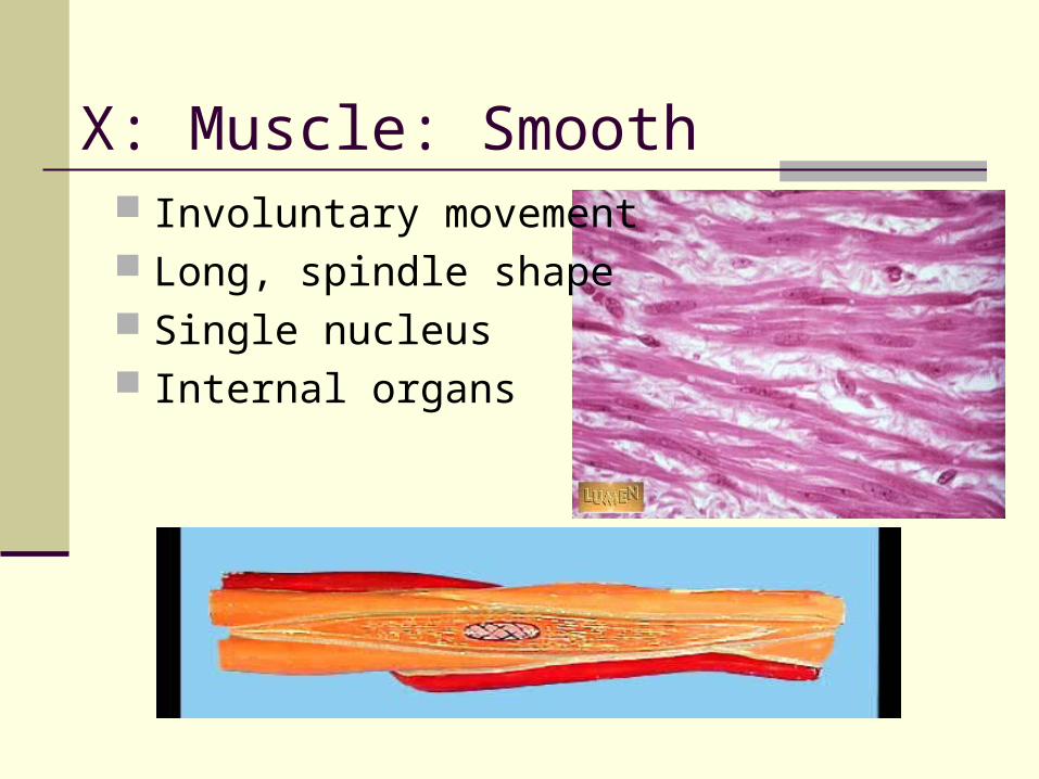
X: Muscle: Smooth Involuntary movement Long, spindle shape Single nucleus Internal organs

X: Muscle: Cardiac Striations Involuntary One nucleus Intercalated disks Heart muscle

XI: Nervous Cells very high ability to
Respond to stimuli Transmit impulses
Two types of cells Neurons – conduct nerve impulses Neuroglia – provide physical support, maintain
chemical composition of tissue fluids, nutrients…

XI: Nervous Cell Body(3) Dendrites (5) Axon(1)
Can’t be replaced Very LONG cells Create the human
“electrical system”