histology and embryology - cbo - oddělení histologie a...
TRANSCRIPT
Modul IB
Nerve tissueNerve tissuehistology and histology and embryologyembryology
Martin ŠpačekMartin Špaček(m.(m.spacekspacek@[email protected]))
• Pictures from:• Junqueira et al.: Basic histology• Rarey, Romrell: Clinical human embryology• Young, Heath: Wheather’s functional histology• http://www.med.unc.edu/embryo_images• http://www.meddean.luc.edu/lumen/meded/Histo/frames/
histo_frames.html
http://www.lf3.cuni.cz/histologie
Development
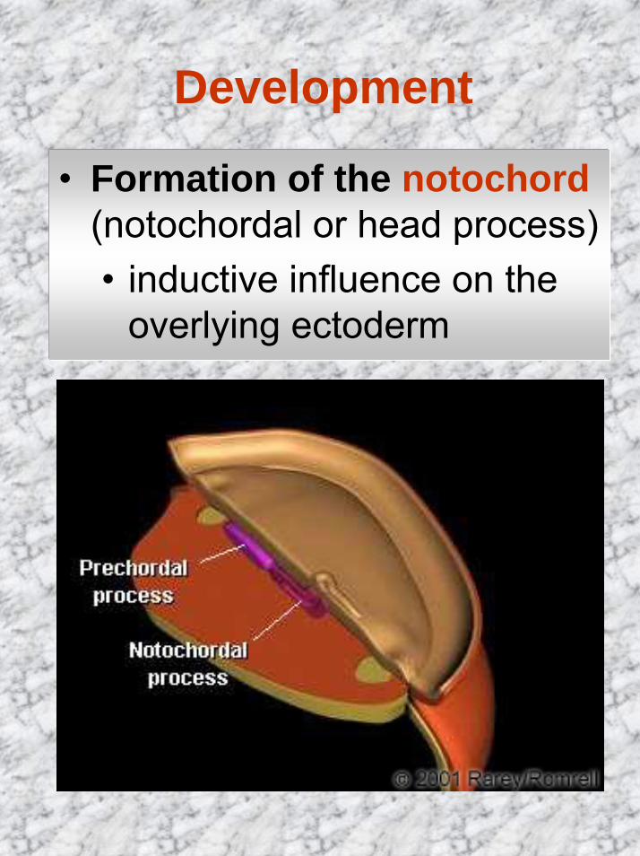
• Formation of the notochord (notochordal or head process)• inductive influence on the
overlying ectoderm

Development• Nerve tissue develops from
the ectoderm• At the beginning of the 3rd
week the ectoderm overlying the notochord forms the neural plate
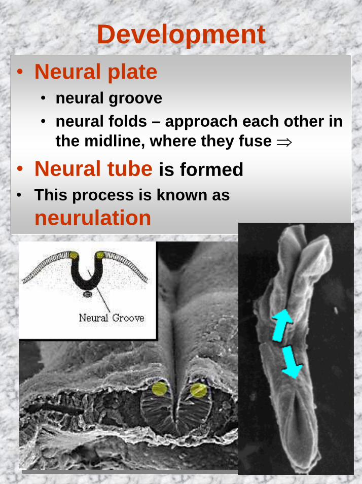
Development• Neural plate
• neural groove• neural folds – approach each other in
the midline, where they fuse ⇒
• Neural tube is formed• This process is known as
neurulation
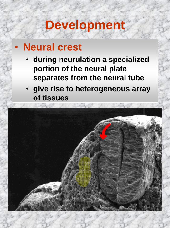
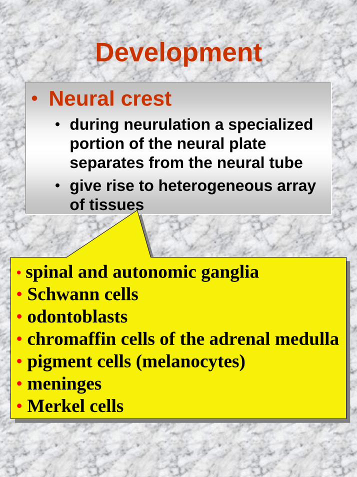
Development• Neural crest
• during neurulation a specialized portion of the neural plate separates from the neural tube
• give rise to heterogeneous array of tissues
Development• Neural crest
• during neurulation a specialized portion of the neural plate separates from the neural tube
• give rise to heterogeneous array of tissues
• spinal and autonomic ganglia • Schwann cells• odontoblasts• chromaffin cells of the adrenal medulla • pigment cells (melanocytes)• meninges• Merkel cells
• spinal and autonomic ganglia • Schwann cells• odontoblasts• chromaffin cells of the adrenal medulla • pigment cells (melanocytes)• meninges• Merkel cells
Development
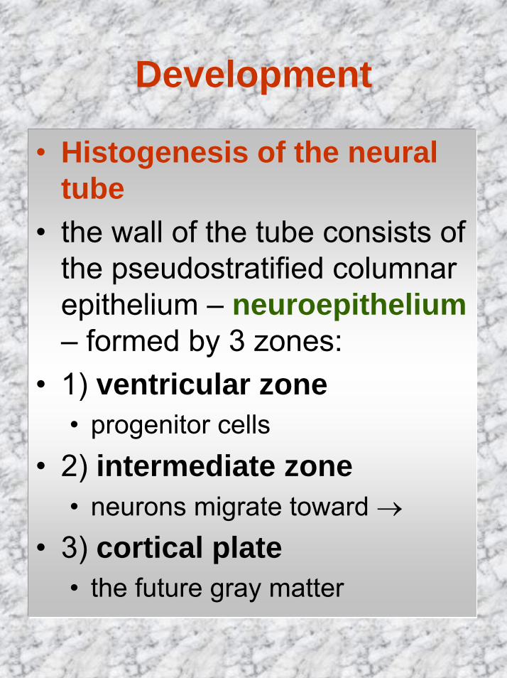
• Histogenesis of the neural tube
• the wall of the tube consists of the pseudostratified columnar epithelium – neuroepithelium– formed by 3 zones:
• 1) ventricular zone• progenitor cells
• 2) intermediate zone• neurons migrate toward →
• 3) cortical plate• the future gray matter
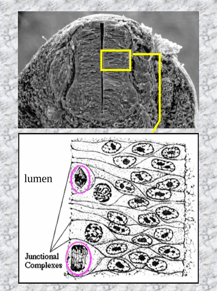
lumen
Development
• Histological differentiation
1. nerve cells (neurons)2. glia cells
„connective tissue of the CNS“
3. neural crest cells
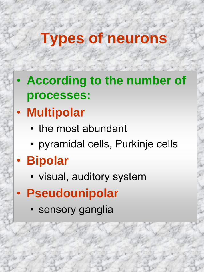
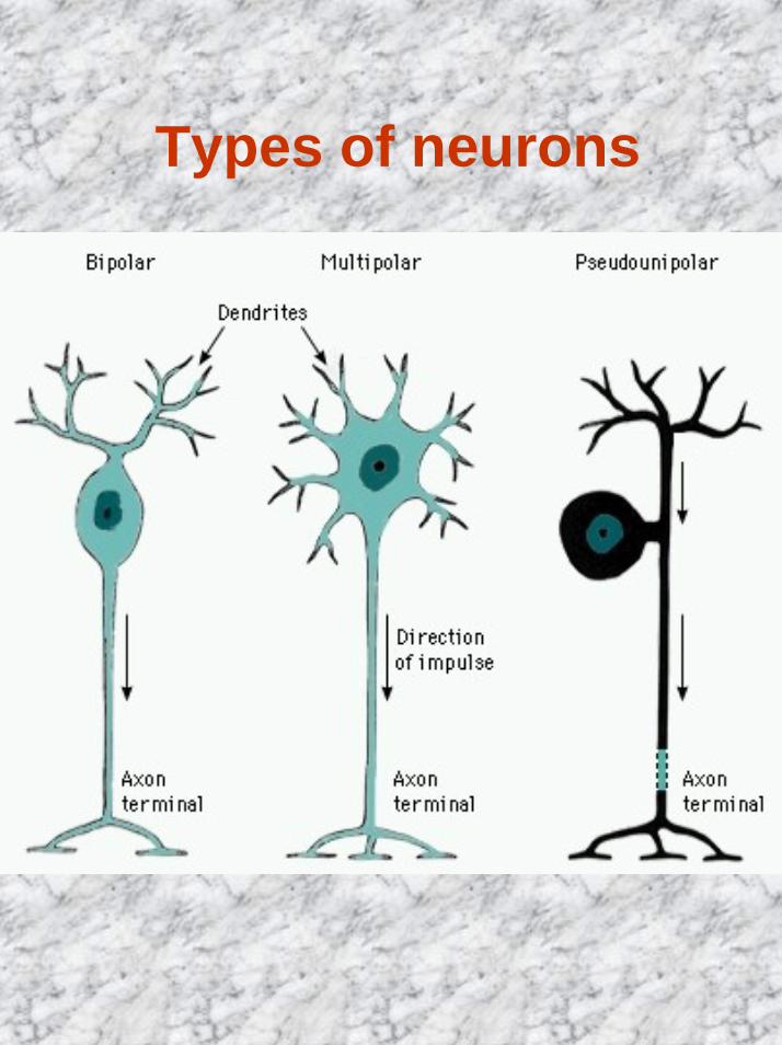
Types of neurons
• According to the number ofprocesses:
• Multipolar• the most abundant• pyramidal cells, Purkinje cells
• Bipolar• visual, auditory system
• Pseudounipolar• sensory ganglia
Types of neurons
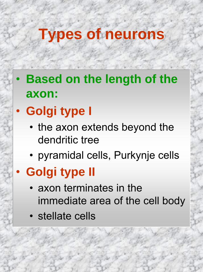
Types of neurons
• Based on the length of the axon:
• Golgi type I• the axon extends beyond the
dendritic tree• pyramidal cells, Purkynje cells
• Golgi type II• axon terminates in the
immediate area of the cell body• stellate cells
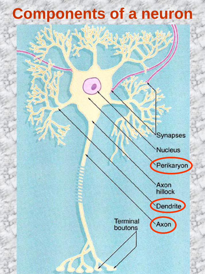
Components of a neuron
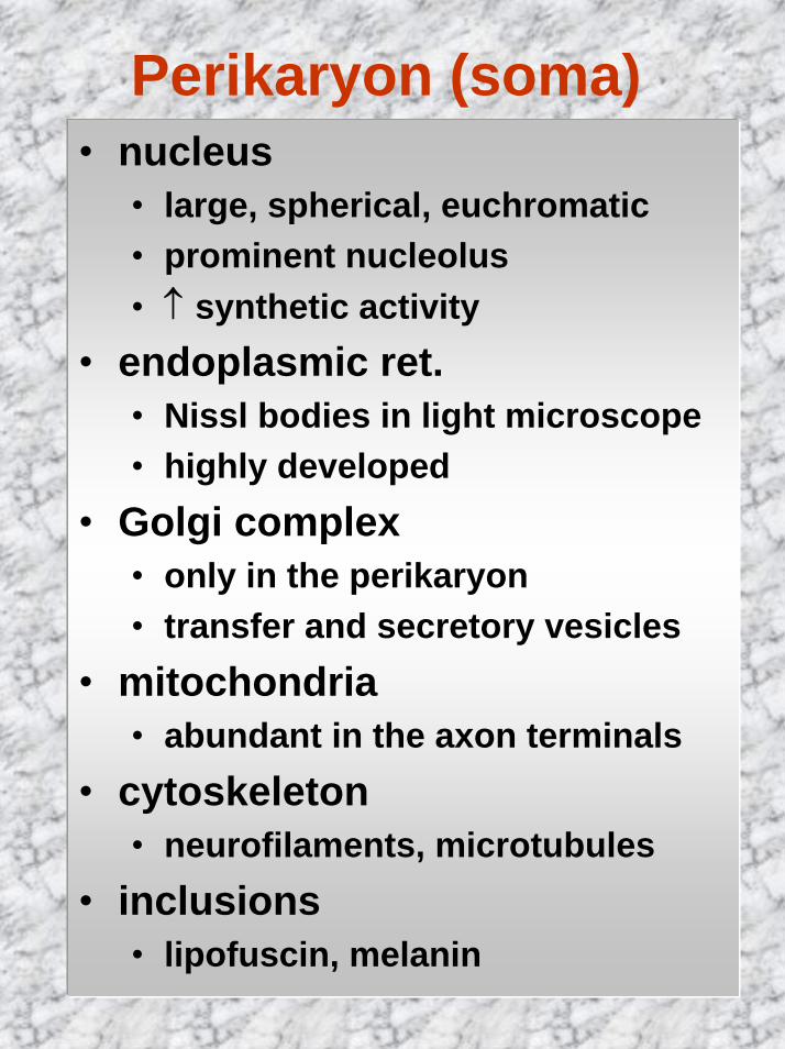
Perikaryon (soma)• nucleus
• large, spherical, euchromatic• prominent nucleolus• ↑ synthetic activity
• endoplasmic ret.• Nissl bodies in light microscope• highly developed
• Golgi complex• only in the perikaryon• transfer and secretory vesicles
• mitochondria• abundant in the axon terminals
• cytoskeleton• neurofilaments, microtubules
• inclusions• lipofuscin, melanin
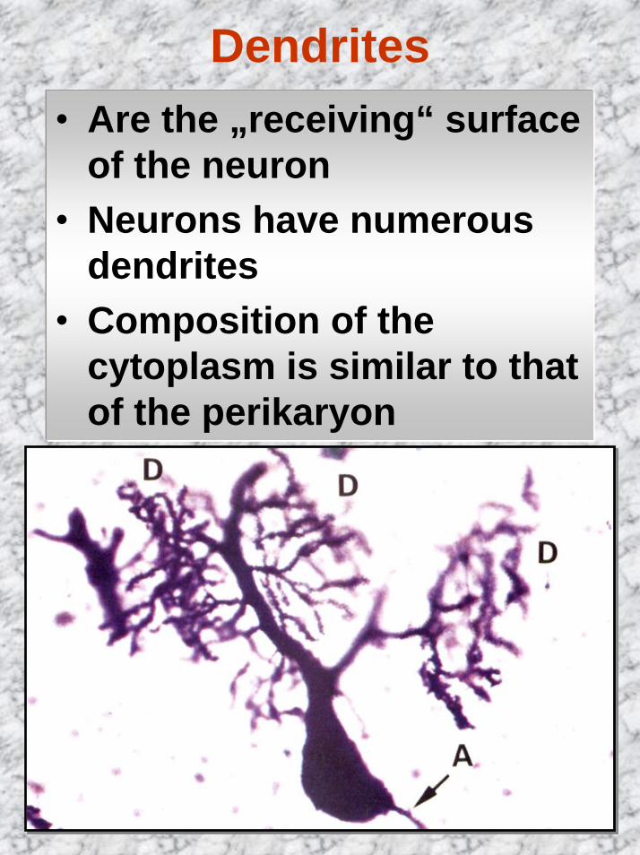
Dendrites• Are the „receiving“ surface
of the neuron• Neurons have numerous
dendrites • Composition of the
cytoplasm is similar to that of the perikaryon
Axons
• Most neurons have only one axon
• Originate from axon hillock • Dependent on the
perikaryon for its maintenance – axonal transport
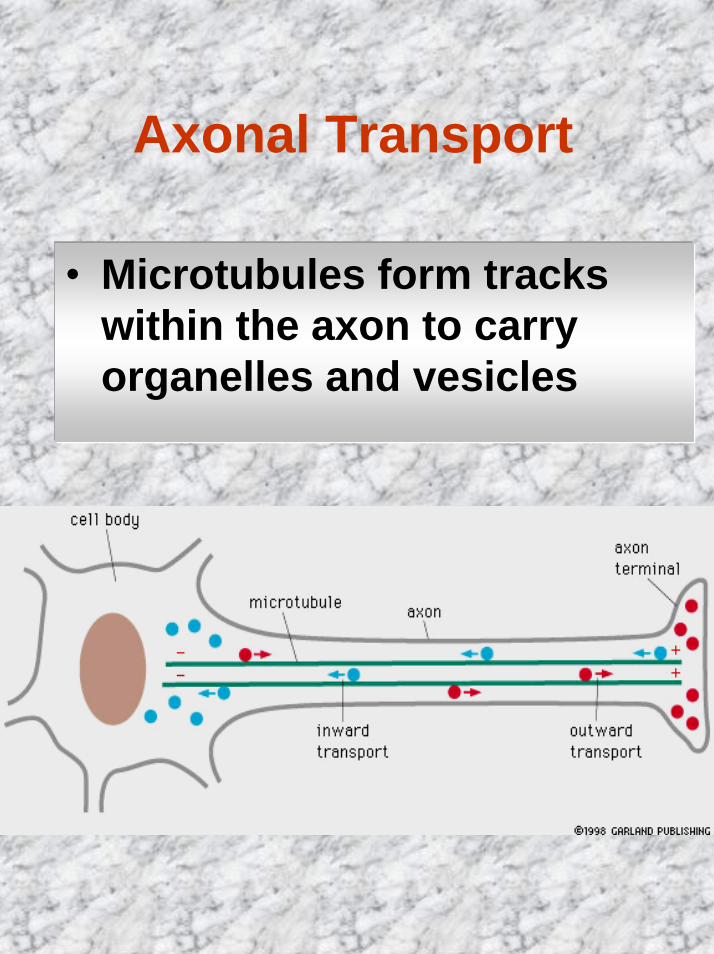
Axonal Transport
• Microtubules form tracks within the axon to carry organelles and vesicles
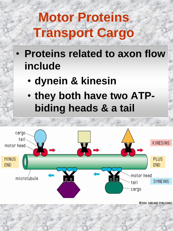
Motor Proteins Transport Cargo
• Proteins related to axon flow include• dynein & kinesin• they both have two ATP-
biding heads & a tail
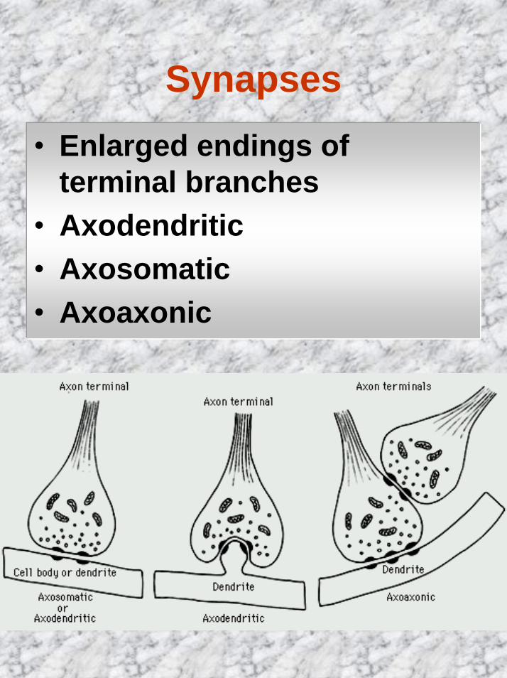
Synapses• Enlarged endings of
terminal branches • Axodendritic• Axosomatic• Axoaxonic
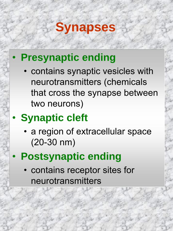
Synapses
• Presynaptic ending• contains synaptic vesicles with
neurotransmitters (chemicals that cross the synapse between two neurons)
• Synaptic cleft• a region of extracellular space
(20-30 nm)• Postsynaptic ending
• contains receptor sites for neurotransmitters
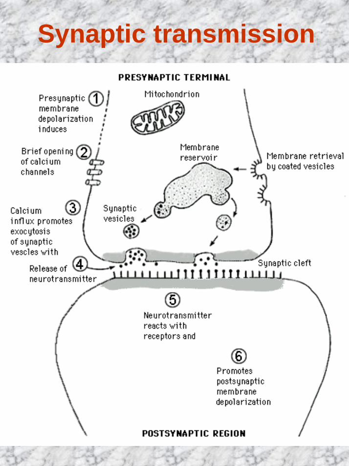
Synaptic transmission
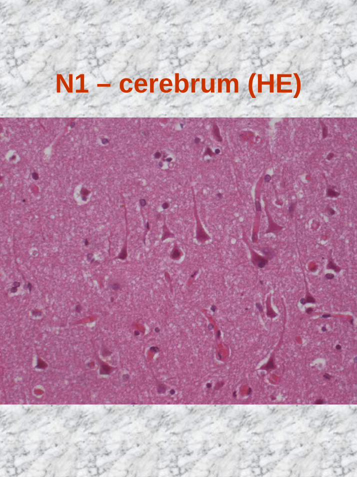
N1 – cerebrum (HE)
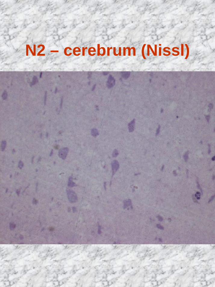
N2 – cerebrum (Nissl)
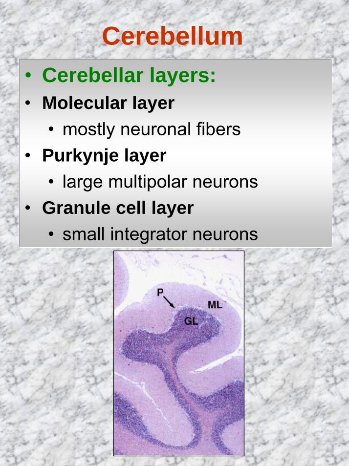
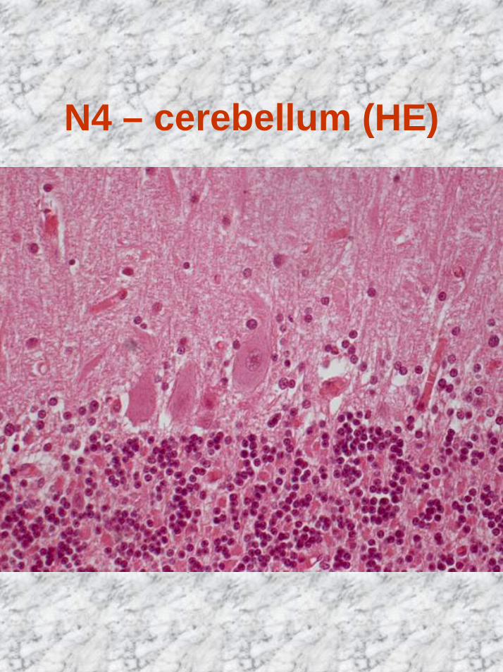
Cerebellum• Cerebellar layers:• Molecular layer
• mostly neuronal fibers• Purkynje layer
• large multipolar neurons• Granule cell layer
• small integrator neurons
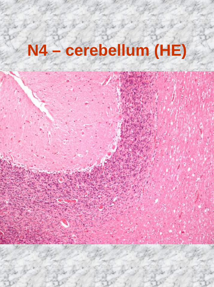
N4 – cerebellum (HE)
N4 – cerebellum (HE)
Neuroglia• 10 glial cells for each neuron• About half of the volume of
nerve tissue• Function: provide neurons
with structural support and maintain local conditions for neuronal function
• Staining: silver or gold impregnation, histochemicaltechnique
• 4 morphologic types
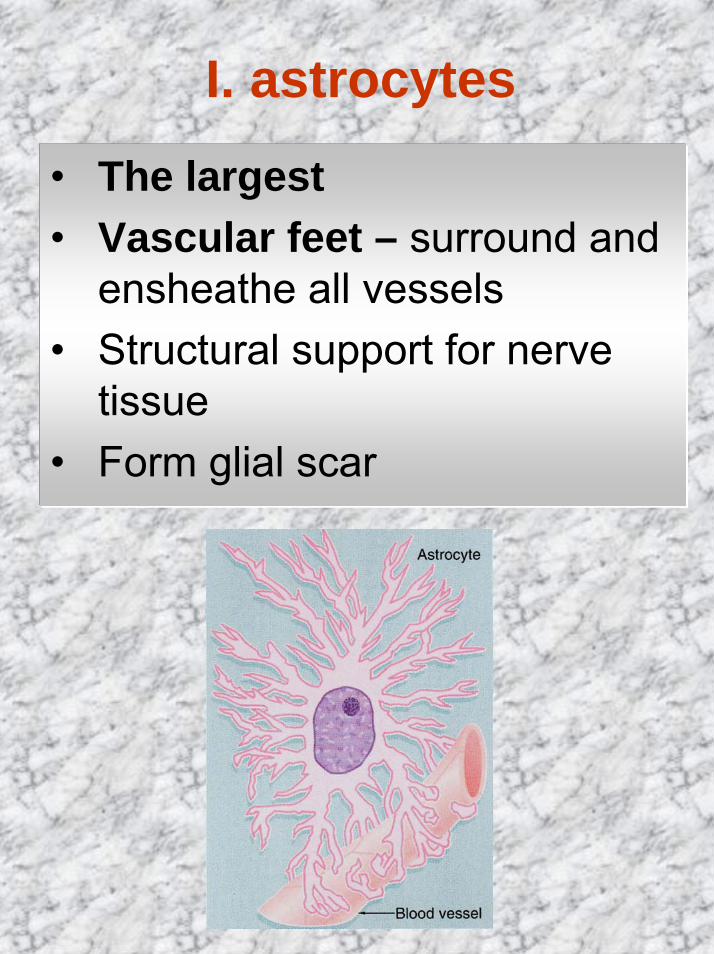
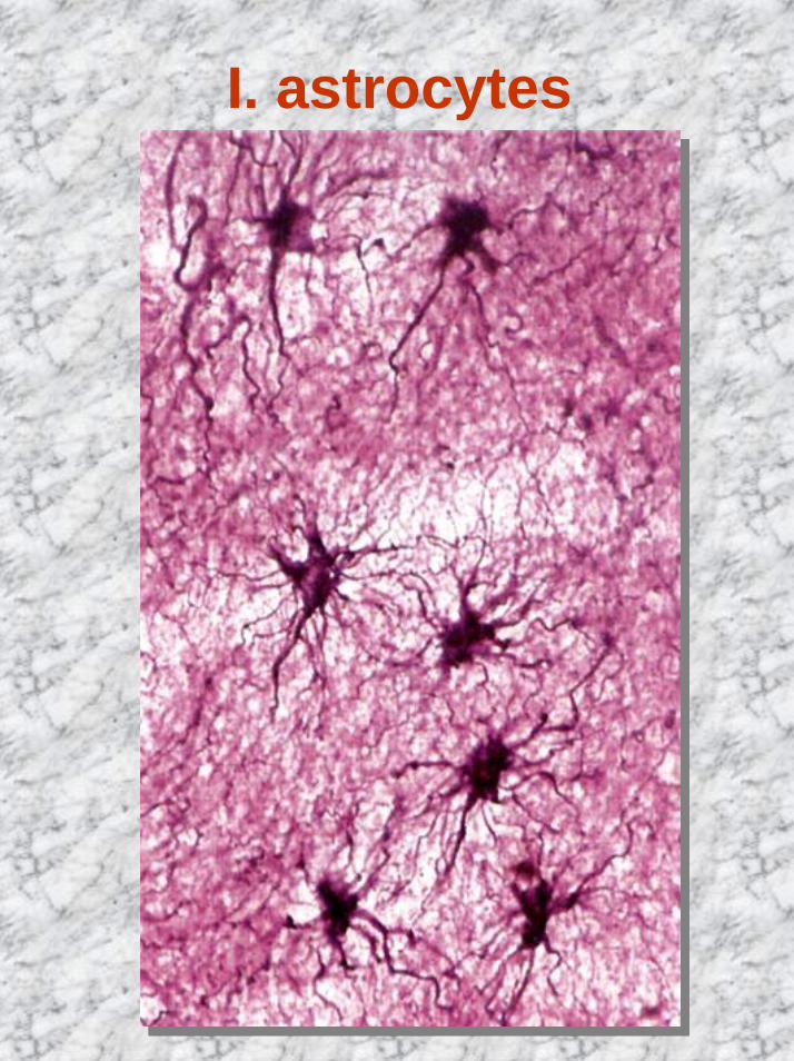
I. astrocytes
• The largest • Vascular feet – surround and
ensheathe all vessels• Structural support for nerve
tissue• Form glial scar
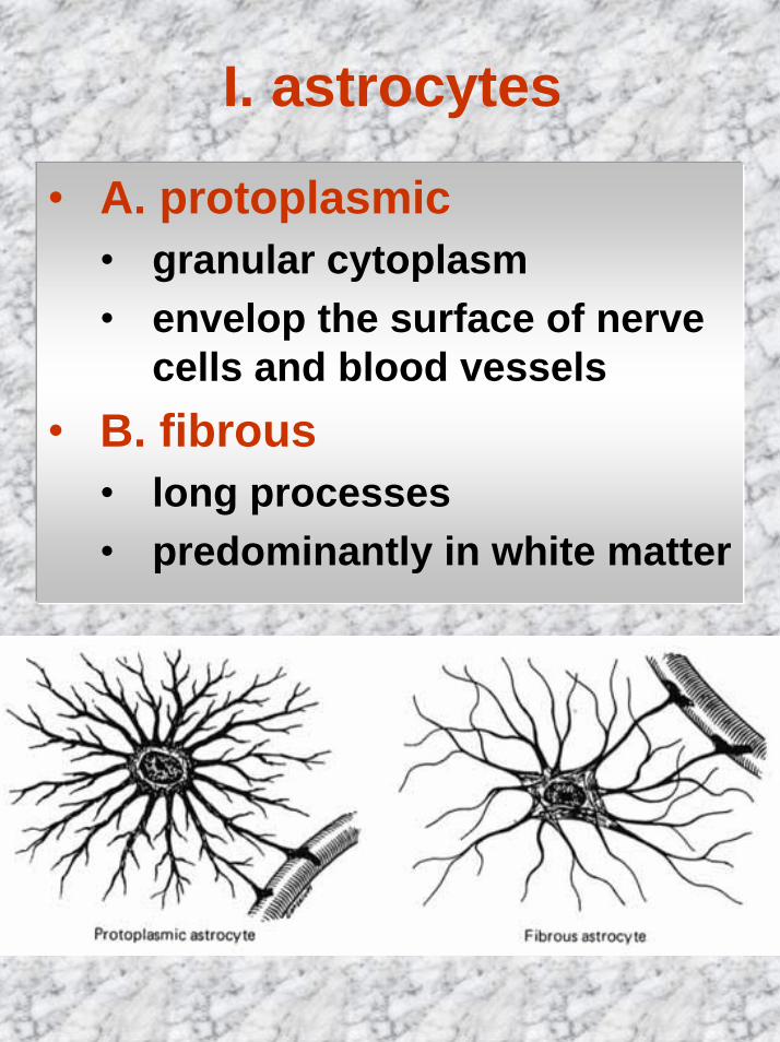
I. astrocytes
• A. protoplasmic • granular cytoplasm• envelop the surface of nerve
cells and blood vessels• B. fibrous
• long processes• predominantly in white matter
I. astrocytes
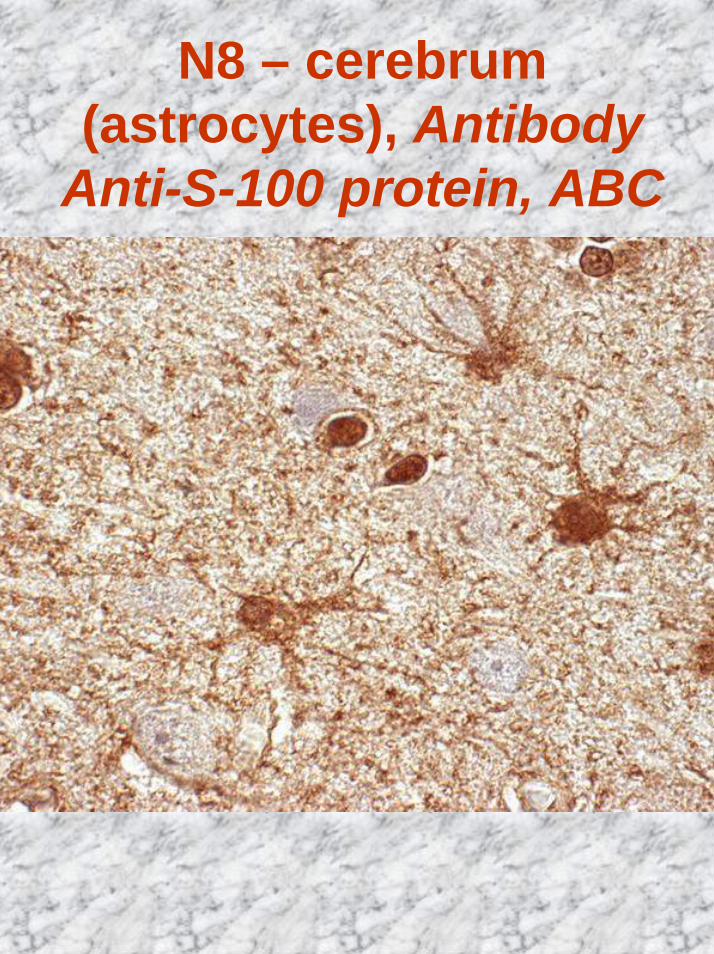
N8 – cerebrum(astrocytes), Antibody
Anti-S-100 protein, ABC
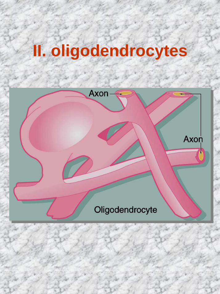
II. oligodendrocytes
• Smaller, ↓processes• Processes envelop axons
and form myelin sheath• Are found in both gray and
white matter• The formation of the myelin
sheath is similar to that of Schwann cells in peripheral nerves
II. oligodendrocytes
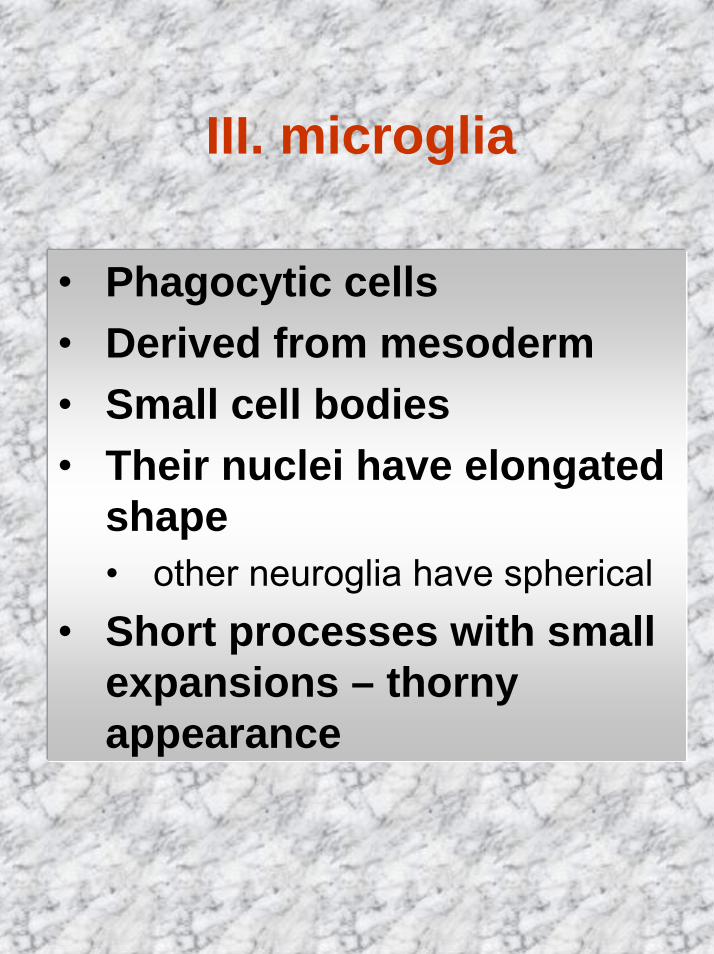
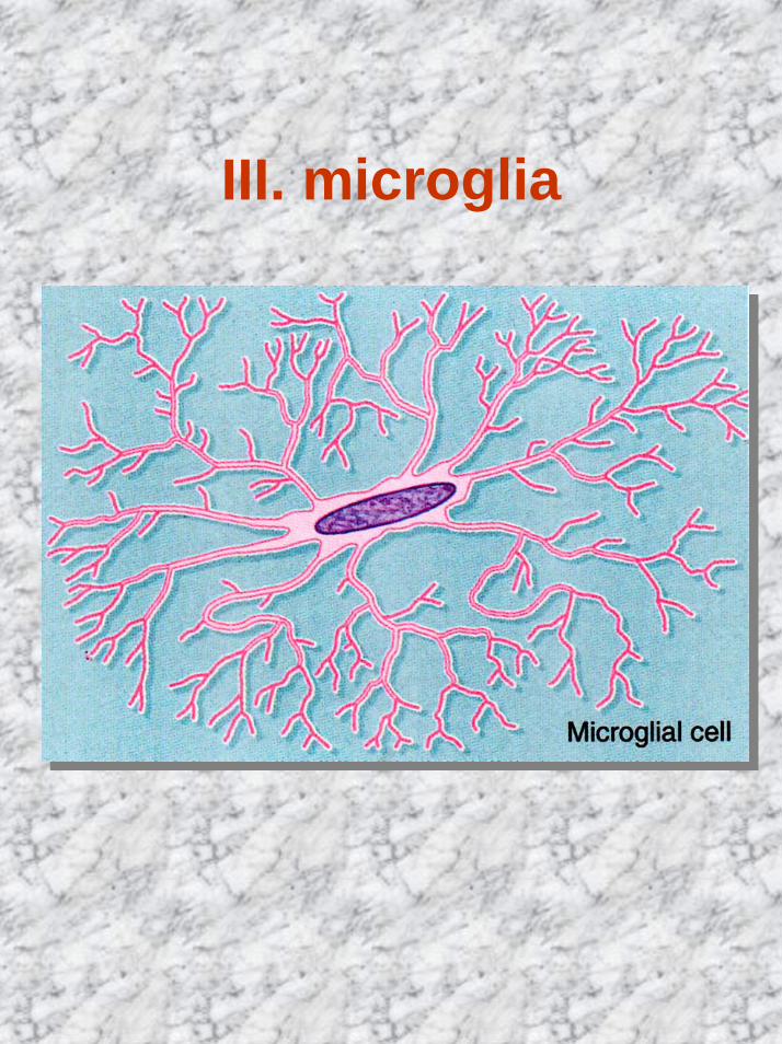
III. microglia
• Phagocytic cells• Derived from mesoderm• Small cell bodies• Their nuclei have elongated
shape • other neuroglia have spherical
• Short processes with small expansions – thorny appearance
III. microglia
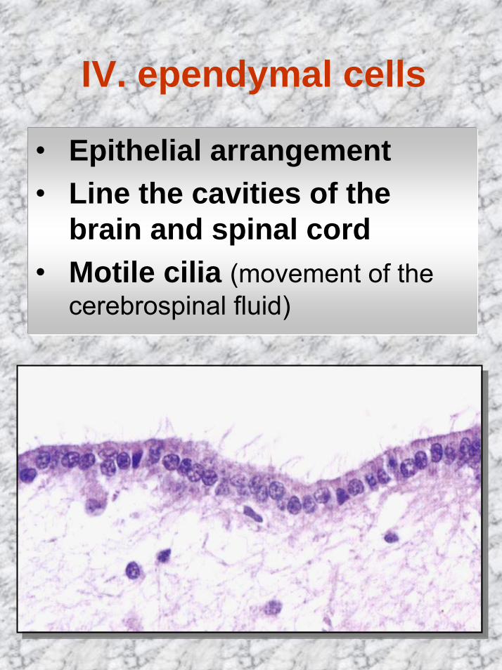
IV. ependymal cells
• Epithelial arrangement• Line the cavities of the
brain and spinal cord• Motile cilia (movement of the
cerebrospinal fluid)
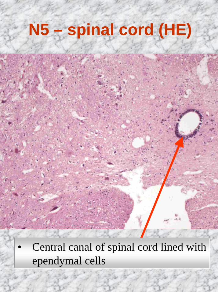
N5 – spinal cord (HE)
• Central canal of spinal cord lined with ependymal cells
Nerve fibres
• Consist of axons enveloped by special sheaths of ectodermal origin
• Groups of nerve fibresconstitute:• the tracts of the brain
(oligodendrocytes) • peripheral nerves (Schwann
cells)• Fibres:
• unmyelinated• myelinated
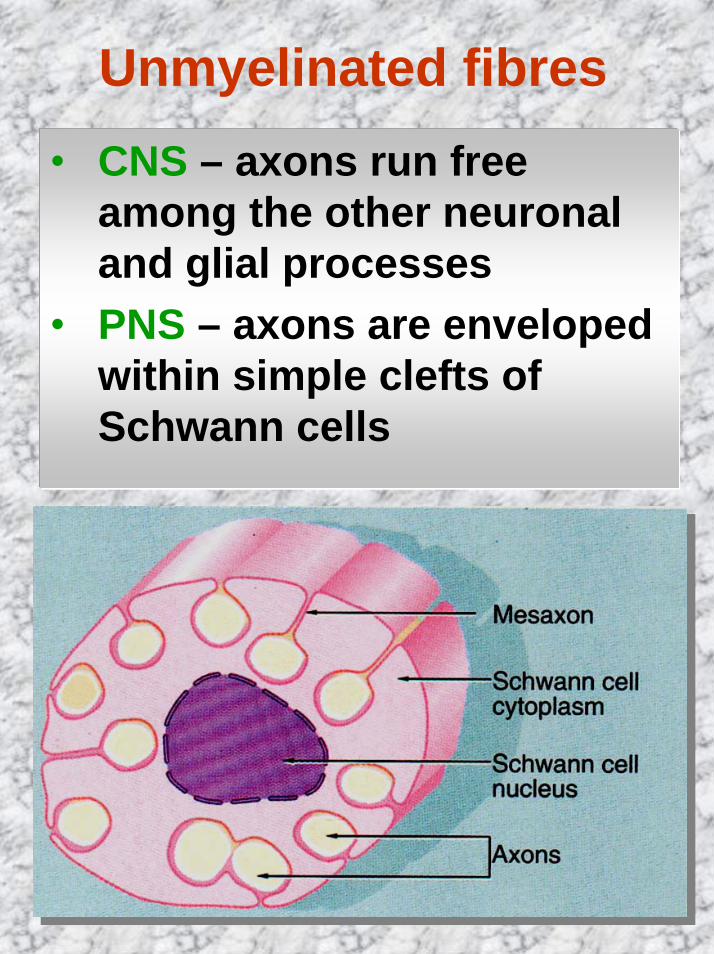
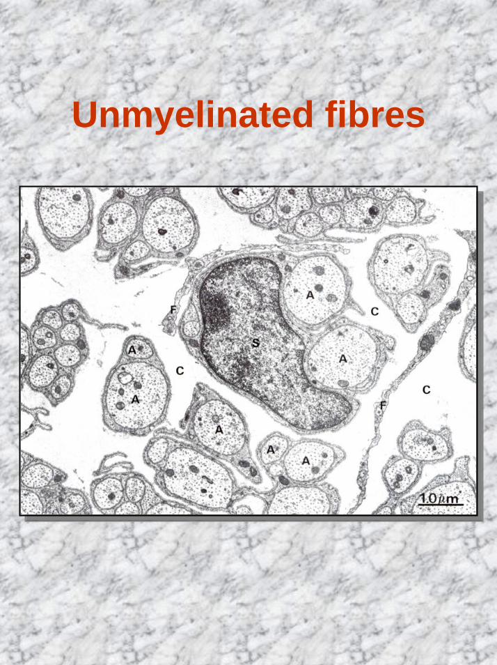
Unmyelinated fibres• CNS – axons run free
among the other neuronal and glial processes
• PNS – axons are enveloped within simple clefts of Schwann cells
Unmyelinated fibres
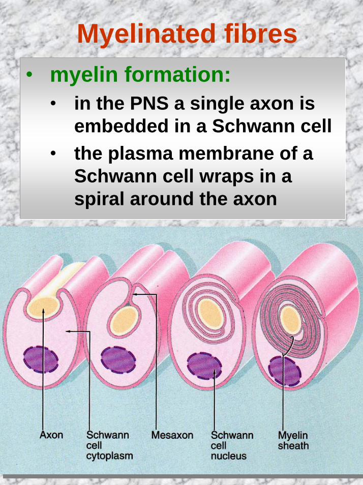
Myelinated fibres• myelin formation:
• in the PNS a single axon is embedded in a Schwann cell
• the plasma membrane of a Schwann cell wraps in a spiral around the axon
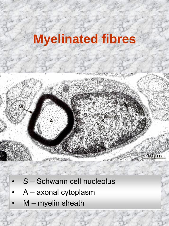
Myelinated fibres
• S – Schwann cell nucleolus• A – axonal cytoplasm• M – myelin sheath
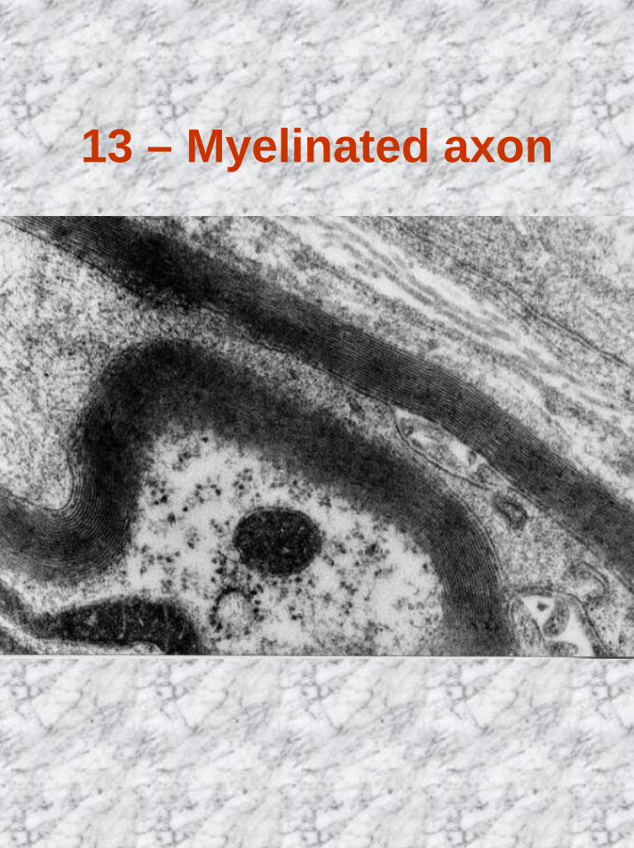
13 – Myelinated axon
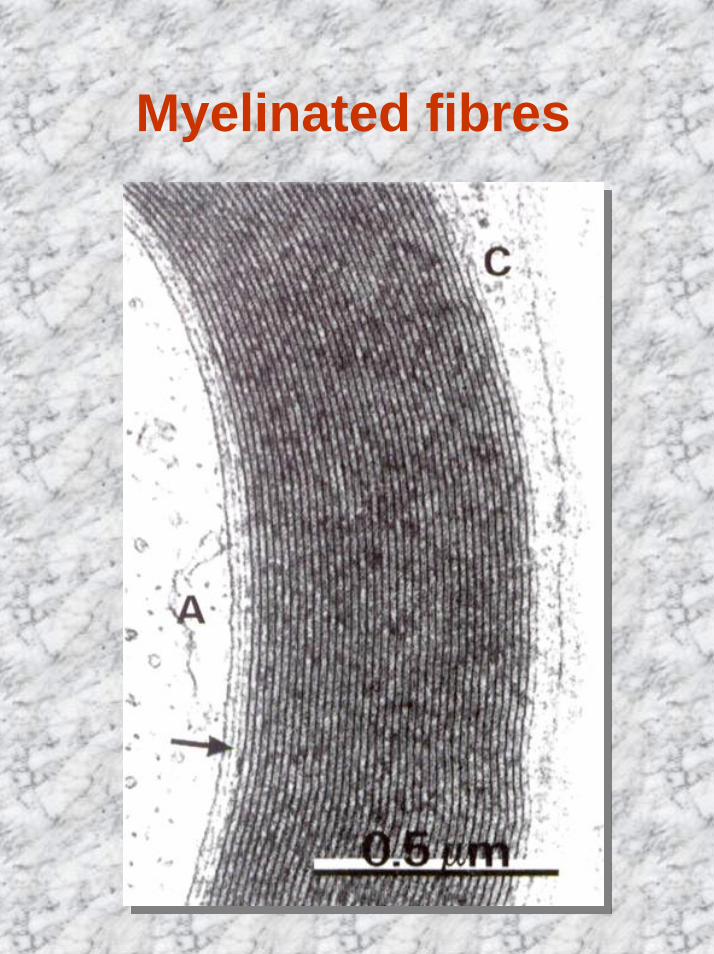
Myelinated fibres
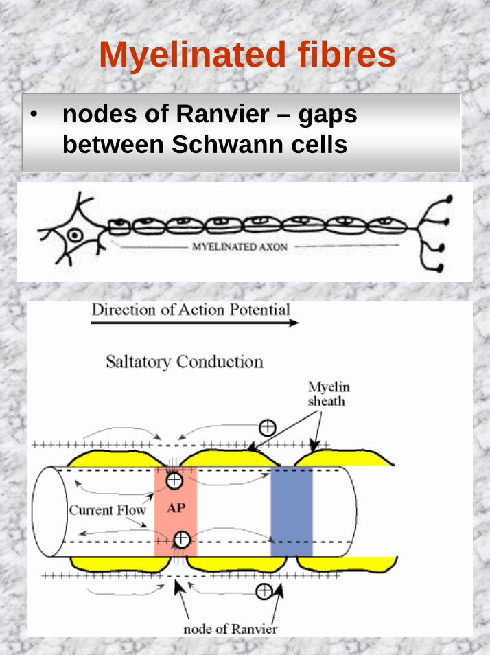
Myelinated fibres• nodes of Ranvier – gaps
between Schwann cells
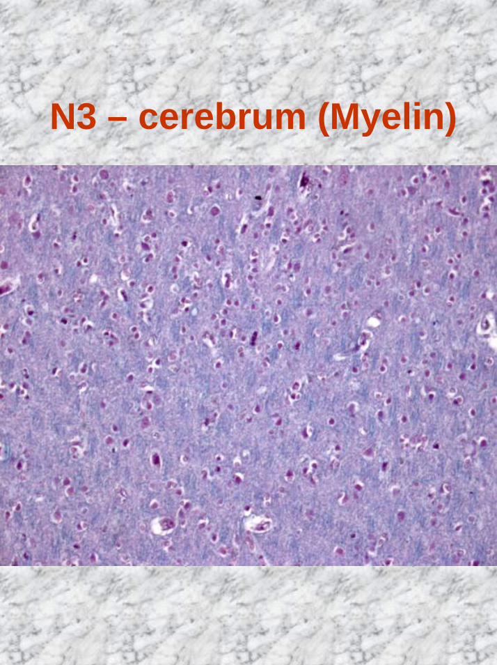
N3 – cerebrum (Myelin)
Peripheral nervous system
• nerves• nerve fibres grouped in bundles• connective tissue coverings:
• endoneurium• perineurium• epineurium
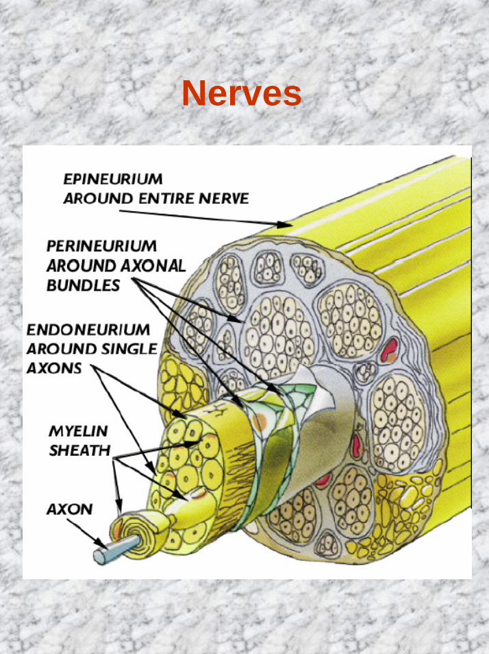
Nerves
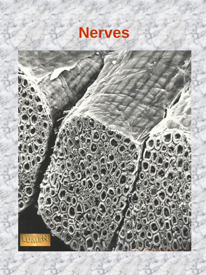
Nerves
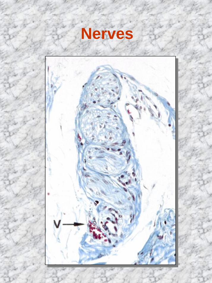
Nerves
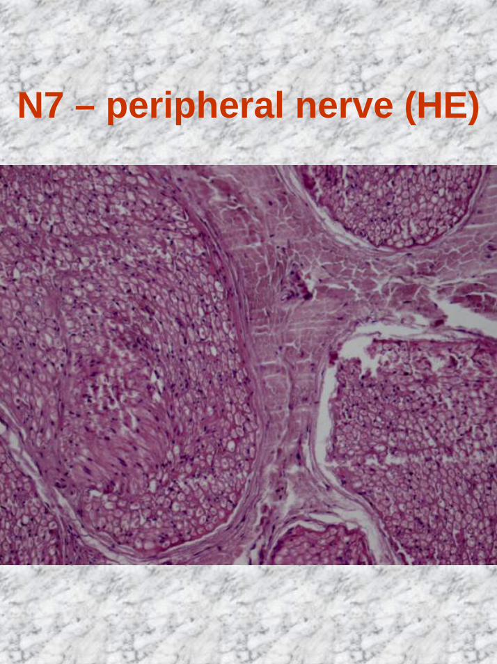
N7 – peripheral nerve (HE)
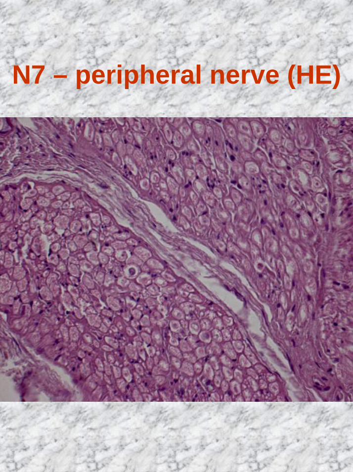
N7 – peripheral nerve (HE)
Peripheral nervoussystem
• Ganglia• aggregations of nerve cell
bodies outside the CNS• a connective tissue capsule• each neuronal cell body is
surrounded by Schwann cell-like satellite cell
• Two main classes• craniospinal (sensory)• autonomic (motor)
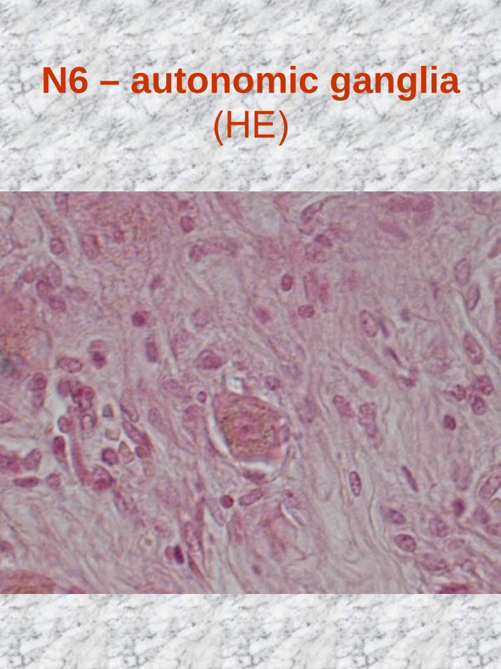
N6 – autonomic ganglia (HE)
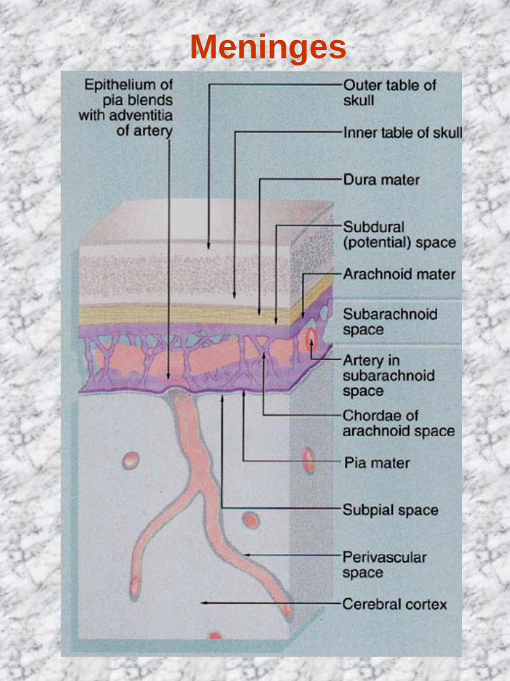
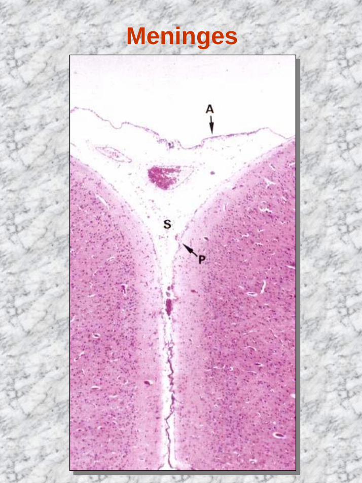
Meninges
Meninges• Dura mater
• external meninx• dense connective tissue• subdural space
• Arachnoid• connective tissue devoid of
blood vessels• a layer in contact with dura
mater• system of trabeculae –
cavities form subarachnoidspace (filled with CSF)
• Pia mater• loose connective tissue
containing many blood vessels
Meninges