h.influenza, yersinia, bordetella, brucella,...
TRANSCRIPT
MBBS
H.influenza, Yersinia, Bordetella, Brucella,
B.anthracis, Leginellae
Chen Niu (牛辰) MOH&MOE Key Lab of Medical Molecular Virology
Shanghai Medical College, Fudan University 复旦大学上海医学院分子病毒学教育部/卫生部重点实验室
Most important bacteria in zoonosis
• Brucella • Yersinia • Bacillus
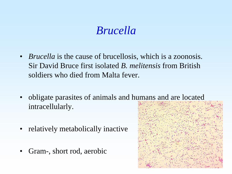
Brucella
• Brucella is the cause of brucellosis, which is a zoonosis. Sir David Bruce first isolated B. melitensis from British soldiers who died from Malta fever.
• obligate parasites of animals and humans and are located
intracellularly.
• relatively metabolically inactive
• Gram-, short rod, aerobic
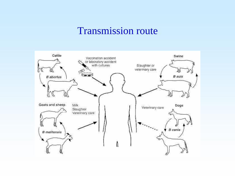
Transmission route
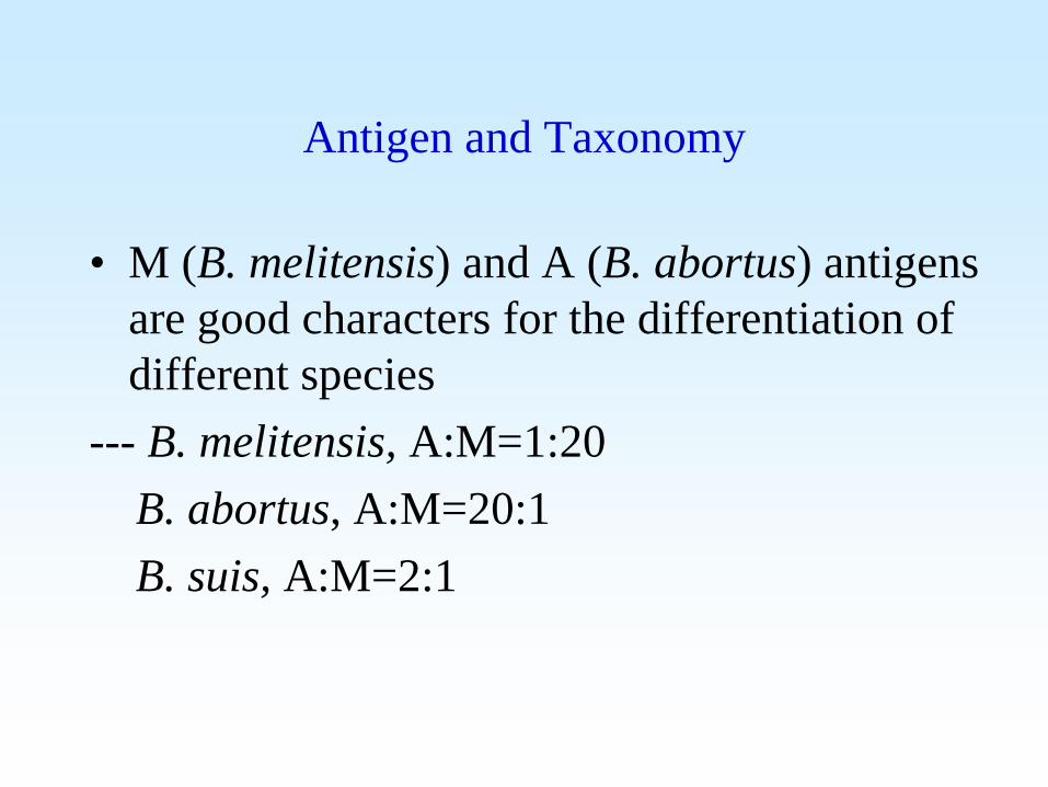
Antigen and Taxonomy
• M (B. melitensis) and A (B. abortus) antigens are good characters for the differentiation of different species
--- B. melitensis, A:M=1:20 B. abortus, A:M=20:1 B. suis, A:M=2:1
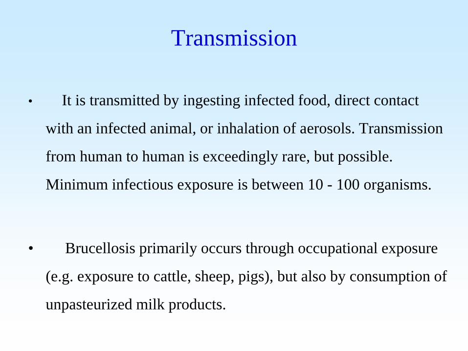
Transmission
• It is transmitted by ingesting infected food, direct contact
with an infected animal, or inhalation of aerosols. Transmission
from human to human is exceedingly rare, but possible.
Minimum infectious exposure is between 10 - 100 organisms.
• Brucellosis primarily occurs through occupational exposure
(e.g. exposure to cattle, sheep, pigs), but also by consumption of
unpasteurized milk products.
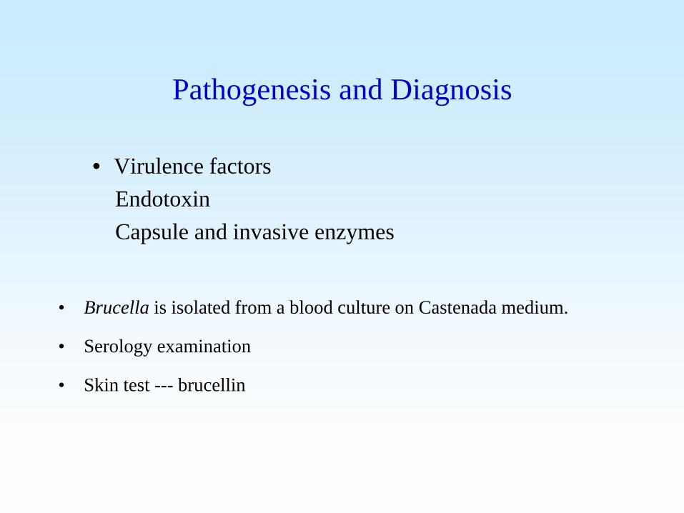
Pathogenesis and Diagnosis
• Virulence factors Endotoxin Capsule and invasive enzymes
• Brucella is isolated from a blood culture on Castenada medium.
• Serology examination
• Skin test --- brucellin
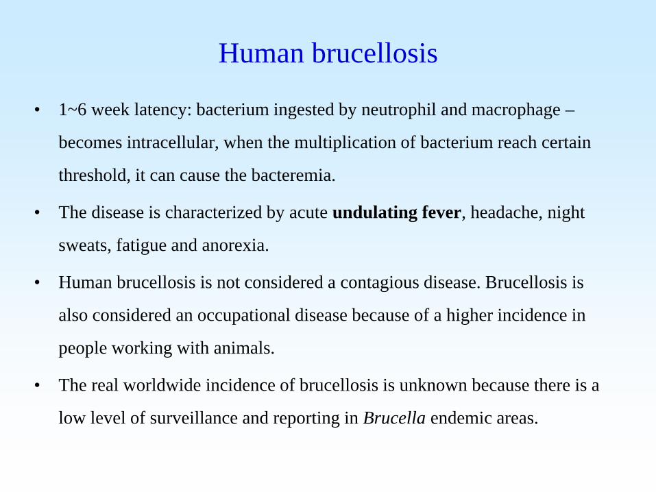
Human brucellosis
• 1~6 week latency: bacterium ingested by neutrophil and macrophage –
becomes intracellular, when the multiplication of bacterium reach certain
threshold, it can cause the bacteremia.
• The disease is characterized by acute undulating fever, headache, night
sweats, fatigue and anorexia.
• Human brucellosis is not considered a contagious disease. Brucellosis is
also considered an occupational disease because of a higher incidence in
people working with animals.
• The real worldwide incidence of brucellosis is unknown because there is a
low level of surveillance and reporting in Brucella endemic areas.
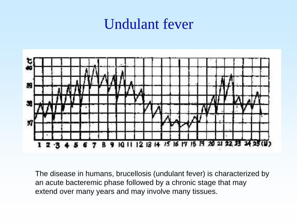
Undulant fever
The disease in humans, brucellosis (undulant fever) is characterized by an acute bacteremic phase followed by a chronic stage that may extend over many years and may involve many tissues.
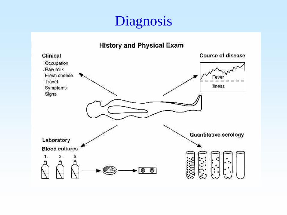
Diagnosis
Treatment
• Tetracyclines, Rifampicin or ampicillin
• Symptomatic relief may occur within a few days after
treatment. However, because of their intracellular location,
treatment must be prolonged.
• Combined treatment with a tetracycline and either
streptomycin for 2-3 weeks or rifampin for 6 weeks is
recommended.
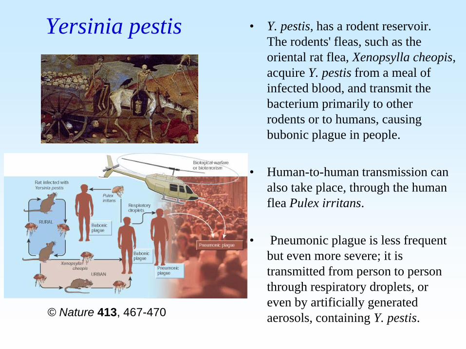
Yersinia pestis • Y. pestis, has a rodent reservoir. The rodents' fleas, such as the oriental rat flea, Xenopsylla cheopis, acquire Y. pestis from a meal of infected blood, and transmit the bacterium primarily to other rodents or to humans, causing bubonic plague in people.
• Human-to-human transmission can
also take place, through the human flea Pulex irritans.
• Pneumonic plague is less frequent but even more severe; it is transmitted from person to person through respiratory droplets, or even by artificially generated aerosols, containing Y. pestis.
© Nature 413, 467-470
Y. pestis and plaque
• Plaque is an infection of wild rodents, transmitted from
one rodent to another and occasionally from rodents to
humans by the bites of fleas.
• the pandemics of “black death” with millions of fatalities
• The ability of this organism to be transmitted by aerosol,
and the severity and high mortality associated with
pneumonic plaque, make Y. pestis a potential biological
weapon.
Biological traits
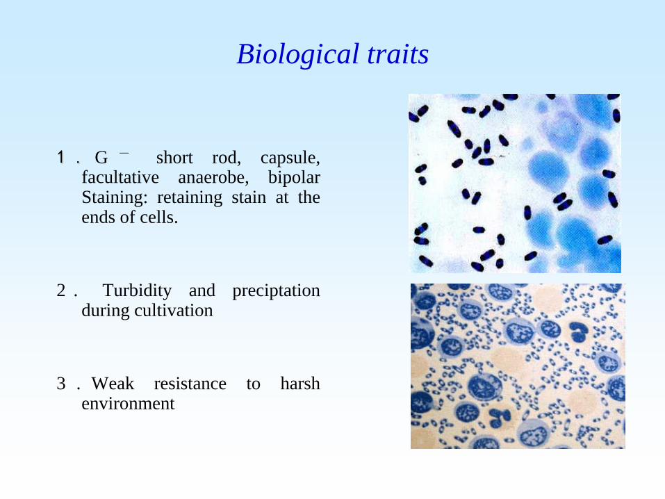
1 . G - short rod, capsule, facultative anaerobe, bipolar Staining: retaining stain at the ends of cells.
2 . Turbidity and preciptation
during cultivation 3 . Weak resistance to harsh
environment
Yersinia: antigenic structure
• F1 Antigen: capsule antigen
• V,W Antigen: anti-phagcytosis
• Yersinia Outer membrane Protein (Yop)
• Murine Toxin (MT), good antigenicity, could be used to
prepare toxoid, and to immunize animals to get anti-toxin
• Endotoxin (LPS)
Pathogenesis & Pathology I
• Flea infected with Y. pestis hungry bites ferociously. The inoculated organisms may be phagocytosed by polymorphonuclear cells and macrophages, they produce the antiphagocytic protein and subsequently are able to resist phagocytosis.
• The pathogens rapidly reach the lymphatics and an intense hemorrhagic inflammation develops in the enlarged lymph nodes, which may undergo necrosis and become fluctuant.
Pathogenesis & Pathology II
• Hemorrhagic and necrotic lesions may develop in all organs; meningitis, pneumonia, and serosanguineous pleuropericarditis are prominent features.
• Primary pneumonic plague results from inhalation of
infective droplets (usually from a coughing patient), with hemorrhagic consolidation, sepsis, and death.
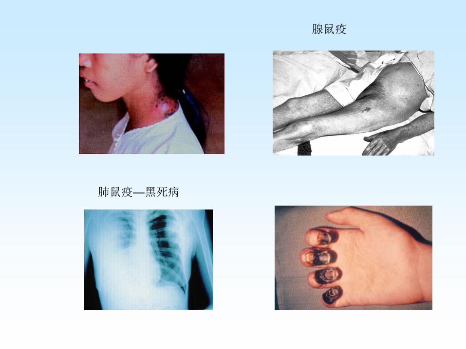
腺鼠疫
肺鼠疫—黑死病
Clinical findings
• High fever, painful lymphadenopathy, commonly with greatly enlarged, tender nodes in the groin or axillae.
• Vomiting and diarrhea may develop with early sepsis. • Later, disseminated intravascular coagulation leads to
hypotension, altered mental status, and renal and cardiac failure.
• Terminally, signs of pneumonia and meningitis can appear, and Y pestis multiplies intravascularly and can be seen in blood smears.
Diagnostic laboratory tests
A. Specimens: – Aspirates of lymph nodes – Cerebrospinal fluid – Blood – Sputum
B. Smears: -Giemsa’s stain -immunofluorescent stain
C. Culture D. Serology
Treatment
• Unless promptly treated, plague may have a mortality rate
of nearly 50%; pneumonic plague, nearly 100%.
• The drug of choice is streptomycin, but gentamicin is also
as effective.
• Dooxycycline is an alternative drug and is sometimes
given in combination with streptomycin.
Epidemiology & control • Plague is an infection of wild rodents (field mice, gerbils,
moles, skunks, and other animals) that occur in many parts of the world. The chief enzootic areas are India, Southeast Asia (especially Vietnam), Africa, and North and South America.
• The control of plague requires surveys of infected animals, vectors, and human contacts.
• Killed whole cell vaccines are no longer available. Because of concern for bioterrorism, numerous vaccines are currently under development. In China, avirulent EV vaccine is available.
Bacillus
• B. anthracis and B. cereus. • B. anthracis is responsible for the disease anthrax. • B. cereus is predominantly responsible for food
poisoning in humans.
• Spores of many Bacillus species are resistant to heat, radiation, disinfectants and desiccation
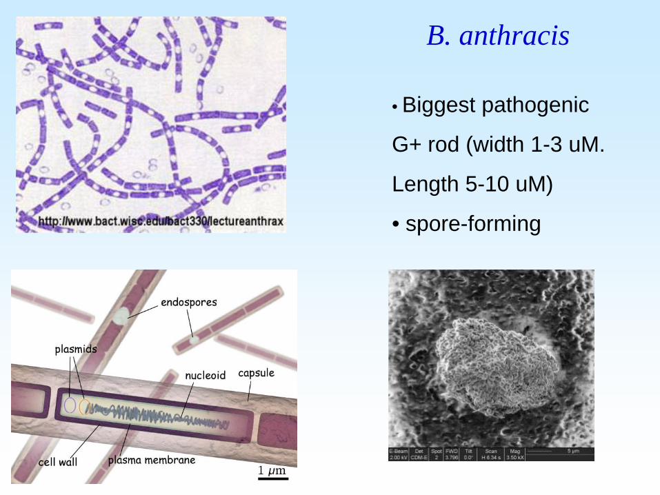
B. anthracis
• Biggest pathogenic
G+ rod (width 1-3 uM.
Length 5-10 uM)
• spore-forming
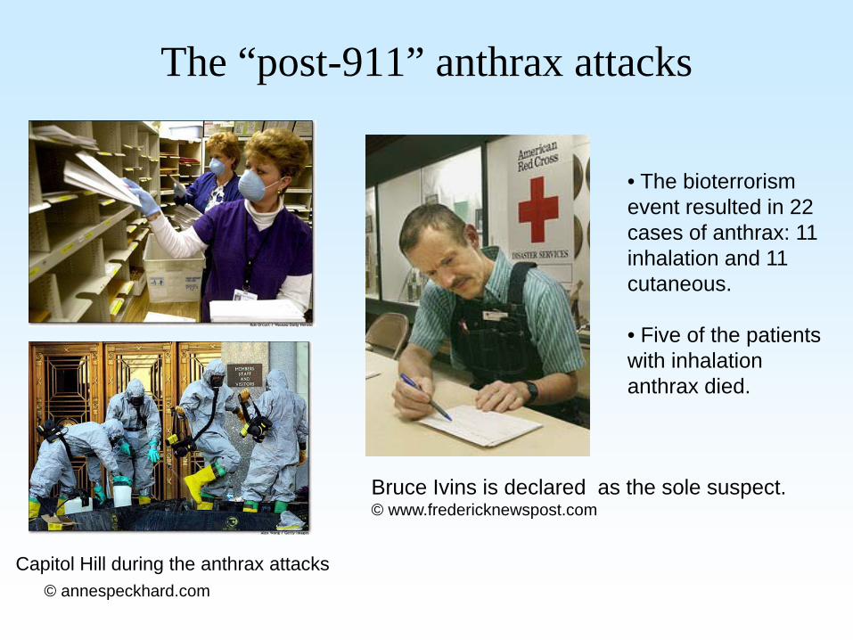
The “post-911” anthrax attacks
Capitol Hill during the anthrax attacks
Bruce Ivins is declared as the sole suspect. © www.fredericknewspost.com
• The bioterrorism event resulted in 22 cases of anthrax: 11 inhalation and 11 cutaneous. • Five of the patients with inhalation anthrax died.
© annespeckhard.com
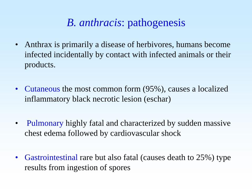
B. anthracis: pathogenesis
• Anthrax is primarily a disease of herbivores, humans become infected incidentally by contact with infected animals or their products.
• Cutaneous the most common form (95%), causes a localized
inflammatory black necrotic lesion (eschar)
• Pulmonary highly fatal and characterized by sudden massive chest edema followed by cardiovascular shock
• Gastrointestinal rare but also fatal (causes death to 25%) type results from ingestion of spores
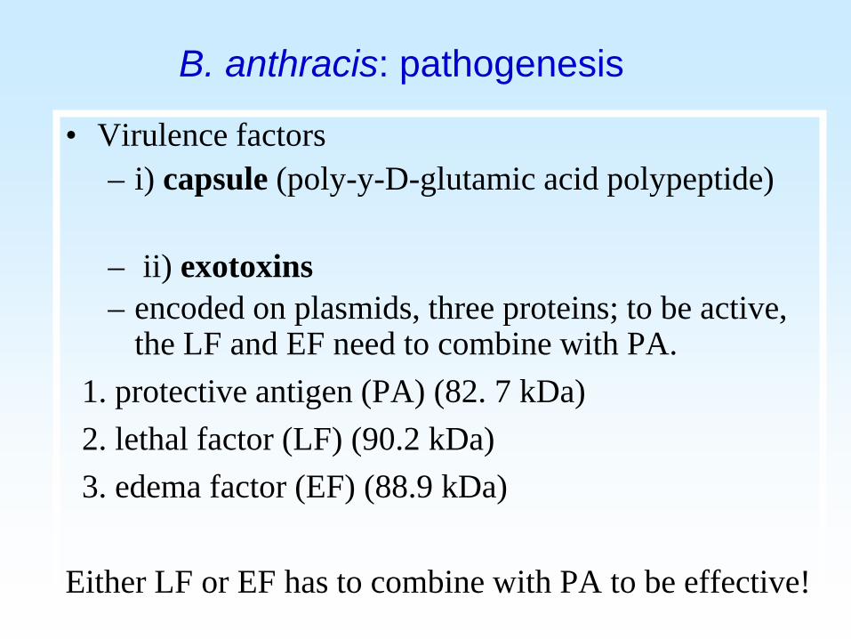
• Virulence factors – i) capsule (poly-y-D-glutamic acid polypeptide) – ii) exotoxins – encoded on plasmids, three proteins; to be active,
the LF and EF need to combine with PA. 1. protective antigen (PA) (82. 7 kDa) 2. lethal factor (LF) (90.2 kDa) 3. edema factor (EF) (88.9 kDa) Either LF or EF has to combine with PA to be effective!
B. anthracis: pathogenesis
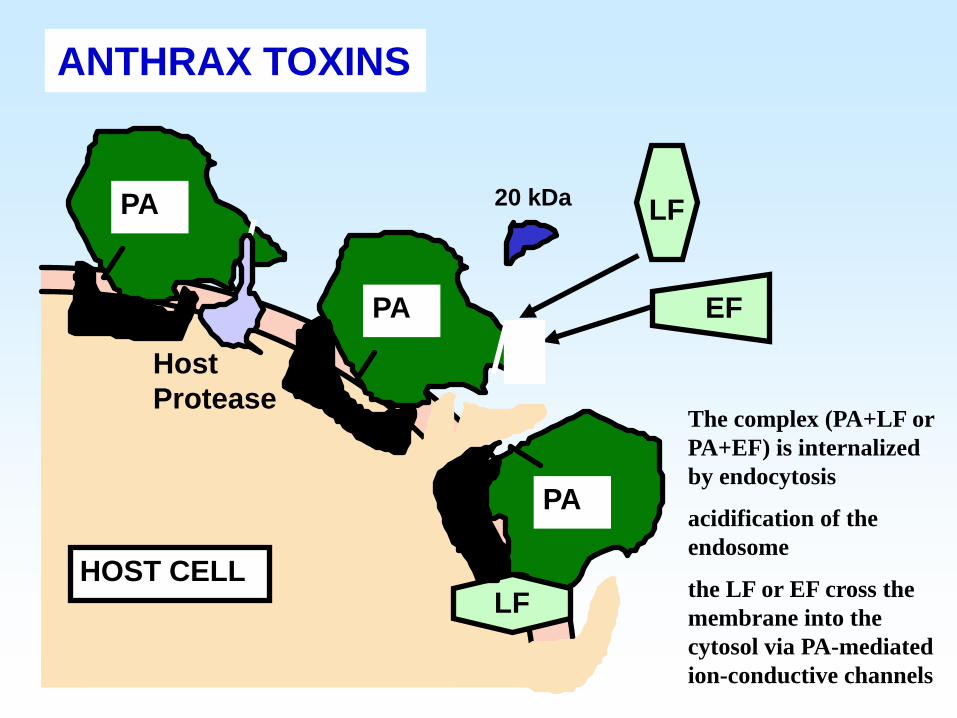
ANTHRAX TOXINS
LF
EF
PA
PA
PA
LF
Host Protease
HOST CELL
20 kDa
The complex (PA+LF or PA+EF) is internalized by endocytosis
acidification of the endosome
the LF or EF cross the membrane into the cytosol via PA-mediated ion-conductive channels
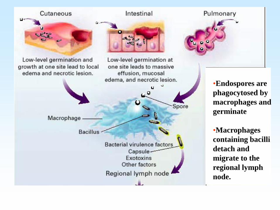
•Endospores are phagocytosed by macrophages and germinate
•Macrophages containing bacilli detach and migrate to the regional lymph node.
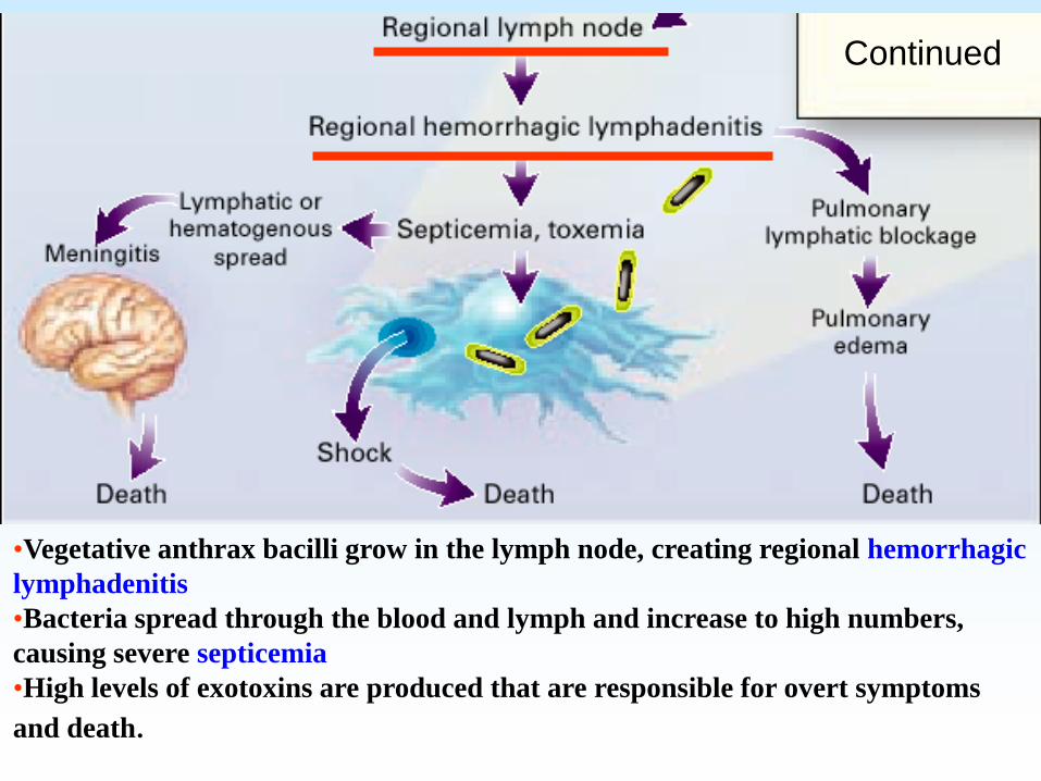
•Vegetative anthrax bacilli grow in the lymph node, creating regional hemorrhagic lymphadenitis •Bacteria spread through the blood and lymph and increase to high numbers, causing severe septicemia •High levels of exotoxins are produced that are responsible for overt symptoms and death.
Continued
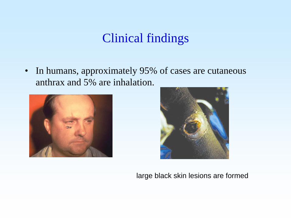
Clinical findings
• In humans, approximately 95% of cases are cutaneous anthrax and 5% are inhalation.
large black skin lesions are formed
Diagnostic laboratory tests
• Stained smears from the local lesion or of blood from dead animals often show chains of large gram positive rods. Anthrax can be identified in dried smears by immunofluorescence staining techniques.
• An ELISA has been developed to measure antibodies
against edema and lethal toxins, but the test has not been extensively studied.
• Some public health laboratories may also have nucleic acid
amplification assays available.
Treatment
• Many antibiotics are effective against anthrax in humans, but treatment must be started early. Ciprofloxacin is recommended.
• After the potential exposure to B. Anthracis as an agent of
biological warfare, prophylaxis with ciprofloxacin or doxycycline should be continued for 4 weeks while three doses of vaccine are being given, or for 8 weeks if no vaccine is administered.
Epidemiology and control
• Soil is contaminated with anthrax spores from the carcasses of dead animals. These spores remain viable for decades.
• Grazing animals infected through injured mucous membranes
serve to perpetuate the chain of infection. • Control measures include 1) disposal of animal carcasses, 2)
decontamination of animal products, 3) protective clothing and gloves for handling potentially infected materials, 4) active immunization of domestic animals with live attenuated vaccines. Persons with high occupational risk should be immunized.
Several other medical important bacteria
• Haemophilus
• Bordetella
• Leginellae
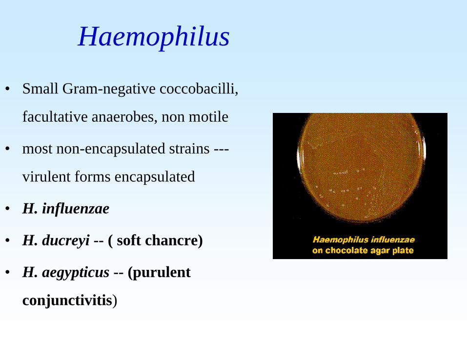
Haemophilus
• Small Gram-negative coccobacilli,
facultative anaerobes, non motile
• most non-encapsulated strains ---
virulent forms encapsulated
• H. influenzae
• H. ducreyi -- ( soft chancre)
• H. aegypticus -- (purulent
conjunctivitis)
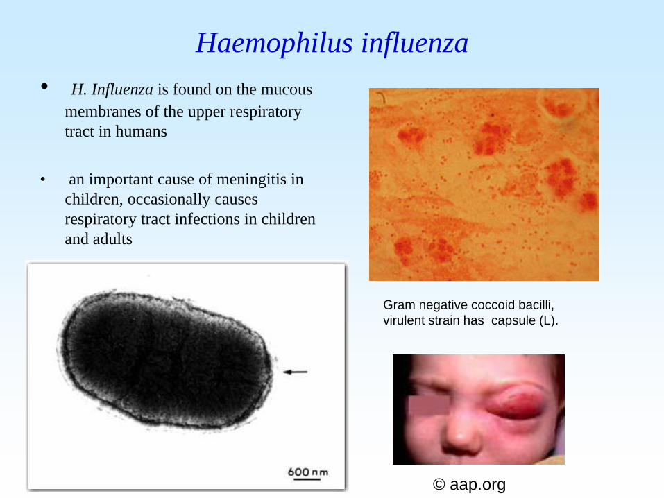
Haemophilus influenza • H. Influenza is found on the mucous
membranes of the upper respiratory tract in humans
• an important cause of meningitis in children, occasionally causes respiratory tract infections in children and adults
Gram negative coccoid bacilli, virulent strain has capsule (L).
© aap.org
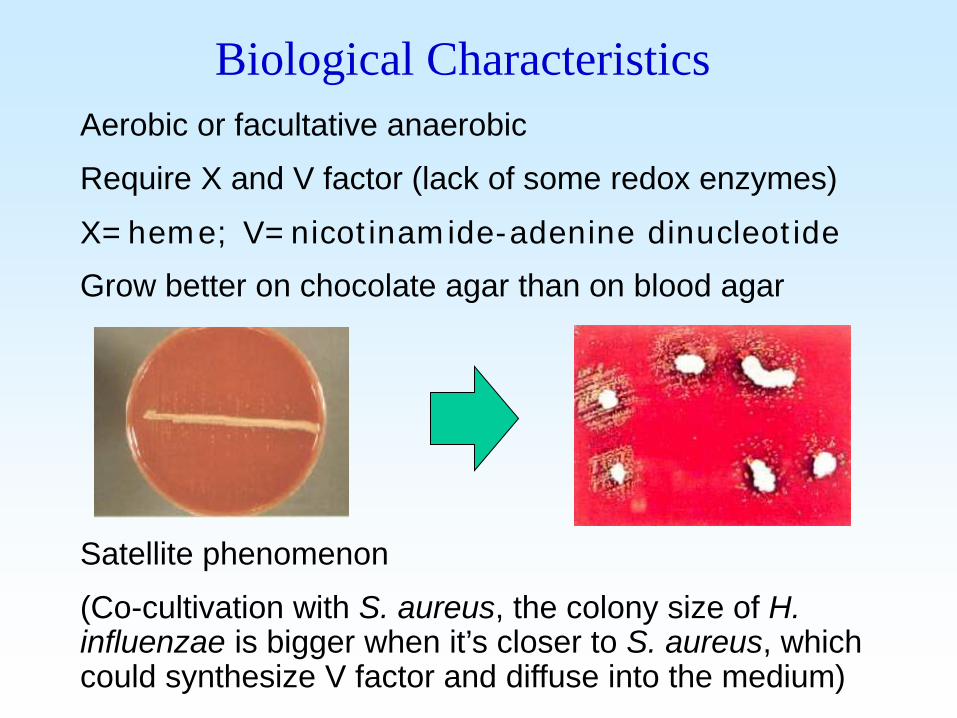
Biological Characteristics Aerobic or facultative anaerobic
Require X and V factor (lack of some redox enzymes)
X=heme; V=nicotinamide-adenine dinucleotide Grow better on chocolate agar than on blood agar
Satellite phenomenon
(Co-cultivation with S. aureus, the colony size of H. influenzae is bigger when it’s closer to S. aureus, which could synthesize V factor and diffuse into the medium)
H. Influenza: antigenic structure
• Capsular polysaccharides, capsule is the antigen used for “typing” H. Influenza
• Somatic antigens consist of outer membrane proteins • Lipoooligosaccharides (endotoxins) share many structures
with those of neisseriae
Virulence factors
• Endotoxin
• Lipooligosaccharide
• Neuraminidase
• IgA protease
• Fimbriae
• Polyribosyl ribitol phosphate (PRP) capsule
(the most important)
Pathogenesis
• no exotoxin, the polyribose phosphate capsule of type b H. influenzae is the major virulence factor.
• Type b H. influenzae is most virulent, which causes meningitis, pneumonia and empyema, epiglottitis, cellulitis, septic arthritis, etc.
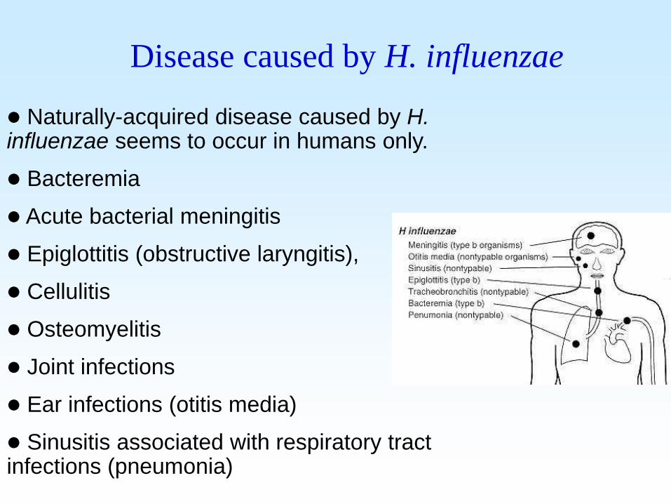
Disease caused by H. influenzae
Naturally-acquired disease caused by H. influenzae seems to occur in humans only.
Bacteremia
Acute bacterial meningitis
Epiglottitis (obstructive laryngitis),
Cellulitis
Osteomyelitis
Joint infections
Ear infections (otitis media)
Sinusitis associated with respiratory tract infections (pneumonia)
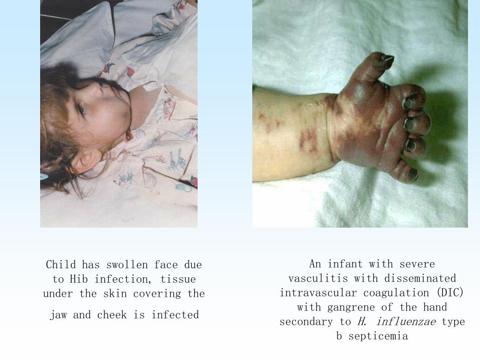
An infant with severe vasculitis with disseminated
intravascular coagulation (DIC) with gangrene of the hand
secondary to H. influenzae type b septicemia
Child has swollen face due to Hib infection, tissue
under the skin covering the
jaw and cheek is infected
Diagnostic laboratory tests
• Specimens -- consist of nasopharyngeal swabs, pus, blood, and spinal fluid for smears and cultures.
• Direct identification -- a positive test indicates that the
fluid contains high concentrations of specific polysaccharide from H. influenzae type b.
• Culture – Specimens grown on IsoVitaleX-enriched
chocolate agar until typical colonies appear. Tests for X (heme) and V (nicotinamide-adenine dinucleotide) factor
Treatment
• Essentially all strains are susceptible to the third-generation cephalosporins.
• Prompt diagnosis and antimicrobial therapy are essential to
minimize late neurologic and intellectual impairment.
Epidemiology, prevention and control
• Encapsulated H. influenzae type b is transmitted from person to person by the respiratory route.
• H. influenzae type b disease can be prevented by administration of Haemophilus b conjugate vaccine to children.
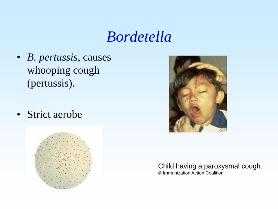
Bordetella • B. pertussis, causes
whooping cough (pertussis).
• Strict aerobe
Child having a paroxysmal cough. © Immunization Action Coalition
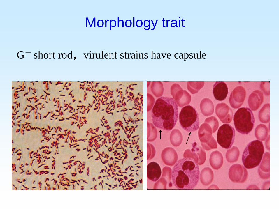
G- short rod,virulent strains have capsule
Morphology trait
Bordetella: pathogenesis
• Common routes of infection: intestinal tract (ingestion of
infected milk), mucous membranes (droplets), and skin
(contact with infected tissues of animals).
Clinical findings
• First, 2 weeks post exposure, the “catarrhal stage”, with
mild coughing and sneezing --- highly infectious but not
very ill
• Then, the “paroxysmal” stage, characteristic “whoop”
upon inhalation, explosive cough. This leads to rapid
exhaustion and may be associated with vomiting, cyanosis,
and convulsions. The white blood count is high (16,000-
30,000/uL)
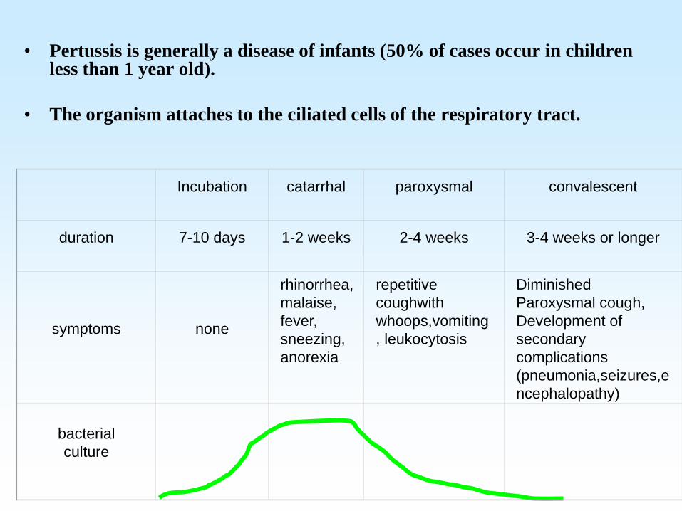
Incubation
catarrhal
paroxysmal
convalescent
duration
7-10 days
1-2 weeks
2-4 weeks
3-4 weeks or longer
symptoms
none
rhinorrhea, malaise, fever, sneezing, anorexia
repetitive coughwith whoops,vomiting, leukocytosis
Diminished Paroxysmal cough, Development of secondary complications (pneumonia,seizures,encephalopathy)
bacterial culture
• Pertussis is generally a disease of infants (50% of cases occur in children less than 1 year old).
• The organism attaches to the ciliated cells of the respiratory tract.
Diagnostic laboratory tests
• A nasopharyngeal or an oropharynx swab is sent to the bacteriology laboratory for Gram stain (Gram negative, coccobacilli, no arrangement), growth on Bordet-Gengou agar or BCYE plate with added cephalosporin to select for the organism, which shows mercury-drop-like colonies.
• The organism is oxidase positive, but urease, nitrase, and
citrate negative. It is also non-motile.
Treatment
• The drug of choice is streptomycin, aminoglycoside
gentamicin to be as effective. Doxycyline could be used in
combination with streptomycin.
• Treatment after onset of the paroxysmal phase rerely alters
the clinical course.
• Oxygen inhalation and sedation may prevent anoxic
damage to the brain.
Epidemiology and control • Whooping cough is endemic in most densely populated areas worldwide
and also occurs intermittently in epidemics.
• The source of infection is usually a patient in the early catarrhal stage of the
disease. Communicability is high, ranging from 30% to 90%. Most cases
occur in children under age 5 years; most deaths occur in the first year of
life.
• Control of whooping cough rests mainly on adequate active immunization
(DPT) of all infants.
Legionella
• 46 species of Legionella and 68 serogroups.
• 1976 outbreak of pneumonia occurred among
persons attending a convention of the American
Legion in Philadelphia, is defined as Legionella
pneumophila.
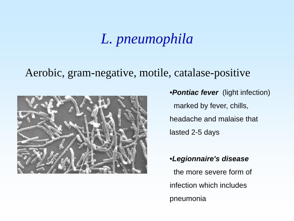
L. pneumophila
Aerobic, gram-negative, motile, catalase-positive •Pontiac fever (light infection)
marked by fever, chills,
headache and malaise that
lasted 2-5 days
•Legionnaire's disease
the more severe form of
infection which includes
pneumonia
Leginellae: antigens & cell products
• At least 16 serogroups of L. pneumophila, serogroup 1 was
the cause of the 1976 outbreak of Legionnaires’ disease
and remains the most common serogroup isolated from
humans.
• The Leginellae produce distinctive 14- to 17-carbon
branched-chain fatty acids; make proteases, phosphatase,
lipase, DNase, and RNase.
Leginellae: pathogenesis
• Legionellae are ubiquitous in warm moist environments, found in lakes, streams, and other bodies of water. They can multiply in free-living amebas and can coexist with them in biofilms.
• L. pneumophila usually produces a lobar, segmental, or patchy
pulmonary infiltration. It readily enters and grows within human alveolar macrophages and monocytes and is not effectively killed by polymorphonuclear leukocytes.
• Phagosomes containing L. pneumophila do not acidify as much
as phagosomes containing other ingested particles.
Clinical findings
• Asymptomatic infection is common in all age groups, as shown by elevated titers of specific antibodies.
• Factors associated with high risk include smoking, chronic bronchitis and emphysema, steroid and other immunosuppressive treatment, cancer chemotherapy, and diabetes mellitus.
• Infection may result in nondescript febrile illness of short duration or in a severe, rapidly progressive illness.
• There may be leukocytosis, hyponatremia, hematuria, or abnormal liver function.
Diagnostic laboratory tests • Specimens --- the organisms can be recovered from bronchial
washings, pleural fluid, lung biopsy specimens, or blood. • Smears --- not demonstrable in Gram-stained smears of
clinical specimens. Direct fluorescent antibody, low detect sensitivity; silver stains are sometimes used
• Culture --- on BCYE agar. Cultured organisms can be rapidly
identified by immunofluorescence staining. • Serology --- Levels of antibodies to legionellae rise slowly
during the illness. Serologic tests have a sensitivity of 60-80% and a specificity of 95-99%.
Treatment
• L. pneumophila are intracellular parasites. Thus, antimicrobials
usable to treat Leginellae infections must enter the phagocytes
and have biological activity there.
• Macrolides quinolones, and tetracyclines are effective. β-
Lactams, monobactams, and aminoglycosides are not effective;
many legionellae make β-Lactamase.
• 3-week prolonged therapy may be required depending upon the
clinical situation.
Epidemiology and control
• The natural habitats are lakes, streams, rivers, and especially thermally heated bodies of water and soil. Leginellae grow best in warm water in the presence of amebas and water bacteria.
• The legionellae survive water treatment processes, and small numbers enter
the water distribution systems where they proliferate. • Similarly, there are links between contamination of residental water
systems and community acquired Legionnaires’ disease and between contamination of hospital water systems and nosocomial L. pneumophila infection.
• Hyperchlorination and superheating of water can help control the
multiplication of legionellae in water and in air-conditioning systems.

References • Jawetz, Melnick, & Adelberg‘s Medical Microbiology, 25th
(McGraw-Hill ) • http://en.wikipedia.org • http://www.google.com
• basic.shsmu.edu.cn/jpkc/micro2
• Teaching PPT from multi-resources, including Prof. Huang M,
Liao F, etc.
Review Questions 1. Select one zoonotic bacterium covered in this lecture,
summarize the related knowledge you have learned.
2. Summarize the knowledge you have learned on one of the following bacteria: H. Influenza, B. pertussis, and L. pneumophila.