higher-order aberrations after corneal collagen crosslinking for keratoconus and corneal ectasia
TRANSCRIPT
ARTICLE
Higher-order aberratio
ns after corneal collagencrosslinking for keratoconus and corneal ectasiaSteven A. Greenstein, MD, Kristen L. Fry, OD, MS, Matthew J. Hersh, Peter S. Hersh, MD
Q
P
292
2012 Aublished
PURPOSE: To determine changes in higher-order aberrations (HOAs) after corneal collagencrosslinking (CXL).
SETTING: Cornea and refractive surgery practice.
DESIGN: Prospective randomized controlled clinical trial.
METHODS: Corneal and ocular HOAs were measured and analyzed using the Pentacam device andLadarwave aberrometer, respectively, at baseline and 12 months after CXL.
RESULTS: Ninety-six eyes (64 keratoconus, 32 ectasia) of 73 patients had CXL. A fellow-eye controlgroup comprised 42 eyes. The mean preoperative total anterior corneal HOAs, total coma, 3rd-ordercoma, and vertical coma were 4.68 mmG 2.33 (SD), 4.40G 2.32 mm, 4.36G 2.30 mm, and 4.04G2.27mm, respectively. At 1 year, themean values decreased significantly to 4.27G 2.25mm, 4.01G2.29 mm, 3.96G 2.27 mm, and 3.66G 2.22 mm, respectively (all P<.001). There were no significantchanges in posterior corneal HOAs. The mean preoperative total ocular HOAs, total coma, 3rd-ordercoma, trefoil, and spherical aberration were 2.80 G 1.0 mm, 2.60 G 1.03 mm, 2.57 G 1.03 mm,0.98 G 0.46 mm, and 0.90 G 0.42 mm, respectively. At 1 year, the mean values decreasedsignificantly to 2.59 G 1.06 mm, 2.42 G 1.07 mm, 2.39 G 1.07 mm, 0.88 G 0.49 mm, and0.83 G 0.38 mm, respectively (all PZ.01). After CXL, HOAs were significantly improvedcompared with the control group. Changes in HOAs were not statistically associated with animprovement in visual acuity or most subjective visual symptoms, however.
CONCLUSION: Corneal and ocular HOAs decreased after CXL, suggesting an improvement incorneal shape.
Financial Disclosure: Dr. Hersh is medical monitor for Avedro, Inc. No author has a financial orproprietary interest in any material or method mentioned.
J Cataract Refract Surg 2012; 38:292–302 Q 2012 ASCRS and ESCRS
Keratoconus and corneal ectasia after laser in situ ker-atomileusis (LASIK) are noninflammatory processes inwhich the corneal architecture deforms in associationwith thinning.1 The progressive distortion of thecornea results in irregular astigmatism, progressivemyopia, and increased higher-order aberrations(HOAs),2–6 with consequent loss of visual function.
Recently, corneal collagen crosslinking (CXL) wasintroduced as a new therapy to mitigate the progres-sion of these ectatic corneal disorders.7,8 Findings inrecent studies suggest that CXL can also have benefi-cial visual and optical effects,9–14 with few reportedcomplications.15–17 In our previous reports of 1-yearCXL outcomes,11,18 patients had an improvement incorrected distance visual acuity (CDVA), uncorrecteddistance visual acuity (UDVA), maximum and aver-age keratometry (K) values, and several quantitativeindices of corneal topography.
SCRS and ESCRS
by Elsevier Inc.
In this study, to further assess optical quality afterCXL, we evaluated the effect of CXL on HOAs byanalyzing changes in anterior corneal HOAs, posteriorcorneal HOAs, and total ocular HOAs 1 year aftertreatment. In addition, changes in HOAs were corre-lated with changes in visual acuity (UDVA andCDVA) and patient-reported visual symptoms.
PATIENTS AND METHODS
Patients with progressive keratoconus and ectasia afterLASIK, were enrolled as part of a multicenter prospectiverandomized controlled clinical trial.A,B This study wasapproved andmonitored by an investigational review boardand complied with the U.S. Health Insurance Portability andAccountability Act. Informed consent was obtained from allpatients.
The inclusion criteria included 14 years of age or older andaxial topography consistent with keratoconus or cornealectasia. Progressive keratoconus or ectasia was defined as
0886-3350/$ - see front matter
doi:10.1016/j.jcrs.2011.08.041
293HIGHER-ORDER ABERRATIONS AFTER CXL
1 or more of the following changes over 24 months: an in-crease of 1.00 diopter (D) or more in the steepest K value,an increase of 1.00 D or more in manifest cylinder, or an in-crease of 0.50 D or more in manifest refraction sphericalequivalent. Exclusion criteria included a history of cornealsurgery (except previous intrastromal corneal ring segmentremoval), chemical injury, delayed epithelial healing, anda corneal pachymetry less than 300 mm.
Treatment Group
Collagen crosslinking was performed according to themethodology described byWollensak et al.1 Topical anesthe-sia was administered, and the corneal epithelium wasremoved by mechanical debridement over the central9.0 mm. Riboflavin (0.1% in 20% dextran T500 solution,Medio-Cross, Peschke Meditrade GmbH) was then adminis-tered topically every 2 minutes for 30 minutes. After ribofla-vin administration, riboflavin absorption throughoutthe corneal stroma and anterior chamber was confirmedon slitlamp examination. Ultrasound (US) pachymetry wasperformed and if the cornea was less than 400 mm, hypotonicriboflavin (0.1% in sterile water, Medio-Cross hypotonic)was administered, 1 drop every 10 seconds for 2-minutesessions, after which US pachymetry was performed to con-firm that the stroma had swollen to more than 400 mm. Thiswas repeated until adequate corneal thickness was obtained.
The cornea was exposed to ultraviolet-A (UVA) 365 nmlight (UV-X system, IROC AG) for 30 minutes at an irradi-ance of 3.0 mW/cm2. During UV exposure, riboflavin dropswere continued every 2 minutes.
Postoperatively, antibiotic and corticosteroid drops wereadministered and a therapeutic soft contact lens (AccuvueOasys, Vistakon) was placed. The contact lens was removedafter epithelial healing, typically 3 to 5 days postoperatively.Antibiotic drops were continued for 1 week and corticoste-roid drops for 2 weeks.
Control Group
In this study, a fellow-eye control group was analyzed.This group comprised the fellow eyes of patients who didnot have CXL bilaterally and included eyes with frank kera-toconus or ectasia that did not have CXL, eyes with evidenceof disease that did notmeet the study’s inclusion criteria, and
Submitted: February 14, 2011.Final revision submitted: August 24, 2011.Accepted: August 27, 2011.
From the Cornea and Laser Eye Institute–Hersh Vision Group(Greenstein, Fry, M. Hersh, P. Hersh), CLEI Center for Keratoconus,Teaneck, and the Department of Ophthalmology (Greenstein, P. Hersh)UMDNJ–New Jersey Medical School, Newark, New Jersey, USA.
Supported in part by Peschke Meditrade, GmbH, Zurich,Switzerland, and by an unrestricted grant to the Department ofOphthalmology, UMDNJ–New Jersey Medical School, fromResearch to Prevent Blindness, Inc., New York, New York, USA.
Corresponding author: Peter S. Hersh, MD, Cornea and Laser EyeInstitute–Hersh Vision Group, CLEI Center for Keratoconus, 300Frank W. Burr Boulevard, Teaneck, New Jersey 07666, USA.E-mail: [email protected].
J CATARACT REFRACT SURG - V
eyes with no evidence of disease. Anterior and posteriorcorneal HOAs were measured and analyzed at baselineand 12 months. Unlike the treated eyes, fellow eyes werenot dilated at the 12-month follow-up examination. There-fore, ocular HOA data were not available for the controlgroup.
Higher-Order Aberrations Measurements
Anterior and posterior corneal aberrations over the central6.5 mm were measured preoperatively and at 12 monthspostoperatively using the Pentacam device (Oculus Inc.).The device extrapolates anterior corneal HOA and posteriorcorneal HOA Zernike coefficients from corneal elevationdata obtained by Scheimpflug imagery.
Ocular HOAs were measured through a 6.5 mm pupilusing a Ladarwave wavefront aberrometer (Alcon Laborato-ries, Inc.). This Shack-Hartmann aberrometer measures totalocular HOAs. Measurements were performed after the eyeswere dilated preoperatively and 12 months postoperatively.If ocular HOAs could not be measured after multipleattempts (usually on the basis of a markedly distortedcornea), the patient was removed from the ocular HOAanalysis.
For corneal and ocular HOAs, the changes in total HOAs(3rd to 6th order), total coma (3rd and 5th order), 3rd-ordercoma, vertical coma, horizontal coma, spherical aberration(4th and 6th), and trefoil aberrations were analyzed.
Visual Acuity and Symptoms
The UDVA and CDVA were measured preoperativelyand 1 year postoperatively. High-contrast visual acuitymeasurements were obtained under controlled lightingconditions using a modified Lighthouse Early Treatment ofDiabetic Retinopathy Study (ETDRS) visual acuity test(2nd edition) with Sloan letters. Patients were tested 4 mfrom the visual acuity chart. If patients could not read anyletters at 4 m, they were tested at 2 m.
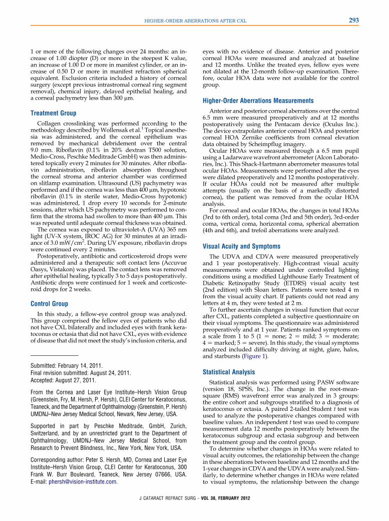
To further ascertain changes in visual function that occurafter CXL, patients completed a subjective questionnaire ontheir visual symptoms. The questionnaire was administeredpreoperatively and at 1 year. Patients ranked symptoms ona scale from 1 to 5 (1 Z none; 2 Z mild; 3 Z moderate;4Zmarked; 5Z severe). In this study, the visual symptomsanalyzed included difficulty driving at night, glare, halos,and starbursts (Figure 1).
Statistical Analysis
Statistical analysis was performed using PASW software(version 18, SPSS, Inc.). The change in the root-mean-square (RMS) wavefront error was analyzed in 3 groups:the entire cohort and subgroups stratified to a diagnosis ofkeratoconus or ectasia. A paired 2-tailed Student t test wasused to analyze the postoperative changes compared withbaseline values. An independent t test was used to comparemeasurement data 12 months postoperatively between thekeratoconus subgroup and ectasia subgroup and betweenthe treatment group and the control group.
To determine whether changes in HOAs were related tovisual acuity outcomes, the relationship between the changein these aberrations between baseline and 12months and the1-year changes in CDVAand theUDVAwere analyzed. Sim-ilarly, to determine whether changes in HOAs were relatedto visual symptoms, the relationship between the change
OL 38, FEBRUARY 2012
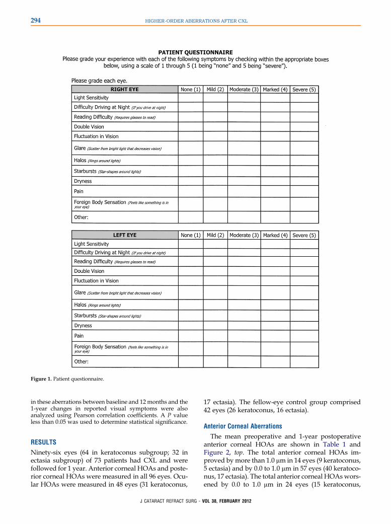
Figure 1. Patient questionnaire.
294 HIGHER-ORDER ABERRATIONS AFTER CXL
in these aberrations between baseline and 12months and the1-year changes in reported visual symptoms were alsoanalyzed using Pearson correlation coefficients. A P valueless than 0.05 was used to determine statistical significance.
RESULTS
Ninety-six eyes (64 in keratoconus subgroup; 32 inectasia subgroup) of 73 patients had CXL and werefollowed for 1 year. Anterior corneal HOAs and poste-rior corneal HOAs were measured in all 96 eyes. Ocu-lar HOAs were measured in 48 eyes (31 keratoconus,
J CATARACT REFRACT SURG - V
17 ectasia). The fellow-eye control group comprised42 eyes (26 keratoconus, 16 ectasia).
Anterior Corneal Aberrations
The mean preoperative and 1-year postoperativeanterior corneal HOAs are shown in Table 1 andFigure 2, top. The total anterior corneal HOAs im-proved by more than 1.0 mm in 14 eyes (9 keratoconus,5 ectasia) and by 0.0 to 1.0 mm in 57 eyes (40 keratoco-nus, 17 ectasia). The total anterior corneal HOAswors-ened by 0.0 to 1.0 mm in 24 eyes (15 keratoconus,
OL 38, FEBRUARY 2012
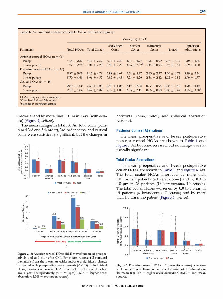
Table 1. Anterior and posterior corneal HOAs in the treatment group.
Mean (mm) G SD
Parameter Total HOAs Total Coma*3rd-OrderComa
VerticalComa
HorizontalComa Trefoil
SphericalAberrations
Anterior corneal HOAs (n Z 96)Preop 4.68 G 2.33 4.40 G 2.32 4.36 G 2.30 4.04 G 2.27 1.26 G 0.99 0.37 G 0.36 1.40 G 0.761 year postop 4.27 G 2.25† 4.01 G 2.29† 3.96 G 2.27† 3.66 G 2.22† 1.14 G 0.95 0.42 G 0.41 1.29 G 0.60
Posterior corneal HOAs (n Z 96)Preop 8.87 G 5.05 8.15 G 4.76 7.98 G 4.67 7.24 G 4.37 2.60 G 2.37 1.00 G 0.75 3.19 G 2.241 year postop 8.70 G 4.68 8.06 G 4.52 7.92 G 4.45 7.23 G 4.20 2.54 G 2.12 1.02 G 0.82 2.99 G 1.77
Ocular HOAs (N Z 48)Preop 2.80 G 1.00 2.60 G 1.03 2.57 G 1.03 2.17 G 2.23 0.57 G 0.94 0.98 G 0.46 0.90 G 0.421 year postop 2.59 G 1.06† 2.42 G 1.07† 2.39 G 1.07† 2.05 G 2.11 0.56 G 0.98 0.88 G 0.49† 0.83 G 0.38†
HOAs Z higher-order aberrations*Combined 3rd and 5th orders†Statistically significant change
295HIGHER-ORDER ABERRATIONS AFTER CXL
8 ectasia) and by more than 1.0 mm in 1 eye (with ecta-sia) (Figure 2, bottom).
The mean changes in total HOAs, total coma (com-bined 3rd and 5th order), 3rd-order coma, and verticalcoma were statistically significant, but the changes in
A
B
Figure 2.A: Anterior corneal HOAs (RMSwavefront error) preoper-atively and at 1 year after CXL. Error bars represent 2 standarddeviations from the mean. Asterisks indicate a significant changecompared with preoperative measurements (P!.05). B: Individualchanges in anterior corneal HOA wavefront error between baselineand 1 year postoperatively (n Z 96 eyes) (HOA Z higher-orderaberration; RMS Z root mean square).
J CATARACT REFRACT SURG - V
horizontal coma, trefoil, and spherical aberrationwere not.
Posterior Corneal Aberrations
The mean preoperative and 1-year postoperativeposterior corneal HOAs are shown in Table 1 andFigure 3. All but one decreased, but no change was sta-tistically significant.
Total Ocular Aberrations
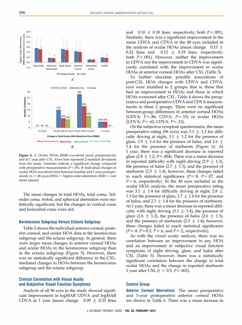
The mean preoperative and 1-year postoperativeocular HOAs are shown in Table 1 and Figure 4, top.The total ocular HOAs improved by more than1.0 mm in 5 patients (all keratoconus) and by 0.0 to1.0 mm in 28 patients (18 keratoconus, 10 ectasia).The total ocular HOAs worsened by 0.0 to 1.0 mm in15 patients (8 keratoconus, 7 ectasia) and by morethan 1.0 mm in no patient (Figure 4, bottom).
Figure 3. Posterior corneal HOAs (RMS wavefront error) preopera-tively and at 1 year. Error bars represent 2 standard deviations fromthe mean () (HOA Z higher-order aberration; RMS Z root meansquare).
OL 38, FEBRUARY 2012
A
B
Figure 4. A: Ocular HOAs (RMS wavefront error) preoperativelyand at 1 year after CXL. Error bars represent 2 standard deviationsfrom the mean. Asterisks indicate a significant change comparedwith preoperative measurements (P!.05). B: Individual changes inocular HOA wavefront error between baseline and 1 year postoper-atively (nZ 48 eyes) (HOAZ higher-order aberration; RMSZ rootmean square).
296 HIGHER-ORDER ABERRATIONS AFTER CXL
The mean changes in total HOAs, total coma, 3rd-order coma, trefoil, and spherical aberration were sta-tistically significant, but the changes in vertical comaand horizontal coma were not.
Keratoconus Subgroup Versus Ectasia Subgroup
Table 2 shows the individual anterior corneal, poste-rior corneal, and ocular HOA data in the keratoconussubgroup and the ectasia subgroup. In general, therewere larger mean changes in anterior corneal HOAsand ocular HOAs in the keratoconus subgroup thanin the ectasia subgroup (Figure 5). However, therewas no statistically significant difference in the CXL-mediated changes in HOAs between the keratoconussubgroup and the ectasia subgroup.
Clinical Correlation with Visual Acuityand Subjective Visual Function Symptoms
Analysis of all 96 eyes in the study showed signifi-cant improvement in logMAR UDVA and logMARCDVA at 1 year (mean change �0.09 G 0.25 lines
J CATARACT REFRACT SURG - V
and �0.10 G 0.18 lines, respectively; both PZ.001).Similarly, there was a significant improvement in themean UDVA and CDVA in the 48 eyes included inthe analysis of ocular HOAs (mean change �0.13 G0.22 lines and �0.12 G 0.19 lines, respectively;both P!.001). However, neither the improvementin UDVA nor the improvement in CDVA was signifi-cantly correlated with the improvement in ocularHOAs or anterior corneal HOAs after CXL (Table 3).
To further elucidate possible associations ofpost-CXL HOA changes with UDVA and CDVA,eyes were stratified to 2 groups; that is, those thathad an improvement in HOAs and those in whichHOAs worsened after CXL. Table 4 shows the preop-erative and postoperative UDVA andCDVAmeasure-ments in these 2 groups. There were no significantbetween-group differences in anterior corneal HOAs(UDVA: PZ.86; CDVA: PZ.15) or ocular HOAs(UDVA: PZ.62; CDVA: PZ.13).On the subjective symptom questionnaire, the mean
preoperative rating (96 eyes) was 3.1 G 1.3 for diffi-culty driving at night, 3.1 G 1.2 for the presence ofglare, 2.9 G 1.4 for the presence of halos, and 2.6 G1.4 for the presence of starbursts (Figure 6). At1 year, there was a significant decrease in reportedglare (2.8 G 1.2; PZ.004). There was a mean decreasein reported difficulty with night driving (2.9 G 1.3),the presence of halos (2.7 G 1.3), and the presence ofstarbursts (2.5 G 1.4); however, these changes failedto reach statistical significance (PZ.8, PZ.07, andPZ.6, respectively). In the 48 eyes included in theocular HOA analysis, the mean preoperative ratingwas 3.1 G 1.4 for difficulty driving at night, 2.8 G1.3 for the presence of glare, 2.7G 1.4 for the presenceof halos, and 2.3 G 1.4 for the presence of starbursts.At 1 year, there was a mean decrease in reported diffi-culty with night driving (3.1 G 1.4), the presence ofglare (2.6 G 1.2), the presence of halos (2.6 G 1.3),and the presence of starbursts (2.5 G 1.4); however,these changes failed to reach statistical significance(PZ.8, PZ0.3, PZ.6, and PZ.2, respectively).
As with the visual acuity analysis, there was nocorrelation between an improvement in any HOAand an improvement in subjective visual functionsymptoms of night driving, glare, and halos afterCXL (Table 5). However, there was a statisticallysignificant correlation between the change in totalocular HOAs and the change in reported starbursts1 year after CXL (r Z 0.5, P!.001).
Control Group
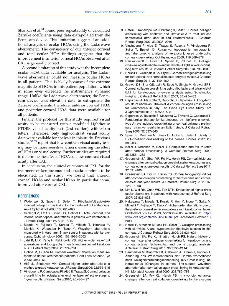
Anterior Corneal Aberrations The mean preoperativeand 1-year postoperative anterior corneal HOAsare shown in Table 6. There was a mean increase in
OL 38, FEBRUARY 2012
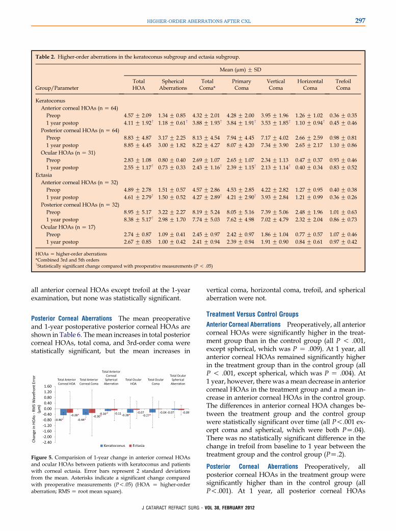
Table 2. Higher-order aberrations in the keratoconus subgroup and ectasia subgroup.
Mean (mm) G SD
Group/ParameterTotalHOA
SphericalAberrations
TotalComa*
PrimaryComa
VerticalComa
HorizontalComa
TrefoilComa
KeratoconusAnterior corneal HOAs (n Z 64)
Preop 4.57 G 2.09 1.34 G 0.85 4.32 G 2.01 4.28 G 2.00 3.95 G 1.96 1.26 G 1.02 0.36 G 0.351 year postop 4.11 G 1.92† 1.18 G 0.61† 3.88 G 1.93† 3.84 G 1.91† 3.53 G 1.85† 1.10 G 0.94† 0.45 G 0.46
Posterior corneal HOAs (n Z 64)Preop 8.83 G 4.87 3.17 G 2.25 8.13 G 4.54 7.94 G 4.45 7.17 G 4.02 2.66 G 2.59 0.98 G 0.811 year postop 8.85 G 4.45 3.00 G 1.82 8.22 G 4.27 8.07 G 4.20 7.34 G 3.90 2.65 G 2.17 1.10 G 0.86
Ocular HOAs (n Z 31)Preop 2.83 G 1.08 0.80 G 0.40 2.69 G 1.07 2.65 G 1.07 2.34 G 1.13 0.47 G 0.37 0.93 G 0.461 year postop 2.55 G 1.17† 0.73 G 0.33 2.43 G 1.16† 2.39 G 1.15† 2.13 G 1.14† 0.40 G 0.34 0.83 G 0.52
EctasiaAnterior corneal HOAs (n Z 32)
Preop 4.89 G 2.78 1.51 G 0.57 4.57 G 2.86 4.53 G 2.85 4.22 G 2.82 1.27 G 0.95 0.40 G 0.381 year postop 4.61 G 2.79† 1.50 G 0.52 4.27 G 2.89† 4.21 G 2.90† 3.93 G 2.84 1.21 G 0.99 0.36 G 0.26
Posterior corneal HOAs (n Z 32)Preop 8.95 G 5.17 3.22 G 2.27 8.19 G 5.24 8.05 G 5.16 7.39 G 5.06 2.48 G 1.96 1.01 G 0.631 year postop 8.38 G 5.17† 2.98 G 1.70 7.74 G 5.03 7.62 G 4.98 7.02 G 4.79 2.32 G 2.04 0.86 G 0.73
Ocular HOAs (n Z 17)Preop 2.74 G 0.87 1.09 G 0.41 2.45 G 0.97 2.42 G 0.97 1.86 G 1.04 0.77 G 0.57 1.07 G 0.461 year postop 2.67 G 0.85 1.00 G 0.42 2.41 G 0.94 2.39 G 0.94 1.91 G 0.90 0.84 G 0.61 0.97 G 0.42
HOAs Z higher-order aberrations*Combined 3rd and 5th orders†Statistically significant change compared with preoperative measurements (P ! .05)
297HIGHER-ORDER ABERRATIONS AFTER CXL
all anterior corneal HOAs except trefoil at the 1-yearexamination, but none was statistically significant.
Posterior Corneal Aberrations The mean preoperativeand 1-year postoperative posterior corneal HOAs areshown in Table 6. Themean increases in total posteriorcorneal HOAs, total coma, and 3rd-order coma werestatistically significant, but the mean increases in
Figure 5. Comparision of 1-year change in anterior corneal HOAsand ocular HOAs between patients with keratoconus and patientswith corneal ectasia. Error bars represent 2 standard deviationsfrom the mean. Asterisks indicate a significant change comparedwith preoperative measurements (P!.05) (HOA Z higher-orderaberration; RMS Z root mean square).
J CATARACT REFRACT SURG - V
vertical coma, horizontal coma, trefoil, and sphericalaberration were not.
Treatment Versus Control Groups
Anterior Corneal Aberrations Preoperatively, all anteriorcorneal HOAs were significantly higher in the treat-ment group than in the control group (all P ! .001,except spherical, which was P Z .009). At 1 year, allanterior corneal HOAs remained significantly higherin the treatment group than in the control group (allP ! .001, except spherical, which was P Z .004). At1 year, however, there was a mean decrease in anteriorcorneal HOAs in the treatment group and a mean in-crease in anterior corneal HOAs in the control group.The differences in anterior corneal HOA changes be-tween the treatment group and the control groupwere statistically significant over time (all P!.001 ex-cept coma and spherical, which were both PZ.04).There was no statistically significant difference in thechange in trefoil from baseline to 1 year between thetreatment group and the control group (PZ.2).
Posterior Corneal Aberrations Preoperatively, allposterior corneal HOAs in the treatment group weresignificantly higher than in the control group (allP!.001). At 1 year, all posterior corneal HOAs
OL 38, FEBRUARY 2012
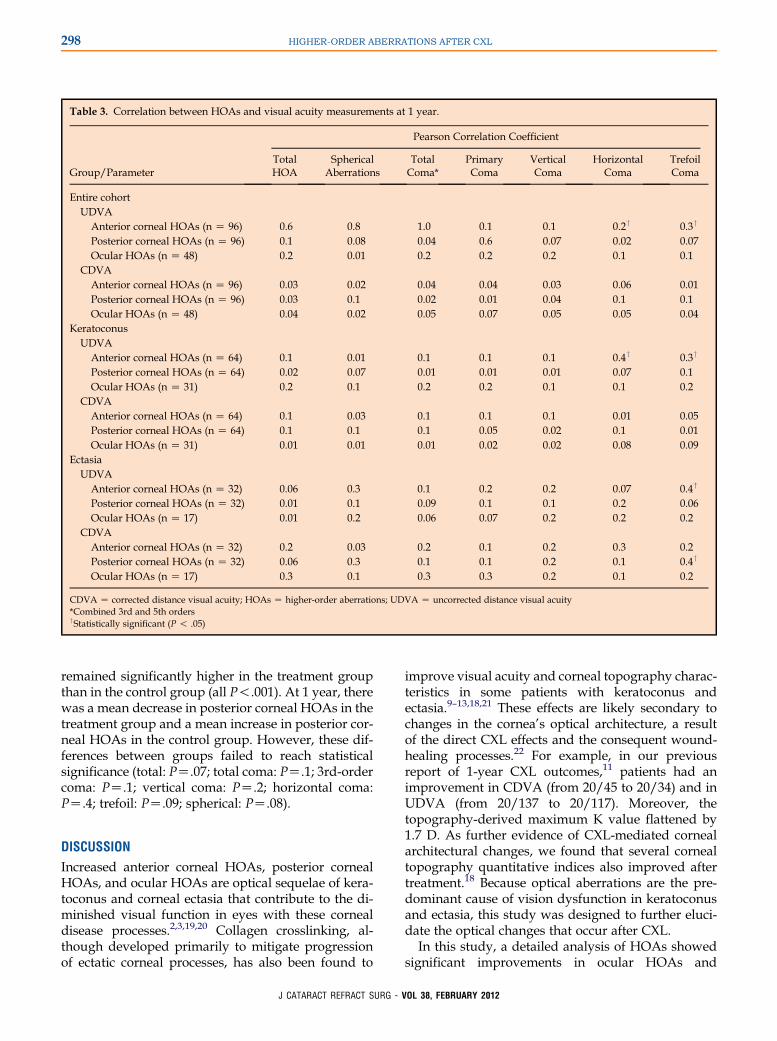
Table 3. Correlation between HOAs and visual acuity measurements at 1 year.
Pearson Correlation Coefficient
Group/ParameterTotalHOA
SphericalAberrations
TotalComa*
PrimaryComa
VerticalComa
HorizontalComa
TrefoilComa
Entire cohortUDVA
Anterior corneal HOAs (n Z 96) 0.6 �0.8 1.0 0.1 0.1 0.2† �0.3†
Posterior corneal HOAs (n Z 96) 0.1 �0.08 0.04 0.6 0.07 0.02 �0.07Ocular HOAs (n Z 48) �0.2 �0.01 �0.2 �0.2 �0.2 �0.1 �0.1
CDVAAnterior corneal HOAs (n Z 96) 0.03 �0.02 0.04 0.04 0.03 0.06 �0.01Posterior corneal HOAs (n Z 96) 0.03 0.1 0.02 0.01 �0.04 0.1 0.1Ocular HOAs (n Z 48) 0.04 0.02 0.05 0.07 0.05 �0.05 �0.04
KeratoconusUDVA
Anterior corneal HOAs (n Z 64) 0.1 0.01 0.1 0.1 0.1 0.4† �0.3†
Posterior corneal HOAs (n Z 64) �0.02 �0.07 �0.01 0.01 0.01 0.07 �0.1Ocular HOAs (n Z 31) �0.2 �0.1 �0.2 �0.2 �0.1 0.1 �0.2
CDVAAnterior corneal HOAs (n Z 64) 0.1 �0.03 0.1 0.1 0.1 �0.01 0.05Posterior corneal HOAs (n Z 64) 0.1 0.1 0.1 0.05 0.02 0.1 �0.01Ocular HOAs (n Z 31) �0.01 0.01 �0.01 �0.02 �0.02 �0.08 �0.09
EctasiaUDVA
Anterior corneal HOAs (n Z 32) 0.06 �0.3 0.1 0.2 0.2 �0.07 �0.4†
Posterior corneal HOAs (n Z 32) 0.01 �0.1 0.09 0.1 0.1 �0.2 �0.06Ocular HOAs (n Z 17) 0.01 0.2 �0.06 �0.07 �0.2 �0.2 0.2
CDVAAnterior corneal HOAs (n Z 32) �0.2 �0.03 �0.2 �0.1 �0.2 0.3 �0.2Posterior corneal HOAs (n Z 32) �0.06 0.3 �0.1 �0.1 �0.2 0.1 0.4†
Ocular HOAs (n Z 17) 0.3 0.1 0.3 0.3 0.2 �0.1 0.2
CDVA Z corrected distance visual acuity; HOAs Z higher-order aberrations; UDVA Z uncorrected distance visual acuity*Combined 3rd and 5th orders†Statistically significant (P ! .05)
298 HIGHER-ORDER ABERRATIONS AFTER CXL
remained significantly higher in the treatment groupthan in the control group (all P!.001). At 1 year, therewas a mean decrease in posterior corneal HOAs in thetreatment group and a mean increase in posterior cor-neal HOAs in the control group. However, these dif-ferences between groups failed to reach statisticalsignificance (total: PZ.07; total coma: PZ.1; 3rd-ordercoma: PZ.1; vertical coma: PZ.2; horizontal coma:PZ.4; trefoil: PZ.09; spherical: PZ.08).
DISCUSSION
Increased anterior corneal HOAs, posterior cornealHOAs, and ocular HOAs are optical sequelae of kera-toconus and corneal ectasia that contribute to the di-minished visual function in eyes with these cornealdisease processes.2,3,19,20 Collagen crosslinking, al-though developed primarily to mitigate progressionof ectatic corneal processes, has also been found to
J CATARACT REFRACT SURG - V
improve visual acuity and corneal topography charac-teristics in some patients with keratoconus andectasia.9–13,18,21 These effects are likely secondary tochanges in the cornea’s optical architecture, a resultof the direct CXL effects and the consequent wound-healing processes.22 For example, in our previousreport of 1-year CXL outcomes,11 patients had animprovement in CDVA (from 20/45 to 20/34) and inUDVA (from 20/137 to 20/117). Moreover, thetopography-derived maximum K value flattened by1.7 D. As further evidence of CXL-mediated cornealarchitectural changes, we found that several cornealtopography quantitative indices also improved aftertreatment.18 Because optical aberrations are the pre-dominant cause of vision dysfunction in keratoconusand ectasia, this study was designed to further eluci-date the optical changes that occur after CXL.
In this study, a detailed analysis of HOAs showedsignificant improvements in ocular HOAs and
OL 38, FEBRUARY 2012
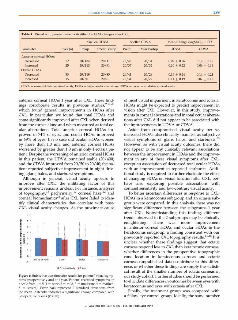
Table 4. Visual acuity measurements stratified by HOA changes after CXL.
Snellen UDVA Snellen CDVA Mean Change (logMAR) G SD
Parameter Eyes (n) Preop 1 Year Postop Preop 1 Year Postop UDVA CDVA
Anterior corneal HOAsDecreased 72 20/134 20/110 20/45 20/34 �0.09 G 0.26 �0.12 G 0.19Increased 25 20/113 20/91 20/37 20/32 �0.10 G 0.22 �0.06 G 0.14
Ocular HOAsDecreased 33 20/119 20/85 20/41 20/29 �0.15 G 0.24 �0.14 G 0.21Increased 15 20/80 20/61 20/31 20/27 �0.11 G 0.19 �0.07 G 0.12
CDVA Z corrected distance visual acuity; HOAs Z higher-order aberrations; UDVA Z uncorrected distance visual acuity
299HIGHER-ORDER ABERRATIONS AFTER CXL
anterior corneal HOAs 1 year after CXL. These find-ings corroborate results in previous studies,9,13,23
which found general improvements in HOAs afterCXL. In particular, we found that total HOAs andcoma significantly improved after CXL when derivedfrom the cornea alone and when measured as total oc-ular aberrations. Total anterior corneal HOAs im-proved in 74% of eyes, and ocular HOAs improvedin 69% of eyes. In no case did ocular HOAs worsenby more than 1.0 mm, and anterior corneal HOAsworsened by greater than 1.0 mm in only 1 ectasia pa-tient. Despite the worsening of anterior corneal HOAsin this patient, the UDVA remained stable (20/400)and the CDVA improved from 20/50 to 20/40; the pa-tient reported subjective improvement in night driv-ing, glare, halos, and starburst symptoms.
Although in general, visual acuity appears toimprove after CXL, the militating factor of thisimprovement remains unclear. For instance, analysesof topography,18 pachymetry,17 corneal haze,22 andcorneal biomechanics24 after CXL have failed to iden-tify clinical characteristics that correlate with post-CXL visual acuity changes. As the proximate cause
Figure 6. Subjective questionnaire results for patients’ visual symp-toms preoperatively and at 1 year. Patients recorded symptoms ona scale from 1 to 5 (1Z none, 2Zmild, 3Zmoderate, 4Zmarked,5 Z severe). Error bars represent 2 standard deviations fromthe mean. Asterisks indicates a significant change compared withpreoperative results (P!.05).
J CATARACT REFRACT SURG - V
of most visual impairment in keratoconus and ectasia,HOAs might be expected to predict improvement invision after CXL. However, in this study, improve-ments in corneal aberrations and in total ocular aberra-tions after CXL did not appear to be associated withthe improvements in UDVA or CDVA.
Aside from compromised visual acuity per se,increased HOAs also clinically manifest as subjectivevisual symptoms of glare, halos, and starbursts.25
However, as with visual acuity outcomes, there didnot appear to be any clinically relevant associationsbetween the improvement in HOAs and the improve-ment in any of these visual symptoms after CXL,except an association of decreased total ocular HOAswith an improvement in reported starbursts. Addi-tional study is required to further elucidate the effectof changing HOAs on visual function after CXL, per-haps also exploring possible associations withcontrast sensitivity and low-contrast visual acuity.
To better ascertain differences in response to CXL,HOAs in a keratoconus subgroup and an ectasia sub-group were compared. In this analysis, there was nosignificant difference between the subgroups 1 yearafter CXL. Notwithstanding this finding, differenttrends observed in the 2 subgroups may be clinicallyenlightening. There was more improvementin anterior corneal HOAs and ocular HOAs in thekeratoconus subgroup, a finding consistent with ourpreviously reported CXL topography results.11,18 It isunclear whether these findings suggest that ectaticcorneas respond less to CXL than keratoconic corneas,whether differences in the preoperative topographiccone location in keratoconus corneas and ectaticcorneas (unpublished data) contribute to this differ-ence, or whether these findings are simply the statisti-cal result of the smaller number of ectatic corneas inour study cohort. Further studies should be performedto elucidate differences in outcomes between eyeswithkeratoconus and eyes with ectasia after CXL.
Finally, the treatment group was compared witha fellow-eye control group. Ideally, the same number
OL 38, FEBRUARY 2012
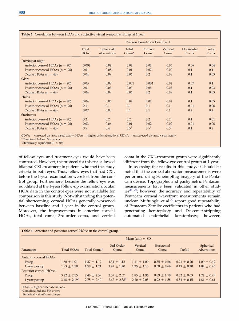
Table 5. Correlation between HOAs and subjective visual symptoms ratings at 1 year.
Pearson Correlation Coefficient
TotalHOA
SphericalAberrations
TotalComa*
PrimaryComa
VerticalComa
HorizontalComa
TrefoilComa
Driving at nightAnterior corneal HOAs (n Z 96) �0.002 0.02 �0.02 �0.01 �0.03 0.06 0.04Posterior corneal HOAs (n Z 96) 0.01 �0.05 0.01 0.02 0.02 �0.1 0.1Ocular HOAs (n Z 48) 0.04 �0.09 0.06 0.2 0.08 0.1 0.03
GlareAnterior corneal HOAs (n Z 96) 0.03 0.08 �0.001 �0.004 �0.02 0.07 �0.1Posterior corneal HOAs (n Z 96) �0.01 0.03 �0.03 �0.05 �0.03 �0.1 �0.03Ocular HOAs (n Z 48) 0.04 �0.09 0.06 0.2 0.08 0.1 0.03
HalosAnterior corneal HOAs (n Z 96) 0.04 0.05 0.02 0.02 �0.02 0.1 �0.05Posterior corneal HOAs (n Z 96) 0.1 0.1 0.1 0.1 0.1 �0.01 �0.06Ocular HOAs (n Z 48) 0.07 �0.08 0.1 0.1 0.1 0.2 �0.2
StarburstsAnterior corneal HOAs (n Z 96) 0.2† 0.2 0.2 0.2 0.2 0.1 �0.01Posterior corneal HOAs (n Z 96) 0.03 0.06 �0.01 �0.02 0.02 �0.01 �0.06Ocular HOAs (n Z 48) 0.5† 0.4 0.5† 0.5† 0.5† 0.1 0.2
CDVA Z corrected distance visual acuity; HOAs Z higher-order aberrations; UDVA Z uncorrected distance visual acuity*Combined 3rd and 5th orders†Statistically significant (P ! .05)
300 HIGHER-ORDER ABERRATIONS AFTER CXL
of fellow eyes and treatment eyes would have beencompared. However, the protocol for this trial allowedbilateral CXL treatment in patients who met the studycriteria in both eyes. Thus, fellow eyes that had CXLbefore the 1-year examination were lost from the con-trol group. Furthermore, because the fellow eye wasnot dilated at the 1-year follow-up examination, ocularHOA data in the control eyes were not available forcomparison in this study. Notwithstanding this poten-tial shortcoming, corneal HOAs generally worsenedbetween baseline and 1 year in the control group.Moreover, the improvements in anterior cornealHOAs, total coma, 3rd-order coma, and vertical
Table 6. Anterior and posterior corneal HOAs in the control group.
Parameter Total HOAs Total Coma*3rd-OrderComa
Anterior corneal HOAsPreop 1.80 G 1.01 1.37 G 1.12 1.34 G 1.121 year postop 1.93 G 1.10 1.50 G 1.21 1.47 G 1.20
Posterior corneal HOAsPreop 3.22 G 2.15 2.46 G 2.39 2.37 G 2.371 year postop 3.48 G 2.19† 2.75 G 2.40† 2.67 G 2.38
HOAs Z higher-order aberrations*Combined 3rd and 5th orders†Statistically significant change
J CATARACT REFRACT SURG - V
coma in the CXL-treatment group were significantlydifferent from the fellow-eye control group at 1 year.
In assessing the results in this study, it should benoted that the corneal aberration measurements wereperformed using Scheimpflug imagery of the Penta-cam device. Topographic and pachymetric Pentacammeasurements have been validated in other stud-ies26–29; however, the accuracy and repeatability ofPentacam corneal wavefront measurements remainunclear. Muftuoglu et al.30 report good repeatabilityof Pentacam Zernike coefficients in patients who hadpenetrating keratoplasty and Descemet-strippingautomated endothelial keratoplasty; however,
Mean (mm) G SD
VerticalComa
HorizontalComa Trefoil
SphericalAberrations
1.11 G 1.00 0.55 G 0.66 0.21 G 0.20 1.00 G 0.421.25 G 1.10 0.58 G 0.66 0.19 G 0.20 1.02 G 0.45
1.85 G 1.96 0.89 G 1.58 0.52 G 0.63 1.74 G 0.49† 2.20 G 2.05 0.92 G 1.58 0.54 G 0.45 1.81 G 0.61
OL 38, FEBRUARY 2012
301HIGHER-ORDER ABERRATIONS AFTER CXL
Shankar et al.31 found poor repeatability of calculatedZernike coefficients using data extrapolated from thePentacam device. This limitation suggested an addi-tional analysis of ocular HOAs using the Ladarwaveaberrometer. The consistency of our anterior cornealand total ocular HOA findings suggests that theimprovement in anterior corneal HOAs observed afterCXL is generally correct.
A second limitation of this studywas the incompleteocular HOA data available for analysis. The Ladar-wave aberrometer could not measure ocular HOAsin all patients. This is likely because of the extrememagnitude of HOAs in this patient population, whichin some eyes exceeded the instrument’s dynamicrange. Unlike the Ladarwave aberrometer, the Penta-cam device uses elevation data to extrapolate theZernike coefficients; therefore, anterior corneal HOAand posterior corneal HOA data were available forall patients.
Finally, the protocol for this study required visualacuity to be measured with a modified LighthouseETDRS visual acuity test (2nd edition) with Sloanletters. Therefore, only high-contrast visual acuitydata were available for analysis in this study. Previousstudies32,33 report that low-contrast visual acuity test-ing may be more sensitive when measuring the effectof HOAs on visual acuity. Further studies are requiredto determine the effect of HOAs on low-contrast visualacuity after CXL.
In conclusion, the clinical outcomes of CXL for thetreatment of keratoconus and ectasia continue to beelucidated. In this study, we found that anteriorcorneal HOAs and ocular HOAs, in particular coma,improved after corneal CXL.
REFERENCES1. Wollensak G, Spoerl E, Seiler T. Riboflavin/ultraviolet-A-
induced collagen crosslinking for the treatment of keratoconus.
Am J Ophthalmol 2003; 135:620–627
2. Schlegel Z, Lteif Y, Bains HS, Gatinel D. Total, corneal, and
internal ocular optical aberrations in patients with keratoconus.
J Refract Surg 2009; 25:S951–S957
3. Maeda N, Fujikado T, Kuroda T, Mihashi T, Hirohara Y,
Nishida K, Watanabe H, Tano Y. Wavefront aberrations
measured with Hartmann-Shack sensor in patients with kerato-
conus. Ophthalmology 2002; 109:1996–2003
4. Jafri B, Li X, Yang H, Rabinowitz YS. Higher order wavefront
aberrations and topography in early and suspected keratoco-
nus. J Refract Surg 2007; 23:774–781
5. Gobbe M, Guillon M. Corneal wavefront aberration measure-
ments to detect keratoconus patients. Cont Lens Anterior Eye
2005; 28:57–66
6. Ali�o JL, Shabayek MH. Corneal higher order aberrations: a
method to grade keratoconus. J Refract Surg 2006; 22:539–545
7. VinciguerraP,CamesascaFI,Alb�eE,TrazzaS.Corneal collagen
cross-linking for ectasia after excimer laser refractive surgery:
1-year results. J Refract Surg 2010; 26:486–497
J CATARACT REFRACT SURG - V
8. Hafezi F, Kanellopoulos J,WiltfangR, Seiler T. Corneal collagen
crosslinking with riboflavin and ultraviolet A to treat induced
keratectasia after laser in situ keratomileusis. J Cataract
Refract Surg 2007; 33:2035–2040
9. Vinciguerra P, Alb�e E, Trazza S, Rosetta P, Vinciguerra R,
Seiler T, Epstein D. Refractive, topographic, tomographic,
and aberrometric analysis of keratoconic eyes undergoing
corneal cross-linking. Ophthalmology 2009; 116:369–378
10. Raiskup-Wolf F, Hoyer A, Spoerl E, Pillunat LE. Collagen
crosslinking with riboflavin and ultraviolet-A light in keratoconus:
long-term results. J Cataract Refract Surg 2008; 34:796–801
11. Hersh PS, Greenstein SA, Fry KL. Corneal collagen crosslinking
for keratoconus and corneal ectasia: one year results. J Cataract
Refract Surg 2011; 37:149–160
12. Grewal DS, Brar GS, Jain R, Sood V, Singla M, Grewal SPS.
Corneal collagen crosslinking using riboflavin and ultraviolet-A
light for keratoconus; one-year analysis using Scheimpflug
imaging. J Cataract Refract Surg 2009; 35:425–432
13. Caporossi A, Mazzotta C, Baiocchi S, Caporossi T. Long-term
results of riboflavin ultraviolet A corneal collagen cross-linking
for keratoconus in Italy: The Siena Eye Cross Study. Am
J Ophthalmol 2010; 149:585–593
14. Caporossi A, Baiocchi S, Mazzotta C, Traversi C, Caporossi T.
Parasurgical therapy for keratoconus by riboflavin-ultraviolet
type A rays induced cross-linking of corneal collagen; prelimi-
nary refractive results in an Italian study. J Cataract Refract
Surg 2006; 32:837–845
15. Spoerl E, Mrochen M, Sliney D, Trokel S, Seiler T. Safety of
UVA-riboflavin cross-linking of the cornea. Cornea 2007; 26:
385–389
16. Koller T, Mrochen M, Seiler T. Complication and failure rates
after corneal crosslinking. J Cataract Refract Surg 2009;
35:1358–1362
17. Greenstein SA, Shah VP, Fry KL, Hersh PS. Corneal thickness
changes after corneal collagen crosslinking for keratoconus and
corneal ectasia: one-year results. J Cataract Refract Surg 2011;
37:691–700
18. Greenstein SA, Fry KL, Hersh PS. Corneal topography indices
after corneal collagen crosslinking for keratoconus and corneal
ectasia: one-year results. J Cataract Refract Surg 2011; 37:
1282–1290
19. Lim L, Wei RH, Chan WK, Tan DTH. Evaluation of higher order
ocular aberrations in patients with keratoconus. J Refract Surg
2007; 23:825–828
20. Nakagawa T, Maeda N, Kosaki R, Hori Y, Inoue T, Saika M,
Mihashi T, Fujikado T, Tano Y. Higher-order aberrations due to
the posterior corneal surface in patients with keratoconus. Invest
Ophthalmol Vis Sci 2009; 50:2660–2665. Available at: http://
www.iovs.org/content/50/6/2660.full.pdf. Accessed October 13,
2011
21. Hafezi F, Mrochen M, Iseli HP, Seiler T. Collagen crosslinking
with ultraviolet-A and hypoosmolar riboflavin solution in thin
corneas. J Cataract Refract Surg 2009; 35:621–624
22. Greenstein SA, Fry KL, Bhatt J, Hersh PS. Natural history of
corneal haze after collagen crosslinking for keratoconus and
corneal ectasia: Scheimpflug and biomicroscopic analysis.
J Cataract Refract Surg 2010; 36:2105–2114
23. Baumeister M, Klaproth OK, Gehmlich J, B€uhren J, Kohnen T.€Anderung des Wellenfrontfehlers der Hornhautvorderfl€achenach Kollagenvernetzungsbehandlung (UV-Crosslinking) bei
Keratokonus [Changes in corneal first-surface wavefront
aberration after corneal collagen cross-linking in keratoconus].
Klin Monatsbl Augenheilkd 2009; 226:752–756
24. Greenstein SA, Fry KL, Hersh PS. In vivo biomechanical
changes after corneal collagen crosslinking for keratoconus
OL 38, FEBRUARY 2012
302 HIGHER-ORDER ABERRATIONS AFTER CXL
and corneal ectasia: 1-year analysis of a randomized, controlled,
clinical trial. In press, Cornea 2011
25. Seiler T, Kaemmerer M, Mierdel P, Krinke H-E. Ocular optical
aberrations after photorefractive keratectomy for myopia and
myopic astigmatism. Arch Ophthalmol 2000; 118:17–21. Avail-
able at: http://archopht.ama-assn.org/cgi/reprint/118/1/17.pdf.
Accessed October 13, 2011
26. Emre S, Doganay S, Yologlu S. Evaluation of anterior
segment parameters in keratoconic eyes measured with
the Pentacam system. J Cataract Refract Surg 2007; 33:
1708–1712
27. Bourges J-L, Alfonsi N, Lalibert�e J-F, Chagnon M, Renard G,
Legeais J-M, Brunette I. Average 3-dimensional models
for the comparison of Orbscan II and Pentacam pachymetry
maps in normal corneas. Ophthalmology 2009; 116:2064–2071
28. Mih�altz K, Kov�acs I, Tak�acs �A, Nagy ZZ. Evaluation of
keratometric, pachymetric, and elevation parameters of
keratoconic corneas with Pentacam. Cornea 2009; 28:976–980
29. de Sanctis U, Missolungi A, Mutani B, Richiardi L, Grignolo FM.
Reproducibility and repeatability of central corneal thickness
measurement in keratoconus using the rotating Scheimpflug
camera and ultrasound pachymetry. Am J Ophthalmol 2007;
144:712–718
30. Muftuoglu O, Prasher P, Bowman RW, McCulley JP,
Mootha VV. Corneal higher-order aberrations after Descemet’s
stripping automated endothelial keratoplasty. Ophthalmology
2010; 117:878–884.e6
31. Shankar H, Taranath D, Santhirathelagan CT, Pesudovs K.
Repeatability of corneal first-surface wavefront aberrations
J CATARACT REFRACT SURG - V
measured with Pentacam corneal topography. J Cataract
Refract Surg 2008; 34:727–734
32. Pepose JS, Applegate RA. Making sense out of wavefront
sensing. Am J Ophthalmol 2005; 139:335–343
33. Pesudovs K, Marsack JD, Donnelly WJ III, Thibos LN,
Applegate RA. Measuring visual acuity�mesopic or photopic
conditions, and high or low contrast letters? J Refract Surg
2004; 20:S508–S514
OTHER CITED MATERIALA. Avedro, Inc. Corneal Collagen Cross-linking for Progressive
Keratoconus (CXL). Identifier NCT00647699. Available at:
http://www.clinicaltrials.gov/ct2/show/record/NCT00647699?idZNCT00647699&rankZ1. Accessed October 13, 2011
B. Avedro, Inc. Corneal Collagen Cross-Linking for Ectasia (CXL).
Identifier NCT00674661. Available at: http://www.clinicaltrials.
gov/ct2/show/record/NCT00674661?idZNCT00674661&rankZ1.
Accessed October 13, 2011
OL
38, FEBRUARY 2012First author:Steven A. Greenstein, MD
Cornea and Laser Eye Institute–HershVision Group, CLEI Center forKeratoconus, Teaneck,New Jersey, USA