high-salt diet downregulates trem2 expression and blunts
TRANSCRIPT
RESEARCH Open Access
High-salt diet downregulates TREM2expression and blunts efferocytosis ofmacrophages after acute ischemic strokeMengyan Hu1†, Yinyao Lin1†, Xuejiao Men1†, Shisi Wang1, Xiaobo Sun1, Qiang Zhu1, Danli Lu1, Sanxin Liu1,Bingjun Zhang1, Wei Cai1,2* and Zhengqi Lu1*
Abstract
Background: A high-salt diet (HSD) is one of the major risk factors for acute ischemic stroke (AIS). As a potentialmechanism, surplus salt intake primes macrophages towards a proinflammatory phenotype. In this study, whetherHSD could blunt the efferocytic capability of macrophages after ischemic stroke, thus exacerbating post-strokeneural inflammation, was investigated.
Methods: Wild-type male C57BL/6 mice were fed with fodder containing 8% sodium chloride for 4 weeks andsubjected to transient middle cerebral occlusion (tMCAO). Disease severity, macrophage polarization as well asefferocytic capability were evaluated. Bone marrow-derived macrophages were cultured in vitro, and the impact ofhigh salinity on their efferocytic activity, as well as their expression of phagocytic molecules, were analyzed. Therelationships among sodium concentration, macrophage phenotype, and disease severity in AIS patients wereexplored.
Results: HSD-fed mice displayed increased infarct volume and aggravated neurological deficiency. Mice fed withHSD suffered exacerbated neural inflammation as shown by higher inflammatory mediator expression and immunecell infiltration levels. Infiltrated macrophages within stroke lesions in HSD-fed mice exhibited a shift towardsproinflammatory phenotype and impaired efferocytic capability. As assessed with a PCR array, the expression oftriggering receptor expressed on myeloid cells 2 (TREM2), a receptor relevant to phagocytosis, was downregulatedin high-salt-treated bone marrow-derived macrophages. Enhancement of TREM2 signaling restored the efferocyticcapacity and cellular inflammation resolution of macrophages in a high salinity environment in vitro and in vivo. Ahigh concentration of urine sodium in AIS patients was found to be correlated with lower TREM2 expression anddetrimental stroke outcomes.
Conclusions: HSD inhibited the efferocytic capacity of macrophages by downregulating TREM2 expression, thusimpeding inflammation resolution after ischemic stroke. Enhancing TREM2 signaling in monocytes/macrophagescould be a promising therapeutic strategy to enhance efferocytosis and promote post-stroke inflammation resolution.
Keywords: High-salt diet, Stroke, Macrophage, Phagocytosis, Triggering receptor expressed on myeloid cells 2
© The Author(s). 2021 Open Access This article is licensed under a Creative Commons Attribution 4.0 International License,which permits use, sharing, adaptation, distribution and reproduction in any medium or format, as long as you giveappropriate credit to the original author(s) and the source, provide a link to the Creative Commons licence, and indicate ifchanges were made. The images or other third party material in this article are included in the article's Creative Commonslicence, unless indicated otherwise in a credit line to the material. If material is not included in the article's Creative Commonslicence and your intended use is not permitted by statutory regulation or exceeds the permitted use, you will need to obtainpermission directly from the copyright holder. To view a copy of this licence, visit http://creativecommons.org/licenses/by/4.0/.The Creative Commons Public Domain Dedication waiver (http://creativecommons.org/publicdomain/zero/1.0/) applies to thedata made available in this article, unless otherwise stated in a credit line to the data.
* Correspondence: [email protected]; [email protected]†Mengyan Hu, Yinyao Lin and Xuejiao Men contributed equally to this work.1Department of Neurology, Mental and Neurological Disease ResearchCenter, The Third Affiliated Hospital of Sun Yat-sen University, 600 TianheRoad, Guangzhou, Guangdong 510630, People’s Republic of ChinaFull list of author information is available at the end of the article
Hu et al. Journal of Neuroinflammation (2021) 18:90 https://doi.org/10.1186/s12974-021-02144-9
BackgroundHigh-salt intake is positively correlated with blood pres-sure, blood lipid concentration, circulating alarmins, andother factors leading to detrimental stroke prognosis.The habit of high-salt diet (HSD) has long been consid-ered as a risk factor for acute ischemic stroke (AIS) [1–3]. Therefore, salt restriction is widely accepted as a vitalstep in efficient lifestyle intervention to prevent new vas-cular events, especially AIS [4–6]. Nevertheless, in dailyclinical practice, we found that it was not always prac-tical for patients to change the long-established high-saltdiet. Therefore, there is an unmet need to develop thera-peutic strategies to tackle the already existing high salin-ity and the associated pathophysiology.Macrophage, as an innate immune cell, has a high de-
gree of parallelism and flexibility [7, 8]. Macrophagescould release multiple proinflammatory mediators or ef-ficiently sweep cell debris and promote neural recovery.The phenotypic shift of macrophages depends largely onthe cues of the microenvironment [9, 10]. Recent re-search has elucidated that surplus dietary salt shifts mac-rophages/microglia towards the classical activatedproinflammatory phenotype, which is often referred toas M1 [11], indicating that excessive salt intake breaksthe M1/M2 macrophages balance and further aggravatesthe inflammatory response. In vivo, the proinflammatoryproperty of macrophages in HSD-fed mice contributedto blood-brain barrier (BBB) disruption after stroke andexacerbated stroke outcomes [12]. Efferocytosis repre-sents a vital anti-inflammatory process of macrophages.Timely clearance of cell debris and cell corpses is essen-tial for subsequent tissue reconstruction of lesions [9,13, 14]. Nevertheless, the impact of HSD on phagocyticactivity and the subsequent anti-inflammatory functionsof macrophages remain elusive.The current study investigated the impact of excessive
salt intake on the efferocytic capacity of macrophagesafter ischemic stroke. Our data indicated that HSDdownregulated the expression of the phagocytic mol-ecule triggering receptor expressed on myeloid cells 2(TREM2) in macrophages, thus impeding their debrisclearing activities. Enhancement of TREM2 signaling inmacrophages rescued the inflammation resolving func-tions and displayed delightful therapeutic effects.TREM2 could be a promising therapeutic target of AIS,especially in patients with un-converted HSD habits.
MethodsEthical statementThe clinical and experimental animal studies were ap-proved by the Medical Ethics Committee of the ThirdAffiliated Hospital of Sun Yat-Sen University and theAnimal Care and Use Committee of Sun Yat-Sen Uni-versity, respectively. All participants had signed the
informed consent according to the principles illustratedin the Declaration of Helsinki.
PatientsIn this study, a total of 38 stroke patients recruited inThe Third Affiliated Hospital of Sun Yat-Sen Universityfrom July 2018 to October 2019 consecutively had an in-dependently documented primary stroke event in com-bination with confirmed magnetic resonance imaging(MRI) evidence showing ischemic stroke. All patients re-cruited in the study met the following inclusion criteria:(i) onset age ≥ 18 years; (ii) first symptomatic ischemicstroke; (iii) clinical evidence of motor, language, atten-tion, visual, or memory deficits based on neurologicalexamination; (iv) time of enrolment: < 7 days fromstroke onset. Exclusion criteria included (i) prior stroke,(ii) cerebral hemorrhage, (iii) malignant tumors, (iv) pa-tent foramen ovale, (v) vasculitis, (vi) autoimmune dis-ease, (vii) chronic kidney disease with hemodialysis, (viii)Parkinson’s disease, (ix) treatment with systemic gluco-corticoids or other immunosuppressive agents within 14days of admission, and (x) thromboembolism, collagendisease, disseminated intravascular coagulation, and ad-vanced liver disease [15, 16]. Clinical data, including age,gender, and scores of the National Institute of HealthStroke Scale (NIHSS), were recorded. We estimated diet-ary sodium intake by measurements of a 24-h urinaryexcretion of sodium. Patients were instructed to collecttheir 24-h urine void on the day of their clinic visit, andthe 24-h sodium excretion was calculated by multiplyingthe sodium concentration in the urine by the total urinevolume during this period. Patient demographics, in-cluding comorbidities, were summarized in Supplemen-tary Table 1.
MRI scanning and infarct volume analysis of patientsMagnetic resonance imaging (MRI) was performed within24h of admission using 1.5- or 3.0-T magnetic resonanceimaging (Sigma; GE Medical Systems, Milwaukee, WI,USA). In this study, the diffusion-weighted imaging(DWI) spin-echo planar sequence included 20 contiguousaxial oblique slices (b = 0 and 1000 s/mm2 isotropicallyweighted; repetition time/echo time, 6000/60.4 ms; acqui-sition matrix, 128 × 128; slice thickness, 5mm; intersticegap, 1 mm; field of view, 24 cm). DWI lesions in 38 pa-tients were measured with Analyze 7.0 software (AnalyzeDirect, KS). Cerebral infarct size was assessed based onthe largest infarct diameter determined on the image dem-onstrating the largest lesion [17–19]. MRI scans of pa-tients were assessed by the experienced neurologistZhengqi Lu, who was blinded to the patients’ clinical fea-tures. All images were interpreted with the same windowsettings, monitor types, and lighting conditions.
Hu et al. Journal of Neuroinflammation (2021) 18:90 Page 2 of 16
PBMC isolationAnti-coagulated blood (3 mL) was collected and then di-luted 2-fold with PBS, pipetted into a centrifuge tubeprefilled with Ficoll lymphocyte separation solution(TBDscience), followed by centrifuged at 2000 rpm for25 min at room temperature without deceleration.PBMCs from the buffy coat were washed twice with PBSand then stored at − 80°C until further analysis.
AnimalsC57BL/6 wild-type mice (8 weeks old, weight 18–25 g)were purchased from the Guangdong Medical Labora-tory Animal Center (Guangzhou, China) and housed ina humidity- and temperature-controlled animal facilityat Sun Yat-sen University with a 12-h light-dark cycle.Mice received normal chow (0.5% NaCl) and tap waterad libitum (normal diet) or sodium-rich chow (8% NaCl)and tap water containing 1% NaCl ad libitum (HSD) for4 weeks, according to the experiment.
Model of acute ischemic strokeMice were subjected to focal acute ischemic stroke in-duced with transient middle cerebral artery occlusion(tMCAO). Procedures for tMCAO were described previ-ously [15]. Briefly, mice were anesthetized with 1.5–2.0%isoflurane under conditions of spontaneous breathing. Afilament was inserted into the external carotid artery(ECA) and was directed to the middle cerebral artery(MCA) through the internal carotid artery (ICA). Fila-ment insertion into the ICA was maintained for 60 minfollowed by reperfusion with the maintenance of corebody temperatures. Cerebral blood flow (CBF) duringsurgery was measured by laser Doppler flow cytometry.Mice with a more than 70% reduction of blood flow inthe ischemic core were included in the study, and micethat died during surgery were excluded. Survival of micewas recorded.
Infarct volume analysisFor immunologic staining of NeuN, six equally spacedcoronal brain sections encompassing the MCA territorywere stained with NeuN antibodies. Infarct volume inNeuN-stained sections was analyzed with NIH ImageJsoftware. The infarct area was determined as the differ-ence between the NeuN-positive area of contralateralhemispheres and ipsilateral hemispheres. Brain infarctwas determined by multiplying the mean area of tissueloss by the distances between two adjacent stained brainslices.
Primary macrophage enriched culture and stimulationPrimary macrophage-enriched cultures were preparedfrom the bone marrow of 6- to 8-week-old healthyC57BL/6 wild-type mice using the EasySep Mouse
Monocyte Enrichment Kit (Stem Cell) according tomanufacturer’s instructions. Macrophages were inducedwith MCSF (50 ng/ml) for 6 days in macrophage culturemedium (RPMI1640 + 10%FBS). For polarization, mac-rophages were treated with lipopolysaccharide (LPS, 100ng/mL, Sigma) or IL-4 (20 ng/ml, Peprotech) for 24 h.
Macrophage depletion and transferA single dose of clodronate liposomes (Liposoma, 10ml/kg, i.p.) was administered to mice at 3 days beforetMCAO to deplete peripheral monocytes/macrophages.Empty liposomes were used as control (Liposoma, 10ml/kg, i.p.). Depletion efficacy of clodronate liposomeswas confirmed with flow cytometric analysis. Bonemarrow-derived macrophages (BMDM, 2 × 10⁶) weretransferred to the tMCAO model intravenously immedi-ately after reperfusion.
Primary microglia culturePrimary mouse microglia were obtained from BLUE-FBIO company and cultured in culture medium(DMEM-HG + 10%FBS) until treatment.
Primary cortical neuron culture and oxygen-glucosedeprivationPrimary cortical neuronal cultures were prepared fromE16-18 embryos of C57BL/6 mice as previously de-scribed [20].Neuronal ischemia was induced with oxygen-glucose
deprivation (OGD). Briefly, the culture medium (neuralbasal medium + B27 + 2% glutamate) was removed andreplaced by EBSS (Gibco). Neurons were then incubatedin 95% N2 + 5% CO2 for 90 min.
Phagocytosis assayFor evaluation of efferocytic capacity, apoptotic neuronswere labeled with the dead cell marker propidium iodide(PI) in PBS (1 μg/ml, 37 °C, 15 min) and treated withmacrophages at a ratio of dead neurons:macrophages =5:1, for the indicated time periods. For in vitro immuno-staining experiments, macrophages were pre-grown onpoly-l-lysine-coated coverslips. The coverslips of macro-phages were washed two times to remove unengulfedneurons and fixed with 4% paraformaldehyde. The cov-erslips were then subjected to immunostaining and re-moved from wells using tweezers and mounted to theslides. F-actin in macrophage was then stained withAlexa Fluor488 phalloidin (A12379, 1:500 in PBS; Invi-trogen) at room temperature in the dark for 30 min. Forthe flow cytometry experiment, macrophages were pre-seeded on 24-well plates and treated with the same ratioof dead neurons for indicated time points. Macrophageswere washed with PBS, detached from wells with trypsin,and subjected to flow cytometric analysis. The
Hu et al. Journal of Neuroinflammation (2021) 18:90 Page 3 of 16
percentage of efferocytic macrophages (PI+) was calcu-lated via flow cytometric analysis.
Lentiviral infection of macrophageLenti virus was constructed and packaged by FenghBio(Changsha, China). The cultured macrophages were in-fected for 3 days with Lenti-TREM2 or the control vec-tors. Overexpression of TREM2 was confirmed bywestern blot and flow cytometry.
Flow cytometric analysisThe brain tissue was homogenized and prepared assingle-cell suspensions for flow cytometric analysis(FACS). Briefly, brains were dissected, and ipsilateralhemispheres were collected. Each hemisphere was sub-jected to digestion with 0.25% trypsin-EDTA (ThermoFisher, Carlsbad, CA, USA) at 37 °C for 25 min. Thebrain tissue was then pressed through a cell strainer (70μm). Brain cells were separated from myelin debris bycentrifugation in 30%/70% Percoll solution (GE Health-care Biosciences AB, Uppsala, Sweden). Brain cells at theinterface were collected, washed with HBSS, and sub-jected to further staining. The following antibodies wereused: CD45-PE-Texas Red (1:400, BioLegend), CD11b-PE (1:400, BioLegend), CD3-PerCp/Cy5.5 (1:400, BioLe-gend), CD19-FITC (1:400, BioLegend), Ly6G-APC/Cy7(1:400, BioLegend), TREM2-PE (1:200, R&D Systems),TNFα-PE (1:200, BioLegend), CD206-Alexa Fluor 647(1:200, BD bioscience), and Arg1-APC (1:200, R&D Sys-tems). FACS was performed using a fluorescence-activated cell sorter flow cytometer (BD bioscience, SanDiego, CA), and data were analyzed using FlowJo X10.0.7r2 software. Appropriate isotype controls werestained following the manufacturer’s instructions(Thermo Fisher, Carlsbad, CA, USA). Fluorochromecompensation was performed with single-stained One-Comp eBeads (Thermo Fisher, Carlsbad, CA, USA). Fordata presentation, when cells could be divided into nega-tive or positive populations, the percentage of cells wascalculated. When the expression of the coordinatedmarker was consecutive and population separation wasobscure, data were presented as mean fluorescence in-tensity (MFI).
Immunofluorescence staining and cell quantificationAnimals were euthanized and perfused with PBSfollowed by 4% paraformaldehyde. After sufficient perfu-sion, the brains were removed and then cut into 25-μmfrozen cryo-sections using a microtome. Brain sectionswere incubated with primary antibodies at 4°C overnight.After being washed with PBS, sections were incubatedwith secondary antibodies for 1h at room temperature.Sections were then washed and mounted with DAPIFluoromount-G (Thermo Fisher, Carlsbad, CA, USA).
The following primary antibodies were used: rabbit anti-NeuN (1:500, Abcam), rabbit anti-Iba1 (1:1000, WakoPure Chemical Industries), goat anti-Iba1 (1:500, Abcam),rabbit anti-SIK1 (1:500, Proteintech), goat anti-CD206 (1:500, R&D Systems), and rat anti-CD16 (1:500, Santa CruzBiotechnology) antibodies. The following secondary anti-bodies were applied: anti-rabbit secondary antibody conju-gated with Cy3 (1:1000, Jackson ImmunoResearchLaboratories), anti-rabbit secondary antibody conjugatedwith Alexa Fluor 488 (1:1000, Jackson ImmunoResearchLaboratories), anti-rabbit secondary antibody conjugatedwith Alexa Fluor 405 (1:1000, Jackson ImmunoResearchLaboratories), anti-goat secondary antibody conjugatedwith Cy3 (1:1000, Jackson ImmunoResearch Laboratories),anti-goat secondary antibody conjugated with Alexa Fluor488 (1:1000, Jackson ImmunoResearch Laboratories), andanti-rat secondary antibody conjugated with Alexa Fluor488 (1:1000, Jackson ImmunoResearch Laboratories). Forneuronal apoptosis analysis, terminal deoxynucleotidyltransferase dUTP nick end labeling (TUNEL) was proc-essed after NeuN labeling according to the manufacturer’sinstructions (Thermo Fisher). Confocal microscopy im-ages were acquired using a Leica SP confocal microscopeand Leica confocal software. Immunopositive cell quantifi-cation and area analysis were performed with the NIHImageJ software by an investigator who was blinded to theexperimental design. In quantification of cells in the strokepenumbra, the stroke core was identified as the region inwhich the majority of DAPI-stained nuclei were shrunken,and the stroke penumbra was defined as the region ofgenerally morphologically normal cells, approximately450–500 μm wide, surrounding the stroke core.
Quantitative determination of mRNA expressionTotal RNA from cells was extracted with a commercialkit (ESscience) according to the manufacturer’s instruc-tions. A total of 1ug RNA (OD 260 nm/280 nm = 1.8–2.2)was utilized for the first-strand cDNA synthesis in a 40-μlsystem using a PrimeScript RT reagent kit (Takara). Real-time polymerase chain reaction (RT-PCR) was performedon a QuantStudio 5 (ABI) quantitative PCR machine usinga TB green Premix Ex Taq kit (Takara) with 1 μl of thesynthesized cDNA in each reaction with the addition ofROX. The following program was performed: 95 °C for 30s; 95 °C for 5 s and 60 °C for 34 s, repeated for 40 cycles;95 °C for 15 s, 60 °C for 1 min and 95 °C for 15 s (Meltcurve). Primers used in the study are listed in Supplemen-tary Table 2. Double delta CT was calculated, and the datawere presented as fold change normalized to PBS-treatedcontralateral brain, PBS-treated macrophage, or negativecontrol lentivirus-treated macrophage. Glyceraldehyde-3-phosphate dehydrogenase (GAPDH) was used as a house-keeping gene for normalization. In the data analysis in Fig.2, Fig. 5, and Supplementary Figure 1D, the mRNA
Hu et al. Journal of Neuroinflammation (2021) 18:90 Page 4 of 16
expression level was visualized with the heat map andclustered with the software of R using the “pheatmap”package.
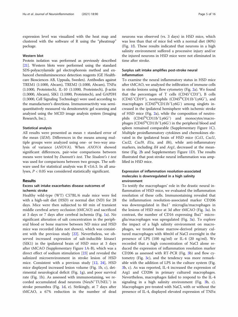
Western blotProtein isolation was performed as previously described[21]. Western blots were performed using the standardSDS-polyacrylamide gel electrophoresis method and en-hanced chemiluminescence detection reagents (GE Health-care Biosciences AB, Uppsala, Sweden). Antibodies againstTREM1 (1:1000, Abcam), TREM2 (1:1000, Abcam), TNFα(1:1000, Proteintech), IL-10 (1:1000, Proteintech), β-actin(1:3000, Abcam), SIK1 (1:1000, Proteintech), and GAPDH(1:3000, Cell Signaling Technology) were used according tothe manufacturer’s directions. Immunoreactivity was semi-quantitatively measured via densitometric gel scanning andanalyzed using the MCID image analysis system (ImagingResearch, Inc.).
Statistical analysisAll results were presented as mean ± standard error ofthe mean (SEM). Differences in the means among mul-tiple groups were analyzed using one- or two-way ana-lysis of variance (ANOVA). When ANOVA showedsignificant differences, pair-wise comparisons betweenmeans were tested by Dunnett’s test. The Student’s t testwas used for comparisons between two groups. The soft-ware used for statistical analysis was R v3.6.3. In all ana-lyses, P < 0.05 was considered statistically significant.
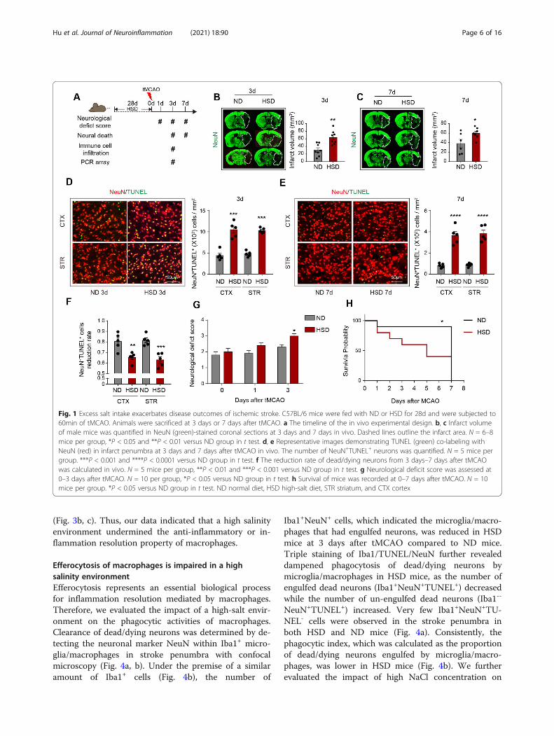
ResultsExcess salt intake exacerbates disease outcomes ofischemic strokeHealthy wild-type (WT) C57BL/6 male mice were fedwith a high-salt diet (HSD) or normal diet (ND) for 28days. Mice were then subjected to 60 min of transientmiddle cerebral artery occlusion (tMCAO) and sacrificedat 3 days or 7 days after cerebral ischemia (Fig. 1a). Nosignificant alteration of salt concentration in the periph-eral blood or bone marrow between ND mice and HSDmice was recorded (data not shown), which was consist-ent with the previous study [22]. Nevertheless, we ob-served increased expression of salt-inducible kinase1(SIK1) in the ipsilateral brain of HSD mice at 3 daysafter tMCAO (Supplementary Figure 1A-B), which was adirect effect of sodium stimulation [23] and revealed thesalinized microenvironment in stroke lesion of HSDmice. Consistent with a previous study [12, 24], HSDmice displayed increased lesion volume (Fig. 1b, c), det-rimental neurological deficit (Fig. 1g), and poor survivalrate (Fig. 1h). As assessed with immunostaining, we re-corded accumulated dead neurons (NeuN+TUNEL+) instroke penumbra (Fig. 1d, e). Strikingly, at 7 days aftertMCAO, a 67% reduction in the number of dead
neurons was observed (vs. 3 days) in HSD mice, whichwas less than that of mice fed with a normal diet (80%)(Fig. 1f). These results indicated that neurons in a highsalinity environment suffered a processive injury and/orthe injured neurons in HSD mice were not eliminated intime after stroke.
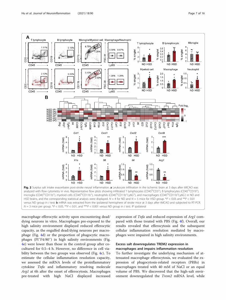
Surplus salt intake amplifies post-stroke neuralinflammationTo examine the neural inflammatory status in HSD miceafter tMCAO, we analyzed the infiltration of immune cellsin stroke lesions using flow cytometry (Fig. 2a). We foundthat the percentages of T cells (CD45+CD3+), B cells(CD45+CD19+), neutrophils (CD45hiCD11b+Ly6G+), andmacrophages (CD45hiCD11b+Ly6G-) among singlets in-creased in the ipsilateral hemisphere with ischemic strokeof HSD mice (Fig. 2a), while the composition of neutro-phils (CD45hiCD11b+Ly6G+) and monocytes/macro-phages (CD45hiCD11b+Ly6G-) in the peripheral blood andspleen remained comparable (Supplementary Figure 1C).Multiple proinflammatory cytokines and chemokines ele-vated in the ipsilateral brain of HSD mice (Ccl1, Cxcl1,Cxcl2, Cxcl9, Il1a, and Il6), while anti-inflammatorymarkers, including Il4 and Arg1, decreased at the mean-time (Fig. 2b and Supplementary Figure 1D). The resultsillustrated that post-stroke neural inflammation was amp-lified in HSD mice.
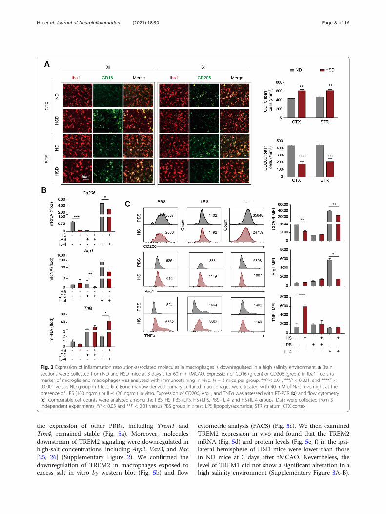
Expression of inflammation resolution-associatedmolecules is downregulated in a high salinityenvironmentTo testify the macrophages’ role in the drastic neural in-flammation of HSD mice, we evaluated the inflammationresolution of these cells. Immunostaining revealed thatthe inflammation resolution-associated marker CD206was downregulated in Iba1+ microglia/macrophages inthe lesions of HSD mice at 3d after tMCAO (Fig. 3a). Incontrast, the number of CD16 expressing Iba1+ micro-glia/macrophages was upregulated (Fig. 3a). To explorethe impact of a high salinity environment on macro-phages, we treated bone marrow-derived primary cul-tured macrophages with 40mM of NaCl overnight in thepresence of LPS (100 ng/ml) or IL-4 (20 ng/ml). Werecorded that a high concentration of NaCl alone re-duced the expression of inflammation resolution markerCD206 as assessed with RT-PCR (Fig. 3b) and flow cy-tometry (Fig. 3c), and the tendency was more remark-able with the addition of LPS in the culture system (Fig.3b, c). As was reported, IL-4 increased the expression ofArg1 and CD206 in primary cultured macrophages.Nevertheless, macrophages failed to respond to the IL-4signaling in a high salinity environment (Fig. 3b, c).Macrophages pre-treated with NaCl, with or without thepresence of IL-4, displayed elevated expression of TNFα
Hu et al. Journal of Neuroinflammation (2021) 18:90 Page 5 of 16
(Fig. 3b, c). Thus, our data indicated that a high salinityenvironment undermined the anti-inflammatory or in-flammation resolution property of macrophages.
Efferocytosis of macrophages is impaired in a highsalinity environmentEfferocytosis represents an essential biological processfor inflammation resolution mediated by macrophages.Therefore, we evaluated the impact of a high-salt envir-onment on the phagocytic activities of macrophages.Clearance of dead/dying neurons was determined by de-tecting the neuronal marker NeuN within Iba1+ micro-glia/macrophages in stroke penumbra with confocalmicroscopy (Fig. 4a, b). Under the premise of a similaramount of Iba1+ cells (Fig. 4b), the number of
Iba1+NeuN+ cells, which indicated the microglia/macro-phages that had engulfed neurons, was reduced in HSDmice at 3 days after tMCAO compared to ND mice.Triple staining of Iba1/TUNEL/NeuN further revealeddampened phagocytosis of dead/dying neurons bymicroglia/macrophages in HSD mice, as the number ofengulfed dead neurons (Iba1+NeuN+TUNEL+) decreasedwhile the number of un-engulfed dead neurons (Iba1--
NeuN+TUNEL+) increased. Very few Iba1+NeuN+TU-NEL- cells were observed in the stroke penumbra inboth HSD and ND mice (Fig. 4a). Consistently, thephagocytic index, which was calculated as the proportionof dead/dying neurons engulfed by microglia/macro-phages, was lower in HSD mice (Fig. 4b). We furtherevaluated the impact of high NaCl concentration on
Fig. 1 Excess salt intake exacerbates disease outcomes of ischemic stroke. C57BL/6 mice were fed with ND or HSD for 28d and were subjected to60min of tMCAO. Animals were sacrificed at 3 days or 7 days after tMCAO. a The timeline of the in vivo experimental design. b, c Infarct volumeof male mice was quantified in NeuN (green)-stained coronal sections at 3 days and 7 days in vivo. Dashed lines outline the infarct area. N = 6–8mice per group, *P < 0.05 and **P < 0.01 versus ND group in t test. d, e Representative images demonstrating TUNEL (green) co-labeling withNeuN (red) in infarct penumbra at 3 days and 7 days after tMCAO in vivo. The number of NeuN+TUNEL+ neurons was quantified. N = 5 mice pergroup. ***P < 0.001 and ****P < 0.0001 versus ND group in t test. f The reduction rate of dead/dying neurons from 3 days–7 days after tMCAOwas calculated in vivo. N = 5 mice per group, **P < 0.01 and ***P < 0.001 versus ND group in t test. g Neurological deficit score was assessed at0–3 days after tMCAO. N = 10 per group, *P < 0.05 versus ND group in t test. h Survival of mice was recorded at 0–7 days after tMCAO. N = 10mice per group. *P < 0.05 versus ND group in t test. ND normal diet, HSD high-salt diet, STR striatum, and CTX cortex
Hu et al. Journal of Neuroinflammation (2021) 18:90 Page 6 of 16
macrophage efferocytic activity upon encountering dead/dying neurons in vitro. Macrophages pre-exposed to thehigh salinity environment displayed reduced efferocyticcapacity, as the engulfed dead/dying neurons per macro-phage (Fig. 4d) or the proportion of phagocytic macro-phages (PI+F4/80+) in high salinity environments (Fig.4e) were lower than those in the control group after co-cultured for 0.5–4 h. However, no difference in cell via-bility between the two groups was observed (Fig. 4c). Toestimate the cellular inflammation resolution capacity,we assessed the mRNA levels of the proinflammatorycytokine Tnfα and inflammatory resolving moleculeArg1 at 6h after the onset of efferocytosis. Macrophagespre-treated with high NaCl displayed increased
expression of Tnfα and reduced expression of Arg1 com-pared with those treated with PBS (Fig. 4f). Overall, ourresults revealed that efferocytosis and the subsequentcellular inflammation resolution mediated by macro-phages were impaired in high salinity environments.
Excess salt downregulates TREM2 expression inmacrophages and impairs inflammation resolutionTo further investigate the underlying mechanism of at-tenuated macrophage efferocytosis, we evaluated the ex-pression of phagocytosis-related receptors (PRRs) inmacrophages treated with 40 mM of NaCl or an equalvolume of PBS. We discovered that the high-salt envir-onment downregulated the Trem2 mRNA level, while
Fig. 2 Surplus salt intake exacerbates post-stroke neural inflammation. a Leukocyte infiltration in the ischemic brain at 3 days after tMCAO wasanalyzed with flow cytometry in vivo. Representative flow plots showing infiltrated T lymphocytes (CD45hiCD3+), B lymphocytes (CD45hiCD19+),microglia (CD45inCD11b+), myeloid cells (CD45hiCD11b+), neutrophils (CD45hiCD11b+Ly6G+), and macrophages (CD45hiCD11b+Ly6G-) in ND andHSD brains, and the corresponding statistical analysis were displayed. N = 4 for ND and N = 5 mice for HSD group. *P < 0.05 and **P < 0.01versus ND group in t test. b mRNA was extracted from the ipsilateral hemisphere of stroke mice at 3 days after tMCAO and subjected to RT-PCR.N = 3 mice per group. *P < 0.05, **P < 0.01, and ***P < 0.001 versus ND group in t test. IP ipsilateral
Hu et al. Journal of Neuroinflammation (2021) 18:90 Page 7 of 16
the expression of other PRRs, including Trem1 andTim4, remained stable (Fig. 5a). Moreover, moleculesdownstream of TREM2 signaling were downregulated inhigh-salt concentrations, including Arp2, Vav3, and Rac[25, 26] (Supplementary Figure 2). We confirmed thedownregulation of TREM2 in macrophages exposed toexcess salt in vitro by western blot (Fig. 5b) and flow
cytometric analysis (FACS) (Fig. 5c). We then examinedTREM2 expression in vivo and found that the TREM2mRNA (Fig. 5d) and protein levels (Fig. 5e, f) in the ipsi-lateral hemisphere of HSD mice were lower than thosein ND mice at 3 days after tMCAO. Nevertheless, thelevel of TREM1 did not show a significant alteration in ahigh salinity environment (Supplementary Figure 3A-B).
Fig. 3 Expression of inflammation resolution-associated molecules in macrophages is downregulated in a high salinity environment. a Brainsections were collected from ND and HSD mice at 3 days after 60-min tMCAO. Expression of CD16 (green) or CD206 (green) in Iba1+ cells (amarker of microglia and macrophage) was analyzed with immunostaining in vivo. N = 3 mice per group. **P < 0.01, ***P < 0.001, and ****P <0.0001 versus ND group in t test. b, c Bone marrow-derived primary cultured macrophages were treated with 40 mM of NaCl overnight at thepresence of LPS (100 ng/ml) or IL-4 (20 ng/ml) in vitro. Expression of CD206, Arg1, and TNFα was assessed with RT-PCR (b) and flow cytometry(c). Comparable cell counts were analyzed among the PBS, HS, PBS+LPS, HS+LPS, PBS+IL-4, and HS+IL-4 groups. Data were collected from 3independent experiments. *P < 0.05 and **P < 0.01 versus PBS group in t test. LPS lipopolysaccharide, STR striatum, CTX cortex
Hu et al. Journal of Neuroinflammation (2021) 18:90 Page 8 of 16
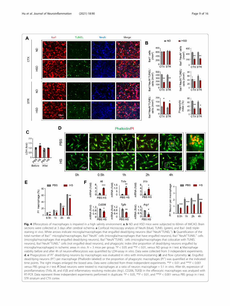
Fig. 4 Efferocytosis of macrophages is impaired in a high salinity environment. a, b ND and HSD mice were subjected to 60min of tMCAO. Brainsections were collected at 3 days after cerebral ischemia. a Confocal microscopy analysis of NeuN (blue), TUNEL (green), and Iba1 (red) triple-staining in vivo. White arrows indicate microglia/macrophages that engulfed dead/dying neurons (Iba1+NeuN+TUNEL+). b Quantification of thetotal number of Iba1+ microglia/macrophages, Iba1+NeuN+ cells (microglia/macrophages that have engulfed neurons), Iba1+NeuN+TUNEL+ cells(microglia/macrophages that engulfed dead/dying neurons), Iba1+NeuN+TUNEL- cells (microglia/macrophages that colocalize with TUNEL-
neurons), Iba1-NeuN+TUNEL+ cells (not engulfed dead neurons), and phagocytic index (the proportion of dead/dying neurons engulfed bymicroglia/macrophages) in ischemic areas in vivo. N = 3 mice per group. *P < 0.05 and **P < 0.01, versus ND group in t test. c Macrophageviability before and after 4h of neuron-efferocytosis was quantified by LDH-assay in vitro. Data were collected from 3 independent experiments.d, e Phagocytosis of PI+ dead/dying neurons by macrophages was evaluated in vitro with immunostaining (d) and flow cytometry (e). Engulfeddead/dying neurons (PI+) per macrophage (Phalloidin labeled) or the proportion of phagocytic macrophages (PI+) was quantified at the indicatedtime points. The right images enlarged the boxed area. Data were collected from three independent experiments. **P < 0.01 and ***P < 0.001versus PBS group in t test. f Dead neurons were treated to macrophages at a ratio of neuron: macrophage = 5:1 in vitro. After 6h, expression ofproinflammatory (Tnfα, Il6, and Il1β) and inflammatory resolving molecules (Arg1, CD206, TGFβ) in the efferocytic macrophages was analyzed withRT-PCR. Data represent three independent experiments performed in duplicate. *P < 0.05, **P < 0.01, and ***P < 0.001 versus PBS group in t test.STR striatum and CTX cortex
Hu et al. Journal of Neuroinflammation (2021) 18:90 Page 9 of 16
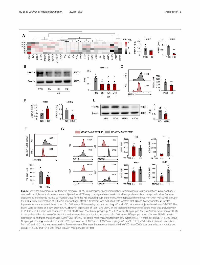
Fig. 5 Excess salt downregulates efferocytic molecule TREM2 in macrophages and impairs their inflammation resolution functions. a Macrophagescultured in a high-salt environment were subjected to a PCR array to analyze the expression of efferocytosis-associated receptors in vitro. Data aredisplayed as fold change relative to macrophages from the PBS-treated group. Experiments were repeated three times. **P < 0.01 versus PBS group int test. b, c Protein expression of TREM2 in macrophages after HS treatment was evaluated with western blot (b) and flow cytometry (c) in vitro.Experiments were repeated three times. *P < 0.05 versus PBS-treated group in t test. d–g ND and HSD mice were subjected to 60min of tMCAO. Thebrains were collected at 3 days after tMCAO. d mRNA expression of Trem1 and Trem2 in the ipsilateral hemisphere of stroke mice was analyzed withRT-PCR in vivo. CT value was normalized to that of ND mice. N = 3 mice per group. *P < 0.05 versus ND group in t test. e Protein expression of TREM2in the ipsilateral hemisphere of stroke mice with western blot. N = 6 mice per group. *P < 0.05, versus ND group in t test. f In vivo, TREM2 proteinexpression in infiltrated macrophages (CD45hiCD11b+Ly6G-) of stroke mice was analyzed with flow cytometry. N = 4 mice per group. *P < 0.05 versusND group in t test. g In vivo CD16 and CD206 expression in TREM2lo and TREM2hi macrophages (CD45hiCD11b+Ly6G-) in the ipsilateral hemispherefrom ND and HSD mice was measured via flow cytometry. The mean fluorescence intensity (MFI) of CD16 or CD206 was quantified. N = 4 mice pergroup. *P < 0.05 and **P < 0.01 versus TREM2lo macrophages in t test
Hu et al. Journal of Neuroinflammation (2021) 18:90 Page 10 of 16
When investigating the relationship between TREM2 ex-pression and inflammatory phenotype of macrophageswith FACS, we found that macrophages with highTREM2 expression (CD45+F4/80+TREM2hi) displayedhigher levels of the anti-inflammatory marker CD206than those with low TREM2 expression (CD45+F4/80+TREMlo), while the CD16-MFI showed no differencebetween macrophages with high and low TREM2 ex-pression in either HSD or ND mice (Fig. 5g).
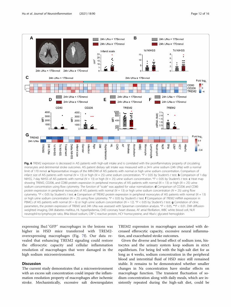
Decreased TREM2 expression is correlated with aproinflammatory property of circulating monocytes anddetrimental stroke outcomes in AIS patientsWe then tested the TREM2 level in monocytes of AISpatients and evaluated the relationship between TREM2expression and stroke outcomes. AIS patients’ dietarysalt intake was measured with a 24-h urine sodium witha normal limit of 170 mmol [27]. Thereafter, we foundthat patients with high urine sodium concentrations hadlarger infarct sizes (Fig. 6a) and higher NIHSS scores(Fig. 6b) than those with normal urine sodium concen-trations. To assess the impact of excessive salt on thephenotypic shift of circulating monocytes in AIS patientsduring the acute phase (0–3 days after disease onset), weanalyzed the expression of the proinflammatory markerCD80 and the anti-inflammatory marker CD206 [28–30]in monocytes (CD11b+CD14+) of patient peripheralblood with FACS. Detailed gating strategy is displayed inSupplementary Figure 4. As expected, monocytes fromstroke patients with high urine sodium concentrationexpressed less CD206 than normal monocytes from dietstroke patients, while no differential expression of CD80was recorded (Fig. 6c, d and Supplementary Figure 5).TREM2 expression in monocytes was downregulated instroke patients with high urine sodium concentrationcompared with those with normal diets using FACS (Fig.6c, e and Supplementary Figure 5). Moreover, we foundthat the TREM2 mRNA level decreased in the peripheralblood mononuclear cells (PBMC) of patients with highurine sodium concentration (Fig. 6f). At the same time,the expression of other PRRs remained to be stable(Supplementary Figure 6). Since PRRs are mainlyexpressed in monocytes in PBMC [31], our data indi-cated that a high salinity environment specifically down-regulated TREM2 expression in monocytes of AISpatients. Through Spearman correlation analysis, we de-termined that the CD206 MFI of peripheral bloodmonocytes showed a significant positive correlation withTREM2 MFI in stroke patients, while the CD80 MFIshowed a negative correlation with the TREM2 MFI(Supplementary Figure 7), which was consistent with ourdata in animal models. Interestingly, we found thatTREM2 expression in the circulating monocytes of AISpatients was negatively correlated with a 24-h urine
excretion (Fig. 6g and Supplementary Figure 7), while de-creased TREM2 level of macrophages was associated withincreased NIHSS scores (Fig. 6g and Supplementary Fig-ure 7). The results indicated that TREM2 expression inmonocytes/macrophages favored efferocytosis and thesubsequent inflammation resolution after ischemic stroke.
Enhancement of TREM2 signaling restores the efferocyticcapacity and cellular inflammation resolution ofmacrophages in the high salinity environmentTREM2 is a vital functional molecule implicated in thephagocytosis activity of macrophages. The efferocytosiscapacity of macrophages plays a decisive role in inflam-mation resolution after stroke and affects the diseaseoutcomes. Therefore, we hypothesized that enhancingTREM2 signaling in macrophages could restore theirefferocytic capacity and promote inflammation reso-lution. Macrophages were infected with lent viral vectorscarrying TREM2 cDNA or empty vector (NC) for 2 daysbefore treatment. Efficacy of transfection was confirmedwith flow cytometry (Supplementary Figure 8A) andwestern blot (Supplementary Figure 8B). Transfectedmacrophages were treated with or without the additionof NaCl (40mM) and incubated with PI-labeled post-OGD neurons. Gratifyingly, TREM2 overexpression inmacrophages exposed to excess salt restored the effero-cytic capacity, as the engulfed dead/dying neurons permacrophage (Fig. 7a), or the proportion of phagocyticmacrophages (PI+F4/80+) (Fig. 7b) recovered to thelevels of those treated with PBS at 1h after co-culture.Moreover, at 24 h after co-culture, protein levels ofCD206 and Arg1 in TREM2-overexpressing macrophagestreated with a high concentration of NaCl resembled thosetreated with PBS (Fig. 7c). To further evaluate the effect ofTREM2-overexpressing macrophages on high-salt-affordedneuroinflammation after ischemic stroke, mice with HSDor ND were first injected with clodronate liposomes to de-plete monocytes/macrophages (Supplementary figure 9).Mice were then subjected to 60 min of tMCAO. Immedi-ately after reperfusion, a single dose of 2x10⁶ TREM2-overexpressing macrophages or NC macrophages wereintravenously transferred. In ND mice, we recorded thattransfer of TREM2-overexpressing macrophages reducedthe infarct volume compared with those transferred withNC macrophages (Fig. 7d). In addition, transfer of TREM2-overexpressing macrophages offered protection to HSDmice. Infarct volume decreased in HSD mice transferredwith TREM2-overexpressing macrophages. We recordedrestored efferocytic capacity of the infiltrated macrophagesin HSD mice transferred with TREM2-overexpressing mac-rophages, as the number of phagocytic microglia/macro-phages (Iba1+NeuN+) and phagocytic index in ischemiclesions increased compared with those transferred with NCmacrophages (Fig. 7e). The percentage of CD206
Hu et al. Journal of Neuroinflammation (2021) 18:90 Page 11 of 16
expressing Iba1+GFP+ macrophages in the lesions washigher in HSD mice transferred with TREM2-overexpressing macrophages (Fig. 7f). Our data re-vealed that enhancing TREM2 signaling could restorethe efferocytic capacity and cellular inflammationresolution of macrophages that were damaged in thehigh sodium microenvironment.
DiscussionThe current study demonstrates that a microenvironmentwith an excess salt concentration could impair the inflam-mation resolution property of macrophages after ischemicstroke. Mechanistically, excessive salt downregulates
TREM2 expression in macrophages associated with de-creased efferocytic capacity, excessive neural inflamma-tion, and exacerbated stroke outcomes.Given the diverse and broad effect of sodium ions, his-
tocytes and the urinary system keep sodium in strictequilibrium. For being fed with the high-salt diet for aslong as 4 weeks, sodium concentration in the peripheralblood and interstitial fluid of HSD mice still remainedstable. It remains to be demonstrated whether smallerchanges in Na concentration have similar effects onmacrophage function. The transient fluctuation of so-dium concentration along with daily meals, which are in-sistently repeated during the high-salt diet, could be
Fig. 6 TREM2 expression is decreased in AIS patients with high-salt intake and is correlated with the proinflammatory property of circulatingmonocytes and detrimental stroke outcomes. AIS patient dietary salt intake was measured with a 24-h urine sodium (24h UNa) with a normallimit of 170 mmol. a Representative images of the MRI-DWI of AIS patients with normal or high urine sodium concentration. Comparison ofinfarct size of AIS patients with normal (N = 13) or high (N = 25) urine sodium concentration. *P < 0.05 by Student’s t test. b Comparison of 1-dayNIHSS, 7-day NIHSS of AIS patients with normal (N = 13) or high (N = 25) urine sodium concentration. *P < 0.05 by Student’s t test. c Heat mapshowing TREM2, CD206, and CD80 protein expression in peripheral monocytes of AIS patients with normal (N = 13) or high (N = 25) urinesodium concentration using flow cytometry. The function of “scale” was applied for value normalization. d Comparison of CD206 and CD80protein expression in peripheral monocytes of AIS patients with normal (N = 13) or high urine sodium concentration (N = 25) using flowcytometry. *P < 0.05 by Student’s t test. e Comparison of TREM2 protein expression in peripheral monocytes of AIS patients with normal (N = 13)or high urine sodium concentration (N = 25) using flow cytometry. *P < 0.05 by Student’s t test. f Comparison of TREM2 mRNA expression inPBMCs of AIS patients with normal (N = 6) or high urine sodium concentration (N = 12). *P < 0.05 by Student’s t test. g Correlation of clinicparameters, the protein expression of TREM2 and 24h UNa was assessed with Spearman correlation analysis. *P < 0.05, **P < 0.01. DWI diffusion-weighted imaging, DM diabetes mellitus, HL hyperlipidemia, CHD coronary heart disease, AF atrial fibrillation, WBC white blood cell, NLRneutrophil-to-lymphocyte ratio, BNa blood sodium, CRP C-reactive protein, HCY homocysteine, and Hba1c glycated hemoglobin
Hu et al. Journal of Neuroinflammation (2021) 18:90 Page 12 of 16
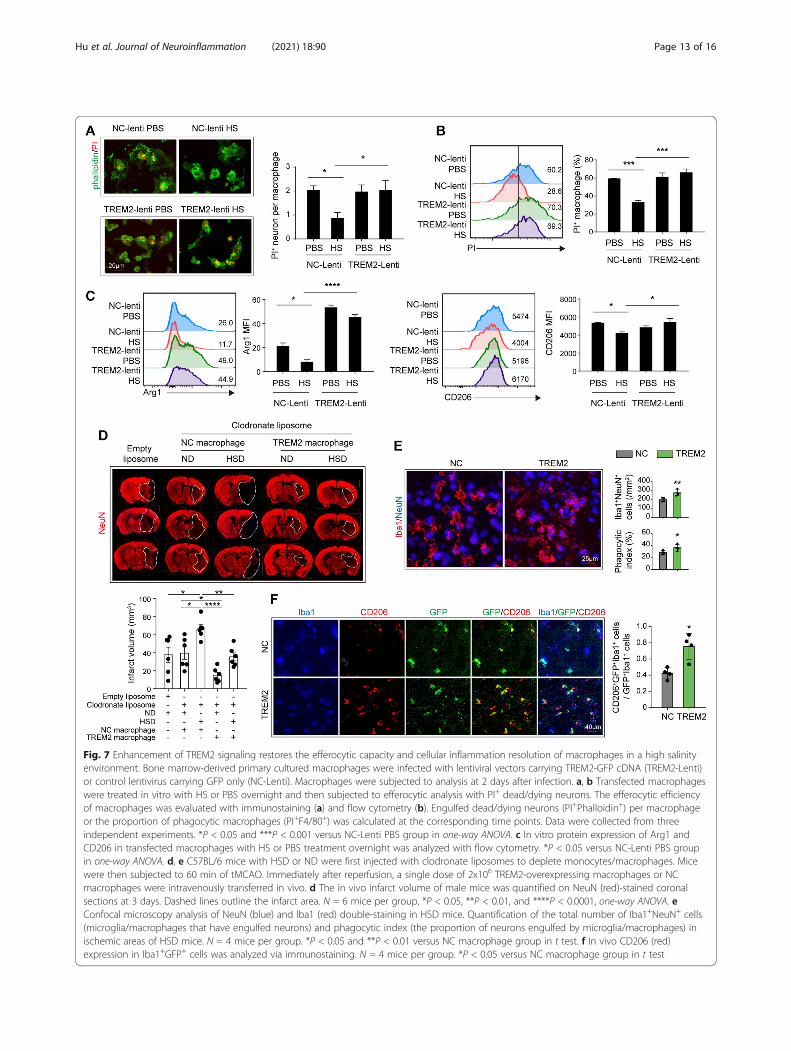
Fig. 7 Enhancement of TREM2 signaling restores the efferocytic capacity and cellular inflammation resolution of macrophages in a high salinityenvironment. Bone marrow-derived primary cultured macrophages were infected with lentiviral vectors carrying TREM2-GFP cDNA (TREM2-Lenti)or control lentivirus carrying GFP only (NC-Lenti). Macrophages were subjected to analysis at 2 days after infection. a, b Transfected macrophageswere treated in vitro with HS or PBS overnight and then subjected to efferocytic analysis with PI+ dead/dying neurons. The efferocytic efficiencyof macrophages was evaluated with immunostaining (a) and flow cytometry (b). Engulfed dead/dying neurons (PI+Phalloidin+) per macrophageor the proportion of phagocytic macrophages (PI+F4/80+) was calculated at the corresponding time points. Data were collected from threeindependent experiments. *P < 0.05 and ***P < 0.001 versus NC-Lenti PBS group in one-way ANOVA. c In vitro protein expression of Arg1 andCD206 in transfected macrophages with HS or PBS treatment overnight was analyzed with flow cytometry. *P < 0.05 versus NC-Lenti PBS groupin one-way ANOVA. d, e C57BL/6 mice with HSD or ND were first injected with clodronate liposomes to deplete monocytes/macrophages. Micewere then subjected to 60 min of tMCAO. Immediately after reperfusion, a single dose of 2x10⁶ TREM2-overexpressing macrophages or NCmacrophages were intravenously transferred in vivo. d The in vivo infarct volume of male mice was quantified on NeuN (red)-stained coronalsections at 3 days. Dashed lines outline the infarct area. N = 6 mice per group, *P < 0.05, **P < 0.01, and ****P < 0.0001, one-way ANOVA. eConfocal microscopy analysis of NeuN (blue) and Iba1 (red) double-staining in HSD mice. Quantification of the total number of Iba1+NeuN+ cells(microglia/macrophages that have engulfed neurons) and phagocytic index (the proportion of neurons engulfed by microglia/macrophages) inischemic areas of HSD mice. N = 4 mice per group. *P < 0.05 and **P < 0.01 versus NC macrophage group in t test. f In vivo CD206 (red)expression in Iba1+GFP+ cells was analyzed via immunostaining. N = 4 mice per group. *P < 0.05 versus NC macrophage group in t test
Hu et al. Journal of Neuroinflammation (2021) 18:90 Page 13 of 16
sufficient to exert effective impacts. In accordance, werecorded that circulating monocytes in stroke patientswith high-salt consumption displayed proinflammatoryinclination compared with those in patients with appro-priate salt intake, which was presented in culture macro-phages directly exposed to a high-salt environmentin vitro. Further, in the arena of stroke lesion, ongoingneural inflammation and local metabolic disturbancecould facilitate sodium accumulation. Excessive expres-sion of SIK1 in stroke lesion of HSD mice revealed thesalinized microenvironment encountered by the infil-trated macrophages. To explore the molecular mecha-nisms of functional alterations of macrophages whenencountering sodium stimulation, bone marrow-derivedmacrophages were cultured and threatened with high so-dium treatment. Although the in vitro culture systemfailed to perfectly reproduce the complicated and dy-namic pathophysiological process in vivo, we recordeddistinctive proinflammatory activations in high-salt-treated macrophages, which were further verified inHSD stroke mice and AIS patients.It has been reported that the HSD could promote BBB
injury after ischemic stroke [12]. Consistently, we re-corded that increased infiltration of multiple leukocytes,including macrophages, neutrophils, T lymphocytes, andB lymphocytes, in the stroke lesions of HSD mice at 3days after stroke, could be attributed to the exacerbatedBBB damage. It was found that surplus dietary salt di-rected macrophages/microglia towards the classical acti-vated “M1” phenotype, which further exacerbated strokeoutcomes [24]. In accordance, our data indicated thatthe inflammation resolution property of macrophageswas downregulated by excess salt, which led to the post-poned recovery of stroke lesions.Efferocytosis represents an essential process of inflamma-
tion resolution [32, 33]. Elimination of the dead or injuredcomponents within stroke lesion arrests amplification ofneural inflammation. We demonstrated that the efferocyticcapacity, together with the subsequent cellular inflamma-tion resolution of macrophages, was impaired in the highsalinity environment, which could be a rational explanationfor accumulated dead cells in the stroke penumbra. It hasbeen demonstrated that the function of TREM2 is indis-pensable for phagocytic activities of microglia and macro-phages [34]. Our data indicated that TREM2 wasdownregulated in macrophages in the high salinity environ-ment. Decreased TREM2 expression was correlated withrobust post-stroke neural inflammation and exacerbatedstroke outcomes, which indicated that inhibition of TREM2signaling in macrophages was the potential mechanism in-volved in the detrimental impact of the high-saltmicroenvironment.It has been recognized that HSD is a crucial risk factor
for ischemic stroke. Restriction of dietary salt intake
serves as an efficient and practical method for prevent-ing new vascular events. Nevertheless, no niched therapythat targets the already impaired inflammation reso-lution property of macrophages in the high-salt environ-ment has been reported. In our study, utilization ofTREM2-overexpressing macrophages offered neuropro-tection to HSD mice. In addition, overexpression ofTREM2 restored the efferocytic capacity and cellular in-flammation resolution of macrophages in a high salinityenvironment. The data encourage further research onthe therapeutic potential of enhancing TREM2 signalingin patients with ischemic stroke, especially those withhigh-salt intake.
ConclusionsConclusively, HSD aggravates ischemic stroke outcomesby exacerbating neural inflammation, which is associatedwith the impaired inflammation resolution property ofmacrophages. TREM2 expression in macrophages isdownregulated by high-salt environments and enhancingTREM2 signaling could restore the efferocytic capacityand cellular inflammation resolution of macrophages.Further study on the value of TREM2 signaling as atherapeutic target in AIS is warranted.
Supplementary InformationThe online version contains supplementary material available at https://doi.org/10.1186/s12974-021-02144-9.
Additional file 1: Table S1. Clinic characteristics of the whole cohort.Table S2. Primers used in the study. Figure S1. Comparison ofinflammatory mediator expression in peripheral blood and contralateralbrains of ND and HSD mice. Figure S2. Excess salt downregulatesefferocytic molecules in macrophages. Figure S3. Impact of high salt onTREM1 expression in primary culture macrophage and ischemic brain.Figure S4. Gating strategy in flow cytometric analysis of TREM2, CD80,and CD206 expression in monocytes of AIS patients. Figure S5.Representative images of TREM2, CD206, and CD80 expression inperipheral monocyte of AIS patients with normal or high urine sodiumconcentration. Figure S6. PRRs mRNA expression remained no differentother than TREM2 in AIS patients with high salt intake. Figure S7.Spearman correlation analysis of TREM2 expression, monocytephenotypic marker, AIS outcomes and clinic parameters. Figure S8.Validation of TREM2 overexpression efficacy in primary culturedmacrophages. Figure S9. Circulating macrophages and monocytes weredepleted by clodronate liposome treatment.
AcknowledgementsWe want to sincerely express our special thanks to Dr. Xining Wang (SunYat-sen Memorial Hospital) for his hard work coding in R language and pol-ishing the English in the manuscript.
Authors’ contributionsMH designed and performed the experiments, collected and analyzed thedata, and drafted the manuscript. YL and XM contributed to theexperimental design and revised the manuscript. SW and XS contributed tothe experimental design and the manuscript. QZ and DL performed theanimal experiments and collected the data. SL and BZ contributed to theexperimental design and the manuscript. WC and ZL designed andsupervised the study and critically revised the manuscript. The authors readand approved the final manuscript.
Hu et al. Journal of Neuroinflammation (2021) 18:90 Page 14 of 16
FundingThis work was supported by grants from the Youth Program of NationalNatural Science Foundation of China (81901201 to W. C), National NaturalScience Foundation of China (81971110 to Z. L), Guangzhou Science andTechnology Program Key Project (202007030010), Guangzhou Science andTechnology Plan Project (201904010444 to Z. L), China Postdoctoral ScienceFoundation Grant (2019T120776 to W. C), and China Postdoctoral ScienceFoundation Grant (2018 M643332 to W. C).
Availability of data and materialsThe datasets used and/or analyzed during the current study are availablefrom the corresponding author on reasonable request.
Declarations
Ethics approval and consent to participateThe clinical and experimental animal studies were approved by the MedicalEthics Committee of the Third Affiliated Hospital of Sun Yat-Sen University andthe Animal Care and Use Committee of Sun Yat-Sen University, respectively. Allparticipants had signed the informed consent according to the principlesillustrated in the Declaration of Helsinki. All animal experiments were approvedby The Third Affiliated Hospital of Sun Yat-sen University and performedfollowing the Guide for the Care and Use of Laboratory Animals andStroke Treatment.
Consent for publicationWe have obtained consent to publish from the participant to reportindividual patient data.
Competing interestsThe authors declare that they have no competing interests.
Author details1Department of Neurology, Mental and Neurological Disease ResearchCenter, The Third Affiliated Hospital of Sun Yat-sen University, 600 TianheRoad, Guangzhou, Guangdong 510630, People’s Republic of China. 2Centerof Clinical Immunology, Mental and Neurological Disease Research Center,The Third Affiliated Hospital of Sun Yat-sen University, 600 Tianhe Road,Guangzhou, Guangdong 510630, People’s Republic of China.
Received: 27 November 2020 Accepted: 29 March 2021
References1. Kono Y, Yamada S, Yamaguchi J, Hagiwara Y, Iritani N, Ishida S, et al.
Secondary prevention of new vascular events with lifestyle intervention inpatients with noncardioembolic mild ischemic stroke: a single-centerrandomized controlled trial. Cerebrovasc Dis. 2013;36(2):88–97.
2. Kono Y, Yamada S, Kamisaka K, Araki A, Fujioka Y, Yasui K, et al. Recurrencerisk after noncardioembolic mild ischemic stroke in a Japanese population.Cerebrovasc Dis. 2011;31(4):365–72.
3. Gardener H, Rundek T, Wright CB, Elkind MS, Sacco RL. Dietary sodium andrisk of stroke in the Northern Manhattan study. Stroke. 2012;43(5):1200–5.
4. He FJ, MacGregor GA. Role of salt intake in prevention of cardiovasculardisease: controversies and challenges. Nat Rev Cardiol. 2018;15(6):371–7.
5. O’Donnell M, Mente A, Yusuf S. Sodium intake and cardiovascular health.Circ Res. 2015;116(6):1046–57.
6. Appel LJ. Reducing sodium intake to prevent stroke: time for action, nothesitation. Stroke. 2014;45(3):909–11.
7. Planas AM. Role of immune cells migrating to the ischemic brain. Stroke.2018;49(9):2261–7.
8. Jian Z, Liu R, Zhu X, Smerin D, Zhong Y, Gu L, et al. The involvement andtherapy target of immune cells after ischemic stroke. Front Immunol. 2019;10:2167.
9. Cai W, Dai X, Chen J, Zhao J, Xu M, Zhang L, et al. STAT6/Arg1 promotesmicroglia/macrophage efferocytosis and inflammation resolution in strokemice. JCI Insight. 2019;4:20.
10. Cai W, Liu S, Hu M, Sun X, Qiu W, Zheng S, et al. Post-stroke DHA treatmentprotects against acute ischemic brain injury by skewing macrophagepolarity toward the M2 phenotype. Transl Stroke Res. 2018;9(6):669–80.
11. Zhang WC, Zheng XJ, Du LJ, Sun JY, Shen ZX, Shi C, et al. High salt primes aspecific activation state of macrophages, M(Na). Cell Res. 2015;25(8):893–910.
12. Zhang T, Fang S, Wan C, Kong Q, Wang G, Wang S, et al. Excess saltexacerbates blood-brain barrier disruption via a p38/MAPK/SGK1-dependent pathway in permanent cerebral ischemia. Sci Rep. 2015;5:16548.
13. Ting SM, Zhao X, Sun G, Obertas L, Ricote M, Aronowski J. Brain cleanup asa potential target for poststroke recovery: the role of RXR (retinoic Xreceptor) in phagocytes. Stroke. 2020;51(3):958–66.
14. Neher JJ, Emmrich JV, Fricker M, Mander PK, Thery C, Brown GC.Phagocytosis executes delayed neuronal death after focal brain ischemia.Proc Natl Acad Sci U S A. 2013;110(43):E4098–107.
15. Cai W, Liu S, Hu M, Huang F, Zhu Q, Qiu W, et al. Functionaldynamics of neutrophils after ischemic stroke. Transl Stroke Res. 2020;11(1):108–21.
16. Tan S, Shan Y, Wang Y, Lin Y, Liao S, Deng Z, et al. Exacerbation of oxygen-glucose deprivation-induced blood-brain barrier disruption: potentialpathogenic role of interleukin-9 in ischemic stroke. Clin Sci (Lond). 2017;131(13):1499–513.
17. Jackson C, Sudlow C. Comparing risks of death and recurrent vascularevents between lacunar and non-lacunar infarction. Brain. 2005;128(Pt 11):2507–17.
18. Traylor M, Rutten-Jacobs LC, Thijs V, Holliday EG, Levi C, Bevan S, et al.Genetic associations with white matter hyperintensities confer risk oflacunar stroke. Stroke. 2016;47(5):1174–9.
19. Norrving B. Long-term prognosis after lacunar infarction. Lancet Neurol.2003;2(4):238–45.
20. Stetler RA, Cao G, Gao Y, Zhang F, Wang S, Weng Z, et al. Hsp27 protectsagainst ischemic brain injury via attenuation of a novel stress-responsecascade upstream of mitochondrial cell death signaling. J Neurosci. 2008;28(49):13038–55.
21. Cai W, Wang J, Hu M, Chen X, Lu Z, Bellanti JA, et al. All trans-retinoic acidprotects against acute ischemic stroke by modulating neutrophil functionsthrough STAT1 signaling. J Neuroinflammation. 2019;16(1):175.
22. Faraco G, Brea D, Garcia-Bonilla L, Wang G, Racchumi G, Chang H, et al.Dietary salt promotes neurovascular and cognitive dysfunction through agut-initiated TH17 response. Nat Neurosci. 2018;21(2):240–9.
23. Wein MN, Foretz M, Fisher DE, Xavier RJ, Kronenberg HM. Salt-induciblekinases: physiology, regulation by cAMP, and therapeutic potential. TrendsEndocrinol Metab. 2018;29(10):723–35.
24. Zhang T, Wang D, Li X, Jiang Y, Wang C, Zhang Y, et al. Excess salt intakepromotes M1 microglia polarization via a p38/MAPK/AR-dependentpathway after cerebral ischemia in mice. Int Immunopharmacol. 2020;81:106176.
25. Peng Q, Malhotra S, Torchia JA, Kerr WG, Coggeshall KM, Humphrey MB.TREM2- and DAP12-dependent activation of PI3K requires DAP10 and isinhibited by SHIP1. Sci Signal. 2010;3(122):ra38.
26. Wang Y, Grainger DW. RNA therapeutics targeting osteoclast-mediated excessive bone resorption. Adv Drug Deliv Rev. 2012;64(12):1341–57.
27. Olde Engberink RHG, van den Hoek TC, van Noordenne ND, van denBorn BH, Peters-Sengers H, Vogt L. Use of a single baseline versusmultiyear 24-hour urine collection for estimation of long-term sodiumintake and associated cardiovascular and renal risk. Circulation. 2017;136(10):917–26.
28. Pinto BF, Medeiros NI, Teixeira-Carvalho A, Eloi-Santos SM, Fontes-Cal TCM,Rocha DA, et al. CD86 Expression by monocytes influences animmunomodulatory profile in asymptomatic patients with chronic chagasdisease. Front Immunol. 2018;9:454.
29. Gundra UM, Girgis NM, Gonzalez MA, San Tang M, Van Der Zande HJP, LinJD, et al. Vitamin A mediates conversion of monocyte-derived macrophagesinto tissue-resident macrophages during alternative activation. NatImmunol. 2017;18(6):642–53.
30. Gubin MM, Esaulova E, Ward JP, Malkova ON, Runci D, Wong P, et al. High-dimensional analysis delineates myeloid and lymphoid compartmentremodeling during successful immune-checkpoint cancer therapy. Cell.2018;175(4):1014–30 e19.
31. Turnbull IR, Gilfillan S, Cella M, Aoshi T, Miller M, Piccio L, et al. Cuttingedge: TREM-2 attenuates macrophage activation. J Immunol. 2006;177(6):3520–4.
Hu et al. Journal of Neuroinflammation (2021) 18:90 Page 15 of 16
32. Doran AC, Yurdagul A Jr, Tabas I. Efferocytosis in health and disease. NatRev Immunol. 2020;20(4):254–67.
33. Proto JD, Doran AC, Gusarova G, Yurdagul A Jr, Sozen E, Subramanian M,et al. Regulatory T cells promote macrophage efferocytosis duringinflammation resolution. Immunity. 2018;49(4):666–77 e6.
34. Zhao Y, Wu X, Li X, Jiang LL, Gui X, Liu Y, et al. TREM2 is a receptor for beta-amyloid that mediates microglial function. Neuron. 2018;97(5):1023–31 e7.
Publisher’s NoteSpringer Nature remains neutral with regard to jurisdictional claims inpublished maps and institutional affiliations.
Hu et al. Journal of Neuroinflammation (2021) 18:90 Page 16 of 16