hibi.qxd 1/31/06 1:53 pm page 141 distraction osteogenesis...
TRANSCRIPT
The International Journal of Oral & Maxillofacial Implants 141
Distraction Osteogenesis Assisted by Tissue Engineering in an Irradiated Mandible:
A Case ReportHideharu Hibi, DDS, PhD1/Yoichi Yamada, DDS, PhD2/Hideaki Kagami, DDS, PhD3/Minoru Ueda, DDS, PhD4
Distraction osteogenesis (DO) can provide predictable bone regeneration without grafting proceduresbut requires long treatment time and forms less bone transverse to the direction of distraction. To pro-mote 3-dimensional bone formation and shorten the consolidation period, tissue-engineeredosteogenic material (injectable bone) was applied in a patient who was being treated with vertical DOwith an osteocutaneous fibular flap to reconstruct the mandible. The material, which comprised autol-ogous mesenchymal stem cells culture-expanded then induced to be osteogenic in character andplatelet-rich plasma (PRP) activated with thrombin and calcium chloride, was infiltrated into the dis-tracted tissue at the end of distraction and injected into a space created labially with a titanium meshat implant placement. The infiltration contributed to full consolidation of the regenerate for 3 months,and the injection thickened the regenerated ridge and bridged a gap between the native mandible anddistracted fibula. The reconstructed mandible was expanded from 10 mm to 25 mm in height despitea lacerated and opened labial periosteum in the distracted area. Six implants 18 mm in length wereplaced and subsequently achieved osseointegration. The cutaneous flap covering the implants wastrimmed, and the palatal mucosa was transplanted to the regenerated ridge for vestibuloplasty. Theseraw surfaces were covered with PRP; within 3 weeks, they had attained an epithelium. The implantshave supported a fixed prosthesis with adequate surrounding bone and attached mucosa. DO wasassisted by tissue engineering and became effective in restoring the compromised mandible. INT JORAL MAXILLOFAC IMPLANTS 2006;21:141–147
Key words: distraction osteogenesis, injectable bone, platelet-rich plasma, stem cells, tissue engineering
Distraction osteogenesis (DO) has become awidely accepted technique for reconstructing
bone defects in the maxillofacial region. This tech-nique provides predictable bone formation without
grafting procedures but requires a long healing timewhich includes latent, lengthening, and consolida-tion periods. To promote bone formation andshorten the consolidation period, some attempts atapplying hyperbaric oxygenation or electrical, ultra-sonic, or chemical stimulation have been made.1 Sev-eral recent studies have shown that injecting cellswith osteogenic potential into distracted callusenhances its consolidation.2–5
The present authors have recently reported on atissue-engineered osteogenic material called“injectable bone,” which comprises culture-expanded mesenchymal stem cells (MSCs) andplatelet-rich plasma (PRP).6 Not only animal studiesbut also clinical trials have demonstrated that thismaterial can effectively regenerate osseous tissue. Itwas therefore decided to apply the material to DOand present this case of the reconstruction of amandible with damaged healing potential.
1Associate Professor, Center for Genetic and Regenerative Medi-cine, Nagoya University School of Medicine, Nagoya, Japan.
2Assistant Professor, Center for Genetic and Regenerative Medi-cine, Nagoya University School of Medicine, Nagoya, Japan.
3Associate Professor, Department of Tissue Engineering, NagoyaUniversity School of Medicine, Nagoya, Japan.
4Professor, Department of Oral and Maxillofacial Surgery, NagoyaUniversity Graduate School of Medicine, Nagoya, Japan.
Correspondence to: Dr Hideharu Hibi, Center for Genetic andRegenerative Medicine, Nagoya University School of Medicine, 65Tsurumai-cho, Showa-ku, Nagoya 466-8550 Japan. Fax: +81 52744 2352. E-mail: [email protected]
Hibi.qxd 1/31/06 1:53 PM Page 141
142 Volume 21, Number 1, 2006
Hibi et al
MATERIALS AND METHODS
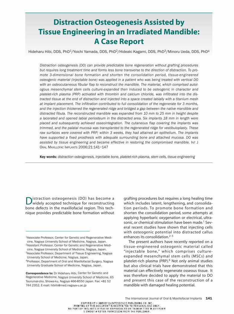
The MSCs and PRP are prepared as described previ-ously.6 The MSCs are isolated from iliac marrow aspi-rates, expanded in culture media for 3 weeks, and dif-ferentiated in supplemented osteogenesis inductionmedia for another week. The PRP is isolated fromautologous blood using density gradient centrifuga-tion and a selective collection technique (Figs 1a and1b). A 3-way stopcock connects 2 syringes; one con-tains 1 mL of air, 1 mL of 10% calcium chloride, and1,000 units of human thrombin; the other contains 6mL of PRP and all of the induced MSCs. This formulais standard except for the MSCs; the amount of thosevaries according to need. With the stopcock open,the contents of the 2 syringes are completely mixedfor 5 seconds. The injectable bone mixture thenmaintains its gel form for about 20 seconds (Figs 1cand 1d).
CASE REPORT
A 54-year-old male patient was referred to theauthors’ hospital for rehabilitation of his recon-structed edentulous mandible. Two years earlier, thepatient had undergone a segmental resection andimmediate reconstruction of the mandible in con-junction with the oral floor resultant from squamouscell carcinoma, following chemotherapy and irradia-
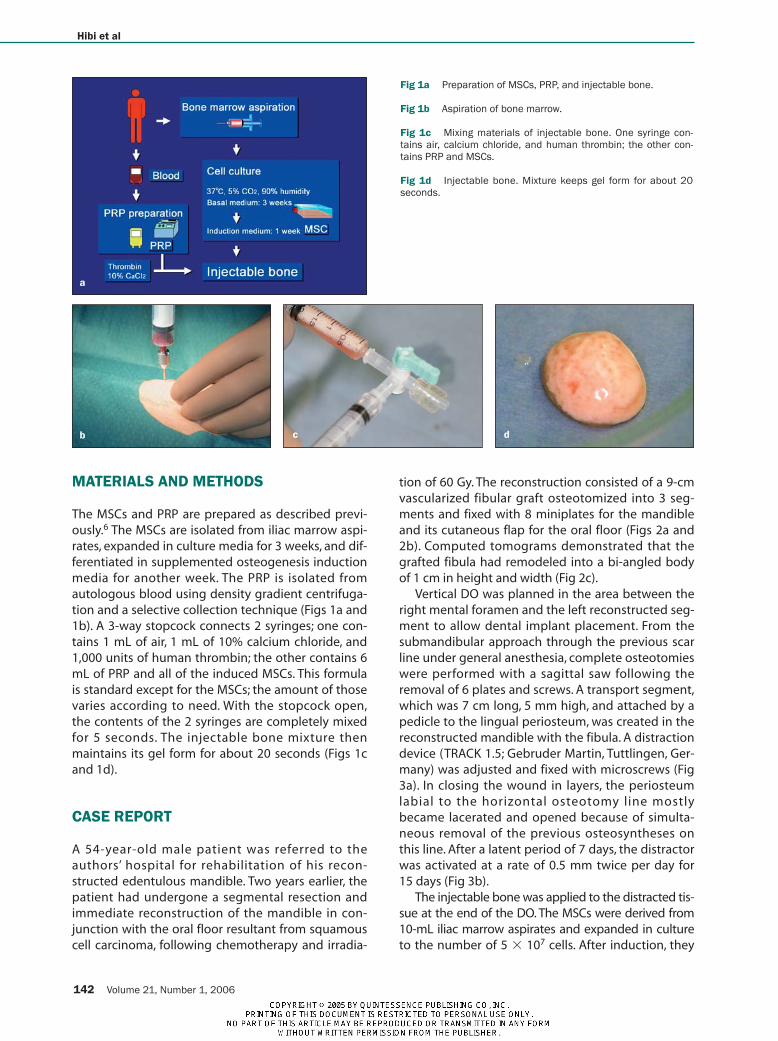
tion of 60 Gy. The reconstruction consisted of a 9-cmvascularized fibular graft osteotomized into 3 seg-ments and fixed with 8 miniplates for the mandibleand its cutaneous flap for the oral floor (Figs 2a and2b). Computed tomograms demonstrated that thegrafted fibula had remodeled into a bi-angled bodyof 1 cm in height and width (Fig 2c).
Vertical DO was planned in the area between theright mental foramen and the left reconstructed seg-ment to allow dental implant placement. From thesubmandibular approach through the previous scarline under general anesthesia, complete osteotomieswere performed with a sagittal saw following theremoval of 6 plates and screws. A transport segment,which was 7 cm long, 5 mm high, and attached by apedicle to the lingual periosteum, was created in thereconstructed mandible with the fibula. A distractiondevice (TRACK 1.5; Gebruder Martin, Tuttlingen, Ger-many) was adjusted and fixed with microscrews (Fig3a). In closing the wound in layers, the periosteumlabial to the horizontal osteotomy line mostlybecame lacerated and opened because of simulta-neous removal of the previous osteosyntheses onthis line. After a latent period of 7 days, the distractorwas activated at a rate of 0.5 mm twice per day for15 days (Fig 3b).
The injectable bone was applied to the distracted tis-sue at the end of the DO. The MSCs were derived from10-mL iliac marrow aspirates and expanded in cultureto the number of 5 � 107 cells. After induction, they
Fig 1a Preparation of MSCs, PRP, and injectable bone.
Fig 1b Aspiration of bone marrow.
Fig 1c Mixing materials of injectable bone. One syringe con-tains air, calcium chloride, and human thrombin; the other con-tains PRP and MSCs.
Fig 1d Injectable bone. Mixture keeps gel form for about 20seconds.
a
b c d
Hibi.qxd 1/31/06 1:53 PM Page 142
The International Journal of Oral & Maxillofacial Implants 143
Hibi et al
expressed high alkaline phosphatase activity in assay.Twenty milliliters of PRP were isolated from 200 mLof blood; this PRP contained 1.6 � 109 platelets/mL, aconcentration 8.3 times stronger than that of theoriginal whole blood. With a C-arm fluoroscope forguidance, while the patient was under intravenoussedation, a 18-gauge needle was placed into the dis-traction gap (Fig 4a). The 3 mL of injectable bone wasprepared and infiltrated for 15 seconds (Fig 4b). Theneedle was left in place for an additional minute toallow the gel to increase in viscosity and to prevent
the injected material from leaking out of the punc-ture. No complications were observed during theinjection, and the subsequent course was unevent-ful.
A series of monthly panoramic radiographsshowed that radiopacity in the distraction gap hadbegun to appear at 1 month. After 2 to 3 months,during which the transport segment resorbed mar-ginally (Fig 5a), the area became wholly radiopaque.Computed tomograms at 3 months revealed thatnewly formed bone in the distraction gap had
Figs 2a and 2b Reconstructed mandible and oral floor with vascularized osteocutaneousfibular flap.
Fig 3a Distraction device. The periosteumlacerated and opened due to simultaneousremoval of the previous osteosyntheticplates and screws.
Fig 3b Immediately after distraction.Transport segment was repositioned 15mm superiorly.
Figs 4a and 4b Application of injectablebone to distracted tissue with fluoroscopicguide.
Fig 2c Grafted fibula remodeling into a bi-angled body of 1 cm in height and width.
Hibi.qxd 1/31/06 1:53 PM Page 143
144 Volume 21, Number 1, 2006
Hibi et al
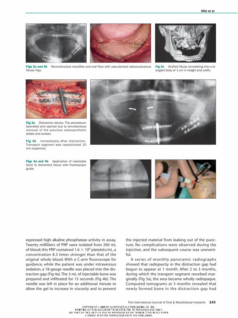
unclear labial surfaces but clear lingual cortical sur-faces. The area in between, which was relatively evenwith respect to density, scored higher in Hounsfieldunits than the cancellous bone areas in the neigh-boring mandibular and fibular bone (Fig 5b).
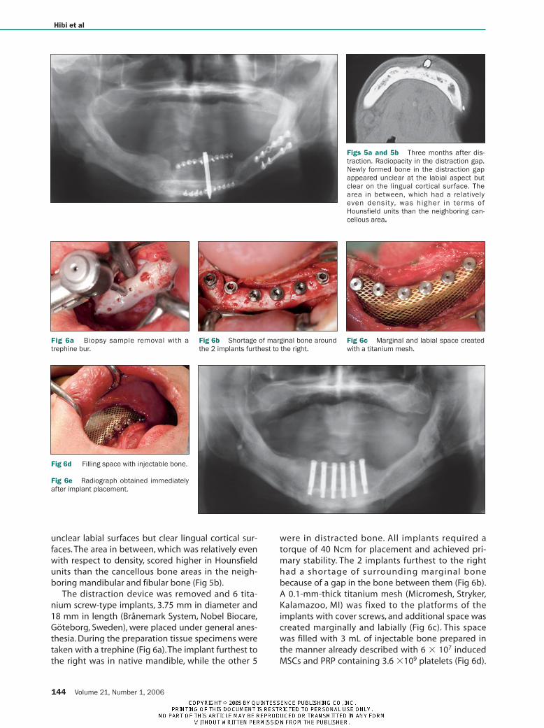
The distraction device was removed and 6 tita-nium screw-type implants, 3.75 mm in diameter and18 mm in length (Brånemark System, Nobel Biocare,Göteborg, Sweden), were placed under general anes-thesia. During the preparation tissue specimens weretaken with a trephine (Fig 6a). The implant furthest tothe right was in native mandible, while the other 5
were in distracted bone. All implants required atorque of 40 Ncm for placement and achieved pri-mary stability. The 2 implants furthest to the righthad a shortage of surrounding marginal bonebecause of a gap in the bone between them (Fig 6b).A 0.1-mm-thick titanium mesh (Micromesh, Stryker,Kalamazoo, MI) was fixed to the platforms of theimplants with cover screws, and additional space wascreated marginally and labially (Fig 6c). This spacewas filled with 3 mL of injectable bone prepared inthe manner already described with 6 � 107 inducedMSCs and PRP containing 3.6 �109 platelets (Fig 6d).
Figs 5a and 5b Three months after dis-traction. Radiopacity in the distraction gap.Newly formed bone in the distraction gapappeared unclear at the labial aspect butclear on the lingual cortical surface. Thearea in between, which had a relativelyeven density, was higher in terms ofHounsfield units than the neighboring can-cellous area.
Fig 6a Biopsy sample removal with atrephine bur.
Fig 6b Shortage of marginal bone aroundthe 2 implants furthest to the right.
Fig 6c Marginal and labial space createdwith a titanium mesh.
Fig 6d Filling space with injectable bone.
Fig 6e Radiograph obtained immediatelyafter implant placement.
Hibi.qxd 1/31/06 1:53 PM Page 144
The International Journal of Oral & Maxillofacial Implants 145
Hibi et al
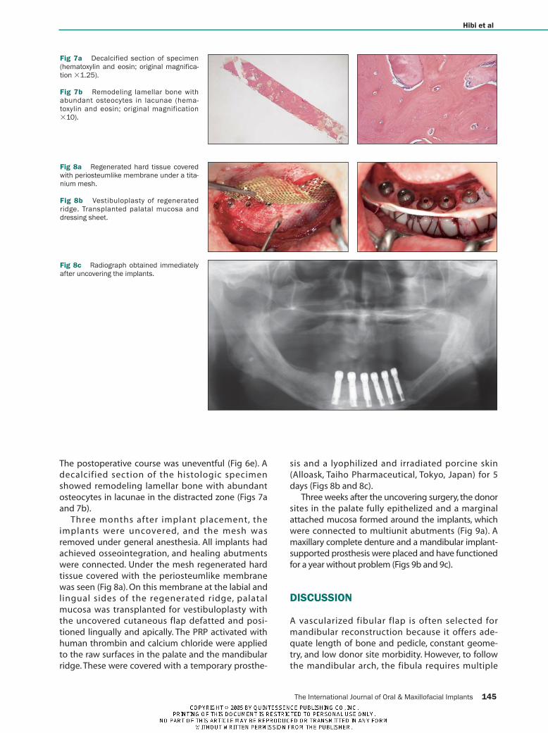
The postoperative course was uneventful (Fig 6e). Adecalcified section of the histologic specimenshowed remodeling lamellar bone with abundantosteocytes in lacunae in the distracted zone (Figs 7aand 7b).
Three months after implant placement, theimplants were uncovered, and the mesh wasremoved under general anesthesia. All implants hadachieved osseointegration, and healing abutmentswere connected. Under the mesh regenerated hardtissue covered with the periosteumlike membranewas seen (Fig 8a). On this membrane at the labial andlingual sides of the regenerated ridge, palatalmucosa was transplanted for vestibuloplasty withthe uncovered cutaneous flap defatted and posi-tioned lingually and apically. The PRP activated withhuman thrombin and calcium chloride were appliedto the raw surfaces in the palate and the mandibularridge. These were covered with a temporary prosthe-
sis and a lyophilized and irradiated porcine skin(Alloask, Taiho Pharmaceutical, Tokyo, Japan) for 5days (Figs 8b and 8c).
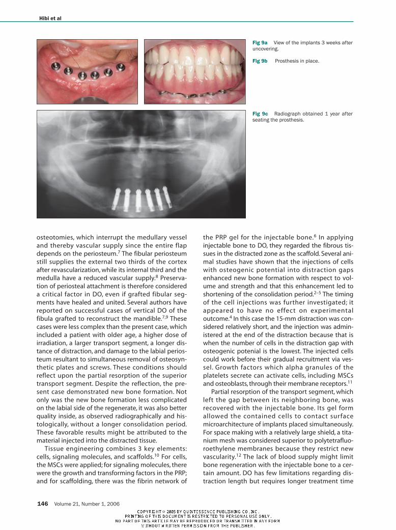
Three weeks after the uncovering surgery, the donorsites in the palate fully epithelized and a marginalattached mucosa formed around the implants, whichwere connected to multiunit abutments (Fig 9a). Amaxillary complete denture and a mandibular implant-supported prosthesis were placed and have functionedfor a year without problem (Figs 9b and 9c).
DISCUSSION
A vascularized fibular flap is often selected formandibular reconstruction because it offers ade-quate length of bone and pedicle, constant geome-try, and low donor site morbidity. However, to followthe mandibular arch, the fibula requires multiple
Fig 7a Decalcified section of specimen(hematoxylin and eosin; original magnifica-tion �1.25).
Fig 7b Remodeling lamellar bone withabundant osteocytes in lacunae (hema-toxylin and eosin; original magnification�10).
Fig 8a Regenerated hard tissue coveredwith periosteumlike membrane under a tita-nium mesh.
Fig 8b Vestibuloplasty of regeneratedridge. Transplanted palatal mucosa anddressing sheet.
Fig 8c Radiograph obtained immediatelyafter uncovering the implants.
Hibi.qxd 1/31/06 1:53 PM Page 145
osteotomies, which interrupt the medullary vesseland thereby vascular supply since the entire flapdepends on the periosteum.7 The fibular periosteumstill supplies the external two thirds of the cortexafter revascularization, while its internal third and themedulla have a reduced vascular supply.8 Preserva-tion of periosteal attachment is therefore considereda critical factor in DO, even if grafted fibular seg-ments have healed and united. Several authors havereported on successful cases of vertical DO of thefibula grafted to reconstruct the mandible.7,9 Thesecases were less complex than the present case, whichincluded a patient with older age, a higher dose ofirradiation, a larger transport segment, a longer dis-tance of distraction, and damage to the labial perios-teum resultant to simultaneous removal of osteosyn-thetic plates and screws. These conditions shouldreflect upon the partial resorption of the superiortransport segment. Despite the reflection, the pre-sent case demonstrated new bone formation. Notonly was the new bone formation less complicatedon the labial side of the regenerate, it was also betterquality inside, as observed radiographically and his-tologically, without a longer consolidation period.These favorable results might be attributed to thematerial injected into the distracted tissue.
Tissue engineering combines 3 key elements:cells, signaling molecules, and scaffolds.10 For cells,the MSCs were applied; for signaling molecules, therewere the growth and transforming factors in the PRP;and for scaffolding, there was the fibrin network of
the PRP gel for the injectable bone.6 In applyinginjectable bone to DO, they regarded the fibrous tis-sues in the distracted zone as the scaffold. Several ani-mal studies have shown that the injections of cellswith osteogenic potential into distraction gapsenhanced new bone formation with respect to vol-ume and strength and that this enhancement led toshortening of the consolidation period.2–5 The timingof the cell injections was further investigated; itappeared to have no effect on experimentaloutcome.4 In this case the 15-mm distraction was con-sidered relatively short, and the injection was admin-istered at the end of the distraction because that iswhen the number of cells in the distraction gap withosteogenic potenial is the lowest. The injected cellscould work before their gradual recruitment via ves-sel. Growth factors which alpha granules of theplatelets secrete can activate cells, including MSCsand osteoblasts, through their membrane receptors.11
Partial resorption of the transport segment, whichleft the gap between its neighboring bone, wasrecovered with the injectable bone. Its gel formallowed the contained cells to contact surfacemicroarchitecture of implants placed simultaneously.For space making with a relatively large shield, a tita-nium mesh was considered superior to polytetrafluo-roethylene membranes because they restrict newvascularity.12 The lack of blood supply might limitbone regeneration with the injectable bone to a cer-tain amount. DO has few limitations regarding dis-traction length but requires longer treatment time
146 Volume 21, Number 1, 2006
Hibi et al
Fig 9a View of the implants 3 weeks afteruncovering.
Fig 9b Prosthesis in place.
Fig 9c Radiograph obtained 1 year afterseating the prosthesis.
Hibi.qxd 1/31/06 1:53 PM Page 146
than grafting. These innovative methods in combina-tion can allow more effective bone regeneration foradequate implant placement.
ACKNOWLEDGMENTS
This work was partly supported by a grant from the Ministry ofEducation, Culture, Sports, Science and Technology, Japan. Theauthors wish to thank Mr Tomio Kuno, JOEL, Nagoya, Japan forhis excellent laboratory work.
REFERENCES
1. Swennen G, Dempf R, Schliephake H. Cranio-facial distractionosteogenesis: A review of the literature. Part 2: experimentalstudies. Int J Oral Maxillofac Surg 2002;31:123–135.
2. Takushima A, Kitano Y, Harii K. Osteogenic potential of cul-tured periosteal cells in a distracted gap in rabbits. J Surg Res1998;78:68–77.
3. Tsubota S, Tsuchiya H, Shinokawa Y, Tomita K, Minato H.Trans-plantation of osteoblast-like cells to the distracted callus inrabbits. J Bone Joint Surg Br 1999;81-B:125–129.
4. Richards M, Huibregtse BA, Caplan AI, Goulet JA, Goldstein SA.Marrow-derived progenitor cell injections enhance new boneformation during distraction. J Orthop Res 1999;17:900–908.
5. Takamine Y, Tsuchiya H, Kitakoji T, et al. Distraction osteogene-sis enhanced by osteoblastlike cells and collagen gel. ClinOrthop 2002;399:240–246.
6. Yamada Y, Ueda M, Hibi H, Nagasaka T.Translational reseachfor injectable tissue-engineered bone regeneration usingmesenchymal stem cells and platelet-rich plasma: From basicresearch to clinical application. Cell Transplant2004;13:343–355.
7. Nocini PF, Wangerin K, Albanese M, Kretschmer W, Cortelazzi R.Vertical distraction of a free vascularized fibula flap in a recon-structed hemimandible: Case report. J Craniomaxillofac Surg2000;28:20–24.
8. Bähr W. Blood supply of small fibula segments: An experimen-tal study on human cadavers. J Craniomaxillofac Surg1998;26:148–152.
9. Klesper B, Lazar F, Sießegger M, Hidding J, Zöller JE. Verticaldistraction osteogenesis of fibula transplants for mandibularreconstruction—A preliminary study. J Craniomaxillofac Surg2002;30:280–285.
10. Lynch SE, Genco RJ, Marx RE (eds).Tissue Engineering: Applica-tions in Maxillofacial Surgery and Periodontics. Chicago: Quin-tessence, 1999:3–286.
11. Marx RE. Platelet-rich plasma: Evidence to support its use. JOral Maxillofac Surg 2004;62:489–496.
12. von Arx T, Hardt N, Wallkamm B.The TIME technique: A newmethod for localized alveolar ridge augmentation prior toplacement of dental implants. Int J Oral Maxillofac Implants1996;11:387–394.
The International Journal of Oral & Maxillofacial Implants 147
Hibi et al
SABRA DENTAL PRODUCTS - Eastern US Dealer1-800-888-4435 - Fax 631-206-9140 - www.sabradental.com
SAFE AND EASYThe very first
comprehensive
system to perform
safe maxillary sinus
floor elevation
with minimal,
controlled
invasiveness.
PHYSIOLINE PRESENTS THE ORIGINAL SINUS LIFTING SYSTEM KIT
Dr. A. Giordano
DDS MS
RAPID APPROACH ANDRELIABLE RESULTS
www.physioline.it
Hibi.qxd 1/31/06 1:53 PM Page 147