heterologous immunoassay for screening macrolide antibiotics residues in milk based on the...
TRANSCRIPT
This article was downloaded by: [The University Of Melbourne Libraries]On: 13 October 2014, At: 03:28Publisher: Taylor & FrancisInforma Ltd Registered in England and Wales Registered Number: 1072954 Registeredoffice: Mortimer House, 37-41 Mortimer Street, London W1T 3JH, UK
Food and Agricultural ImmunologyPublication details, including instructions for authors andsubscription information:http://www.tandfonline.com/loi/cfai20
Heterologous immunoassay forscreening macrolide antibioticsresidues in milk based on themonoclonal antibody of tylosinJian Kang Zhangab, Yong Hua Qic, Ju Xiang Liuab & Jian Ping Wangab
a College of Veterinary Medicine, Agricultural University of Hebei,Baoding, Hebei 071000, Chinab Hebei Engineering and Technology Research Center of VeterinaryBiological Products, Agricultural University of Hebei, Baoding,Hebei 071000, Chinac College of Animal Science, Henan Institute of Science andTechnology, Xinxiang, Henan 453003, ChinaPublished online: 17 Jul 2012.
To cite this article: Jian Kang Zhang, Yong Hua Qi, Ju Xiang Liu & Jian Ping Wang (2013)Heterologous immunoassay for screening macrolide antibiotics residues in milk based onthe monoclonal antibody of tylosin, Food and Agricultural Immunology, 24:4, 419-431, DOI:10.1080/09540105.2012.705820
To link to this article: http://dx.doi.org/10.1080/09540105.2012.705820
PLEASE SCROLL DOWN FOR ARTICLE
Taylor & Francis makes every effort to ensure the accuracy of all the information (the“Content”) contained in the publications on our platform. However, Taylor & Francis,our agents, and our licensors make no representations or warranties whatsoever as tothe accuracy, completeness, or suitability for any purpose of the Content. Any opinionsand views expressed in this publication are the opinions and views of the authors,and are not the views of or endorsed by Taylor & Francis. The accuracy of the Contentshould not be relied upon and should be independently verified with primary sourcesof information. Taylor and Francis shall not be liable for any losses, actions, claims,proceedings, demands, costs, expenses, damages, and other liabilities whatsoever orhowsoever caused arising directly or indirectly in connection with, in relation to or arisingout of the use of the Content.
This article may be used for research, teaching, and private study purposes. Anysubstantial or systematic reproduction, redistribution, reselling, loan, sub-licensing,
systematic supply, or distribution in any form to anyone is expressly forbidden. Terms &Conditions of access and use can be found at http://www.tandfonline.com/page/terms-and-conditions
Dow
nloa
ded
by [
The
Uni
vers
ity O
f M
elbo
urne
Lib
rari
es]
at 0
3:28
13
Oct
ober
201
4
Heterologous immunoassay for screening macrolide antibiotics residuesin milk based on the monoclonal antibody of tylosin
Jian Kang Zhanga,b, Yong Hua Qic, Ju Xiang Liua,b and Jian Ping Wanga,b*
aCollege of Veterinary Medicine, Agricultural University of Hebei, Baoding, Hebei 071000,China; bHebei Engineering and Technology Research Center of Veterinary Biological Products,Agricultural University of Hebei, Baoding, Hebei 071000, China; cCollege of Animal Science,Henan Institute of Science and Technology, Xinxiang, Henan 453003, China
(Received 7 March 2012; final version received 20 June 2012)
In this study, tylosin (TYL) was derivatized with 4-aminophenylacetic acid tosynthesise a new hapten and the hapten was used to produce the monoclonalantibody. The obtained antibody simultaneously recognised TYL, tilmicosin,acetylisovaleryltylosin and the metabolite of TYL (desmycosin) with cross-reactivities of 100%, 62%, 97% and 93%, respectively. After evaluation of twocoating antigens, a heterologous competitive indirect enzyme linked immunosor-bent assay was developed to determine the four analytes in milk simultaneously.The limits of detection for the four analytes were in the range of 1.5�3.1 ng mL�1.The recoveries from fortified milk were in the range of 76.3�97.4% with coefficientsof variation of 5.3�15.7%.
Keywords: tylosin; monoclonal antibody; heterologous ELISA; milk
1. Introduction
Macrolide antibiotics (MACs) are a class of drugs highly active against Gram-positive
bacterial and Mycroplasma species. Therefore, MACs are widely used to treat various
diseases in swine, cattle, sheep and poultry (Christodoulopoulos, Warnick, Papaioan-
nou, & Fthenakis, 2002; Fajt et al., 2003; Laven and Andrews, 1991; Shryock, Staples,
& DeRosa, 2002; Zhang et al., 2004). The commonly used MACs include tylosin
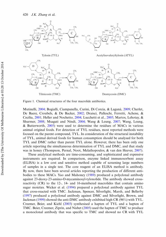
(TYL), tilmicosin (TMC) and acetylisovaleryltylosin (ATYL; Figure 1). TYL is a
traditional MAC that is generated by Streptomyces fradiae. ATYL and TMC are both
semi-synthetic drugs derived from TYL. TYL is unstable and it can be degraded in
sugar syrup (Kochansky, Knox, & Shimanuki, 1999), honey (Kochansky, 2004) and
acidic media (Paesen, Cypers, Pauwels, Roets, & Hoogmartens, 1995) to yield its
predominant metabolite, desmycosin (DMC), also referred to as tylosin B (Figure 1).
The wide use of MACs in farm animals may produce residues in food of
animal origin and induce the resistance of bacterial strains to antimicrobials in
human use. For protection of consumer health, it is very important to monitor the
residues of these MACs in foods of animal origin. In the last decade, high-
performance liquid chromatography (Prats, Francesch, Arboix, & Perez, 2001) and
liquid chromatography�mass spectrometry (Benetti, Dainese, Biancotto, Piro, &
*Corresponding author. Email: [email protected] Kang Zhang and Yong Hua Qi contributed equally to this work.
Food and Agricultural Immunology, 2013
Vol. 24, No. 4, 419�431, http://dx.doi.org/10.1080/09540105.2012.705820
# 2012 Taylor & Francis
Dow
nloa
ded
by [
The
Uni
vers
ity O
f M
elbo
urne
Lib
rari
es]
at 0
3:28
13
Oct
ober
201
4
Mutinelli, 2004; Bogialli, Ciampanella, Curini, Di Corcia, & Lagana, 2009; Cherlet,
De Baere, Croubels, & De Backer, 2002; Draisci, Palleschi, Ferretti, Achene, &
Cecilia, 2001; Heller and Nochetto, 2004; Lucchetti et al., 2005; Martos, Lehotay, &
Shurmer, 2008; Msagati and Nindi, 2004; Wang & Leung, 2007; Wang, Leung,
& Butterworth, 2005) were used to determine the residues of MACs in various
animal original foods. For detection of TYL residues, most reported methods were
focused on the parent compound, TYL. In consideration of the structural instability
of TYL, animal derived foods for human consumption should be analysed for both
TYL and DMC rather than parent TYL alone. However, there has been only one
article reporting the simultaneous determination of TYL and DMC, and that study
was in honey (Thompson, Pernal, Noot, Melathopoulos, & van den Heever, 2007).
These analytical methods are time-consuming, and sophisticated and expensive
instruments are required. In comparison, enzyme linked immunosorbent assay
(ELISA) is a low cost and sensitive method capable of screening large numbers
of samples in a single test. The core reagent of an ELISA method is antibody.
By now, there have been several articles reporting the production of different anti-
bodies to these MACs. Yao and Mahoney (1989) produced a polyclonal antibody
against 23-deoxy-23-amino-O-mycaminosyl-tylonolide. The antibody showed cross-
reactivity (CR) to the 12-, 14- and 16-membered macrolides that contain amino
sugar moieties. Wicker et al. (1994) prepared a polyclonal antibody against TYL
that cross-reacted with TMC. Jackman, Spencer, Silverlight, Marsh, and Bellerby
(1997) produced a polyclonal antibody against DMC and Silverlight, Brown, and
Jackman (1999) showed the anti-DMC antibody exhibited high CR (96%) with TYL.
Creemer, Beier, and Kiehl (2003) synthesised a hapten of TYL and a hapten of
TMC. Beier, Creemer, Ziprin, and Nisbet (2005) used the hapten of TMC to produce
a monoclonal antibody that was specific to TMC and showed no CR with TYL
Figure 1. Chemical structures of the four macrolide antibiotics.
420 J.K. Zhang et al.
Dow
nloa
ded
by [
The
Uni
vers
ity O
f M
elbo
urne
Lib
rari
es]
at 0
3:28
13
Oct
ober
201
4
and other MACs that do not contain the structure of 3,5-dimethylpiperidine (the
D ring in TMC molecule, Figure 1). The authors did not determine the antibody
reactivity to ATYL and DMC. Peng et al. (2012) produced a monoclonal antibody
against TYL that showed CR to TMC.Recently, we have produced a monoclonal antibody against TYL that showed
the following cross-reactivities: TYL (100%), ATYL (91%), DMC (76%) and TMC
(49%) (Zhang, Liu, Wang, Chai, & Wang, 2012). Therefore, TYL can be regarded as
a generic hapten of the four analytes to generate a broad-specific antibody. In this
study, TYL was used to synthesise a new hapten with the aims to enhance the anti-
body CR and to develop a multi-determination immunoassay for the four analytes.
2. Materials and methods
2.1. Reagents and chemicals
Tylosin tartrate, ATYL, TMC and DMC, bovine serum albumin (BSA), Ovalbumin
(OA), and Freund’s adjuvant were all from Sigma-Aldrich (St. Louis, MO, USA).
3, 3?, 5, 5?-tetramethylbenzidine (TMB) was purchased from Serva (Heidelberg,Germany). Other chemical reagents were all analytical grade from Beijing Chemical
Company (Beijing, China). The stock solutions of the four analytes were prepared by
dissolving each compound in methanol to obtain a concentration of 100 mg mL�1.
These solutions were stored at �20 8C in amber glass bottles. Working solutions
of the four drugs with series concentrations were prepared by dilution of the
stock solutions with PBS (1, 2, 5, 10, 20, 50, 100 and 200 ng mL�1). PBS (pH 7.2)
was prepared by dissolving 0.2 g KH2PO4, 0.2 g KCl, 1.15 g Na2HPO4, and 8.0 g
NaCl in 1000 mL water. Coating buffer was prepared with sodium carbonate andsodium hydrocarbonate (0.1 M, pH 9.6). Washing buffer was PBS containing 0.05%
Tween (PBST). Substrate buffer was prepared with sodium hydrogen phosphate
and citrate (0.1 M, pH 5.5). The substrate system was prepared by adding 200 mL
1% (w/v) TMB in DMSO (dimethyl sulfoxide) and 64 mL 0.75% (w/v) H2O2 to
20 mL substrate buffer.
2.2. Synthesis of the hapten
The hapten synthesis route is shown in Figure 2. About 470 mg tylosin tartrate
(0.5 mmol) and 75 mg 4-aminophenylacetic acid (0.5 mmol) were added to a mixture
of 20 mL of water and 0.5 mL of acetic acid. The mixture was refluxed under heating
until the solution turned yellow. Then the solvent was dried in drying box at 40 8C and
the dry residue was dissolved in 10 mL of ethanol. After 10 mL of ethyl acetatewas added into the ethanol solution, some lemon yellow sediment appeared. The
mixture was filtered under vacuum and the residue was washed with 50 mL of water,
and subsequently dried to yield the hapten PTYL (melt point of 220 8C;
IR (KBr) Vmax 3426, 3100, 2971, 2933, 1704, 1594, 1375, 1155, 1266, 713, 680 cm�1).
2.3. Preparation of the conjugates
The conjugates were prepared as shown in Figure 2. About 4 mL of N,N-
dimethylformamide dissolving 53 mg hapten and 25 mL triethylamine were added
Food and Agricultural Immunology 421
Dow
nloa
ded
by [
The
Uni
vers
ity O
f M
elbo
urne
Lib
rari
es]
at 0
3:28
13
Oct
ober
201
4
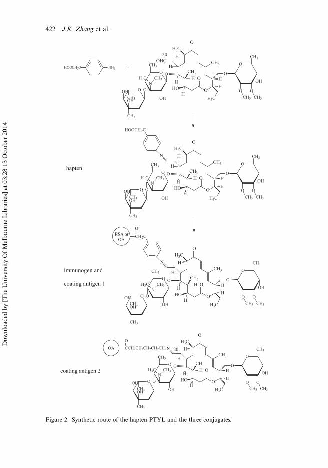
Figure 2. Synthetic route of the hapten PTYL and the three conjugates.
422 J.K. Zhang et al.
Dow
nloa
ded
by [
The
Uni
vers
ity O
f M
elbo
urne
Lib
rari
es]
at 0
3:28
13
Oct
ober
201
4
into a glass jar. Then 20 mL of isobutyl chloroformate was added and the mixture
was stirred for 60 min at 4 8C. Then, the mixture was added dropwise into 2 mL
of sodium bicarbonate solution containing 74 mg BSA or 30 mg OA and stirred
for 12 h at 4 8C. The resulting immunogen (PTYL-BSA) and coating antigen(PTYL-OA) were dialysed against three changes of PBS for 3 days and stored
at �20 8C until used. TYL, PTYL, BSA and the conjugates were all scanned on a
UV-Vis spectrophotometer to verify the conjugation. The hapten/protein coupling
ratios were determined according to the previous 2,4,6-trinitrobenzene sulphonic
acid method (Sashidhar, Capoor, & Ramana, 1994).
2.4. Production of the monoclonal antibody
Six 8-week-old female BALB/c mice were immunised subcutaneously with an
emulsion of the immunogen (50 mg protein per mouse) in Freund’s complete
adjuvant. Beginning two weeks later, mice were boosted at 2-week intervals. The
serum of each mouse was collected and the antibody titre was monitored. The spleenfrom the mouse with the highest titre after six boosters was removed and the
splenocytes were fused with SP2/O myeloma cells and cultured in 96-well plates.
Following a first screening by indirect ELISA, the positive hybridomas were
rescreened using the competitive indirect ELISA described later with TYL as the
competitor. Hybridomas producing the specific monoclonal antibody to TYL were
sub-cloned twice by the limiting dilution method and the sub-cloned hybridoma
cells were collected, centrifuged and frozen in liquid nitrogen. The ascites from
hybridoma-induced mice were purified using saturated ammonium sulphate pre-cipitation and used for development of the competitive indirect ELISA.
2.5. Competitive indirect ELISA
In this study, coating antigen PTYL-OA (coating antigen 1, Figure 2) and a
coating antigen previously prepared in our laboratory (coating antigen 2, Figure 2)
(Zhang et al., 2012) were used to develop the homologous and heterologous ELISA.
The optimal dilutions of coating antigen and antibody were determined by using the
checkerboard procedure, in which the well with an absorbance of 1.0 was defined as
the optimal dilutions of the coating antigen and the antibody. After that, each well of
a microtitre plate was coated with 100 mL of coating antigen, incubated overnight at
4 8C, and then blocked with 1% foetal calf serum. The plate was washed three timeswith PBST, and then, 50 mL of the optimal antibody dilution and 50 mL of TYL
standard with series concentrations were added to the wells (in triplicate) for
incubation for 1 h at 37 8C. The plate was washed as aforementioned. About 100 mL
of horseradish peroxidase labelled goat anti-mouse IgG was added before incubation
for 30 min at 37 8C. After washes, 100 mL of TMB substrate system was added prior
to 15 min incubation at 37 8C. Finally, the reaction was stopped by the addition of 50
mL of 2 M H2SO4, and the plate was read on an ELISA plate reader at 450 nm to
obtain the absorbance (B) of each well.The other three analytes shown in Figure 1 and several other drugs (erythromycin,
spiramycin and avermectin) were all determined by the ELISA. The limits of
detection (LOD) for these analytes were defined as the concentrations showing
10% of inhibition, respectively. The competitive inhibition curves were developed
Food and Agricultural Immunology 423
Dow
nloa
ded
by [
The
Uni
vers
ity O
f M
elbo
urne
Lib
rari
es]
at 0
3:28
13
Oct
ober
201
4
by plotting the B/B0 values (mean absorbance of the standards divided by the
mean absorbance of zero standards) verse the concentrations (Log C). The CR
among these competitors was calculated from the half-maximal inhibition concen-
tration (IC50) as follows: CR (%)�100�IC50 TYL/IC50 competitor.
2.6. Sample preparation
The extraction of MACs from milk sample was modified from the previous reports
(Bogialli et al., 2009; Wang & Leung, 2007). A milk sample (5 mL) and 30 mL of
acetonitrile were added into a 50 mL polypropylene centrifuge tube and the tube
was shaken vigorously on a variable speed reciprocal shaker for 10 min. Themixture was centrifuged at 10,000 rpm for 10 min and the acetonitrile phase was
collected and evaporated to dryness. The dry residue was dissolved in 5 mL of PBS
and filtered through a 0.45 mm Millipore filter for ELISA analysis.
Blank milk samples were obtained from several controlled cows. In order to
evaluate the matrix interference, matrix-matched standards prepared with the
extracts of blank milk sample were used to develop the matrix-matched competitive
curves. The accuracy was evaluated by determination of the recoveries from the
four analytes fortified blank milk at concentrations of 20, 50, 100 ng mL�1. A totalof 35 unknown milk samples from China (20 commercial packaged milk samples
from several supermarkets and 15 raw milk samples from several dairy farms)
were analysed by the developed ELISA method.
3. Results and discussions
3.1. Hapten and immunogen
As shown in Figure 1, the three MAC drugs and DMC all contain a 16-atom
macrocyclic lactone ring (A ring), a 5-O-mycaminosyl ring (B ring), and a
neutral sugar (C ring). The antibody specific to the three rings should recognise
the four analytes simultaneously. In a previous report, B ring and D ring
(3,5-dimethylpiperidin at C20 position in the molecule of TMC) in the hapten of
TMC were presented to the immune system and the resulting antibody only
recognised TMC (Beier et al., 2005). This result is because among the four analytes
only TMC contains the D ring (Figure 1). In other reports, A ring and C ring in themolecule of DMC were presented to the immune system, resulting in an antibody
that simultaneously recognised DMC, TYL and TMC (Jackman et al., 1997;
Silverlight et al., 1999), analytes that contain the two rings. In our recent study, TYL
was derivatized with 6-aminohexanoic acid linker at the C20 position to synthesise
the hapten. The immunogen had A ring and C ring spaced far from the carrier
(similar to the illustration of coating antigen 2, Figure 2) and resulted in an antibody
that simultaneously recognised TYL, ATYL, DMC, and TMC (Zhang et al., 2012).
In the present study, a new hapten of TYL was synthesised by derivatizationof TYL with 4-aminophenylacetic acid utilising the C20 aldehyde group in TYL
molecule (Figure 2). This was equivalent to introduction of a phenylacetic acid
at C20 position and the free carboxyl group was used to couple with the carrier.
This hapten contained the common structures of the four analytes (A ring, B ring
and C ring) to elicit antibodies that recognise all four analytes.
424 J.K. Zhang et al.
Dow
nloa
ded
by [
The
Uni
vers
ity O
f M
elbo
urne
Lib
rari
es]
at 0
3:28
13
Oct
ober
201
4
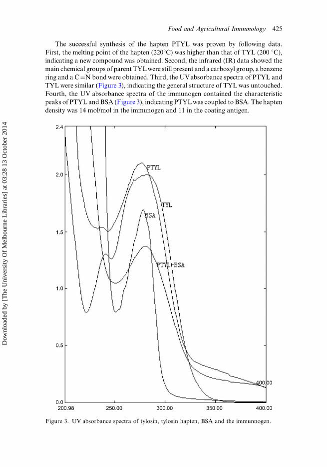
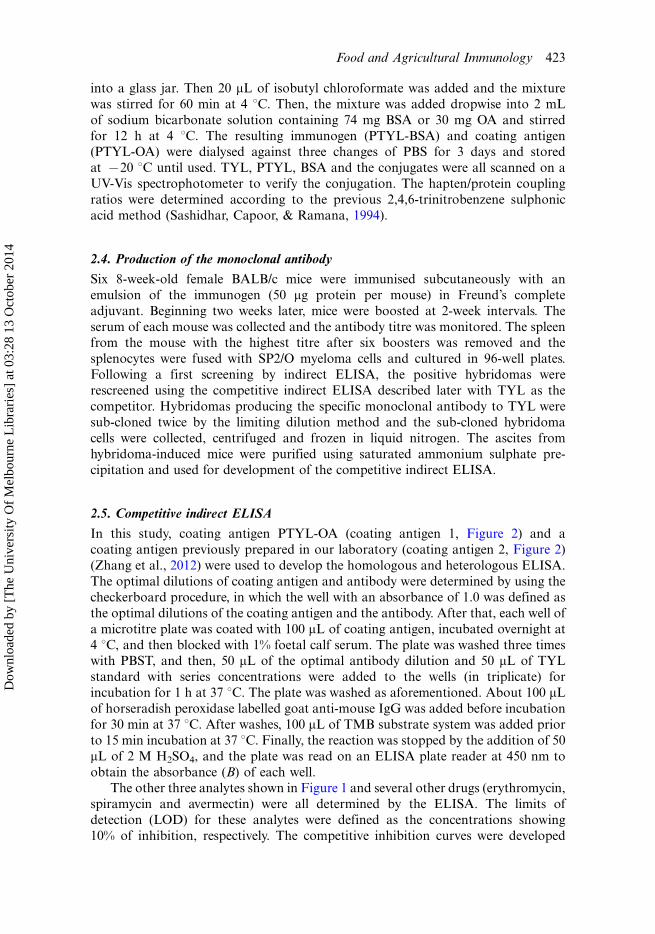
The successful synthesis of the hapten PTYL was proven by following data.
First, the melting point of the hapten (2208C) was higher than that of TYL (200 8C),
indicating a new compound was obtained. Second, the infrared (IR) data showed the
main chemical groups of parent TYLwere still present and a carboxyl group, a benzene
ring and a C�N bond were obtained. Third, the UV absorbance spectra of PTYL and
TYL were similar (Figure 3), indicating the general structure of TYL was untouched.
Fourth, the UV absorbance spectra of the immunogen contained the characteristic
peaks of PTYL and BSA (Figure 3), indicating PTYLwas coupled to BSA. The hapten
density was 14 mol/mol in the immunogen and 11 in the coating antigen.
Figure 3. UV absorbance spectra of tylosin, tylosin hapten, BSA and the immunnogen.
Food and Agricultural Immunology 425
Dow
nloa
ded
by [
The
Uni
vers
ity O
f M
elbo
urne
Lib
rari
es]
at 0
3:28
13
Oct
ober
201
4
3.2. Antibody performance
Six hybridomas producing specific monoclonal antibody to TYL were obtained
and the antibodies were named TY01, TY02, TY07, TY13, TY25 and TY56. Their
IC50 and CRs to the four analytes are shown in Table 1 (coating antigen 1). Antibody
TY01 and TY13 showed high specificity for TYL and showed low CRs to ATYL
(6.4% and 8.1%, respectively) and negligible CRs to TMC and DMC (B 2%). This
pattern was possibly because the two antibodies mainly bind the E ring in the
molecule of TYL (Figure 1). The four other antibodies simultaneously recognised
the four analytes. Antibody TY56 showed the best performance with IC50 in the
range of 14.2�22.9 ng mL�1 and CRs in the range of 62�100% (Table 1). Based
on these results, we speculated that the four antibodies mainly bind A ring and
C ring, because they showed negligible cross-reactivity (CRsB1%) to the MACs that
do not contain the two rings (erythromycin, spiramycin and avermectin, data not
shown). The CRs of these antibodies to the four analytes were better than those
of the previously reported antibodies (Beier et al., 2005; Jackman et al., 1997; Peng
et al., 2012; Silverlight et al., 1999; Wicker et al., 1994; Yao and Mahoney, 1989).
The CRs of the four antibodies to ATYL and DMC (48�97%) were similar to our
previous anti-TYL antibody (37�94%), but the CRs to TMC (29�62%) were higher
than that antibody (16�49%) (Zhang et al., 2012). This was because of the different
molecular structure of the two haptens. In the molecule of the previously described
TYL hapten, there is a simple straight chain at C20 position and the structure at
this position is different from that in TMC molecule (Figures 1 and 2), so the CRs
were low. In the new hapten, PTYL, there is a benzene ring at C20 position and
the general structure near this position is similar to that in the TMC molecule.
The general structure of TMC could be regarded as a part of PTYL, so the CRs
to TMC were high. Therefore, the new hapten of TYL improved the antibody
selectivity, resulting in the antibody that could be used for multi-analyte immu-
noassay of the four analytes.
Table 1. Performances of the six antibodies for the four analytes with different coating
antigens.
TY01 TY13 TY02 TY07 TY25 TY56
IC50a CRb IC50 CR IC50 CR IC50 CR IC50 CR IC50 CR LODa
Homologous format (coating antigen 1)
TYL 13 100 21 100 9.6 100 38.0 100 19.5 100 14.2 100 2.1
ATYL 162 8.1 328 6.4 15.2 63 48.7 78 20.7 94 14.6 97 2.6
DMC �1000 B1.2 �1000 B2 21.3 48 66.5 57 21.9 89 15.3 93 3.0
TMC �1000 B1.2 �1000 B2 27.4 35 131 29 31.9 61 22.9 62 5.4
Heterologous format (coating antigen 2)
TYL NEc NE NE NE 8.4 100 27.3 100 15.0 100 12.6 100 1.8
ATYL NE NE NE NE 11.8 71 33.2 82 15.8 95 11.9 106 1.5
DMC NE NE NE NE 17.0 49 43.3 63 16.5 91 13.4 94 2.7
TMC NE NE NE NE 20.5 41 71.8 38 27.7 54 18.8 67 3.1
aThe unit of IC50 and LOD is ng mL�1.bThe CR is expressed as %.cNE means ‘not evaluated’.
426 J.K. Zhang et al.
Dow
nloa
ded
by [
The
Uni
vers
ity O
f M
elbo
urne
Lib
rari
es]
at 0
3:28
13
Oct
ober
201
4
3.3. ELISA method
Nowadays, heterology in the coating antigen has been commonly used to improve
the sensitivity and specificity of an immunoassay (Franek, Diblikova, Cernoch, Vass,
& Hruska, 2006). Therefore, the four antibodies (TY02, TY07, TY25 and TY56),
PTYL-OA (coating antigen 1) and the previously prepared coating antigen TYL-OA
(coating antigen 2, Figure 2) were arranged into homologous and heterologous
format to optimise the reagent combinations.The performances of these antibodies in homologous format were described
earlier. As shown in Table 1, the specificities and sensitivities of the four antibodies in
heterologous format (using coating antigen 2) were generally better than that in
homologous format (using coating antigen 1). For example, the CRs of antibody
TY56 to the four analytes were in the range of 67�106% and IC50 were in the range
of 11.9�18.8 ng mL�1. In addition, the CRs differences among these competitors in
heterologous format were lower than that in homologous format. In heterologous
format, the coating antigen 2 (containing a long straight chain) eliminated theinfluence of the antibody from the spacer arm in PTYL-BSA to show low
competitive binding to the antibodies; thus increases the antibody binding for the
competitors to achieve broad specificity and high sensitivity. Among these reagent
combinations, antibody TY56 and coating antigen 2 produced the highest sensitivity,
with LODs in the range of 1.5�3.1 ng mL�1 (Table 1). Therefore, this combination
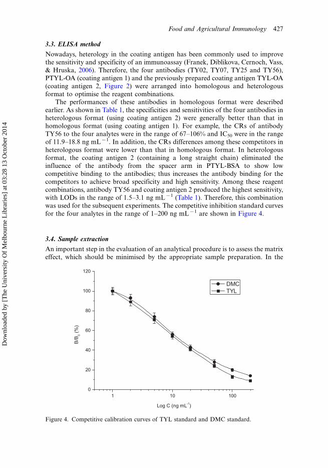
was used for the subsequent experiments. The competitive inhibition standard curves
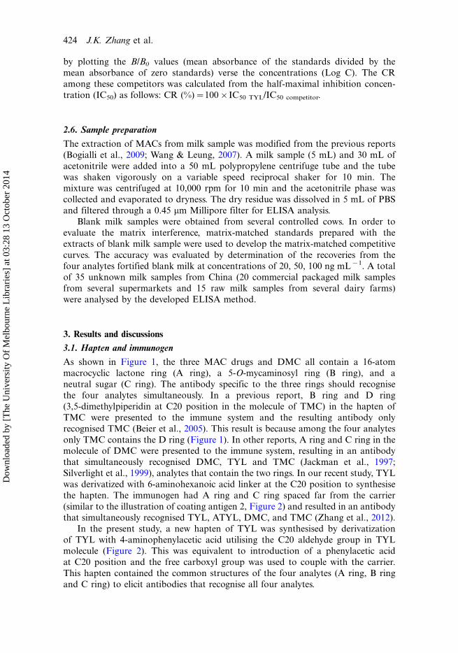
for the four analytes in the range of 1�200 ng mL�1 are shown in Figure 4.
3.4. Sample extraction
An important step in the evaluation of an analytical procedure is to assess the matrix
effect, which should be minimised by the appropriate sample preparation. In the
Figure 4. Competitive calibration curves of TYL standard and DMC standard.
Food and Agricultural Immunology 427
Dow
nloa
ded
by [
The
Uni
vers
ity O
f M
elbo
urne
Lib
rari
es]
at 0
3:28
13
Oct
ober
201
4
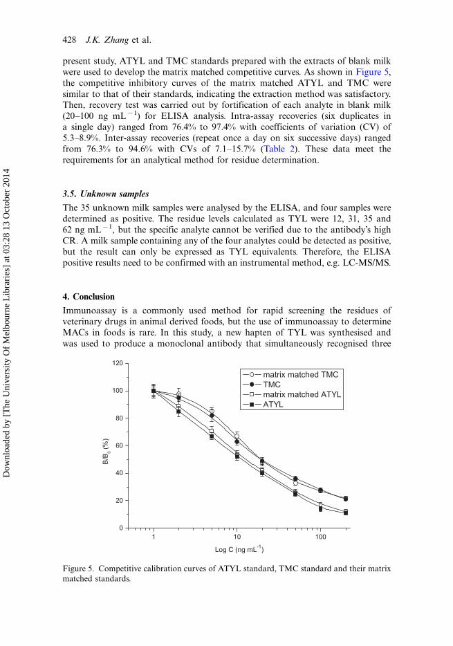
present study, ATYL and TMC standards prepared with the extracts of blank milk
were used to develop the matrix matched competitive curves. As shown in Figure 5,
the competitive inhibitory curves of the matrix matched ATYL and TMC were
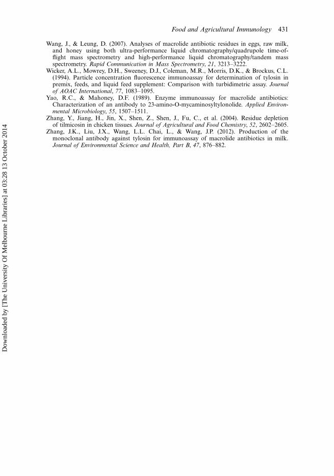
similar to that of their standards, indicating the extraction method was satisfactory.Then, recovery test was carried out by fortification of each analyte in blank milk
(20�100 ng mL�1) for ELISA analysis. Intra-assay recoveries (six duplicates in
a single day) ranged from 76.4% to 97.4% with coefficients of variation (CV) of
5.3�8.9%. Inter-assay recoveries (repeat once a day on six successive days) ranged
from 76.3% to 94.6% with CVs of 7.1�15.7% (Table 2). These data meet the
requirements for an analytical method for residue determination.
3.5. Unknown samples
The 35 unknown milk samples were analysed by the ELISA, and four samples were
determined as positive. The residue levels calculated as TYL were 12, 31, 35 and
62 ng mL�1, but the specific analyte cannot be verified due to the antibody’s high
CR. A milk sample containing any of the four analytes could be detected as positive,
but the result can only be expressed as TYL equivalents. Therefore, the ELISA
positive results need to be confirmed with an instrumental method, e.g. LC-MS/MS.
4. Conclusion
Immunoassay is a commonly used method for rapid screening the residues of
veterinary drugs in animal derived foods, but the use of immunoassay to determine
MACs in foods is rare. In this study, a new hapten of TYL was synthesised and
was used to produce a monoclonal antibody that simultaneously recognised three
Figure 5. Competitive calibration curves of ATYL standard, TMC standard and their matrix
matched standards.
428 J.K. Zhang et al.
Dow
nloa
ded
by [
The
Uni
vers
ity O
f M
elbo
urne
Lib
rari
es]
at 0
3:28
13
Oct
ober
201
4
MACs (TYL, ATYL, TMC) and DMC, a metabolite of TYL. A heterologous
competitive indirect ELISA was developed to detect the four analytes in milk
simultaneously. This method could be used as a practical tool for routine screening
of large numbers of milk samples, with positive results confirmed by instrumental
methods.
Acknowledgements
This study was financed by Hebei Scientific and Technological Project (11221001D).
References
Beier, R.C., Creemer, L.C., Ziprin, R.L., & Nisbet, D.J. (2005). Production and characteriza-tion of monoclonal antibodies against the antibiotic tilmicosin. Journal of Agricultural andFood Chemistry, 53, 9679�9688.
Benetti, C., Dainese, N., Biancotto, G., Piro, R., & Mutinelli, F. (2004). Unauthorisedantibiotic treatments in beekeeping: Development and validation of a method to quantifyand confirm tylosin residues in honey using liquid chromatography-tandem mass spectro-metric detection. Analytica Chimica Acta, 520, 87�92.
Bogialli, S., Ciampanella, C., Curini, R., Di Corcia, A., & Lagana, A. (2009). Developmentand validation of a rapid assay based on liquid chromatogaphy-tandem mass spectrometryfor determining macrolide antibiotic residues in eggs. Journal of Chromatography A, 1216,6810�6815.
Cherlet, M., De Baere, S., Croubels, S., & De Backer, P. (2002). Quantitation of tylosinin swine tissues by liquid chromatography combined with electrospray ionization massspectrometry. Analytica Chimica Acta, 473, 167�175.
Christodoulopoulos, G., Warnick, L.D., Papaioannou, N., & Fthenakis, G.C. (2002).Tilmicosin administration to young lambs with respiratory infection: Safety and efficacyconsiderations. Journal of Veterinary Pharmacology and Therapeutics, 25, 393�397.
Creemer, L.C., Beier, R.C., & Kiehl, D.E. (2003). Facile synthesis of tilmicosin and tylosinrelated haptens for use as protein conjugates. Jounal of Antibiotics, 56, 481�487.
Draisci, R., Palleschi, L., Ferretti, E., Achene, L., & Cecilia, A. (2001). Confirmatory methodfor macrolide residues in bovine tissues by micro-liquid chromatography-tandem massspectrometry. Journal of Chromatography A, 926, 97�104.
Table 2. Recoveries of the four drugs from blank fortified milk.
Intra-assay Inter-assay
Analyte Added (ng mL�1) Recovery (%) CV (%) Recovery (%) CV (%)
TYL 20 80.2 6.6 90.3 9.6
50 89.4 8.9 94.6 9.2
100 92.6 6.2 88.5 7.4
ATYL 20 97.4 7.3 86.2 12.3
50 91.0 7.6 92.4 11.4
100 93.5 8.0 89.0 8.9
TMC 20 84.7 5.4 78.9 7.1
50 82.9 5.9 76.3 8.5
100 81.0 8.7 83.9 15.7
DMC 20 76.4 8.2 83.1 10.6
50 84.1 5.3 77.2 14.3
100 92.1 7.8 91.3 9.1
Food and Agricultural Immunology 429
Dow
nloa
ded
by [
The
Uni
vers
ity O
f M
elbo
urne
Lib
rari
es]
at 0
3:28
13
Oct
ober
201
4
Fajt, V.R., Apley, M.D., Roth, J.A., Frank, D.E., Brogden, K.A., Skogerboe, T.L., et al.(2003). The effects of danofloxacin and tilmicosin on neutrophil function and lungconsolidation in beef heifer calves with induced Pasteurella (Mannheimia) haemolyticapneumonia. Journal of Veterinary Pharmacology and Therapeutics, 26, 173�179.
Franek, M., Diblikova, I., Cernoch, I., Vass, M., & Hruska, K. (2006). Broad-specificityimmunoassays for sulfonamide detection: Immunochemical strategy of generic antibodiesand competitors. Analytical Chemistry, 78, 1559�1567.
Heller, D.N., & Nochetto, C.B. (2004). Development of multiclass methods for drug residuesin eggs: Silica SPE cleanup and LC-MS/MS analysis of ionophore and macrolide residues.Journal of Agricultural and Food Chemistry, 52, 6848�6856.
Jackman, R., Spencer, Y.I., Silverlight, J.J., Marsh, S.A., & Bellerby, P.J. (1997). Developmentof antibodies to tilmicosin and their use in the immunolocalization of the antibiotic inporcine lung tissue. Journal of Agricultural and Food Chemistry, 20(Suppl. 1), 131�132.
Kochansky, J. (2004). Degradation of tylosin residues in honey. Jounal of Apicultural Research,43, 65�68.
Kochansky, J., Knox, D., & Shimanuki, H. (1999). Comparative stability of oxytetracyclineand tylosin in sugar syrup. Apidologie, 30, 321�325.
Laven, R., & Andrews, A.H. (1991). Long-acting antibiotic formulations in the treatmentof calf pneumonia: A comparative study of tilmicosin and oxytetracycline. VeterinaryRecord, 129, 109�111.
Lucchetti, D., Fabrizi, L., Esposito, A., Guandalini, E., Di Pasquale, M., & Coni, E. (2005).Simple confirmatory method for the determination of erythromycin residues in trout:A fast liquid-liquid extraction followed by liquid chromatography-tandem mass spectro-metry. Journal of Agricultural and Food Chemistry, 53, 9689�9694.
Martos, P.A., Lehotay, S.J., & Shurmer, B. (2008). Ultratrace analysis of nine macrolides,including tulathromycin A (Draxxin), in edible animal tissues with minicolumn liquidchromatography tandem mass spectrometry. Journal of Agricultural and Food Chemistry,56, 8844�8850.
Msagati, T.A.M., & Nindi, M.M. (2004). The use of supported liquid membranes in theextraction of macrolides in biomatrices. Microchimica Acta, 148, 199�214.
Paesen, J., Cypers, W., Pauwels, K., Roets, E., & Hoogmartens, J. (1995). Study of the stabilityof tylosin A in aqueous solutions. Journal of Pharmaceutical and Biomedical Analysis,13, 1153�1159.
Peng, D., Ye, S., Wang, Y., Chen, D., Tao, Y., Huang, L., et al. (2012). Development andvalidation of a competitive indirect enzyme-linked immunosorbent assay for the screeningof tylosin and tilmicosin in muscle, liver, milk, honey and eggs. Journal of Agricultural andFood Chemistry, 60, 44�51.
Prats, C., Francesch, R., Arboix, M., & Perez, B. (2001). Determination of tylosin residues indifferent animal tissues by high performance liquid chromatography. Journal of Chromato-graphy B, 766, 57�65.
Sashidhar, R.B., Capoor, A.K., & Ramana, D. (1994). Quantitation of amino groups usingamino acids as reference standards by trinitrobenzene sulfonic acid: A simple spectro-photometric method for the estimation of hapten to carrier protein ratio. Journal ofImmunological Methods, 167, 121�127.
Shryock, T.R., Staples, J.M., & DeRosa, D.C. (2002). Minimum inhibitory concentrationbreakpoints and disk diffusion inhibitory zone interpretive criteria for tilmicosin suscept-ibility testing against Pasteurella multocida and Actinobacillus pleuropneu-moniaeassociated with porcine respiratory disease. Journal of Veterinary Diagnostic Investigation,14, 389�395.
Silverlight, J.J., Brown, A.J., & Jackman, R. (1999). Antisera to tilmicosin for use in ELISAand for immunohistochemistry. Food and Agricultural Immunology, 11, 321�328.
Thompson, T.S., Pernal, S.F., Noot, D.K., Melathopoulos, A.P., & van den Heever, J.P.(2007). Degradation of incurred tylosin to desmycosin-Implications for residue analysisof honey. Analytica Chimica Acta, 586, 304�311.
Wang, J., Leung, D., & Butterworth, F. (2005). Determination of five macrolide antibioticresidues in eggs using liquid chromatography/electrospray ionization tandem mass spectro-metry. Journal of Agricultural and Food Chemistry, 53, 1857�1865.
430 J.K. Zhang et al.
Dow
nloa
ded
by [
The
Uni
vers
ity O
f M
elbo
urne
Lib
rari
es]
at 0
3:28
13
Oct
ober
201
4
Wang, J., & Leung, D. (2007). Analyses of macrolide antibiotic residues in eggs, raw milk,and honey using both ultra-performance liquid chromatography/quadrupole time-of-flight mass spectrometry and high-performance liquid chromatography/tandem massspectrometry. Rapid Communication in Mass Spectrometry, 21, 3213�3222.
Wicker, A.L., Mowrey, D.H., Sweeney, D.J., Coleman, M.R., Morris, D.K., & Brockus, C.L.(1994). Particle concentration fluorescence immunoassay for determination of tylosin inpremix, feeds, and liquid feed supplement: Comparison with turbidimetric assay. Journalof AOAC International, 77, 1083�1095.
Yao, R.C., & Mahoney, D.F. (1989). Enzyme immunoassay for macrolide antibiotics:Characterization of an antibody to 23-amino-O-mycaminosyltylonolide. Applied Environ-mental Microbiology, 55, 1507�1511.
Zhang, Y., Jiang, H., Jin, X., Shen, Z., Shen, J., Fu, C., et al. (2004). Residue depletionof tilmicosin in chicken tissues. Journal of Agricultural and Food Chemistry, 52, 2602�2605.
Zhang, J.K., Liu, J.X., Wang, L.L. Chai, L., & Wang, J.P. (2012). Production of themonoclonal antibody against tylosin for immunoassay of macrolide antibiotics in milk.Journal of Environmental Science and Health, Part B, 47, 876�882.
Food and Agricultural Immunology 431
Dow
nloa
ded
by [
The
Uni
vers
ity O
f M
elbo
urne
Lib
rari
es]
at 0
3:28
13
Oct
ober
201
4