hepatitis c virus triggers mitochondrial fission and attenuates apoptosis to promote viral...
TRANSCRIPT
Hepatitis C virus triggers mitochondrial fission andattenuates apoptosis to promote viral persistenceSeong-Jun Kima,1, Gulam H. Syeda,1, Mohsin Khana, Wei-Wei Chiua, Muhammad A. Sohailb, Robert G. Gishc,and Aleem Siddiquia,2
aDivision of Infectious Diseases and bDivision of Gastroenterology, Department of Medicine, University of California, San Diego, La Jolla, CA 92093;and cHepatitis B Foundation, Doylestown, PA 18901
Edited by Michael Gale, Jr., University of Washington School of Medicine, Seattle, WA, and accepted by the Editorial Board March 18, 2014 (received forreview November 8, 2013)
Mitochondrial dynamics is crucial for the regulation of cellhomeostasis. Our recent findings suggest that hepatitis C virus(HCV) promotes Parkin-mediated elimination of damaged mito-chondria (mitophagy). Here we show that HCV perturbs mito-chondrial dynamics by promoting mitochondrial fission followedby mitophagy, which attenuates HCV-induced apoptosis. HCVinfection stimulated expression of dynamin-related protein 1(Drp1) and its mitochondrial receptor, mitochondrial fission factor.HCV further induced the phosphorylation of Drp1 (Ser616) andcaused its subsequent translocation to the mitochondria, followedby mitophagy. Interference of HCV-induced mitochondrial fissionand mitophagy by Drp1 silencing suppressed HCV secretion, witha concomitant decrease in cellular glycolysis and ATP levels, as wellas enhanced innate immune signaling. More importantly, silencingDrp1 or Parkin caused significant increase in apoptotic signaling,evidenced by increased cytochrome C release from mitochondria,caspase 3 activity, and cleavage of poly(ADP-ribose) polymerase.These results suggest that HCV-induced mitochondrial fission andmitophagy serve to attenuate apoptosis and may contribute topersistent HCV infection.
HCV persistence | innate immunity | autophagy
Hepatitis C virus (HCV) infection often leads to chronichepatitis that can progress to fibrosis, cirrhosis, and hepa-
tocellular carcinoma (1). HCV is a hepatotropic, noncytopathic(2, 3), single-stranded, positive-sense RNA virus that replicatesits RNA genome on the endoplasmic reticulum (ER)-derivedmembranous structures (4, 5). HCV stimulates lipogenesis,leading to the accumulation of lipid droplets that facilitate virionassembly and maturation (5–8). HCV infection also inducesmitochondrial dysfunction via ER and oxidative stress thatresults in mitochondrial Ca2+ overload, collapse of mitochon-drial transmembrane potential (ΔΨm), elevated levels of reactiveoxygen species, and disruption of mitochondrial respiration (9–15). Liver tissues of patients with chronic hepatitis C frequentlyexhibit traits of mitochondrial injury such as swollen, ruptured,and empty mitochondria (16).Mitochondria are dynamic organelles that constantly undergo
fission, fusion, and mitophagy to facilitate mitochondrial qualitycontrol, which is crucial for maintaining cell viability and bio-energetics (17). Aberrant mitochondrial dynamics are associatedwith the pathogenesis of several genetic and neurological disorders,cardiac dysfunctions, cancer, and metabolic diseases such asdiabetes and obesity (18). Depending on their physiological andcellular context, the balance between mitochondrial fission andfusion processes modulates the mitochondrial morphology (17).Mitochondrial fission/fragmentation is mediated by recruitmentof cytosolic Drp1 to the mitochondria, forming spirals thatconstrict both the inner and outer mitochondrial membranes(19). The mitochondrial fission is modulated by mitochondrialouter membrane proteins, which include mitochondrial fission 1(Fis1), mitochondria fission factor (Mff), and mitochondrial dy-namics proteins of 49 and 51 kDa. These proteins coordinateto recruit Drp1 to mitochondria (20, 21). Mitochondrial fusion
involves mitofusin 1 and 2 proteins and the inner mitochondrialmembrane protein optic atrophy 1 (19, 21). More specifically,Drp1 recruitment to mitochondria is regulated by phosphorylationand dephosphorylation of respective serine residues by putativekinases and phosphatases (19). Mitochondrial dynamics is tightlyregulated in response to alterations in cellular physiology such asstress, infections, and nutrient supply, and is also shown to playa critical role in apoptosis (18, 22).In this study, we investigated the HCV-induced modulation of
mitochondrial dynamics, which plays a crucial role in attenuatingapoptosis of infected cells resulting from mitochondrial injuryassociated with infection. We show that HCV stimulates thegene expression of Drp1 and Mff and promotes Drp1 recruit-ment to mitochondria by stimulating the phosphorylation ofDrp1 (Ser616), leading to mitochondrial fission analyzed byconfocal and electron microscopy. By using a dual-fluorescencemito-monomeric red fluorescent protein (mRFP)-EGFP re-porter for monitoring complete mitophagy, we demonstratethat HCV-induced Drp1-mediated mitochondrial fission wasfollowed by mitophagy. Interference of HCV-induced mito-chondrial fission by silencing either Drp1 or Mff led to the ac-cumulation of swollen mitochondria that resisted mitophagicdegradation. Interestingly, interference of mitochondrial fis-sion also suppressed viral secretion and glycolysis paralleled bya concomitant decline in cellular ATP levels and increased IFNsynthesis in the HCV-infected cells. More importantly, inhibitionof HCV-induced aberrant mitochondrial fission and mitophagy
Significance
Persistent hepatitis C virus (HCV) infection is associated withmitochondrial liver injury. Mitochondrial quality control isestablished as a physiological adaptation to mitochondrial in-jury. This study provides a new insight into how HCV disruptsmitochondrial dynamics and evades apoptosis and innate im-munity to sustain persistent viral infection. HCV promoteddynamin-related protein 1-mediated mitochondrial fission, fol-lowed by mitophagy. Interference of HCV-induced mitochon-drial fission and mitophagy led to the suppression of virussecretion, a decrease in glycolysis and ATP generation, an in-crease in interferon synthesis, and an increase in apoptoticdeath of infected cells via enhanced apoptotic signaling. Theseobservations implicate the functional relevance of altered mi-tochondrial dynamics in the pathogenesis of chronic liver dis-ease associated with HCV infection.
Author contributions: S.-J.K., G.H.S., and A.S. designed research; S.-J.K., G.H.S., M.K.,W.-W.C., and M.A.S. performed research; R.G.G. contributed new reagents/analytic tools;S.-J.K., G.H.S., and A.S. analyzed data; and S.-J.K., G.H.S., and A.S. wrote the paper.
The authors declare no conflict of interest.
This article is a PNAS Direct Submission. M.G. is a guest editor invited by the EditorialBoard.1S.-J.K. and G.H.S. contributed equally to this work.2To whom correspondence should be addressed. E-mail: [email protected].
This article contains supporting information online at www.pnas.org/lookup/suppl/doi:10.1073/pnas.1321114111/-/DCSupplemental.
www.pnas.org/cgi/doi/10.1073/pnas.1321114111 PNAS | April 29, 2014 | vol. 111 | no. 17 | 6413–6418
MICRO
BIOLO
GY
triggered robust apoptosis evidenced by a marked increase incytochrome C release, caspase 3 activity, and cleavage of poly(ADP-ribose) polymerase. These observations unambiguously implicatethe functional relevance of mitochondrial dynamics in the main-tenance of persistent HCV infection.
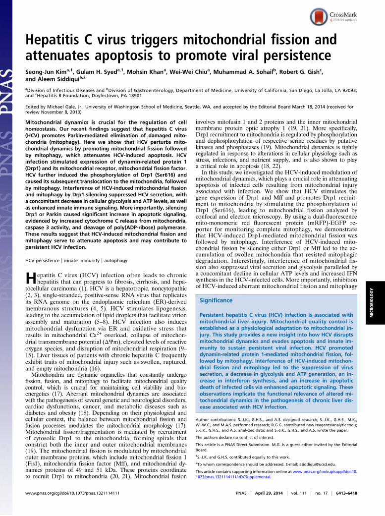
ResultsHCV Promotes Mitochondrial Fission. HCV infection induces ERand oxidative stress and alters calcium homeostasis, which causesmitochondrial dysfunction and damage (23–25). To investigatethe HCV-induced alterations of mitochondrial dynamics, me-tabolism, and physiology, human hepatoma (Huh7) cells wereinfected with cell culture-derived HCV (HCVcc) of JC1 strain[genotype2a chimera of J6 and Japanese fulminant hepatitis(JFH) 1 strains]. As shown in Fig. 1A, HCV-infected cells dis-played distinct fragmented mitochondria (mitochondrial fis-sion), in contrast to uninfected cells, which displayed a typicaltubular mitochondrial network indicative of normal healthy cells.We also observed similar mitochondrial fragmentation in cellsinfected with the HCV JFH1 strain, which replicates with lowerefficiency compared with the HCV JC1 strain (SI Appendix, Fig.S1). This confirms that mitochondrial fission is a consequence ofHCV infection and is not selectively induced in JC1-infected
cells because of its higher replication rates. Ultrastructuralanalysis of HCV-infected cells by transmission electron micros-copy further substantiated the occurrence of fragmented mi-tochondria in HCV-infected cells, in contrast to uninfected cells,which displayed elongated and tubular mitochondria (Fig. 1B).Also seen in Fig. 1B are the membranous web-like structure andfeatures of mitochondrial injury such as swollen mitochondriadevoid of mitochondrial cristae. A quantitative analysis of rela-tive mitochondrial length in uninfected versus HCV-infectedHuh7 cells is presented in Fig. 1C. These analyses demonstratethat HCV infection induces mitochondrial fission.
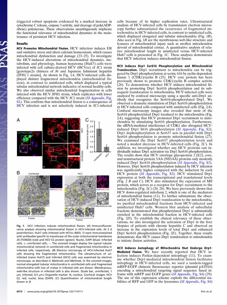
HCV Induces Drp1 Ser616 Phosphorylation and MitochondrialTranslocation. Drp1 recruitment to mitochondria can be trig-gered by Drp1 phosphorylation at serine 616 by cyclin-dependentkinase 1 (CDK1)/cyclin B (25). HCV core protein has beenpreviously shown to promote CDK1/cyclin B complex activity(26). To demonstrate whether HCV induces mitochondrial fis-sion by promoting Drp1 Ser616 phosphorylation and its sub-sequent translocation to mitochondria, HCV-infected cells wereanalyzed by confocal microscopy using a specific anti-Drp1 an-tibody that recognizes the Ser616-phosphorylated Drp1. Weobserved a dramatic stimulation of Drp1 Ser616 phosphorylationin HCV-infected cells compared with uninfected cells (Fig. 2A).Confocal microscopy images also revealed that most of theSer616 phosphorylated Drp1 localized to the mitochondria (Fig.2A), suggesting that HCV promoted Drp1 recruitment to mito-chondria by stimulating Ser616 phosphorylation. Furthermore,the siRNA-mediated interference of CDK1 also abrogated HCV-induced Drp1 S616 phosphorylation (SI Appendix, Fig. S2).Drp1 dephosphorylation at Ser637 acts in parallel with Drp1Ser616 phosphorylation to promote mitochondrial fission (27).We examined the Drp1 Ser637 phosphorylation levels andnoted a modest decrease in HCV-infected cells (Fig. 2C). Inaddition, we investigated whether any HCV proteins can in-dividually induce Drp1 activation via Drp1 Ser616 phosphorylation.The results show that HCV envelope glycoprotein 2 (E2), core,and nonstructural protein 3/4A (NS3/4A) proteins only modestlyinduced Drp1 Ser616 phosphorylation (SI Appendix, Fig. S3).However, Drp1 Ser616 phosphorylation induced by HCV infectionwas significantly higher compared with the induction by eachHCV protein (SI Appendix, Fig. S3). HCV stimulated Drp1expression at both the transcriptional and translational levels(Fig. 2 B and C). HCV also stimulated the expression of Mffprotein, which serves as a receptor for Drp1 recruitment to themitochondria (Fig. 2C) (20, 28). We have previously shown thatHCV down-regulated mitofusin 2, which is one of the mediatorsof mitochondrial fusion (11). To further substantiate the obser-vation of HCV-induced Drp1 translocation to the mitochondria,we purified mitochondrial fractions from HCV-infected anduninfected Huh7 cells. Western blot analysis of subcellularfractions demonstrated that phosphorylated Drp1 is substantiallyenriched in the mitochondrial fraction in HCV-infected cells(Fig. 2D). To establish the clinical relevance of these obser-vations, we also investigated the activation of Drp1 in the liverbiopsies of patients with chronic hepatitis C and observed anincrease in the expression levels of total Drp1 and enhancedDrp1 Ser616 phosphorylation (Fig. 2E). Together, these resultsdemonstrate that HCV causes Drp1 translocation to mitochondriato initiate fission activities.
HCV Induces Autophagy of Mitochondria that Undergo Drp1-Mediated Fission. We have recently reported that HCV in-fection induces Parkin-dependent mitophagy (11). To exam-ine whether Drp1-mediated mitochondrial fission facilitatesmitophagy in HCV-infected cells, we used a tandem-taggedmRFP-EGFP chimeric fluorescence reporter (plasmid pAT016)encoding a mitochondrial targeting signal sequence fused in-frame with mRFP and EGFP genes (SI Appendix, Fig. S4) (29).The use of this expression scheme exploits the differential sta-bilities of RFP and GFP in the lysosomes (SI Appendix, Fig. S4)
VCHdetcefninUB
N
N
DAPI/HCV E2 DAPI/HCV E2
Membranous web
A
12
3
1 2 3
1
2
1 2
C
0.0
0.5
1.0
1.5
2.0
2.5
3.0
Mito
chon
dria
l len
gth
(μm
)
HC
VU
ninf
ecte
d
DAPI/HCV E2/TOM20
TOM20
TOM20
++
-
A
B
A
B
+
-
--
-
--
FusionFission
Fig. 1. HCV infection induces mitochondrial fission. (A) Immunofluores-cence analysis showing mitochondrial fission in HCV-infected cells. At 3 dpostinfection, Huh7 cells infected with HCVcc (MOI, 1) were immunostainedwith antibodies specific to translocase of the outer mitochondrial membrane20 (TOM20) (red) and HCV E2 protein (green). Nuclei, DAPI (blue). Infectedcells, +; uninfected cells, −. The zoomed images display the typical tubularmitochondrial network in uninfected cells and fragmented mitochondria ininfected cells, respectively. (B) Electron microscopy of HCV-infected Huh7cells showing the fragmented mitochondria. The ultrastructure of un-infected (naive Huh7) and infected (HCV) cells was examined by electronmicroscope, as described in Materials and Methods. In the zoomed images,normal elongated tubular mitochondria in uninfected cells and fragmentedmitochondria with loss of cristae in infected cells are shown. Membranous,web-like structure in infected cells is also shown. (Scale bar, uninfected, 1μm; infected, 0.5 μm.) Organelle marker: N, nucleus. Confocal images: HCVE2, red; nuclei, blue (DAPI). (C) Quantification of mitochondrial lengthshown in B.
6414 | www.pnas.org/cgi/doi/10.1073/pnas.1321114111 Kim et al.
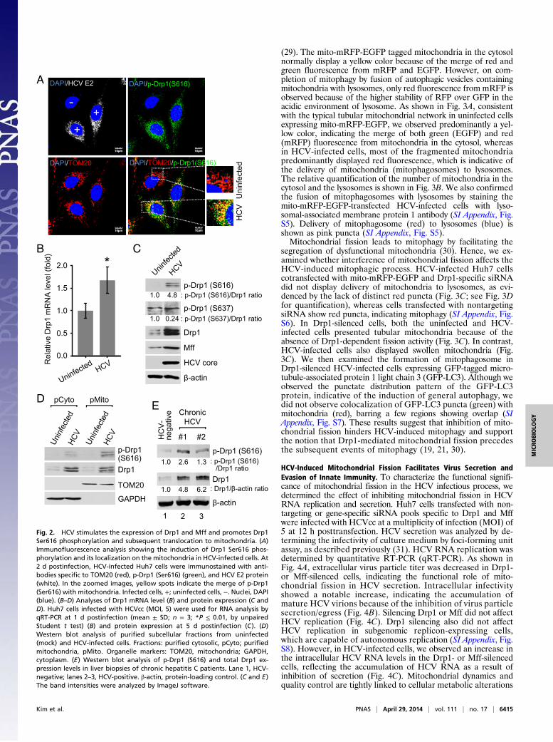
(29). The mito-mRFP-EGFP tagged mitochondria in the cytosolnormally display a yellow color because of the merge of red andgreen fluorescence from mRFP and EGFP. However, on com-pletion of mitophagy by fusion of autophagic vesicles containingmitochondria with lysosomes, only red fluorescence from mRFP isobserved because of the higher stability of RFP over GFP in theacidic environment of lysosome. As shown in Fig. 3A, consistentwith the typical tubular mitochondrial network in uninfected cellsexpressing mito-mRFP-EGFP, we observed predominantly a yel-low color, indicating the merge of both green (EGFP) and red(mRFP) fluorescence from mitochondria in the cytosol, whereasin HCV-infected cells, most of the fragmented mitochondriapredominantly displayed red fluorescence, which is indicative ofthe delivery of mitochondria (mitophagosomes) to lysosomes.The relative quantification of the number of mitochondria in thecytosol and the lysosomes is shown in Fig. 3B. We also confirmedthe fusion of mitophagosomes with lysosomes by staining themito-mRFP-EGFP-transfected HCV-infected cells with lyso-somal-associated membrane protein 1 antibody (SI Appendix, Fig.S5). Delivery of mitophagosome (red) to lysosomes (blue) isshown as pink puncta (SI Appendix, Fig. S5).Mitochondrial fission leads to mitophagy by facilitating the
segregation of dysfunctional mitochondria (30). Hence, we ex-amined whether interference of mitochondrial fission affects theHCV-induced mitophagic process. HCV-infected Huh7 cellscotransfected with mito-mRFP-EGFP and Drp1-specific siRNAdid not display delivery of mitochondria to lysosomes, as evi-denced by the lack of distinct red puncta (Fig. 3C; see Fig. 3Dfor quantification), whereas cells transfected with nontargetingsiRNA show red puncta, indicating mitophagy (SI Appendix, Fig.S6). In Drp1-silenced cells, both the uninfected and HCV-infected cells presented tubular mitochondria because of theabsence of Drp1-dependent fission activity (Fig. 3C). In contrast,HCV-infected cells also displayed swollen mitochondria (Fig.3C). We then examined the formation of mitophagosome inDrp1-silenced HCV-infected cells expressing GFP-tagged micro-tubule-associated protein 1 light chain 3 (GFP-LC3). Although weobserved the punctate distribution pattern of the GFP-LC3protein, indicative of the induction of general autophagy, wedid not observe colocalization of GFP-LC3 puncta (green) withmitochondria (red), barring a few regions showing overlap (SIAppendix, Fig. S7). These results suggest that inhibition of mito-chondrial fission hinders HCV-induced mitophagy and supportthe notion that Drp1-mediated mitochondrial fission precedesthe subsequent events of mitophagy (19, 21, 30).
HCV-Induced Mitochondrial Fission Facilitates Virus Secretion andEvasion of Innate Immunity. To characterize the functional signifi-cance of mitochondrial fission in the HCV infectious process, wedetermined the effect of inhibiting mitochondrial fission in HCVRNA replication and secretion. Huh7 cells transfected with non-targeting or gene-specific siRNA pools specific to Drp1 and Mffwere infected with HCVcc at a multiplicity of infection (MOI) of5 at 12 h posttransfection. HCV secretion was analyzed by de-termining the infectivity of culture medium by foci-forming unitassay, as described previously (31). HCV RNA replication wasdetermined by quantitative RT-PCR (qRT-PCR). As shown inFig. 4A, extracellular virus particle titer was decreased in Drp1-or Mff-silenced cells, indicating the functional role of mito-chondrial fission in HCV secretion. Intracellular infectivityshowed a notable increase, indicating the accumulation ofmature HCV virions because of the inhibition of virus particlesecretion/egress (Fig. 4B). Silencing Drp1 or Mff did not affectHCV replication (Fig. 4C). Drp1 silencing also did not affectHCV replication in subgenomic replicon-expressing cells,which are capable of autonomous replication (SI Appendix, Fig.S8). However, in HCV-infected cells, we observed an increase inthe intracellular HCV RNA levels in the Drp1- or Mff-silencedcells, reflecting the accumulation of HCV RNA as a result ofinhibition of secretion (Fig. 4C). Mitochondrial dynamics andquality control are tightly linked to cellular metabolic alterations
p-Drp1
Drp1
TOM20
GAPDH
D
A
HC
VU
ninf
ecte
d
Rel
ativ
e D
rp1
mR
NA
leve
l (fo
ld)
0.5
1.0
1.5
2.0
B C
p-Drp1 (S616)
HCV core
β-actin
Drp1
+
-+
DAPI/HCV E2
DAPI/TOM20 DAPI/TOM20/p-Drp1(S616)
DAPI/p-Drp1(S616)
Mff0.0
pCyto pMito
*
p-Drp1 (S616)
Drp1
β-actin
EChronic
HCV
#1 #2
(S616)
2 31
1.0 4.8 : p-Drp1 (S616)/Drp1 ratio
nega
tive
HC
V-
p-Drp1 (S637)1.0 0.24 : p-Drp1 (S637)/Drp1 ratio
1.0 2.6 : p-Drp1 (S616)/Drp1 ratio
1.0 4.8 : Drp1/β-actin ratio6.2
1.3
Fig. 2. HCV stimulates the expression of Drp1 and Mff and promotes Drp1Ser616 phosphorylation and subsequent translocation to mitochondria. (A)Immunofluorescence analysis showing the induction of Drp1 Ser616 phos-phorylation and its localization on the mitochondria in HCV-infected cells. At2 d postinfection, HCV-infected Huh7 cells were immunostained with anti-bodies specific to TOM20 (red), p-Drp1 (Ser616) (green), and HCV E2 protein(white). In the zoomed images, yellow spots indicate the merge of p-Drp1(Ser616) with mitochondria. Infected cells, +; uninfected cells, −. Nuclei, DAPI(blue). (B–D) Analyses of Drp1 mRNA level (B) and protein expression (C andD). Huh7 cells infected with HCVcc (MOI, 5) were used for RNA analysis byqRT-PCR at 1 d postinfection (mean ± SD; n = 3; *P ≤ 0.01, by unpairedStudent t test) (B) and protein expression at 5 d postinfection (C). (D)Western blot analysis of purified subcellular fractions from uninfected(mock) and HCV-infected cells. Fractions: purified cytosolic, pCyto; purifiedmitochondria, pMito. Organelle markers: TOM20, mitochondria; GAPDH,cytoplasm. (E) Western blot analysis of p-Drp1 (S616) and total Drp1 ex-pression levels in liver biopsies of chronic hepatitis C patients. Lane 1, HCV-negative; lanes 2–3, HCV-positive. β-actin, protein-loading control. (C and E)The band intensities were analyzed by ImageJ software.
Kim et al. PNAS | April 29, 2014 | vol. 111 | no. 17 | 6415
MICRO
BIOLO
GY
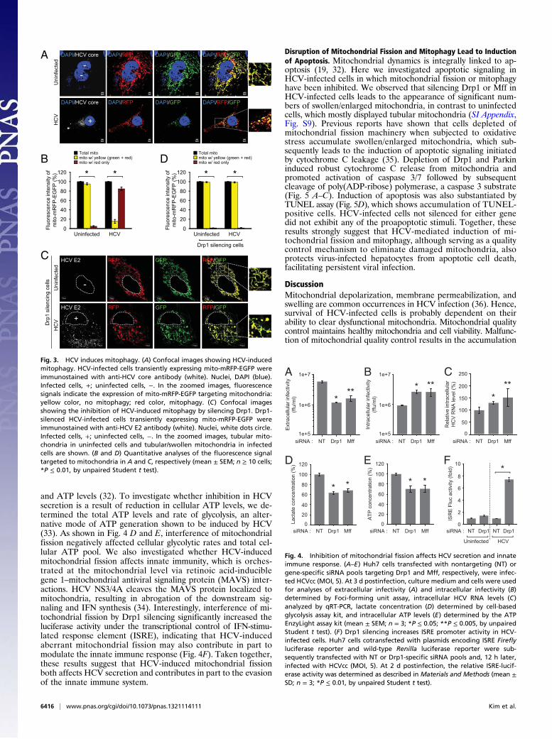
and ATP levels (32). To investigate whether inhibition in HCVsecretion is a result of reduction in cellular ATP levels, we de-termined the total ATP levels and rate of glycolysis, an alter-native mode of ATP generation shown to be induced by HCV(33). As shown in Fig. 4 D and E, interference of mitochondrialfission negatively affected cellular glycolytic rates and total cel-lular ATP pool. We also investigated whether HCV-inducedmitochondrial fission affects innate immunity, which is orches-trated at the mitochondrial level via retinoic acid-induciblegene 1–mitochondrial antiviral signaling protein (MAVS) inter-actions. HCV NS3/4A cleaves the MAVS protein localized tomitochondria, resulting in abrogation of the downstream sig-naling and IFN synthesis (34). Interestingly, interference of mi-tochondrial fission by Drp1 silencing significantly increased theluciferase activity under the transcriptional control of IFN-stimu-lated response element (ISRE), indicating that HCV-inducedaberrant mitochondrial fission may also contribute in part tomodulate the innate immune response (Fig. 4F). Taken together,these results suggest that HCV-induced mitochondrial fissionboth affects HCV secretion and contributes in part to the evasionof the innate immune system.
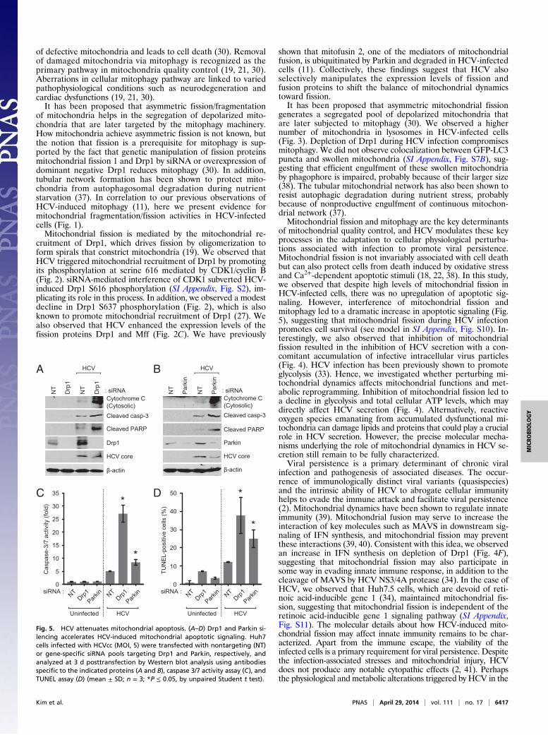
Disruption of Mitochondrial Fission and Mitophagy Lead to Inductionof Apoptosis. Mitochondrial dynamics is integrally linked to ap-optosis (19, 32). Here we investigated apoptotic signaling inHCV-infected cells in which mitochondrial fission or mitophagyhave been inhibited. We observed that silencing Drp1 or Mff inHCV-infected cells leads to the appearance of significant num-bers of swollen/enlarged mitochondria, in contrast to uninfectedcells, which mostly displayed tubular mitochondria (SI Appendix,Fig. S9). Previous reports have shown that cells depleted ofmitochondrial fission machinery when subjected to oxidativestress accumulate swollen/enlarged mitochondria, which sub-sequently leads to the induction of apoptotic signaling initiatedby cytochrome C leakage (35). Depletion of Drp1 and Parkininduced robust cytochrome C release from mitochondria andpromoted activation of caspase 3/7 followed by subsequentcleavage of poly(ADP-ribose) polymerase, a caspase 3 substrate(Fig. 5 A–C). Induction of apoptosis was also substantiated byTUNEL assay (Fig. 5D), which shows accumulation of TUNEL-positive cells. HCV-infected cells not silenced for either genedid not exhibit any of the proapoptotic stimuli. Together, theseresults strongly suggest that HCV-mediated induction of mi-tochondrial fission and mitophagy, although serving as a qualitycontrol mechanism to eliminate damaged mitochondria, alsoprotects virus-infected hepatocytes from apoptotic cell death,facilitating persistent viral infection.
DiscussionMitochondrial depolarization, membrane permeabilization, andswelling are common occurrences in HCV infection (36). Hence,survival of HCV-infected cells is probably dependent on theirability to clear dysfunctional mitochondria. Mitochondrial qualitycontrol maintains healthy mitochondria and cell viability. Malfunc-tion of mitochondrial quality control results in the accumulation
DB
A
Fluo
resc
ence
inte
nsity
of
mito
-mR
FP-E
GFP
(%)
Uninfected HCV
20
40
60
0
80
100
120
Total mitomito w/ yellow (green + red)mito w/ red only
Uni
nfec
ted
HC
VDAPI/HCV core DAPI/GFPDAPI/RFP DAPI/RFP/GFP
DAPI/HCV core DAPI/GFPDAPI/RFP DAPI/RFP/GFP
--
+
Uni
nfec
ted
HC
V
C
Drp
1 si
lenc
ing
cells
HCV E2 GFP PFRPFR /GFP
HCV E2 GFP PFRPFR /GFP
-
+
Fluo
resc
ence
inte
nsity
of
mito
-mR
FP-E
GFP
(%)
Uninfected HCV
20
40
60
0
80
100
120
Total mitomito w/ yellow (green + red)mito w/ red only
Drp1 silencing cells
****
Fig. 3. HCV induces mitophagy. (A) Confocal images showing HCV-inducedmitophagy. HCV-infected cells transiently expressing mito-mRFP-EGFP wereimmunostained with anti-HCV core antibody (white). Nuclei, DAPI (blue).Infected cells, +; uninfected cells, −. In the zoomed images, fluorescencesignals indicate the expression of mito-mRFP-EGFP targeting mitochondria:yellow color, no mitophagy; red color, mitophagy. (C) Confocal imagesshowing the inhibition of HCV-induced mitophagy by silencing Drp1. Drp1-silenced HCV-infected cells transiently expressing mito-mRFP-EGFP wereimmunostained with anti-HCV E2 antibody (white). Nuclei, white dots circle.Infected cells, +; uninfected cells, −. In the zoomed images, tubular mito-chondria in uninfected cells and tubular/swollen mitochondria in infectedcells are shown. (B and D) Quantitative analyses of the fluorescence signaltargeted to mitochondria in A and C, respectively (mean ± SEM; n ≥ 10 cells;*P ≤ 0.01, by unpaired Student t test).
A
Ext
race
llula
r inf
ectiv
ity(ff
u/m
l)
NT Drp11e+5
1e+6
1e+7
Mff
B
NT Drp1 Mff
Intra
cellu
lar i
nfec
tivity
(ffu/
ml)
:ANRis:ANRis1e+5
1e+6
1e+7
*** * **
C
D
Lact
ate
conc
entra
tion
(%)
NT Drp1
20
40
60
0
80
100
120
Mff
ATP
conc
entra
tion
(%)
NT Drp1
20
40
60
0
80
100
120
Mff
NT Drp1
100
0
150
200
250
MffR
elat
ive
intra
cellu
lar
HC
V R
NA
leve
l (%
)
50
:ANRis:ANRis
siRNA :
E
***
* * * *
F
siRNA : NT Drp1 NT Drp10
2
4
6
8
10
ISR
E F
luc
activ
ity (f
old)
HCVUninfected
*
Fig. 4. Inhibition of mitochondrial fission affects HCV secretion and innateimmune response. (A–E) Huh7 cells transfected with nontargeting (NT) orgene-specific siRNA pools targeting Drp1 and Mff, respectively, were infec-ted HCVcc (MOI, 5). At 3 d postinfection, culture medium and cells were usedfor analyses of extracellular infectivity (A) and intracellular infectivity (B)determined by Foci-forming unit assay, intracellular HCV RNA levels (C)analyzed by qRT-PCR, lactate concentration (D) determined by cell-basedglycolysis assay kit, and intracellular ATP levels (E) determined by the ATPEnzyLight assay kit (mean ± SEM; n = 3; *P ≤ 0.05; **P ≤ 0.005, by unpairedStudent t test). (F) Drp1 silencing increases ISRE promoter activity in HCV-infected cells. Huh7 cells cotransfected with plasmids encoding ISRE Fireflyluciferase reporter and wild-type Renilla luciferase reporter were sub-sequently transfected with NT or Drp1-specific siRNA pools and, 12 h later,infected with HCVcc (MOI, 5). At 2 d postinfection, the relative ISRE-lucif-erase activity was determined as described in Materials and Methods (mean ±SD; n = 3; *P ≤ 0.01, by unpaired Student t test).
6416 | www.pnas.org/cgi/doi/10.1073/pnas.1321114111 Kim et al.
of defective mitochondria and leads to cell death (30). Removalof damaged mitochondria via mitophagy is recognized as theprimary pathway in mitochondria quality control (19, 21, 30).Aberrations in cellular mitophagy pathway are linked to variedpathophysiological conditions such as neurodegeneration andcardiac dysfunctions (19, 21, 30).It has been proposed that asymmetric fission/fragmentation
of mitochondria helps in the segregation of depolarized mito-chondria that are later targeted by the mitophagy machinery.How mitochondria achieve asymmetric fission is not known, butthe notion that fission is a prerequisite for mitophagy is sup-ported by the fact that genetic manipulation of fission proteinsmitochondrial fission 1 and Drp1 by siRNA or overexpression ofdominant negative Drp1 reduces mitophagy (30). In addition,tubular network formation has been shown to protect mito-chondria from autophagosomal degradation during nutrientstarvation (37). In correlation to our previous observations ofHCV-induced mitophagy (11), here we present evidence formitochondrial fragmentation/fission activities in HCV-infectedcells (Fig. 1).Mitochondrial fission is mediated by the mitochondrial re-
cruitment of Drp1, which drives fission by oligomerization toform spirals that constrict mitochondria (19). We observed thatHCV triggered mitochondrial recruitment of Drp1 by promotingits phosphorylation at serine 616 mediated by CDK1/cyclin B(Fig. 2). siRNA-mediated interference of CDK1 subverted HCV-induced Drp1 S616 phosphorylation (SI Appendix, Fig. S2), im-plicating its role in this process. In addition, we observed a modestdecline in Drp1 S637 phosphorylation (Fig. 2), which is alsoknown to promote mitochondrial recruitment of Drp1 (27). Wealso observed that HCV enhanced the expression levels of thefission proteins Drp1 and Mff (Fig. 2C). We have previously
shown that mitofusin 2, one of the mediators of mitochondrialfusion, is ubiquitinated by Parkin and degraded in HCV-infectedcells (11). Collectively, these findings suggest that HCV alsoselectively manipulates the expression levels of fission andfusion proteins to shift the balance of mitochondrial dynamicstoward fission.It has been proposed that asymmetric mitochondrial fission
generates a segregated pool of depolarized mitochondria thatare later subjected to mitophagy (30). We observed a highernumber of mitochondria in lysosomes in HCV-infected cells(Fig. 3). Depletion of Drp1 during HCV infection compromisesmitophagy. We did not observe colocalization between GFP-LC3puncta and swollen mitochondria (SI Appendix, Fig. S7B), sug-gesting that efficient engulfment of these swollen mitochondriaby phagophore is impaired, probably because of their larger size(38). The tubular mitochondrial network has also been shown toresist autophagic degradation during nutrient stress, probablybecause of nonproductive engulfment of continuous mitochon-drial network (37).Mitochondrial fission and mitophagy are the key determinants
of mitochondrial quality control, and HCV modulates these keyprocesses in the adaptation to cellular physiological perturba-tions associated with infection to promote viral persistence.Mitochondrial fission is not invariably associated with cell deathbut can also protect cells from death induced by oxidative stressand Ca2+-dependent apoptotic stimuli (18, 22, 38). In this study,we observed that despite high levels of mitochondrial fission inHCV-infected cells, there was no upregulation of apoptotic sig-naling. However, interference of mitochondrial fission andmitophagy led to a dramatic increase in apoptotic signaling (Fig.5), suggesting that mitochondrial fission during HCV infectionpromotes cell survival (see model in SI Appendix, Fig. S10). In-terestingly, we also observed that inhibition of mitochondrialfission resulted in the inhibition of HCV secretion with a con-comitant accumulation of infective intracellular virus particles(Fig. 4). HCV infection has been previously shown to promoteglycolysis (33). Hence, we investigated whether perturbing mi-tochondrial dynamics affects mitochondrial functions and met-abolic reprogramming. Inhibition of mitochondrial fission led toa decline in glycolysis and total cellular ATP levels, which maydirectly affect HCV secretion (Fig. 4). Alternatively, reactiveoxygen species emanating from accumulated dysfunctional mi-tochondria can damage lipids and proteins that could play a crucialrole in HCV secretion. However, the precise molecular mecha-nisms underlying the role of mitochondrial dynamics in HCV se-cretion still remain to be fully characterized.Viral persistence is a primary determinant of chronic viral
infection and pathogenesis of associated diseases. The occur-rence of immunologically distinct viral variants (quasispecies)and the intrinsic ability of HCV to abrogate cellular immunityhelps to evade the immune attack and facilitate viral persistence(2). Mitochondrial dynamics have been shown to regulate innateimmunity (39). Mitochondrial fusion may serve to increase theinteraction of key molecules such as MAVS in downstream sig-naling of IFN synthesis, and mitochondrial fission may preventthese interactions (39, 40). Consistent with this idea, we observedan increase in IFN synthesis on depletion of Drp1 (Fig. 4F),suggesting that mitochondrial fission may also participate insome way in evading innate immune response, in addition to thecleavage of MAVS by HCV NS3/4A protease (34). In the case ofHCV, we observed that Huh7.5 cells, which are devoid of reti-noic acid-inducible gene 1 (34), maintained mitochondrial fis-sion, suggesting that mitochondrial fission is independent of theretinoic acid-inducible gene 1 signaling pathway (SI Appendix,Fig. S11). The molecular details about how HCV-induced mito-chondrial fission may affect innate immunity remains to be char-acterized. Apart from the immune escape, the viability of theinfected cells is a primary requirement for viral persistence. Despitethe infection-associated stresses and mitochondrial injury, HCVdoes not produce any notable cytopathic effects (2, 41). Perhapsthe physiological and metabolic alterations triggered by HCV in the
A
: siRNANT
Drp
1
NT
Drp
1
BHCV
D
β-actin
: siRNA
Cleaved PARP
Cytochrome C
Cleaved casp-3
Parkin
HCV core
NT
Par
kin
NT
Par
kin
C
HCV
Cas
pase
-3/7
act
ivity
(fol
d)
siRNA :
HCV
β-actin
Cleaved PARP
Cytochrome C
Cleaved casp-3
Drp1
HCV core
(Cytosolic) (Cytosolic)
Uninfected
0
5
10
15
20
25
30
35
TUN
EL-
posi
tive
cells
(%)
siRNA :
HCVUninfected
0
10
20
30
40
50*
*
*
*
Fig. 5. HCV attenuates mitochondrial apoptosis. (A–D) Drp1 and Parkin si-lencing accelerates HCV-induced mitochondrial apoptotic signaling. Huh7cells infected with HCVcc (MOI, 5) were transfected with nontargeting (NT)or gene-specific siRNA pools targeting Drp1 and Parkin, respectively, andanalyzed at 3 d posttransfection by Western blot analysis using antibodiesspecific to the indicated proteins (A and B), caspase 3/7 activity assay (C), andTUNEL assay (D) (mean ± SD; n = 3; *P ≤ 0.05, by unpaired Student t test).
Kim et al. PNAS | April 29, 2014 | vol. 111 | no. 17 | 6417
MICRO
BIOLO
GY
infected hepatocyte play a role in abating the stress associatedwith viral infection and prevent the manifestation of viral cyto-pathic effects to sustain persistent infection. The present studyhighlights the physiological adaptation involving up-regulation ofmitochondria quality control in HCV-infected hepatocytes tosustain cellular viability and promote viral persistence. The pri-mary cause of persistent HCV infection is a result of the failureto mount an efficient immune response to eliminate infectedhepatocytes (2). However, HCV-induced aberrant mitochondrialdynamics may also contribute to the viral persistence by atten-uating apoptosis of infected cells. A similar strategy of pertur-bation of mitochondrial dynamics and attenuation of apoptosis isalso used by hepatitis B virus (29). Our findings implicate themitochondrial quality control pathway as a potential therapeutictarget against HCV infection and associated liver diseasepathogenesis.
Materials and MethodsAdditional information is provided in SI Appendix, Materials and Methods.
Cell Culture and Virus. The human hepatoma cell line Huh7 was culturedas described previously (11). Cell culture-derived HCV (HCVcc) of JC1 and
JFH1 strains were used in this study and prepared as described previously(11, 42).
DNA Constructs. The pEGFP-LC3 plasmid DNA was a kind gift from TamotsuYoshimori (National Institute of Genetics, Japan). The p-mito-mRFP-EGFPplasmid DNA (pAT016) was described previously (29).
Immunofluorescence. HCV-infected cells and those transfected with siRNAwere grown on coverslips and used for immunofluorescence assay, as de-scribed previously (11). Images were visualized under a 60× or 100× oilobjectives, using an Olympus FluoView 1000 confocal microscope.
ACKNOWLEDGMENTS. We thank Dr. Charles Rice (Rockefeller University) forproviding HCV JC1 clone and NS5A (9E10) antibody, Dr. Takaji Wakita(National Institute of Infectious Disease, Japan) for providing HCV pJFH1plasmid, Dr. Mansun Law (The Scripps Research Institute) for providing HCVE2 (AR3A) antibody, Dr. Tamotsu Yoshimori (National Institute of Genetics,Japan) for providing pEGFP-LC3 DNA construct, Dr. Marilyn Farquhar foradvice on analysis of electron microscopy, and Ying Jones and Timo Meerloofor technical assistance with electron microscopy. Electron microscopy wascarried out at the University of California, San Diego (UCSD) electron micros-copy facility. Confocal analysis was performed at the UCSD NeuroscienceMicroscopy Shared Facility (Grant P30 NS047101). This work was supportedin whole or in part by National Institutes of Health Grants AI085087,DK077704, and DK08379 (to A.S.), and T32 DK07202 (to M.A.S.).
1. Pawlotsky JM (2004) Pathophysiology of hepatitis C virus infection and related liverdisease. Trends Microbiol 12(2):96–102.
2. Guidotti LG, Chisari FV (2006) Immunobiology and pathogenesis of viral hepatitis.Annu Rev Pathol 1:23–61.
3. Paula T, et al. (2009) New drug targets for hepatitis C and other Flaviviridae viruses.Infect Disord Drug Targets 9(2):133–147.
4. Egger D, et al. (2002) Expression of hepatitis C virus proteins induces distinct mem-brane alterations including a candidate viral replication complex. J Virol 76(12):5974–5984.
5. Moradpour D, Penin F, Rice CM (2007) Replication of hepatitis C virus. Nat Rev Mi-crobiol 5(6):453–463.
6. Miyanari Y, et al. (2007) The lipid droplet is an important organelle for hepatitis Cvirus production. Nat Cell Biol 9(9):1089–1097.
7. Waris G, Felmlee DJ, Negro F, Siddiqui A (2007) Hepatitis C virus induces proteolyticcleavage of sterol regulatory element binding proteins and stimulates their phos-phorylation via oxidative stress. J Virol 81(15):8122–8130.
8. Syed GH, Amako Y, Siddiqui A (2010) Hepatitis C virus hijacks host lipid metabolism.Trends Endocrinol Metab 21(1):33–40.
9. Tardif KD, Mori K, Siddiqui A (2002) Hepatitis C virus subgenomic replicons induceendoplasmic reticulum stress activating an intracellular signaling pathway. J Virol76(15):7453–7459.
10. Gong G, Waris G, Tanveer R, Siddiqui A (2001) Human hepatitis C virus NS5A proteinalters intracellular calcium levels, induces oxidative stress, and activates STAT-3 andNF-kappa B. Proc Natl Acad Sci USA 98(17):9599–9604.
11. Kim SJ, Syed GH, Siddiqui A (2013) Hepatitis C virus induces the mitochondrialtranslocation of Parkin and subsequent mitophagy. PLoS Pathog 9(3):e1003285.
12. Korenaga M, et al. (2005) Hepatitis C virus core protein inhibits mitochondrial elec-tron transport and increases reactive oxygen species (ROS) production. J Biol Chem280(45):37481–37488.
13. Piccoli C, et al. (2007) Hepatitis C virus protein expression causes calcium-mediatedmitochondrial bioenergetic dysfunction and nitro-oxidative stress. Hepatology 46(1):58–65.
14. Waris G, Siddiqui A (2005) Hepatitis C virus stimulates the expression of cyclo-oxygenase-2 via oxidative stress: Role of prostaglandin E2 in RNA replication. J Virol79(15):9725–9734.
15. Waris G, Turkson J, Hassanein T, Siddiqui A (2005) Hepatitis C virus (HCV) constitu-tively activates STAT-3 via oxidative stress: Role of STAT-3 in HCV replication. J Virol79(3):1569–1580.
16. Barbaro G, et al. (1999) Hepatocellular mitochondrial alterations in patients withchronic hepatitis C: Ultrastructural and biochemical findings. Am J Gastroenterol94(8):2198–2205.
17. Chan DC (2012) Fusion and fission: Interlinked processes critical for mitochondrialhealth. Annu Rev Genet 46:265–287.
18. Liesa M, Palacín M, Zorzano A (2009) Mitochondrial dynamics in mammalian healthand disease. Physiol Rev 89(3):799–845.
19. Otera H, Ishihara N, Mihara K (2013) New insights into the function and regulation ofmitochondrial fission. Biochim Biophys Acta 1833(5):1256–1268.
20. Losón OC, Song Z, Chen H, Chan DC (2013) Fis1, Mff, MiD49, and MiD51 mediate Drp1recruitment in mitochondrial fission. Mol Biol Cell 24(5):659–667.
21. Youle RJ, van der Bliek AM (2012) Mitochondrial fission, fusion, and stress. Science337(6098):1062–1065.
22. Suen DF, Norris KL, Youle RJ (2008) Mitochondrial dynamics and apoptosis. Genes Dev22(12):1577–1590.
23. Deng L, et al. (2008) Hepatitis C virus infection induces apoptosis through a Bax-triggered, mitochondrion-mediated, caspase 3-dependent pathway. J Virol 82(21):10375–10385.
24. Li Y, Boehning DF, Qian T, Popov VL, Weinman SA (2007) Hepatitis C virus coreprotein increases mitochondrial ROS production by stimulation of Ca2+ uniporteractivity. FASEB J 21(10):2474–2485.
25. Knott AB, Perkins G, Schwarzenbacher R, Bossy-Wetzel E (2008) Mitochondrial frag-mentation in neurodegeneration. Nat Rev Neurosci 9(7):505–518.
26. Spaziani A, Alisi A, Sanna D, Balsano C (2006) Role of p38 MAPK and RNA-dependentprotein kinase (PKR) in hepatitis C virus core-dependent nuclear delocalization ofcyclin B1. J Biol Chem 281(16):10983–10989.
27. Cereghetti GM, et al. (2008) Dephosphorylation by calcineurin regulates translocationof Drp1 to mitochondria. Proc Natl Acad Sci USA 105(41):15803–15808.
28. Otera H, et al. (2010) Mff is an essential factor for mitochondrial recruitment of Drp1during mitochondrial fission in mammalian cells. J Cell Biol 191(6):1141–1158.
29. Kim SJ, et al. (2013) Hepatitis B virus disrupts mitochondrial dynamics: Induces fissionand mitophagy to attenuate apoptosis. PLoS Pathog 9(12):e1003722.
30. Twig G, Shirihai OS (2011) The interplay between mitochondrial dynamics and mi-tophagy. Antioxid Redox Signal 14(10):1939–1951.
31. Syed GH, Siddiqui A (2011) Effects of hypolipidemic agent nordihydroguaiaretic acidon lipid droplets and hepatitis C virus. Hepatology 54(6):1936–1946.
32. Chen H, Chan DC (2009) Mitochondrial dynamics—fusion, fission, movement, andmitophagy—in neurodegenerative diseases. Hum Mol Genet 18(R2):R169–R176.
33. Ripoli M, et al. (2010) Hepatitis C virus-linked mitochondrial dysfunction promoteshypoxia-inducible factor 1 alpha-mediated glycolytic adaptation. J Virol 84(1):647–660.
34. Sumpter R, Jr., et al. (2005) Regulating intracellular antiviral defense and permis-siveness to hepatitis C virus RNA replication through a cellular RNA helicase, RIG-I.J Virol 79(5):2689–2699.
35. Malhi H, Gores GJ, Lemasters JJ (2006) Apoptosis and necrosis in the liver: A tale oftwo deaths? Hepatology 43(2, Suppl 1):S31–S44.
36. Korenaga M, et al. (2005) Mitochondrial dysfunction in hepatitis C. J Clin Gastro-enterol 39(4, Suppl 2):S162–S166.
37. Rambold AS, Kostelecky B, Elia N, Lippincott-Schwartz J (2011) Tubular networkformation protects mitochondria from autophagosomal degradation during nutrientstarvation. Proc Natl Acad Sci USA 108(25):10190–10195.
38. Kageyama Y, et al. (2012) Mitochondrial division ensures the survival of postmitoticneurons by suppressing oxidative damage. J Cell Biol 197(4):535–551.
39. Castanier C, Garcin D, Vazquez A, Arnoult D (2010) Mitochondrial dynamics regulatethe RIG-I-like receptor antiviral pathway. EMBO Rep 11(2):133–138.
40. West AP, Shadel GS, Ghosh S (2011) Mitochondria in innate immune responses. NatRev Immunol 11(6):389–402.
41. Wieland SF, Chisari FV (2005) Stealth and cunning: Hepatitis B and hepatitis C viruses.J Virol 79(15):9369–9380.
42. Amako Y, Sarkeshik A, Hotta H, Yates J, 3rd, Siddiqui A (2009) Role of oxysterolbinding protein in hepatitis C virus infection. J Virol 83(18):9237–9246.
6418 | www.pnas.org/cgi/doi/10.1073/pnas.1321114111 Kim et al.