head and neck anatomy - usc · 2016-06-27 · head and neck anatomy faculty dr. yang chai...
TRANSCRIPT

Head and Neck Anatomy
Yang Chai, D.D.S., Ph.D.
George and MaryLou Boone Professor
Course Director
Ostrow School of Dentistry USC

Head and Neck Anatomy
Faculty
Dr. Yang Chai
Dr. Rizkalla
Zakhary
Dr. Yau-Shi Lin
Teaching Assistants
Keck School of Medicine USC
Matthew Agam [email protected]
Michelle Connor [email protected]
Jake Del Rosso [email protected]
Jonathan Huang [email protected]
Omid Jalali [email protected]
Andrew Katirai [email protected]
Kayvan Kazerouni [email protected]
Mark Landau [email protected]
William Schwartzmar [email protected]
Guy Talmor [email protected]
Peter Tsou [email protected]
Ben Pirotte [email protected]

Anatomy Lab access code: 11111
Start-11111-lower right key
Authorized people only

Presentation materials:
www.ccmb.usc.edu (teaching resources)
Check-lists for midterm and final
examinations


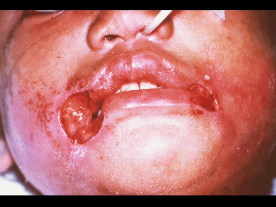
Intra-
oral
I&D

I&D


Anatomy of the Maxilla

Maxillary Sinus

Alignment of posterior implants

Alignment of posterior implants

Important Anatomy for the Posterior Mandible
Lingual Nerve Submandibular Fossa Mandibular Canal Mental Foramen

Anatomy of the Posterior
Mandible
Submandibular (submylohyoid) fossa
Mylohyoid shelf

Neck Anatomy

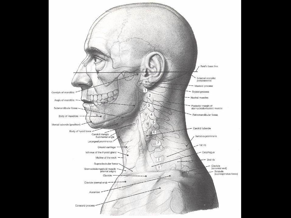

Boundaries of the Neck
•Anterior Aspect:
Superior: Inferior border of the mandible
Inferior: Superior surface of the
manubrium and clavicle
•Posterior Aspect:
Superior: Superior nuchal line
Inferior: Horizontal line between C7 and
T1


Platysma
Covers mainly anterior aspect of the neck.
Origin: deltoid and pectoralis fasciae.
Insertion: inferior border of mandible and
skin as well as hypodermis in this region.
Function: assist depression the lower lip and
corner of mouth.
Innervation: cervical br. of facial N. (VII).


Sensory Innervation of Neck
Ventral primary rami of C1, C2, C3, and
most C4 form the cervical plexus.
Front of neck:
1. Great aruicular N. (C2 & C3)
2. Transverse cervical N. (C2 & C3)
3. Supra clavicular N. (C2 & C4)
a. Anterior (medial)
b. Middle (intermediate)
c. Posterior (lateral)
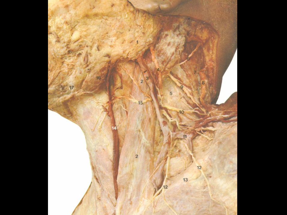


Posterior Aspect of Neck
Greater occipital N. (C2)
Third occipital N. (C3)
C4 and C5


Ansa Cervicalis
Two roots (formed by C1, C2, and C3)
Superior root (travels with CN XII)
Inferior root (from cervical region)
Function: Motor innervation of
infrahyoid muscles


Accessory N. (spinal accessory N.)
Two components of fibers
1. Cranial root
2. Spinal root
Function: motor innervation of
1. Sternocleidomastoid muscle (SCM)
2. Trapezius muscle


Phrenic N.
(C3, C4, and C5)
Motor: Diaphragm
Sensory: Mediastinal pleura and
pericardium of the heart


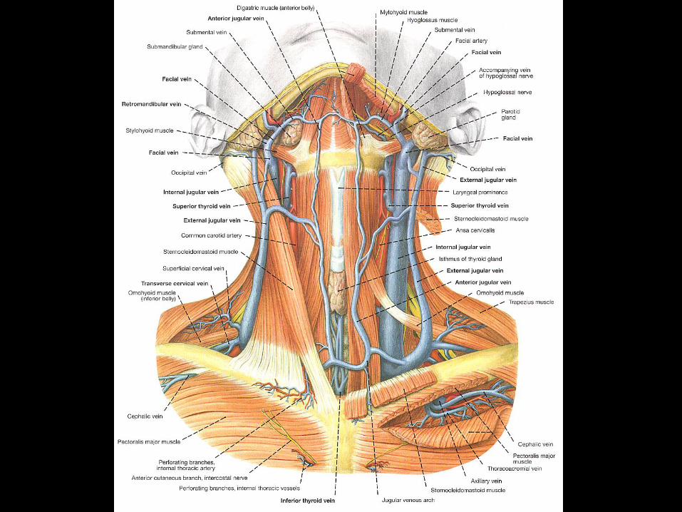
Anterior Triangle of Neck
Boundaries:
Superior: Inferior border of mandible
Posterior: Anterior border of SCM
Anterior: Midline of the neck

Subdivisions of Anterior Tri. 1. Muscular Triangle
Boundaries:
Superior: Superior belly of omohyoid
Inferior: Anterior border of SCM
Anterior: Midline
Contents:
a. sternohyoid (ansa)
b. sternothyroid (ansa)
c. thyrohyoid (C1 via CN XII)
d. omohyoid (ansa)


Continued:
2. Carotid Triangle
Boundaries:
Superior: Posterior belly of digastric
muscle
Inferior: Superior belly of omohyoid
Posterior: Anterior border of SCM

Continued:
Contents:
a. common carotid (carotid sinus-BP
regulator/IX)
internal (carotid body-Chemoreceptor/IX
& X)
external carotid artery
b. internal jugular vein
c. vagus N (CN X)
d. ansa cervicalis


Continued:
3. Submandibular Triangle
Boundaries:
Superior: Inferior border of mandible
Anterior: Anterior belly of digastric muscle
Posterior: Posterior belly of digastric muscle
Contents:
a. SMG (submandibular gland)
b. lymph nodes (submandibular group)
c. facial artery
d. facial vein

Continued:
4. Submental Triangle
Boundaries:
Lateral: both anterior bellies of digastric
muscle
Inferior: hyoid bones


Branches of External
Carotid Artery
1. Superior thyroid AA.
a. infrahyoid
b. sternocleidomastoid
c. superior laryngeal
d. cricothyroid
e. terminal branches to thryoid gland

Continued:
2. Ascending pharyngeal AA.
a. pharyngeal
b. meningeal
c. inferior tympanic (to tympanic
cavity)
3. Lingual AA.
travels deep to hypoglossal nerve (CN
XII) to the muscles of tongue.
4. Facial AA.

5. Ascending palatine AA.
Supplies muscles in the
a. superior pharynx (superior constrictor)
b. soft palate
c. tonsils
d. auditory tube
6. Occipital AA.
7. Posterior auricular AA.
8. Maxillary AA.
9. Superficial temporal AA.


Common
facial vein


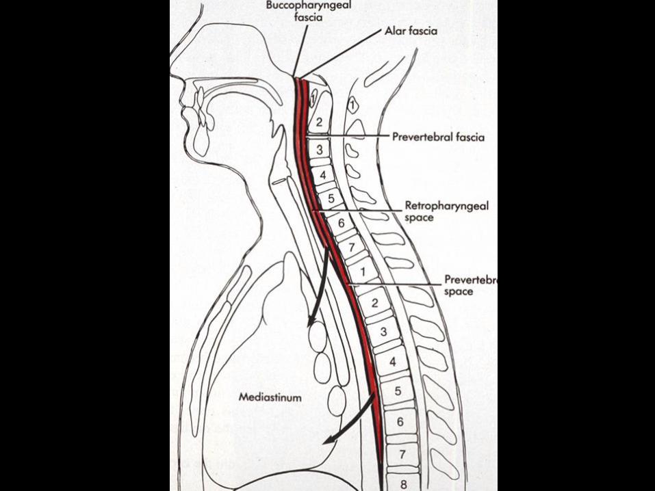
Cervical Fasciae
A. Superficial
1. Superficial cervical fascia
B. Deep
1. External (investing) fascia
2. Middle cervical fascia
3. Cervical visceral fascia
4. Alar fascia
5. Carotid sheath
6. Prevertebral fascia


