hair and fiber identification -...
TRANSCRIPT
1
Chemical Principles Exp. #7
Hair and Fiber Identification
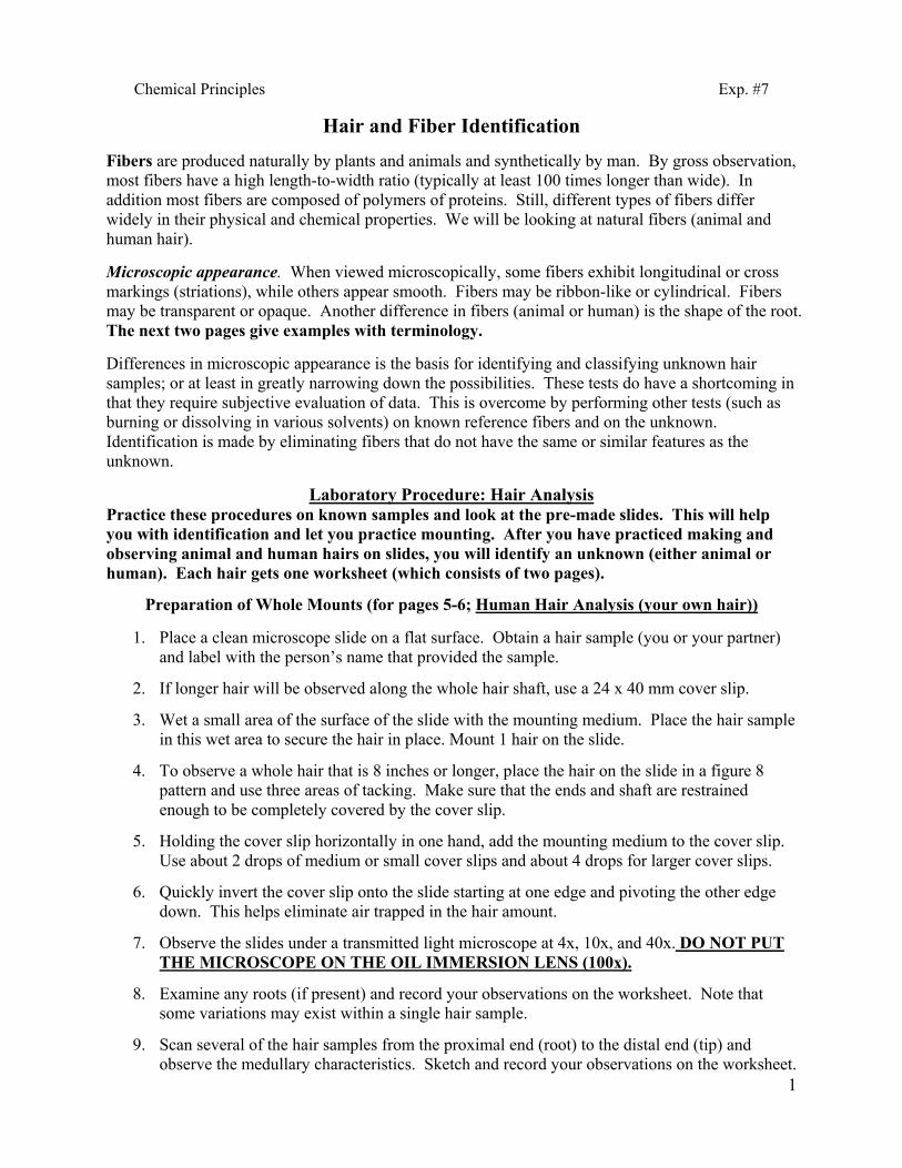
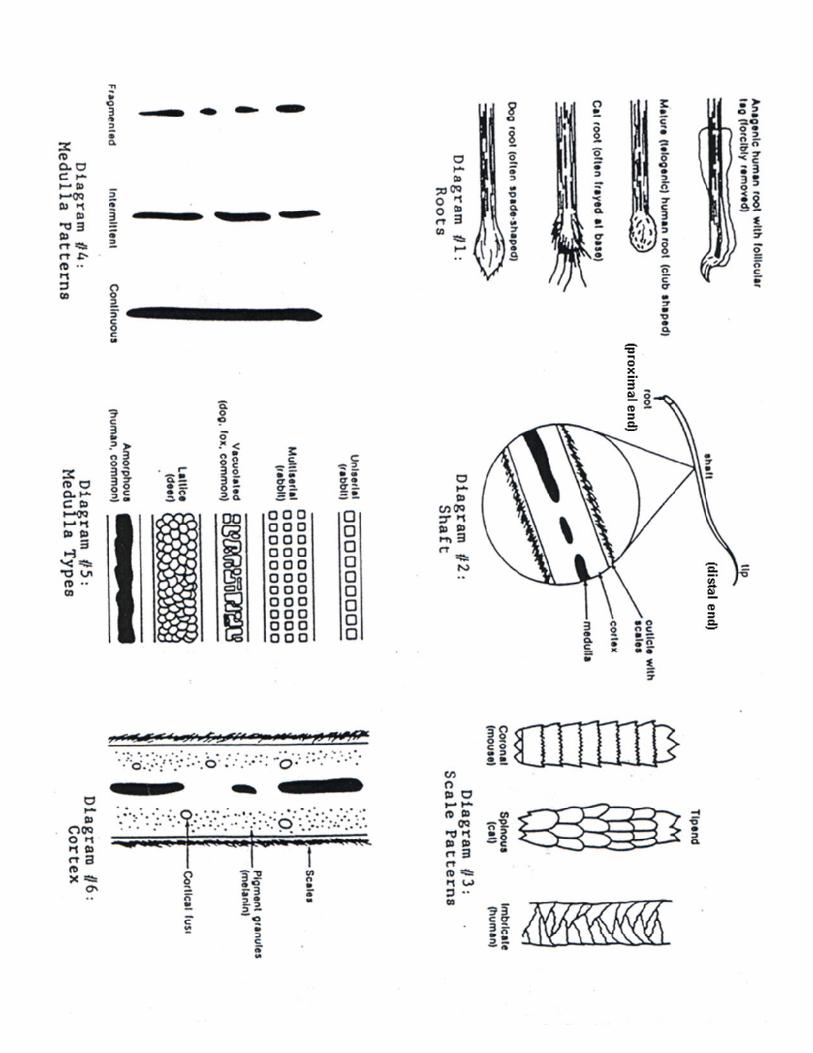
Fibers are produced naturally by plants and animals and synthetically by man. By gross observation, most fibers have a high length-to-width ratio (typically at least 100 times longer than wide). In addition most fibers are composed of polymers of proteins. Still, different types of fibers differ widely in their physical and chemical properties. We will be looking at natural fibers (animal and human hair). Microscopic appearance. When viewed microscopically, some fibers exhibit longitudinal or cross markings (striations), while others appear smooth. Fibers may be ribbon-like or cylindrical. Fibers may be transparent or opaque. Another difference in fibers (animal or human) is the shape of the root. The next two pages give examples with terminology. Differences in microscopic appearance is the basis for identifying and classifying unknown hair samples; or at least in greatly narrowing down the possibilities. These tests do have a shortcoming in that they require subjective evaluation of data. This is overcome by performing other tests (such as burning or dissolving in various solvents) on known reference fibers and on the unknown. Identification is made by eliminating fibers that do not have the same or similar features as the unknown.
Laboratory Procedure: Hair Analysis
Practice these procedures on known samples and look at the pre-made slides. This will help you with identification and let you practice mounting. After you have practiced making and observing animal and human hairs on slides, you will identify an unknown (either animal or human). Each hair gets one worksheet (which consists of two pages).
Preparation of Whole Mounts (for pages 5-6; Human Hair Analysis (your own hair))
1. Place a clean microscope slide on a flat surface. Obtain a hair sample (you or your partner) and label with the person’s name that provided the sample.
2. If longer hair will be observed along the whole hair shaft, use a 24 x 40 mm cover slip.
3. Wet a small area of the surface of the slide with the mounting medium. Place the hair sample in this wet area to secure the hair in place. Mount 1 hair on the slide.
4. To observe a whole hair that is 8 inches or longer, place the hair on the slide in a figure 8 pattern and use three areas of tacking. Make sure that the ends and shaft are restrained enough to be completely covered by the cover slip.
5. Holding the cover slip horizontally in one hand, add the mounting medium to the cover slip. Use about 2 drops of medium or small cover slips and about 4 drops for larger cover slips.
6. Quickly invert the cover slip onto the slide starting at one edge and pivoting the other edge down. This helps eliminate air trapped in the hair amount.
7. Observe the slides under a transmitted light microscope at 4x, 10x, and 40x. DO NOT PUT THE MICROSCOPE ON THE OIL IMMERSION LENS (100x).
8. Examine any roots (if present) and record your observations on the worksheet. Note that some variations may exist within a single hair sample.
9. Scan several of the hair samples from the proximal end (root) to the distal end (tip) and observe the medullary characteristics. Sketch and record your observations on the worksheet.
2
3
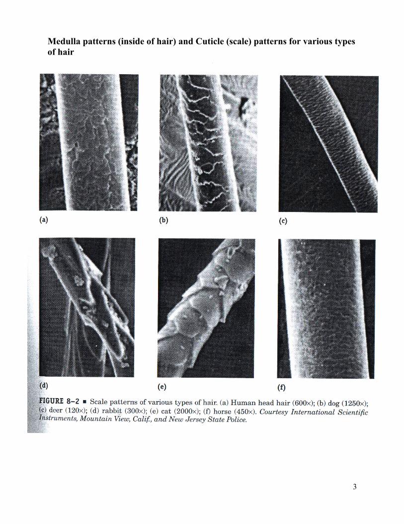
Medulla patterns (inside of hair) and Cuticle (scale) patterns for various types of hair
4
Experiment 7: Hair and Fiber Identification
Name:________________________ Hair and Fibers Prelab
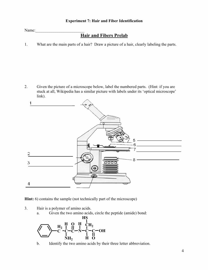
1. What are the main parts of a hair? Draw a picture of a hair, clearly labeling the parts. 2. Given the picture of a microscope below, label the numbered parts. (Hint: if you are
stuck at all, Wikipedia has a similar picture with labels under its ‘optical microscope’ link).
Hint: 6) contains the sample (not technically part of the microscope) 3. Hair is a polymer of amino acids.
a. Given the two amino acids, circle the peptide (amide) bond:
CNH2
CH2C
OHCN C
CH2
O
OH
H
HSH
b. Identify the two amino acids by their three letter abbreviation.
5
Experiment 7: Hair and Fiber Identification Examiner: _____________________ Partner: _____________________ Date: _____________________
Human Hair Analysis (your own hair)
1. Scale Pattern Sketch Scale Type (coronal, spinous, umbricate, etc): ______________ 2. Medulla Sketch Medulla Type (serial, vacuolated, lattice, amorphous, etc): _________________
6
Examiner: _____________________ Partner: _____________________ Date: _____________________
Hair Analysis (your own hair) 1. Length: less than 1 inch _____ 1-3 inches_____ 3-6 inches_____ 6-12 inches_____ 12+ inches_____ 2. Appearance: straight_____ wavy_____ curled_____ 3. Visual Color: colorless ______ dark brown_____ light brown_____ med brown_____ black_____ red_____ other_____ 4. Microscopic Color: colorless ______ dark brown_____ light brown_____ med brown_____ black_____ red_____ other_____ 5. Proximal End: root missing______ club-shaped_____ frayed_____ spade-shaped_____ 6. Distal End: rounded_____ right angle cut_____ oblique cut_____ split ______ taper to point______ cut not easily observed _____ 7. Cuticle (scale): undamaged_____ partially damaged_____ highly damaged_____ 8. Medulla: continuous_____ intermittent_____ fragmented_____ 9. Pigment Distribution: absent_____ even_____ peripheral_____ about medulla_____ 10. Cortical Fusi: absent_____ present_____ 11. Unusual Features:
7
Experiment 7: Hair and Fiber Identification Examiner: _____________________ Partner: _____________________ Date: _____________________
Animal Hair Analysis
1. Scale Pattern Sketch Scale Type (coronal, spinous, umbricate, etc): ______________ 2. Medulla Sketch Medulla Type (serial, vacuolated, lattice, amorphous, etc): _________________
8
Examiner: _____________________ Partner: _____________________ Date: _____________________
Animal Hair Analysis 1. Length: less than 1 inch _____ 1-3 inches_____ 3-6 inches_____ 6-12 inches_____ 12+ inches_____ 2. Appearance: straight_____ wavy_____ curled_____ 3. Visual Color: colorless ______ dark brown_____ light brown_____ med brown_____ black_____ red_____ other_____ 4. Microscopic Color: colorless ______ dark brown_____ light brown_____ med brown_____ black_____ red_____ other_____ 5. Proximal End: root missing______ club-shaped_____ frayed_____ spade-shaped_____ 6. Distal End: rounded_____ right angle cut_____ oblique cut_____ split ______ taper to point______ cut not easily observed _____ 7. Cuticle (scale): undamaged_____ partially damaged_____ highly damaged_____ 8. Medulla: continuous_____ intermittent_____ fragmented_____ 9. Pigment Distribution: absent_____ even_____ peripheral_____ about medulla_____ 10. Cortical Fusi: absent_____ present_____ 11. Unusual Features:
9
Experiment 7: Hair and Fiber Identification Examiner: _____________________ Partner: _____________________ Date: _____________________
Unknown Hair Analysis: Forensic Comparison
Unknown letter: _________________ 1. Scale Pattern Sketch Scale Type (coronal, spinous, umbricate, etc): ______________ 2. Medulla Sketch Medulla Type (serial, vacuolated, lattice, amorphous, etc): _________________
10
Examiner: _____________________ Partner: _____________________ Date: _____________________
Unknown Hair Analysis: Forensic Comparison 1. Length: less than 1 inch _____ 1-3 inches_____ 3-6 inches_____ 6-12 inches_____ 12+ inches_____ 2. Appearance: straight_____ wavy_____ curled_____ 3. Visual Color: colorless ______ dark brown_____ light brown_____ med brown_____ black_____ red_____ other_____ 4. Microscopic Color: colorless ______ dark brown_____ light brown_____ med brown_____ black_____ red_____ other_____ 5. Proximal End: root missing______ club-shaped_____ frayed_____ spade-shaped_____ 6. Distal End: rounded_____ right angle cut_____ oblique cut_____ split ______ taper to point______ cut not easily observed _____ 7. Cuticle (scale): undamaged_____ partially damaged_____ highly damaged_____ 8. Medulla: continuous_____ intermittent_____ fragmented_____ 9. Pigment Distribution: absent_____ even_____ peripheral_____ about medulla_____ 10. Cortical Fusi: absent_____ present_____ 11. Unusual Features: Species of hair (human/not human): ________________ What is your unknown? How can you tell?