gynaecology ultrasound imaging & management · gynaecology ultrasound imaging & management...
TRANSCRIPT

GYNAECOLOGY ULTRASOUND IMAGING & MANAGEMENT
Jean WilsonSchool of MedicineUniversity of Leeds
Dublin November 2015

Back to basics:Normal appearances and common pitfalls

Gynaecological US examinations -observations
The sonographer should demonstrate:nnormal anatomy/variants including age and menstrual status related appearances of the whole organ in at least two planes. This should include assessment of:n size, outline, echotexture and echogenicity.npathological findings.

The anatomical structures which the sonographer should be able to examine correctly are :nuterus: position, size, shape and ultrasound
characteristics of endometrium and myometriumnovaries: position, size, shape and ultrasound
characteristics. Number, size and internal echo pattern of follicles where presentncervixnfallopian tubes where visiblenbroad ligamentsnpelvic musclesnpelvic blood vessels.

Clinical history
nReason for referral, agenMenstrual historynLMPnCycle history – length, regularity, duration, menopause
nSymptoms n Pain, type, duration, ? related to cyclen Bleeding, ? heavy, IMB, PMB, amenorrhoean Relevant medicationn Previous gynaecological surgery / treatment

normal anatomy/variants

menstrual status related appearances

assessment of size

assessment of outline

assessment of echotexture

assessment of echogenicity

GYNAECOLOGICAL EXAMINATION WORKSHEETUterus: AV/RV
longit. trans. APendometrial thickness (mm)
R Ovary: longit. trans. APvol.(mls)
L Ovary: longit. trans. APvol.(mls)
R Adnexa:
L Adnexa:
Free fluid: No/Yes
√= normal U/S appearance & position x = abnormal U/S appearance & position

Observation worksheet
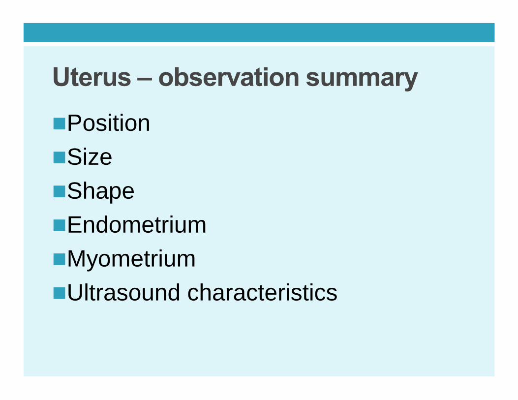
Uterus – observation summarynPositionnSizenShapenEndometriumnMyometriumnUltrasound characteristics

Ovaries – observation summarynPositionnSizenShapenUltrasound characteristicsnFollicle – number, size, echo pattern
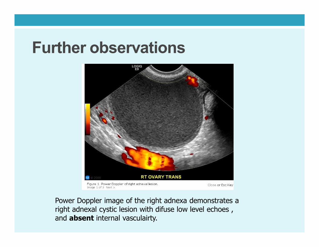
Further observations
Power Doppler image of the right adnexa demonstrates a right adnexal cystic lesion with difuse low level echoes , and absent internal vasculairty.

Further observations
Prominent uterine veins

? IUCD (mirena)

Further observations
TS

Further observations